Review

PDX-1: A Promising Therapeutic Target to Reverse Diabetes

1
Institute of Metabolic Diseases, Guang’anmen Hospital, China Academy of Chinese Medical Sciences, Beijing 100053, China
2
Graduate College, Beijing University of Chinese Medicine, Beijing 100029, China
3
Graduate College, Changchun University of Chinese Medicine, Changchun 130117, China
*
Author to whom correspondence should be addressed.
Biomolecules 2022, 12(12), 1785; https://doi.org/10.3390/biom12121785
Submission received: 17 October 2022 / Revised: 18 November 2022 / Accepted: 19 November 2022 / Published: 30 November 2022

Abstract

:
The pancreatic duodenum homeobox-1 (PDX-1) is a transcription factor encoded by a Hox-like homeodomain gene that plays a crucial role in pancreatic development, β-cell differentiation, and the maintenance of mature β-cell functions. Research on the relationship between PDX-1 and diabetes has gained much attention because of the increasing prevalence of diabetes melitus (DM). Recent studies have shown that the overexpression of PDX-1 regulates pancreatic development and promotes β-cell differentiation and insulin secretion. It also plays a vital role in cell remodeling, gene editing, and drug development. Conversely, the absence of PDX-1 increases susceptibility to DM. Therefore, in this review, we summarized the role of PDX-1 in pancreatic development and the pathogenesis of DM. A better understanding of PDX-1 will deepen our knowledge of the pathophysiology of DM and provide a scientific basis for exploring PDX-1 as a potential target for treating diabetes.

1. Introduction

Diabetes mellitus (DM) is a metabolic disorder with an increasing prevalence worldwide. It is characterized by chronic hyperglycemia and disturbances in protein, carbohydrate, and fat metabolism due to insulin resistance (IR) and/or insulin secretion deficiency [1]. Approximately one billion people suffer from chronic hyperglycemia globally, a major public health problem. The International Diabetes Federation estimates that 10.5% (537 million) of adults aged 20–79 years are currently living with DM, and this prevalence is expected to increase to 11.3% (643 million) by 2030 and 12.2% (783 million) by 2045. With 1.541 million adults having impaired glucose tolerance (IGT), their risk for type 2 diabetes is increased [2]. Diabetes causes a series of complications, including blindness, renal failure, stroke, and coronary artery disease, resulting in a huge medical burden on society [3]. Furthermore, diabetes costs at least 966 billion dollars in health expenditure, a 316% increase over the last 15 years [2].
DM is divided into type 1 diabetes (insulin-dependent diabetes mellitus (T1DM or IDDM)), type 2 diabetes (non-insulin-dependent diabetes mellitus (T2DM or NIDDM)), specific types of diabetes due to other causes, and gestational diabetes mellitus [4,5]. T2DM is the most common presentation of DM, accounting for approximately 90% of DM cases, whereas T1DM constitutes more than 5% [6,7]. T2DM is a chronic multisystem disease characterized by insulin resistance and elevated blood glucose levels [8]. It is the result of a complex interplay between genetic, epigenetic, and environmental factors [9,10]. However, its etiology and pathogenesis have not yet been fully elucidated. The traditional view is that diabetes can only be controlled and not cured. Authoritative guidelines and clinical diabetes research mostly focus on controlling blood glucose levels and improving complications. Blood glucose levels are controlled by promoting insulin secretion, enhancing insulin sensitivity, and promoting glucose absorption by other tissues outside the islets of Langerhans [11,12]. With recent advances in diabetes research and medical technology progress, researchers have discovered new methods for preventing and treating diabetes with promising results. The terms “Diabetes reversal” and “Diabetes remission” have been used in scientific articles. They connote a glycosylated hemoglobin A1c (HbA1c) level of <6.5% (<48 mmol/mol) for at least 3 months without the usual glucose-lowering pharmacotherapy [13]. The reversal strategy, mechanism, and predictors are becoming increasingly clear with further research [14,15,16,17]. One of the possible mechanisms of diabetes reversal is that the removal of excess fat in the liver and pancreas can normalize hepatic glucose production and β-cell redifferentiation [14]. However, more studies are needed on the mechanism of diabetes reversal.
Studies on the role of PDX-1 in reversing diabetes are on the increase. PDX-1 regulates pancreatic development, β-cell differentiation, and the maintenance of mature β-cell function [18,19,20,21,22,23,24]. Non-β-cell can be reprogrammed by the transcription factors PDX-1 and musculoaponeurotic fibrosarcoma oncogene family A (MafA) into functional β-cell, which secretes insulin to restore blood glucose levels [25,26]. In contrast, β-cell-specific removal of PDX-1 resulted in pancreatic agenesis and severe hyperglycemia [27,28]. Thus, promoting PDX-1 expression can be an effective strategy to ameliorate β-cell dysfunction and diabetes progression, making PDX-1 a new target for developing anti-diabetic drugs. Therefore, in this review, we systematically summarized the roles of PDX-1 in pancreatic development and function maintenance. Furthermore, we explored the application and prospects of PDX-1 in diabetes research and treatment.

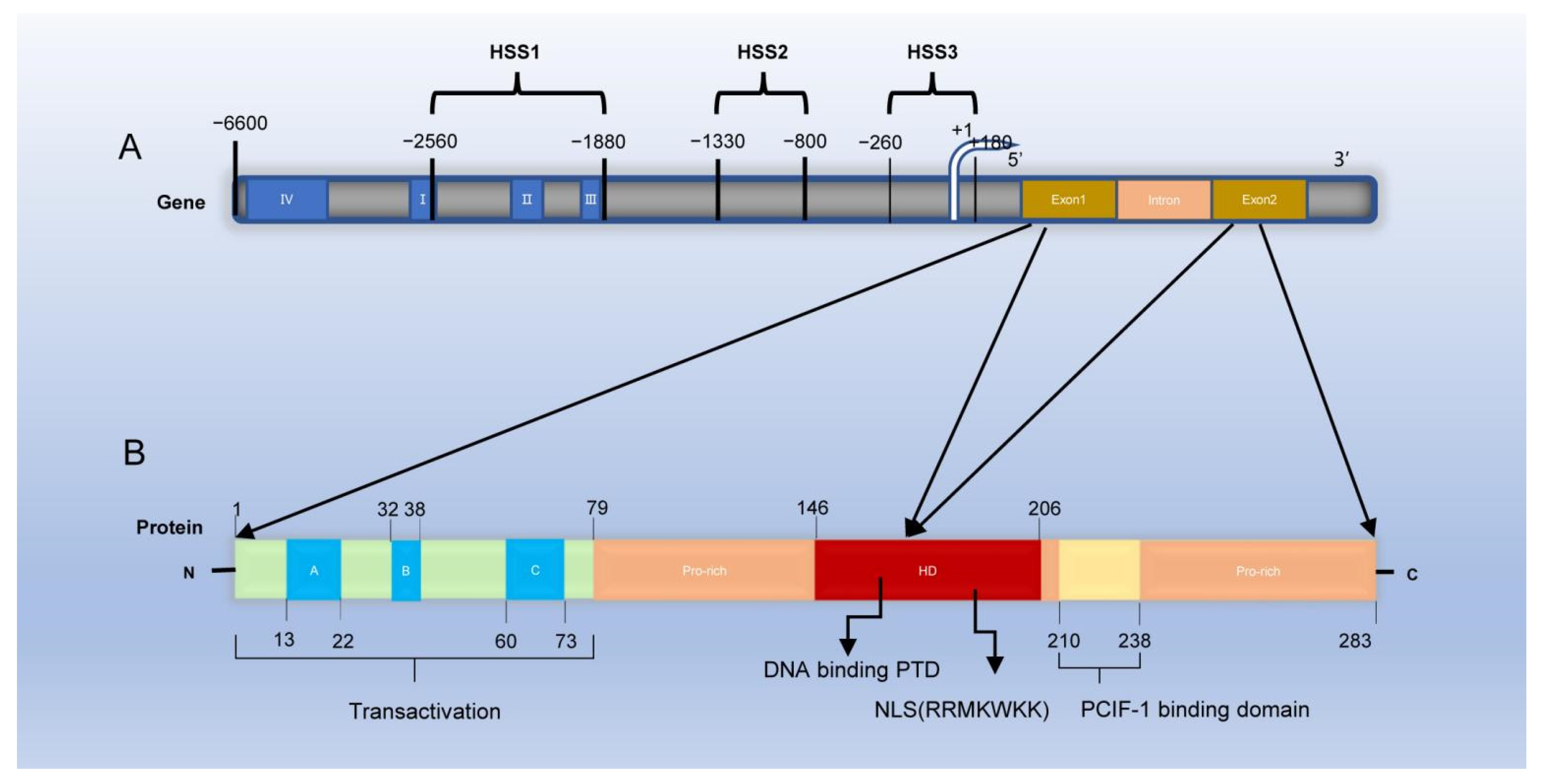
2. Gene Structure and Location, Protein Molecular Structure, Distribution, and Expression of PDX-1

The PDX-1, also known as IUF-1 (insulin upstream factor 1), IPF-1 (insulin promoter factor 1), STF-1 (somatostatin transcription factor 1), and IDX-1 (islet/duodenum homeobox-1), is a member of the homeodomain (HD)-containing transcription factor family and was first found in Xenopus laevis [18,29,30,31,32]. It plays a key role in the genesis, development, and maturation of the pancreas and is also one of the factors necessary for maintaining normal pancreatic islet function.

2.1. Localization and Molecular Structure of PDX-1

Rat and mouse PDX-1 genes are localized on chromosomes 12 and 5, respectively, whereas the human PDX-1 gene is located on chromosome 13q12 (12.1) [33,34,35,36]. The human PDX-1 gene is approximately 6 Kb long with two exons. The first exon encodes the NH2-terminal region and some HD, while the second encodes the remaining HD and the COOH-terminal domain [37]. (Figure 1) Three nuclease-hypersensitive sites were identified within the 5′-flanking region of the endogenous PDX-1 gene: HSS1(−2560~−1880 bp), HSS2(−1330~−800 bp), and HSS3(−260~+180 bp) [38].Among them, HSS1 is an important functional region of PDX-1 gene transcription activation and includes four sub-regions, region I (−2694~−2561 bp), region II (−2139~−1958 bp), and region III (−1879~−1799 bp). The fourth distal enhancer element is region IV, located between −6200 and −5670 bp [39,40,41]. Regions I and II endow endocrine cell expression, region Ⅲ mediates embryonic pancreas-wide expression, and region IV endows pancreas β-cell-specific gene expression and enhancement of proximal enhancer activity [42]. In addition, PDX-1 transcription is also regulated by factors acting upon conserved Area Ⅰ and IV sequences [38]. The proximal and distal promoters contain four conserved regions: I, II, III, and IV, which bind to various transcription factors to regulate cell differentiation [40]. In a study, a child’s pancreas did not develop (pancreatic agenesis) because the child was homozygous for an inactivating cytosine deletion in the protein-coding sequence of PDX-1 (pro63fsdelc), which indicated that PDX-1 might be associated with Type 2 diabetes [43]. In addition, six novel PDX-1 missense mutations (C18R, D76N, R197H, Q59L, G212R and P2390) were identified in patients with type 2 diabetes [44,45].
The human PDX-1 protein comprises 283 amino acids with a predicted molecular weight of 30.77 KDa [46]. The PDX-1 activation domain is contained within the NH2-terminal, its HD is involved in DNA binding, and they both participate in protein–protein interactions [47]. Point mutation analysis showed that the transcriptional activation region was necessary to activate insulin gene transcription [48]. The HD contains the nuclear localization signal (NLS) and an Antennapedia-like protein transduction domain (PTD), the nuclear import of PDX-1 depends on the NLS motif RRMKWKK [49]. PDX-1 mainly exists in the cytoplasm or around the nucleus in the resting state. Changes in the external environment, such as ionizing radiation and the increase in glucose concentration, can activate PDX-1 and translocate it into the nucleus [50]. The NLS of transcription factors is a crucial requirement for its action, possibly because free PDX-1 is modified by phosphorylation, acetylation, and sumoylation, which exposes the NLS and guides the nuclear translocation of PDX-1 [51,52,53]. In addition, Guillemain G et al. found that PDX-1 first interacts with the nuclear input receptor importinβ1 to form a complex; importinβ1 then interacts with the nuclear pore complex on the nucleus surface. It mediates PDX-1 entry into the nucleus. Ras-related nuclear protein (Ran) GTP and importinβ1 dissociate in the nucleus, followed by the reflux of the Ran GTP and importinβ1 complex into the cytoplasm through nuclear pores. Ran GTP is transformed into Ran, GDP, and the dissociated cytoplasm importinβ1 continues to participate in the transport of transcription factors [54]. In addition, it was previously reported that exogenous PDX-1 protein can permeate cells and induce insulin gene expression in pancreatic ducts because its own antennapedia-like protein transduction domain (PTD) sequence in its structure can bind to the insulin promoter and activate its expression [55]. A conserved motif at the C-terminal of PDX-1 mediates the interaction of PDX-1-PCIF1(PDX-1 C-terminal interacting factor-1) and inhibits the transcriptional activity of PDX-1 [56]. In addition, Humphrey et al. reported a novel functional role for the PDX-1 C-terminus in mediating glucose effects. They demonstrated that glucose modulates PDX-1 stability via the AKT-GSK3 (glycogen synthase kinase 3) axis [57]. Therefore, PDX-1 has a dual function. First, it promotes early pancreatic development and late β-cell differentiation. Second, it maintains the morphology and normal function of mature β cells, especially the normal expression of insulin secretion genes.

2.2. Tissue Distribution and Expression of PDX-1

PDX-1 expression in cells at different stages is inconsistent. In the early developmental stage, PDX-1 widely exists in the cell population transformed into endocrine and exocrine parts of the pancreas and some brain cells in the embryonic stage. However, PDX-1 is highly expressed in β, δ, and endocrine cells of the duodenum following maturation. In contrast, its expression is low in some ductal and acinar cells [58,59]. Stoffers et al. found that PDX-1 played an important role in developing the exocrine and endocrine portions of the pancreas, pancreatic ducts, pyloric glands of the distal stomach, common bile and cystic ducts, the intestinal epithelium of the duodenum, Brunner’s glands, and the spleen [60]. In addition to the gastrointestinal system, PDX-1 is expressed in embryonic brain cells during the active nervous system generation phase [61]. Researchers have reported PDX-1 overexpression in various human tumor tissues [62,63,64]. Wang XP et al. used tissue microarray and immunohistochemical techniques to demonstrate PDX-1 overexpression in breast, kidney, pancreatic, and prostate cancers. They suggested that PDX-1 was probably one of the early markers of tumorigenesis [65].

3. Factors Regulating PDX-1 Expression

PDX-1 is critical for maintaining β cells, and its downregulation results in β-cell dedifferentiation. The induction of PDX-1 expression maintains mature and functional β cells. The regulation of PDX-1 gene expression is a complex process (Figure 2) involving nutrient substances, hormones, oxidative stress, and cytokines.

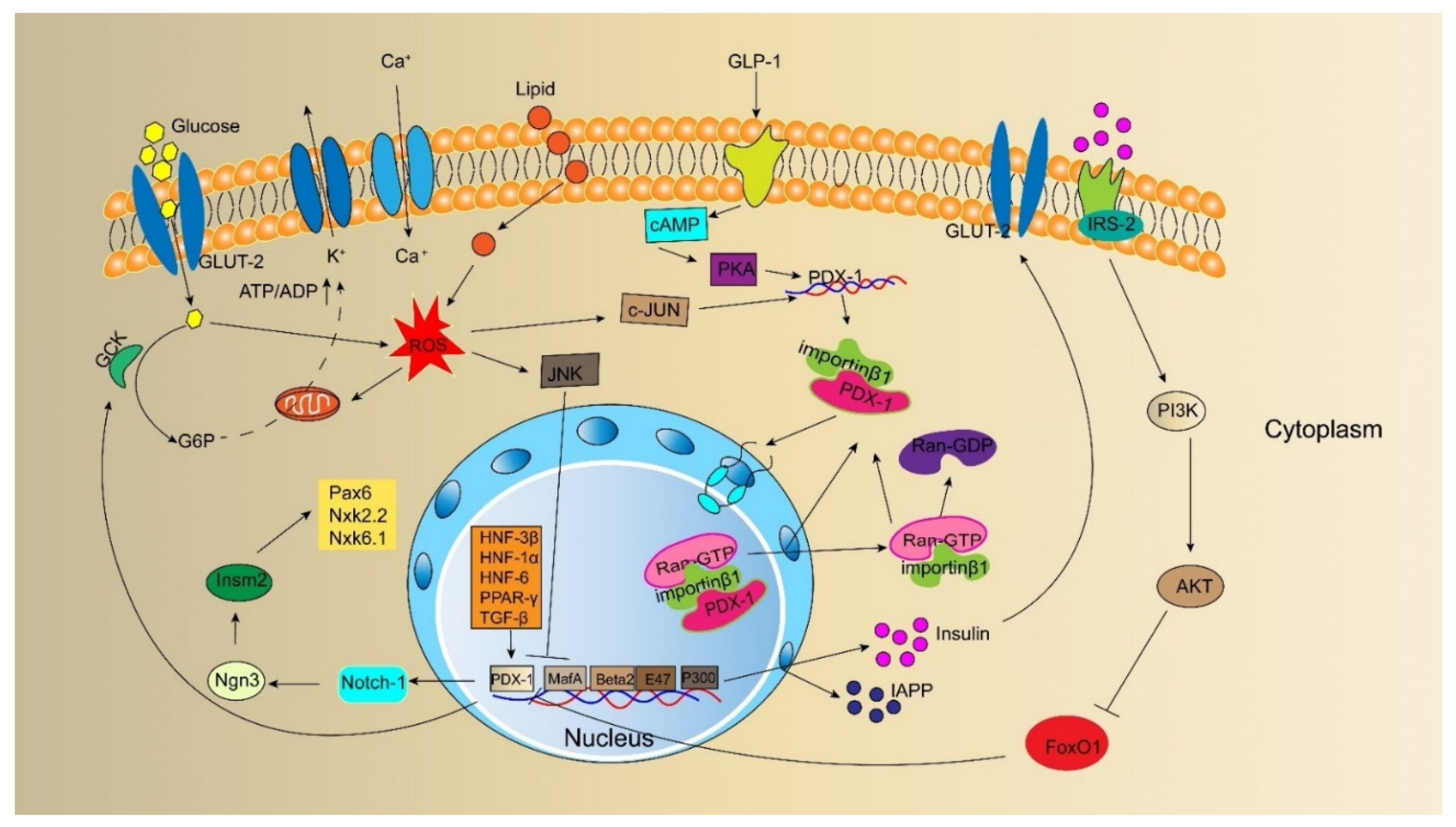
3.1. Nutrient Substances

Glucose and fatty acids regulate PDX-1 expression. Many studies elucidated the underlying molecular mechanisms of glucotoxicity and lipotoxicity. In adult islet β-cell, a short-term hyperglycemic environment promotes the combination of PDX-1 and insulin genes and improves insulin mRNA levels. However, PDX-1 and insulin levels decrease under the cytotoxic effect of long-term hyperglycemia [66]. The inhibition of PDX-1 expression by high glucose concentration is one of the mechanisms of glucotoxicity. Furthermore, chronic hyperglycemia has been reported to deteriorate β-cell function by inducing oxidative stress and reducing PDX-1 DNA-binding activities [67]. In addition to activating the DNA-binding activity of PDX-1, glucose also affects PDX-1 phosphorylation, PDX-1 distribution between the nuclear membrane and nucleoplasm, and the transactivation potential of the amino-terminal active region of PDX-1; however, its mechanism of action remains unclear. High fatty acid concentrations also inhibit PDX-1 expression. Gramlich et al. showed that the co-culture of pancreatic islets with palmitic acid reduced mRNA and protein expression levels of PDX-1 by 70%. The binding force between PDX-1, glucose transporter 2 (GLUT-2), and the insulin gene promoter decreased by 40–65% [68]. The prolonged exposure of islets to palmitate inhibits insulin gene transcription by impairing the nuclear localization of PDX-1 [69]. Shimo N et al. found that PDX-1 expression was significantly reduced during glucotoxicity and lipotoxicity. After treatment with empagliflozin or bezafibrate to selectively improve glucotoxicity or lipotoxicity, PDX-1 showed significantly higher expression levels and enhanced β-cell proliferation [70]. The study provided further evidence that glucose and fatty acids regulate PDX-1 expression.

3.2. Hormones

The incretin hormone glucagon-like peptide-1 (GLP-1) is produced by gut endocrine L cells in a nutrient-dependent manner and secreted in pancreatic islets [71]. Studies have shown that GLP-1 is involved in regulating PDX-1. Wang et al. found that GLP-1 promotes PDX-1 expression in a glucose-dependent manner, increases its intracellular protein content and improves its binding activity with the A1 region of the insulin gene. Simultaneously, GLP-1 activates adenosine cyclase, increases cyclic adenosine monophosphate (cAMP) content in cells, and activates protein kinase A (PKA), which promotes PDX-1 synthesis and increases its content [72,73]. Hwang SL et al. found that treating rat insulinoma cells with GLP-1 significantly increased β-cell translocation gene 2 mRNA expression in dose- and time-dependent manners and subsequently elevated PDX-1 and insulin mRNA levels in pancreatic β cells [74]. The activation of PKA induced by GLP-1 increases PDX-1 level and translocation to the nucleus, where PDX-1 binds to the insulin gene promoter to initiate insulin expression and synthesis [75]. In addition, the runaway signal system of the small Ran GTPase significantly downregulates PDX-1 expression in postnatal mice, resulting in insulin deficiency, decreased cell proliferation rate, and diabetes [76]. In aging animal models of type 2 diabetes, the expression of the PDX-1 gene was decreased, the number of β cells was reduced, and the long-term use of GLP-1 reversed these pathological changes. However, when exendin (9–39) (a specific antagonist of GLP-1) was infused, its effects on the levels of PDX-1 messenger RNA were eliminated [77]. These studies showed that GLP-1 stimulates pancreatic cell proliferation and β-cell differentiation by regulating PDX-1.

3.3. Oxidative Stress

Some studies have shown that reactive oxygen species (ROS) reduce PDX-1 mRNA synthesis, leading to a decline in PDX-1 synthesis and a reduction in the binding of PDX-1 to the insulin gene promoter and the transcription of the insulin gene. Cannabinoids have strong antioxidant properties. Baeeri et al. used 10 μM cannabidiol and tetrahydrocannabinol to treat aged rat islet cells. The results showed that the percentage of ROS was significantly reduced with the elevation of PDX-1 expression and insulin release [78]. However, the results were preliminary, and further studies are needed to elucidate the mechanism. Leenders et al. treated human islets with 200 μM hydrogen peroxide for 90 min and found that the gene and protein expression of the key transcription factor PDX-1 was reduced by over 60% [79]. Furthermore, Matsuoka et al. treated HIT-T15 cells cultured in vitro with the oxidative stress inducer d-ribose. They found that PDX-1 expression in cells was reduced, and the binding of PDX-1 to the insulin gene promoter was significantly reduced. However, 1 mM aminoguanidine or 10 mM N-acetyl-L-cysteine (NAC) prevented the effects of d-ribose [80]. Kawamori et al. also found that oxidative stress affects the nucleocytoplasmic translocation of PDX-1 by activating c-Jun N-terminal kinase (JNK) and pointed out that JNK may induce the translocation of PDX-1 from the nucleus to the cytoplasm by activating the nuclear output signal of PDX-1, reducing the expression of PDX-1 in the nucleus, and also reducing insulin synthesis and secretion [81,82]. After treating diabetes mice (C57BL/KsJ-db/db) with antioxidant drugs (NAC), Kajimoto et al. found that the expression of PDX-1 in the nucleus of pancreatic islets was significantly increased, and the amounts of insulin content and insulin mRNA were preserved [83]. Nucleocytoplasmic translocation of PDX-1 is the key to promoting insulin secretion. Oxidative stress can affect the nucleocytoplasmic translocation of PDX-1, reduce the interaction between PDX-1 and insulin gene promoters, and reduce insulin synthesis and secretion. Baumel-Alterzon S et al. reported that the nuclear factor erythroid 2-related factor (Nrf2) antioxidant pathway controls the redox balance and allows the maintenance of high PDX-1 levels; pharmacological activation of the Nrf2 pathway may alleviate diabetes by preserving PDX-1 levels [84].

3.4. Cytokines

Many cytokines (hepatocyte nuclear factor (HNF-3β), HNF-6, transforming growth factor-beta (TGF-β), peroxisome proliferator-activated receptor gamma (PPAR-γ)) act upstream of PDX-1 in the regulatory hierarchy governing pancreatic development. HNF-3β (also named Foxa2) is a transcription factor in the HNF family. HNF-3β binds to multiple sites in the transcriptional activation functional region of the PDX-1 gene and recruits other transcription factors to the regulatory region to enhance gene transcription [85]. In addition, co-transfection experiments suggested that HNF-3β, HNF-1α, and specificity protein 1 (Sp1) are positive human PDX-1 enhancer elements with mutual coordination [86]. Gao et al. found that compound conditional ablation of Foxa1 and Foxa2 caused near severe pancreatic hypoplasia and total loss of PDX-1 expression, and Foxa2 appeared to predominate. Jacquemin et al. also found that HNF-6 acted upstream of PDX-1 during the development of the pancreas, and HNF-6(-/-) mice were hypoplastic [87]. Sayo et al. used the TGF-β to process pancreatic β cells, and the results showed that TGF-β activates the insulin gene by activating PDX-1 [88]. PPAR-γ agonist rosiglitazone was reported to increase the immunostaining of PDX-1 and Nkx6.1, while PPAR-γ inhibitors reduced the mRNA levels of PDX-1 through RNA interference [89]. In addition, the study found that PDX-1 interacted with the p300 coactivator, β-cell E-box transcription factor (BETA2), and E47 coactivator to mediate insulin gene transcription [58]. These cytokines play a significant role in developing the pancreas; however, further studies are needed to explore their regulation to ensure PDX-1 transcription.

4. PDX-1 and the Pancreas

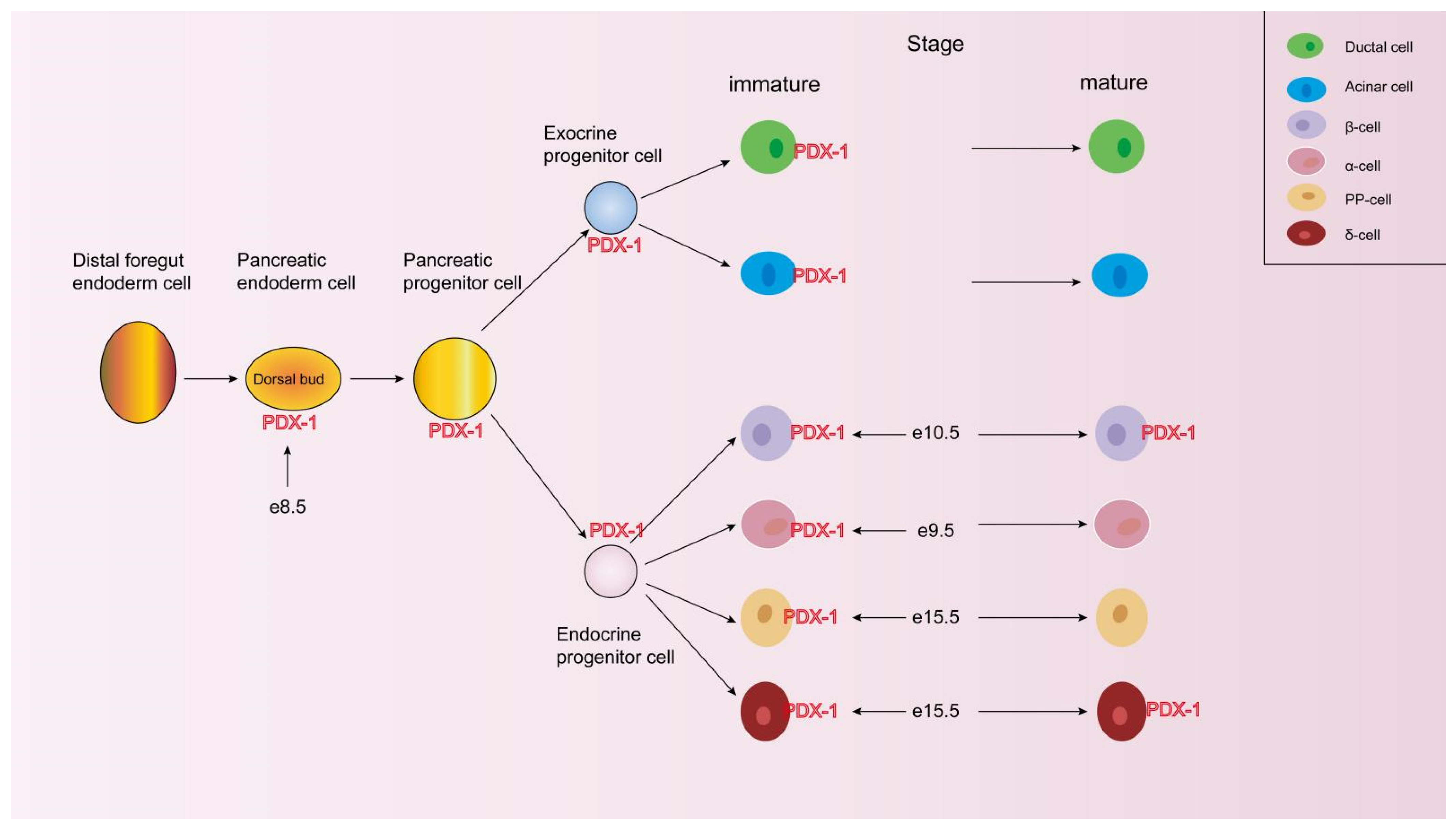
4.1. PDX-1 Promotes Pancreatic Development and Islet Cell Differentiation

Neurogenin 3 (Ngn3) is critical for inducing endocrine islet cell differentiation from embryonic pancreatic progenitors, and all pancreatic endocrine cells arise from endocrine progenitors expressing Ngn 3 [90]. PDX-1 directly regulates Ngn 3 expression [91]. PDX-1 is one of the most important transcription factors for directional development and pancreas maturation. During embryonic development, PDX-1 promotes the early development of the pancreas and β-cell differentiation and is a marker of β-cell formation and maturation.
Before embryonic development and maturation, PDX-1 is expressed in endocrine and exocrine cells. With the development of the pancreas, PDX-1 is only specifically expressed in 90% of β cells and a small amount (10%) of δ cells [92]. (Figure 3).
Jonsson et al. reported pancreatic dysplasia in homozygous mice for a targeted mutation in the PDX-1 gene [19]. In addition, a single nucleotide deletion in the human PDX-1 gene coding sequence resulted in pancreatic agenesis [27,93]. Studies have shown that mice and sheep with PDX-1 gene knockout will have pancreatic development and dysfunction, which further proves the role of PDX-1 in early pancreatic development and the differentiation and maturation of β cells [94,95]. Wiggenhauser reported, for the first time, that PDX-1 knockout caused the development of early diabetic kidney disease features, such as glomerular hypertrophy, filtration barrier impairments corresponding to microalbuminuria, and glomerular basement membrane (GBM) thickening in zebrafish [96]. After a partial resection of rat pancreas overexpressing the PDX-1 gene, regenerative proliferation and repair of residual pancreatic ductal epithelial cells were observed within 24 h, further indicating that PDX-1 promotes the differentiation of pancreatic ductal epithelial cells and pancreatic regeneration and repair [97].

4.2. PDX-1 Adjusts the Dedifferentiation, Redifferentiation, and Transdifferentiation of β-Cell

PDX-1 is important in early pancreatic regulation and is indispensable for β-cell differentiation and identity maintenance. The dysfunction or failure of β-cells occurs in patients with type 2 diabetes even in the early stages of the disease. As the disease progresses, the degree of failure also increases. Previously, apoptosis was believed to be the mechanism underlying β-cell failure [98]. Recently, many studies have shown that β cells in patients with diabetes undergo a process of “dedifferentiation” rather than apoptosis.
The dedifferentiation of β cells is the central link to cell failure. In diabetes, β cells undergo dedifferentiation and return to a progenitor-like state. In 2012, Talchai et al. revealed that β-cell dedifferentiation is a key link in the pathogenesis of diabetes. They observed that in forkhead box O 1 (FoxO1) knockout mice, the expression of pancreatic progenitor cell markers Ngn3 and octamer-binding transcriptional factor 4 was increased, and β cells were dedifferentiated. Furthermore, the characteristics of mature cells were reversed to the state of endocrine precursor cells, which could transdifferentiate into α, PP, and other endocrine cells [99]. In another study, non-islet β cells from deceased donors were transplanted into diabetic mice. The transplanted non-islet β cells were tracked and reprogrammed by transcription factors (PDX-1, MafA) to secrete insulin and control blood glucose. The transplanted cells still produced insulin after six months [26]. Hydrodynamic gene delivery of PDX-1, Ngn3, and MafA to rat liver can initiate transdifferentiation to pancreatic β cells [100]. Ferber S et al. transferred the PDX-1 gene into rat liver cells for the first time, realizing the function of insulin secretion by liver cells and causing the rat’s blood glucose to decrease significantly, which was verified in the experiment of primary hepatocytes in vitro [21]. Other non-islet β cells, such as umbilical cord blood-derived mesenchymal stem cells (UCB-MSCs), bone marrow mesenchymal stem cells (BMSCs), adipose-derived mesenchymal stem cells (ADMSC), and intestinal stem cells have been reshaped into new cells whose morphology and function are similar to those of islet β cells [101]. Pham et al. reported that mRNA PDX-1 transfection improves UCB-MSC differentiation into insulin-producing cells and produces C-peptide and insulin in a glucose-dependent manner [102].
Sun et al. constructed a eukaryotic expression vector containing PDX-1 and transfected it into BMSCs. The results revealed that PDX-1 promoted BMSCs to form functional pancreatic islet-like structures [103]. Gao et al. transfected the PDX-1 gene into ADMSCs and found that it induced the differentiation of ADMSCs into functional islet-like cells [104]. In addition, pluripotent stem cell-derived pancreatic endoderm cells have been used in clinical research on type 1 diabetes [105]. These findings support the potency of PDX-1 in driving β-cell-like differentiation in non-β-cell, which may be a promising approach for future therapeutic use in patients with diabetes.

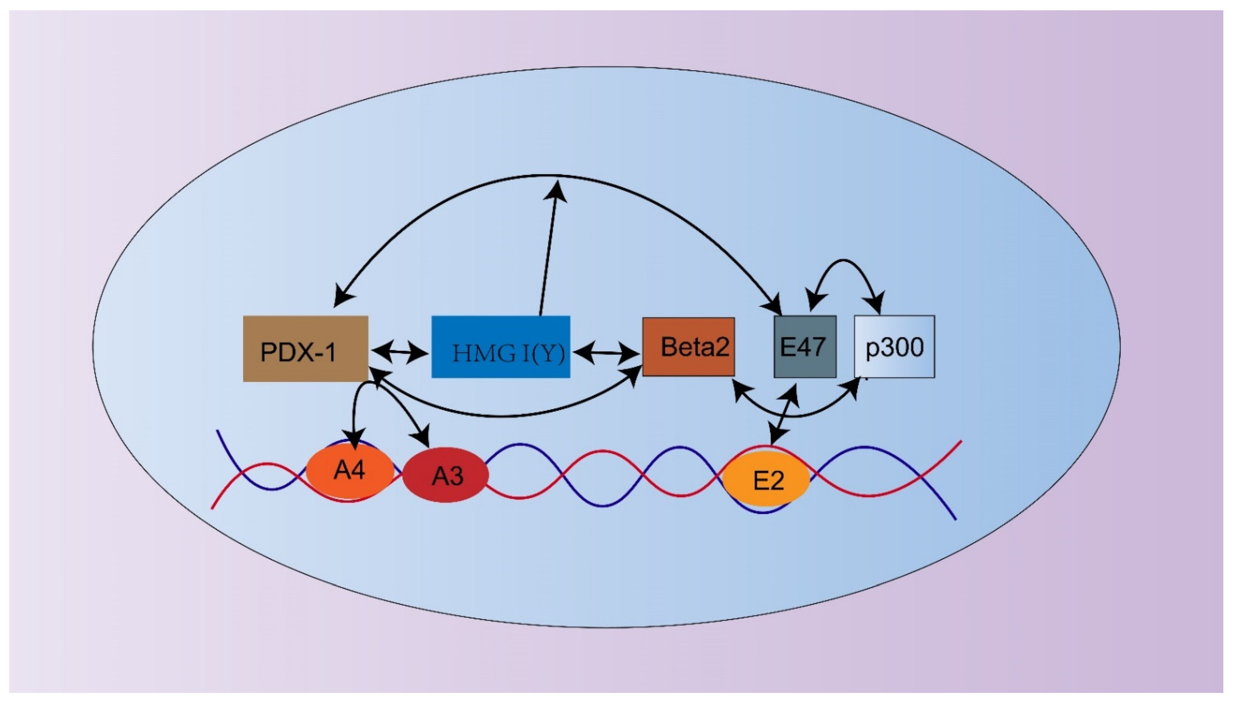
4.3. PDX-1 Regulates the Expression of the Insulin Gene and Insulin Secretion-Related Genes

PDX-1 involves a series of gene transcriptional regulations related to maintaining β-cell characteristics and functions. PDX-1 regulates the insulin gene transcription, glucokinase (GCK), GLUT-2, islet amyloid polypeptide (IAPP), and somatostatin, suggesting that PDX-1 may play an extensive role [36]. (Figure 2).

4.3.1. Insulin

Insulin is the most critical gene expressed explicitly in the β cells of the islet Langerhans. PDX-1, a homeodomain-containing transactivator of the insulin gene, plays a significant role in insulin gene expression and pancreatic development by binding to the enhancer region between nucleotides −340 and −91 [106]. Exposing β cells to a high-glucose environment for a short time enhanced the binding of PDX-1 with insulin gene promoters and increased the level of insulin mRNA in vitro experiments. When β cells are exposed to high glucose for a long time, insulin gene expression defects can occur, accompanied by a decline in proinsulin mRNA levels and a reduction in islet cells, which causes a reduction in insulin synthesis. Defects in insulin gene expression are mainly due to long-term high glucose production; oxidative stress activates the JNK pathway, directly inhibits the expression of the insulin gene, and inhibits the activities of two main transcription factors, PDX-1 and MafA. The latter two are the main participants in the activation of the insulin gene promoter [81,107,108]. Point mutations in the rat insulin gene promoter or the corresponding human sites can significantly decrease promoter activity, affecting insulin gene expression [109]. After silencing the PDX-1 gene in neonatal mouse islet β cells, insulin secretion is defective, causing impaired glucose tolerance and diabetes in adulthood. Some studies also found that following the overexpression of the PDX-1 gene in islets, insulin secretion, and glucose tolerance were enhanced, and the expression of MafA and GCK genes, which maintain normal β-cell function, were restored in maturity-onset diabetes of the young (MODY) mice [110]. However, PDX-1 is a necessary, but not sufficient, key regulator of insulin gene activity. Ohneda et al. reported that PDX-1 combines with E47, neurogenic differentiation 1 (NeuroD1), and other transcription factors at the DNA binding site of the insulin gene promoter to form a transcription activation complex through protein–protein interactions and cooperatively activates multiple fields with the initial complex of the transcription starting point or other related coactivators to regulate the expression of the insulin gene [111] (Figure 4). In addition, as a nuclear architectural factor that can organize chromatin structures, the high-mobility group AT-hook 1 (HMGA1) protein plays a critical role in pancreatic beta-cell function and insulin production by regulating PDX-1- and MafA-induced transactivation of the INS gene promoter [112].

4.3.2. GCK

As the first rate-limiting enzyme, GCK serves as a glucose sensor in pancreatic β cells and is mainly concentrated in pancreatic cells, liver cells, the hypothalamus, and the gastrointestinal tract [113]. PDX-1 is involved in regulating the GCK gene and is crucial in maintaining normal beta cell functions. PDX-1 binds to the hUPE3 site (a primarily important cis-motif of the GCK gene β-cell-type promoter) and activates the transcription of the GCK gene [114]. Li et al. found that differentiated PDX-1 + hMSCs expressed GCK and released insulin/C-peptide in a weak glucose-regulated manner [115]. However, Watada et al. showed that the exogenous expression of PDX-1 in alphaTC1 clone 6 cells alone could not induce GCK expression; when betacellulin was added to the medium, it expressed insulin and GCK mRNAs [116]. This study suggests that PDX-1 can regulate the expression of GCK, and certain regulatory factors contribute to the β-cell specificity of gene expression.

4.3.3. GLUT-2

The glucose transporter GLUT-2 is expressed in the liver, intestine, kidney, pancreatic islet β cells, central nervous system, neurons, astrocytes, and hypothalamic glial cells. It is required for glucose-stimulated insulin secretion in pancreatic β cells [117]. Insulin secretion induced by glucose requires transport, storage, oxidative metabolism, and information transmission. GLUT-2 is a special transmembrane transporter in the β-cell membrane that enters cells through GLUT-2 on the membrane and is phosphorylated by GCK, causing a series of oxidative metabolic processes. In this process, the ATP/ADP ratio is increased, ATP-sensitive potassium channels are closed, cell membrane depolarization occurs, voltage-dependent Ca2+ channels are opened, intracellular Ca2+ is increased, and particle aggregation and exocytosis occur in coordination with other secondary messengers. GLUT-2 and GCK are essential proteins involved in this process. PDX-1 and GTIIa are central islet-specific DNA-binding proteins that regulate GLUT-2 transcription. PDX-1 activates the transcription and expression of GLUT-2 by combining with a highly conserved TAAT sequence (5′-TAATA-ATAACA-3′) located in the GLUT-2 gene promoter, affecting β-cell function [118]. Deleting the PDX-1 gene can reduce the expression of GLUT-2 and decrease GLUT-2 mRNA levels in β cells and the synthesis of GLUT-2, thereby reducing insulin secretion. A study showed that AMP-activated protein kinase (AMPK) activation induced GCK and GLUT-2 downregulation by decreasing PDX-1 protein expression, and neither AICAR (AMPK agonist) nor compound C (AMPK antagonist) could alter GCK or GLUT-2 protein and mRNA expression in the absence of PDX-1 [119]. This indicated that PDX-1 is upstream of GLUT-2 and can directly regulate GLUT-2 expression.

4.3.4. IAPP

IAPP, also known as amylin, is a 37-amino acid peptide of the calcitonin gene family isolated from amyloid deposits of non-insulin-dependent diabetic pancreas and insulinomas. In pancreatic β cells, IAPP is co-stored with insulin in secretory granules and co-released with insulin in response to various secretagogues [120]. The level of IAPP produced must be tightly controlled because its excessive secretion inhibits glucose-induced insulin secretion [121]. The amylin gene contains AT-rich sequences in its regulatory region, similar to the insulin gene promoter. The β-cell-specific expression of the human IAPP gene is principally regulated by promoter-proximal sequences [122]. Watada H et al. reported that all three typical A element-like sequences of the IAPP gene that matched the CT-box consensus (AT-1, −207/−202; AT-2, −154/−142; and AT-3, −88/−83) bind specifically to PDX-1. However, the AT-2 site may not be involved in mediating the PDX-1-dependent expression of IAPP [120]. Watada et al. established a stable alphaTC1 clone 6-derived transfectants expressing PDX-1 and examined the changes in their gene expression patterns. The exogenous expression of PDX-1 in alphaTC1.6 cells alone could induce IAPP mRNA expression in the cells [116]. These results indicated that PDX-1 plays a principal role in defining islet β-cell expression of the IAPP gene.
In addition, PDX-1 regulated the expression of other genes related to insulin secretion, such as somatostatin, synaptophysin, and paired box 4 (Pax4), thereby affecting β cells, stimulating insulin secretion, and changing the insulin secretion phase [123,124].

4.4. Mitochondrial Dysfunction

Mitochondria play a central role in coupling glucose metabolism with insulin secretion. The ability of pancreatic β cells to perform glucose-stimulated insulin secretion depends on the generation of ATP from pyruvate within the mitochondria. Concurrent with reduced mitochondrial membrane potential and cellular ATP content, impaired mitochondrial function reduces GCK expression, decreases insulin secretion, and β-cell apoptosis. Lee et al. found that mitochondrial dysfunction due to metabolic stress reduced GCK expression through PDX-1 downregulation via the production of ROS. This caused a decrease in the association of GCK with mitochondria, resulting in pancreatic β-cell apoptosis and reduced insulin secretion [125]. The loss of PDX-1 leads to abnormal mitochondrial morphology and function and impaired mitochondrial turnover [126]. Islets from constitutive deletion of OGT (βOGTKO) mice displayed swollen mitochondria, reduced glucose-stimulated oxygen consumption rate, ATP production, and glycolysis. PDX-1 overexpression increased insulin content and improved mitochondrial morphology and function in βOGTKO islets [127]. In addition, Gauthier et al. reported that PDX-1 deficiency caused mitochondrial dysfunction and defective insulin secretion in adult mice through reducing islet nuclear-encoded mitochondrial factor A(TFAM) expression [128]. However, the mechanism by which PDX-1 regulates mitochondrial dysfunction has not been fully elucidated, and more detailed studies are needed.

5. PDX-1 Is Involved in the Regulation of the Insulin Signaling Pathway

PDX-1 mediates glucose-stimulated insulin gene transcription and regulates insulin secretion in normal and diabetic mice. We summarized the upstream and downstream targets of PDX-1. (Table 1) However, the signaling pathway of insulin secretion driven by PDX-1 has not been completely elucidated, and many studies have explored possible signaling pathways. Hao et al. found that GLP-1 receptor agonists ameliorate the insulin resistance function of islet β cells via the activation of PDX-1/JAK signal transduction in C57/BL6 mice with high-fat diet-induced diabetes [129]. Kitamura et al. found that insulin and/or insulin-like growth factor regulates β-cell mass by relieving the FoxO1 inhibition of PDX-1 expression [130]. In addition, a study showed that when the activity of the phosphoinositide 3-kinase (PI3K)/AKT/FoxO1/PDX-1 signaling pathway in islets was upregulated, PDX-1 was redistributed from the cytoplasm to the nucleus, and insulin secretion could be improved, thus ameliorating hyperglycemia [131]. Zhang et al. also found that the knockdown of G protein-coupled receptor family C group 6 subtype A (GPRC6A) or the suppression of the PI3K/AKT signaling pathway could reverse the upregulation of GPRC6A/PI3K/AKT/FoxO1/PDX-1 caused by uncarboxylated osteocalcin. Components of osteocalcin activated the FoxO1 signaling pathway to regulate GLUT-2 expression and to improve insulin secretion disorder caused by lipotoxicity [132]. In the complex Ngn3 transcriptional regulation network, the Notch signaling pathway can indirectly regulate the expression of PDX-1 and Ngn3 by inducing Sox9 expression. From the regulation of the PDX-1/Ngn3 signaling pathway, PDX-1 is the upstream regulator of the Ngn3 gene, which can regulate pancreatic stem cell differentiation by activating the expression of Ngn3. Under the synergistic action of PDX-1 and Ngn3, it regulates the promoter of insm2 and activates its expression. Insm2 further differentiated into islet stem cells by upregulating the expression of Pax6, Nkx2.2, and Nkx6.1, which are the key genes involved in islet stem cell differentiation. The cells then continue to differentiate into mature islet β cells [133,134]. Additionally, results obtained in PDX-1, Notch-1, and Ngn3 knockout mice models with gastric bypass surgery (GBS) suggested that GBS in db/db mice resulted in pancreatic islet regeneration through the PDX-1/Notch-1/Ngn3 signaling pathway [135]. PDX-1 and its related signaling pathways are essential for glucose homeostasis. The development of drugs targeting the relevant signaling pathway of PDX-1 in insulin regulation will play a key role in treating diabetes; however, most related studies are animal experiments in the exploratory stage.

6. PDX-1 and the Reversal of T2DM

Based on the importance of PDX-1 in pancreatic development and the maintenance of β cells, more research has begun to explore the application of PDX-1 in treating diabetes. In recent years, preclinical studies have focused on the therapeutic potential of PDX-1 in reversing type 1 diabetes. PDX-1 is also a potential target for treating T2DM. In addition, further progress has been made in cell remodeling, gene therapy, and specific drug development.
Inducing the differentiation of non-islet cells into islet-like cells to produce insulin has shown great potential in diabetes treatment. The Ferber laboratory proposed the concept of β-cell remodeling. They transferred the recombinant adenovirus-mediated gene of PDX-1 into the livers of BALB/C and C57BL/6 mice. They found that the massive expression of PDX-1 promoted the expression of the insulin gene, which resulted in the amelioration of hyperglycemia symptoms in diabetic model mice. This study demonstrated that PDX-1 could reprogram extra-pancreatic tissues into a β-cell phenotype, providing a possible approach to overcome the insufficiency of islet donors. However, the expression of PDX-1 was transient, and the experiment was terminated within 8 days [21]. However, Kojima et al. used the helper-dependent adenovirus-mediated transfer of PDX-1 to treat diabetic mice, which caused fulminant hepatitis. This may be because PDX-1 induces the expression of trypsin and other exocrine enzymes, which causes the self-destruction of target cells. NeuroD (a factor downstream of PDX-1) and betacellulin (Btc) completely reversed diabetes in mice [136]. Bahrebar et al. reported that the delivery of lentiviral PDX-1 can induce hAMSCs to generate islet-like cells and increase their own expression and islet-related genes, such as insulin, Ngn3, and Nkx2.2 [137]. Lima et al. used a cocktail of transcription factors, PDX-1, Ngn3, MafA, and Pax4, in combination with growth factors to reprogram exocrine tissue and found that islet-like cells could be efficiently reprogrammed into islet-like cells. However, the expression of insulin protein in islet-like cells is only 15–30% of that in adult human islets, which needs further studies to resolve [138]. In addition, other non-islet β cells have been reshaped into islet-like cells [102,103,104]. Therefore, PDX-1 can be used as a potential target for the clinical treatment of diabetes, and a treatment strategy based on PDX-1 has great clinical application prospects.
In addition, PDX-1 and MafA can convert islet α cells into β-like cells. Matsuoka et al. successfully constructed transgenic mice that can conditionally target the expression of MafA and PDX-1 genes. They reported that PDX-1 + MafA could transdifferentiate α cells into β-like cells by inducing key features of the β-cell and suppressing those of the α-cell [139]. Furuyama K et al. obtained α and γ cells from deceased non-diabetic or diabetic human donors after the transduction of PDX-1 and MafA with adenoviral vectors into sorted cells and reconstituted pseudo-islets. When transplanted into diabetic mice, converted human α cells reversed diabetes and continued to produce insulin even after 6 months [26]. Xiao X et al. used virus transfection to treat diabetes and found that gene therapy can reverse diabetes by converting the α cells in the pancreas into fully functioning β cells. In addition, insulin gene expression recovered and remained at normal levels for up to 4 months [28]. Krishnamurthy M et al. constructed organoids derived from induced pluripotent stem cells from PDX-1188delC/188delC patients; after the CRISPR-mediated correction of PDX-1 point mutation, all organoid pathologies were reversed [140]. Gene therapy has had remarkable impacts in preclinical research; however, its practical clinical application is limited and requires further studies. A safer and more effective way to accurately deliver targeted genes is an urgent problem to solve.
The development of drugs that target the relevant signaling pathway of PDX-1 in insulin regulation is crucial for diabetes treatment. There are some representative potential PDX-1 inducing drugs/small molecules and natural compounds. (Table 2) Tu et al. reported that exendin-4 can improve T2DM progression by reversing global pancreatic histone H3K9 and H3K23 acetylation, H3K4 mono-methylation, and H3K9 di-methylation and can also reverse the inhibitory state of PDX-1 [141]. DA-1241, a novel small-molecule G protein-coupled receptor 119 agonist, can preserve pancreatic functions by suppressing ER stress and increasing the expression of PDX-1; it may be a promising drug for treating diabetes [142]. Liraglutide can restore the expression of PDX-1 and upregulate mitophagy to restore mitochondrial function and ameliorate β-cell impairment [143]. Acarbose ameliorates spontaneous type-2 diabetes in db/db mice by promoting the proliferation of islet β cells and inhibiting PDX-1 methylation [144]. Natural compound tectorigenin (TG) can enhance the activity of the promoter of the PDX-1 gene and activate extracellular signal-related kinase (ERK) to increase the expression of PDX-1 [145]. Silymarin can induce the expression of PDX-1 gene to increase serum insulin levels and decrease serum glucose levels [146]. Andrographolide, naringin, and Akebia quinata can also enhance insulin secretion and alleviate β-cell dysfunction by targeting PDX-1 [147,148,149].
These results suggest that PDX-1, a novel therapeutic target for diabetes, plays a crucial role. The expression of such a combination of transcription factors is beneficial and efficient for replacing the reduced insulin biosynthesis associated with diabetes and treating the disease.

7. Conclusions and Future Prospects

The treatment of diabetes is a systematic and complex process. Traditional treatment may delay disease progression, but it cannot be cured. PDX-1, the main transcription factor regulating islet β cells, is essential for the transcription of the insulin gene, insulin secretion, and proliferation and differentiation of β cells. Therefore, PDX-1-targeted therapy based on the relevant mechanism of PDX-1 provides a direction for treating diabetes. However, based on existing experimental results, non-β-cell cannot secrete insulin after PDX-1 is introduced. This shows that PDX-1 is not the only element that transforms other cells into β cells; however, it is a crucial factor. Other essential cofactors may exist, such as BETA2, NeuroD1, and Pax4. It also needs the synergistic effect of various inducing conditions. In addition, the expression of insulin gene in islet-like cells is lower than that in adult human islets. It is also unclear whether the insulin secreted by these non-insulin-secreting cells introduced into the body fluctuates with blood glucose levels. This also explains the complexity of insulin gene expression regulation. However, with recent advances in translational medicine research, PDX-1 may reverse diabetes and become a valuable tool for insulin therapy strategy and an effective target of diabetes pharmacogenomics.

Author Contributions

J.T. developed the review question. The initial literature review was performed by Y.Z. Y.Z. wrote the first draft of the manuscript, with X.F., J.W., R.M., H.W. and K.M. commenting on subsequent versions. All authors have read and agreed to the published version of the manuscript.

Funding

This work was supported by the National Natural Science Foundation of China (81904187); Capital Health Development Research Project (CD2020-4-4155); CACMS Outstanding Young Scientific and Technological Talents Program (ZZ13-YQ-026); Scientific and technological innovation project of China Academy of Chinese Medical Sciences (CI2021A01601); Innovation Team and Talents Cultivation Program of National Administration of Traditional Chinese Medicine (ZYYCXTD-D-202001); Open Project of National Facility for Translational Medicine (TMSK-2021-407).

Acknowledgments

We would like to thank all the authors for their contribution to the realization of this manuscript.

Conflicts of Interest

The authors declare that the research was conducted in the absence of any commercial or financial relationships that could be construed as a potential conflict of interest.

References

  1. Vieira, R.; Souto, S.B.; Sánchez-López, E.; Machado, A.L.; Severino, P.; Jose, S.; Santini, A.; Fortuna, A.; García, M.L.; Silva, A.M.; et al. Sugar-Lowering Drugs for Type 2 Diabetes Mellitus and Metabolic Syndrome-Review of Classical and New Compounds: Part-I. Pharmaceuticals 2019, 12, 152. [Google Scholar] [CrossRef] [PubMed] [Green Version]
  2. IDF Diabetes Atlas. Idf Diabetes Atlas; International Diabetes Federation: Brussels, Belgium, 2021. [Google Scholar]
  3. Bjornstad, P.; Drews, K.; Zeitler, P.S. Long-Term Complications in Youth-Onset Type 2 Diabetes. Reply. N. Engl. J. Med. 2021, 385, 2016. [Google Scholar] [CrossRef] [PubMed]
  4. Bolli, G.B.; Gerich, J.E. The “dawn phenomenon”—A common occurrence in both non-insulin-dependent and insulin-dependent diabetes mellitus. N. Engl. J. Med. 1984, 310, 746–750. [Google Scholar] [CrossRef] [PubMed]
  5. American Diabetes Association Professional Practice Committee. Classification and Diagnosis of Diabetes: Standards of Medical Care in Diabetes-2022. Diabetes Care 2022, 45 (Suppl. 1), S17–S38. [Google Scholar] [CrossRef]
  6. Sperling, M.A.; Laffel, L.M. Current Management of Glycemia in Children with Type 1 Diabetes Mellitus. N. Engl. J. Med. 2022, 386, 1155–1164. [Google Scholar] [CrossRef]
  7. Syed, F.Z. Type 1 Diabetes Mellitus. Ann. Intern. Med. 2022, 175, Itc33–Itc48. [Google Scholar] [CrossRef]
  8. Demir, S.; Nawroth, P.P.; Herzig, S.; Ekim Üstünel, B. Emerging Targets in Type 2 Diabetes and Diabetic Complications. Adv. Sci. 2021, 8, e2100275. [Google Scholar] [CrossRef]
  9. Prasad, R.B.; Groop, L. Genetics of type 2 diabetes-pitfalls and possibilities. Genes 2015, 6, 87–123. [Google Scholar] [CrossRef] [Green Version]
  10. Liu, J.; Lang, G.; Shi, J. Epigenetic Regulation of PDX-1 in Type 2 Diabetes Mellitus. Diabetes Metab. Syndr. Obes. Targets Ther. 2021, 14, 431–442. [Google Scholar] [CrossRef]
  11. Kahn, S.E.; Cooper, M.E.; Del Prato, S. Pathophysiology and treatment of type 2 diabetes: Perspectives on the past, present, and future. Lancet 2014, 383, 1068–1083. [Google Scholar] [CrossRef]
  12. Rines, A.K.; Sharabi, K.; Tavares, C.D.; Puigserver, P. Targeting hepatic glucose metabolism in the treatment of type 2 diabetes. Nat. Rev. Drug Discov. 2016, 15, 786–804. [Google Scholar] [CrossRef] [PubMed] [Green Version]
  13. Riddle, M.C.; Cefalu, W.T.; Evans, P.H.; Gerstein, H.C.; Nauck, M.A.; Oh, W.K.; Rothberg, A.E.; le Roux, C.W.; Rubino, F.; Schauer, P.; et al. Consensus Report: Definition and Interpretation of Remission in Type 2 Diabetes. Diabetes Care 2021, 44, 2438–2444. [Google Scholar] [CrossRef] [PubMed]
  14. Taylor, R.; Al-Mrabeh, A.; Sattar, N. Understanding the mechanisms of reversal of type 2 diabetes. Lancet Diabetes Endocrinol. 2019, 7, 726–736. [Google Scholar] [CrossRef] [PubMed]
  15. Al-Mrabeh, A.; Zhyzhneuskaya, S.V.; Peters, C.; Barnes, A.C.; Melhem, S.; Jesuthasan, A.; Aribisala, B.; Hollingsworth, K.G.; Lietz, G.; Mathers, J.C.; et al. Hepatic Lipoprotein Export and Remission of Human Type 2 Diabetes after Weight Loss. Cell Metab. 2020, 31, 233–249.e234. [Google Scholar] [CrossRef]
  16. Wang, Z.; Xiong, H.; Ren, T.Y.S. Repair of Damaged Pancreatic β Cells: New Hope for a Type 2 Diabetes Reversal? J. Transl. Int. Med. 2021, 9, 150–151. [Google Scholar] [CrossRef]
  17. Singla, R.; Gupta, G.; Dutta, D.; Raizada, N.; Aggarwal, S. Diabetes reversal: Update on current knowledge and proposal of prediction score parameters for diabetes remission. Diabetes Metab. Syndr. 2022, 16, 102452. [Google Scholar] [CrossRef]
  18. Leonard, J.; Peers, B.; Johnson, T.; Ferreri, K.; Lee, S.; Montminy, M.R. Characterization of somatostatin transactivating factor-1, a novel homeobox factor that stimulates somatostatin expression in pancreatic islet cells. Mol. Endocrinol. 1993, 7, 1275–1283. [Google Scholar] [CrossRef] [Green Version]
  19. Jonsson, J.; Carlsson, L.; Edlund, T.; Edlund, H. Insulin-promoter-factor 1 is required for pancreas development in mice. Nature 1994, 371, 606–609. [Google Scholar] [CrossRef]
  20. Dutta, S.; Bonner-Weir, S.; Montminy, M.; Wright, C. Regulatory factor linked to late-onset diabetes? Nature 1998, 392, 560. [Google Scholar] [CrossRef]
  21. Ferber, S.; Halkin, A.; Cohen, H.; Ber, I.; Einav, Y.; Goldberg, I.; Barshack, I.; Seijffers, R.; Kopolovic, J.; Kaiser, N.; et al. Pancreatic and duodenal homeobox gene 1 induces expression of insulin genes in liver and ameliorates streptozotocin-induced hyperglycemia. Nat. Med. 2000, 6, 568–572. [Google Scholar] [CrossRef]
  22. Noguchi, H.; Kaneto, H.; Weir, G.C.; Bonner-Weir, S. PDX-1 protein containing its own antennapedia-like protein transduction domain can transduce pancreatic duct and islet cells. Diabetes 2003, 52, 1732–1737. [Google Scholar] [CrossRef] [PubMed] [Green Version]
  23. Holland, A.M.; Góñez, L.J.; Naselli, G.; Macdonald, R.J.; Harrison, L.C. Conditional expression demonstrates the role of the homeodomain transcription factor Pdx1 in maintenance and regeneration of beta-cells in the adult pancreas. Diabetes 2005, 54, 2586–2595. [Google Scholar] [CrossRef] [PubMed] [Green Version]
  24. Spaeth, J.M.; Gupte, M.; Perelis, M.; Yang, Y.P.; Cyphert, H.; Guo, S.; Liu, J.H.; Guo, M.; Bass, J.; Magnuson, M.A.; et al. Defining a Novel Role for the Pdx1 Transcription Factor in Islet β-Cell Maturation and Proliferation During Weaning. Diabetes 2017, 66, 2830–2839. [Google Scholar] [CrossRef] [PubMed] [Green Version]
  25. Xiao, X.; Guo, P.; Shiota, C.; Zhang, T.; Coudriet, G.M.; Fischbach, S.; Prasadan, K.; Fusco, J.; Ramachandran, S.; Witkowski, P.; et al. Endogenous Reprogramming of Alpha Cells into Beta Cells, Induced by Viral Gene Therapy, Reverses Autoimmune Diabetes. Cell Stem Cell 2018, 22, 78–90.e74. [Google Scholar] [CrossRef] [PubMed] [Green Version]
  26. Furuyama, K.; Chera, S.; van Gurp, L.; Oropeza, D.; Ghila, L.; Damond, N.; Vethe, H.; Paulo, J.A.; Joosten, A.M.; Berney, T.; et al. Diabetes relief in mice by glucose-sensing insulin-secreting human α-cells. Nature 2019, 567, 43–48. [Google Scholar] [CrossRef]
  27. Stoffers, D.A.; Zinkin, N.T.; Stanojevic, V.; Clarke, W.L.; Habener, J.F. Pancreatic agenesis attributable to a single nucleotide deletion in the human IPF1 gene coding sequence. Nat. Genet. 1997, 15, 106–110. [Google Scholar] [CrossRef]
  28. Gao, T.; McKenna, B.; Li, C.; Reichert, M.; Nguyen, J.; Singh, T.; Yang, C.; Pannikar, A.; Doliba, N.; Zhang, T.; et al. Pdx1 maintains β cell identity and function by repressing an α cell program. Cell Metab. 2014, 19, 259–271. [Google Scholar] [CrossRef] [Green Version]
  29. Scott, V.; Clark, A.R.; Hutton, J.C.; Docherty, K. Two proteins act as the IUF1 insulin gene enhancer binding factor. FEBS Lett. 1991, 290, 27–30. [Google Scholar] [CrossRef] [Green Version]
  30. Ohlsson, H.; Karlsson, K.; Edlund, T. IPF1, a homeodomain-containing transactivator of the insulin gene. EMBO J. 1993, 12, 4251–4259. [Google Scholar] [CrossRef]
  31. Miller, C.P.; McGehee, R.E., Jr.; Habener, J.F. IDX-1: A new homeodomain transcription factor expressed in rat pancreatic islets and duodenum that transactivates the somatostatin gene. EMBO J. 1994, 13, 1145–1156. [Google Scholar] [CrossRef]
  32. Wright, C.V.; Schnegelsberg, P.; De Robertis, E.M. XlHbox 8: A novel Xenopus homeo protein restricted to a narrow band of endoderm. Development 1989, 105, 787–794. [Google Scholar] [CrossRef] [PubMed]
  33. Yokoi, N.; Serikawa, T.; Walther, R. Pdx1, a homeodomain transcription factor required for pancreas development, maps to rat chromosome 12. Exp. Anim. 1997, 46, 323–324. [Google Scholar] [CrossRef] [PubMed] [Green Version]
  34. Sharma, S.; Leonard, J.; Lee, S.; Chapman, H.D.; Leiter, E.H.; Montminy, M.R. Pancreatic islet expression of the homeobox factor STF-1 relies on an E-box motif that binds USF. J. Biol. Chem. 1996, 271, 2294–2299. [Google Scholar] [CrossRef] [PubMed] [Green Version]
  35. Stoffel, M.; Stein, R.; Wright, C.V.; Espinosa, R., 3rd; Le Beau, M.M.; Bell, G.I. Localization of human homeodomain transcription factor insulin promoter factor 1 (IPF1) to chromosome band 13q12.1. Genomics 1995, 28, 125–126. [Google Scholar] [CrossRef]
  36. Inoue, H.; Riggs, A.C.; Tanizawa, Y.; Ueda, K.; Kuwano, A.; Liu, L.; Donis-Keller, H.; Permutt, M.A. Isolation, characterization, and chromosomal mapping of the human insulin promoter factor 1 (IPF-1) gene. Diabetes 1996, 45, 789–794. [Google Scholar] [CrossRef]
  37. Melloul, D.; Marshak, S.; Cerasi, E. Regulation of pdx-1 gene expression. Diabetes 2002, 51 (Suppl. 3), S320–S325. [Google Scholar] [CrossRef] [Green Version]
  38. Wu, K.L.; Gannon, M.; Peshavaria, M.; Offield, M.F.; Henderson, E.; Ray, M.; Marks, A.; Gamer, L.W.; Wright, C.V.; Stein, R. Hepatocyte nuclear factor 3beta is involved in pancreatic beta-cell-specific transcription of the pdx-1 gene. Mol. Cell. Biol. 1997, 17, 6002–6013. [Google Scholar] [CrossRef] [Green Version]
  39. Gerrish, K.; Gannon, M.; Shih, D.; Henderson, E.; Stoffel, M.; Wright, C.V.E.; Stein, R. Pancreatic beta cell-specific transcription of the pdx-1 gene. The role of conserved upstream control regions and their hepatic nuclear factor 3beta sites. J. Biol. Chem. 2000, 275, 3485–3492. [Google Scholar] [CrossRef] [Green Version]
  40. Gerrish, K.; Van Velkinburgh, J.C.; Stein, R. Conserved transcriptional regulatory domains of the pdx-1 gene. Mol. Endocrinol. 2004, 18, 533–548. [Google Scholar] [CrossRef] [Green Version]
  41. Samaras, S.E.; Cissell, M.A.; Gerrish, K.; Wright, C.V.E.; Gannon, M.; Stein, R. Conserved sequences in a tissue-specific regulatory region of the pdx-1 gene mediate transcription in Pancreatic beta cells: Role for hepatocyte nuclear factor 3 beta and Pax6. Mol. Cell. Biol. 2002, 22, 4702–4713. [Google Scholar] [CrossRef]
  42. Wiebe, P.O.; Kormish, J.D.; Roper, V.T.; Fujitani, Y.; Alston, N.I.; Zaret, K.S.; Wright, C.V.E.; Stein, R.W.; Gannon, M. Ptf1a binds to and activates area III, a highly conserved region of the Pdx1 promoter that mediates early pancreas-wide Pdx1 expression. Mol. Cell. Biol. 2007, 27, 4093–4104. [Google Scholar] [CrossRef] [PubMed] [Green Version]
  43. Stoffers, D.; Stanojevic, V.; Habener, J.F. Insulin promoter factor-1 gene mutation linked to early-onset type 2 diabetes mellitus directs expression of a dominant negative isoprotein. J. Clin. Investig. 1998, 102, 232–241. [Google Scholar] [CrossRef] [PubMed] [Green Version]
  44. Macfarlane, W.M.; Frayling, T.; Ellard, S.; Evans, J.C.; Allen, L.I.; Bulman, M.P.; Ayres, S.; Shepherd, M.; Clark, P.; Millward, A.; et al. Missense mutations in the insulin promoter factor-1 gene predispose to type 2 diabetes. J. Clin. Investig. 1999, 104, R33–R39. [Google Scholar] [CrossRef] [PubMed] [Green Version]
  45. Weng, J.; Macfarlane, W.M.; Lehto, M.; Gu, H.F.; Shepherd, L.M.; Ivarsson, S.A.; Wibell, L.; Smith, T.; Groop, L.C. Functional consequences of mutations in the MODY4 gene (IPF1) and coexistence with MODY3 mutations. Diabetologia 2001, 44, 249–258. [Google Scholar] [CrossRef]
  46. McKinnon, C.M.; Docherty, K. Pancreatic duodenal homeobox-1, PDX-1, a major regulator of beta cell identity and function. Diabetologia 2001, 44, 1203–1214. [Google Scholar] [CrossRef]
  47. Qiu, Y.; Guo, M.; Huang, S.; Stein, R. Insulin gene transcription is mediated by interactions between the p300 coactivator and PDX-1, BETA2, and E47. Mol. Cell Biol. 2002, 22, 412–420. [Google Scholar] [CrossRef] [Green Version]
  48. Hui, H.; Perfetti, R. Pancreas duodenum homeobox-1 regulates pancreas development during embryogenesis and islet cell function in adulthood. Eur. J. Endocrinol. 2002, 146, 129–141. [Google Scholar] [CrossRef] [PubMed] [Green Version]
  49. Moede, T.; Leibiger, B.; Pour, H.G.; Berggren, P.; Leibiger, I.B. Identification of a nuclear localization signal, RRMKWKK, in the homeodomain transcription factor PDX-1. FEBS Lett. 1999, 461, 229–234. [Google Scholar] [CrossRef]
  50. Macfarlane, W.M.; McKinnon, C.M.; Felton-Edkins, Z.A.; Cragg, H.; James, R.F.; Docherty, K. Glucose stimulates translocation of the homeodomain transcription factor PDX1 from the cytoplasm to the nucleus in pancreatic beta-cells. J. Biol. Chem. 1999, 274, 1011–1016. [Google Scholar] [CrossRef] [Green Version]
  51. Elrick, L.J.; Docherty, K. Phosphorylation-dependent nucleocytoplasmic shuttling of pancreatic duodenal homeobox-1. Diabetes 2001, 50, 2244–2252. [Google Scholar] [CrossRef]
  52. Kishi, A.; Nakamura, T.; Nishio, Y.; Maegawa, H.; Kashiwagi, A. Sumoylation of Pdx1 is associated with its nuclear localization and insulin gene activation. Am. J. Physiol. Endocrinol. Metab. 2003, 284, E830–E840. [Google Scholar] [CrossRef] [PubMed] [Green Version]
  53. Hessabi, B.; Ziegler, P.; Schmidt, I.; Hessabi, C.; Walther, R. The nuclear localization signal (NLS) of PDX-1 is part of the homeodomain and represents a novel type of NLS. Eur. J. Biochem. 1999, 263, 170–177. [Google Scholar] [CrossRef] [PubMed]
  54. Guillemain, G.; Da Silva Xavier, G.; Rafiq, I.; Leturque, A.; Rutter, G.A. Importin beta1 mediates the glucose-stimulated nuclear import of pancreatic and duodenal homeobox-1 in pancreatic islet beta-cells (MIN6). Biochem. J. 2004, 378, 219–227. [Google Scholar] [CrossRef] [PubMed] [Green Version]
  55. Noguchi, H.; Matsushita, M.; Matsumoto, S.; Lu, Y.-F.; Matsui, H.; Bonner-Weir, S. Mechanism of PDX-1 protein transduction. Biochem. Biophys. Res. Commun. 2005, 332, 68–74. [Google Scholar] [CrossRef]
  56. Liu, A.; Desai, B.M.; Stoffers, D.A. Identification of PCIF1, a POZ domain protein that inhibits PDX-1 (MODY4) transcriptional activity. Mol. Cell Biol. 2004, 24, 4372–4383. [Google Scholar] [CrossRef] [Green Version]
  57. Humphrey, R.K.; Yu, S.M.; Flores, L.E.; Jhala, U.S. Glucose regulates steady-state levels of PDX1 via the reciprocal actions of GSK3 and AKT kinases. J. Biol. Chem. 2010, 285, 3406–3416. [Google Scholar] [CrossRef] [Green Version]
  58. Guz, Y.; Montminy, M.R.; Stein, R.; Leonard, J.; Gamer, L.W.; Wright, C.V.; Teitelman, G. Expression of murine STF-1, a putative insulin gene transcription factor, in beta cells of pancreas, duodenal epithelium and pancreatic exocrine and endocrine progenitors during ontogeny. Development 1995, 121, 11–18. [Google Scholar] [CrossRef]
  59. Sander, M.; German, M.S. The beta cell transcription factors and development of the pancreas. J. Mol. Med. 1997, 75, 327–340. [Google Scholar] [CrossRef]
  60. Stoffers, D.A.; Heller, R.S.; Miller, C.P.; Habener, J.F. Developmental expression of the homeodomain protein IDX-1 in mice transgenic for an IDX-1 promoter/lacZ transcriptional reporter. Endocrinology 1999, 140, 5374–5381. [Google Scholar] [CrossRef]
  61. Perez-Villamil, B.; Schwartz, P.T.; Vallejo, M. The pancreatic homeodomain transcription factor IDX1/IPF1 is expressed in neural cells during brain development. Endocrinology 1999, 140, 3857–3860. [Google Scholar] [CrossRef]
  62. Sakai, H.; Eishi, Y.; Li, X.L.; Akiyama, Y.; Miyake, S.; Takizawa, T.; Konishi, N.; Tatematsu, M.; Koike, M.; Yuasa, Y. PDX1 homeobox protein expression in pseudopyloric glands and gastric carcinomas. Gut 2004, 53, 323–330. [Google Scholar] [CrossRef] [PubMed] [Green Version]
  63. Ballian, N.; Liu, S.H.; Brunicardi, F.C. Transcription factor PDX-1 in human colorectal adenocarcinoma: A potential tumor marker? World J. Gastroenterol. 2008, 14, 5823–5826. [Google Scholar] [CrossRef] [PubMed]
  64. Liu, T.; Gou, S.M.; Wang, C.Y.; Wu, H.S.; Xiong, J.X.; Zhou, F. Pancreas duodenal homeobox-1 expression and significance in pancreatic cancer. World J. Gastroenterol. 2007, 13, 2615–2618. [Google Scholar] [CrossRef] [PubMed] [Green Version]
  65. Wang, X.P.; Li, Z.J.; Magnusson, J.; Brunicardi, F.C. Tissue MicroArray analyses of pancreatic duodenal homeobox-1 in human cancers. World J. Surg. 2005, 29, 334–338. [Google Scholar] [CrossRef] [PubMed]
  66. Evans-Molina, C.; Garmey, J.C.; Ketchum, R.; Brayman, K.L.; Deng, S.; Mirmira, R.G. Glucose regulation of insulin gene transcription and pre-mRNA processing in human islets. Diabetes 2007, 56, 827–835. [Google Scholar] [CrossRef] [Green Version]
  67. Ahlgren, U.; Jonsson, J.; Jonsson, L.; Simu, K.; Edlund, H. beta-cell-specific inactivation of the mouse Ipf1/Pdx1 gene results in loss of the beta-cell phenotype and maturity onset diabetes. Genes Dev. 1998, 12, 1763–1768. [Google Scholar] [CrossRef] [Green Version]
  68. Gremlich, S.; Bonny, C.; Waeber, G.; Thorens, B. Fatty acids decrease IDX-1 expression in rat pancreatic islets and reduce GLUT2, glucokinase, insulin, and somatostatin levels. J. Biol. Chem. 1997, 272, 30261–30269. [Google Scholar] [CrossRef] [PubMed] [Green Version]
  69. Hagman, D.K.; Hays, L.B.; Parazzoli, S.D.; Poitout, V. Palmitate inhibits insulin gene expression by altering PDX-1 nuclear localization and reducing MafA expression in isolated rat islets of Langerhans. J. Biol. Chem. 2005, 280, 32413–32418. [Google Scholar] [CrossRef] [Green Version]
  70. Shimo, N.; Matsuoka, T.A.; Miyatsuka, T.; Takebe, S.; Tochino, Y.; Takahara, M.; Kaneto, H.; Shimomura, I. Short-term selective alleviation of glucotoxicity and lipotoxicity ameliorates the suppressed expression of key β-cell factors under diabetic conditions. Biochem. Biophys. Res. Commun. 2015, 467, 948–954. [Google Scholar] [CrossRef]
  71. McLean, B.A.; Wong, C.K.; Campbell, J.E.; Hodson, D.J.; Trapp, S.; Drucker, D.J. Revisiting the Complexity of GLP-1 Action from Sites of Synthesis to Receptor Activation. Endocr. Rev. 2021, 42, 101–132. [Google Scholar] [CrossRef]
  72. Wang, X.; Zhou, J.; Doyle, M.E.; Egan, J.M. Glucagon-like peptide-1 causes pancreatic duodenal homeobox-1 protein translocation from the cytoplasm to the nucleus of pancreatic beta-cells by a cyclic adenosine monophosphate/protein kinase A-dependent mechanism. Endocrinology 2001, 142, 1820–1827. [Google Scholar] [CrossRef] [PubMed]
  73. Wang, X.; Cahill, C.M.; Piñeyro, M.A.; Zhou, J.; Doyle, M.E.; Egan, J.M. Glucagon-like peptide-1 regulates the beta cell transcription factor, PDX-1, in insulinoma cells. Endocrinology 1999, 140, 4904–4907. [Google Scholar] [CrossRef] [PubMed]
  74. Hwang, S.L.; Kwon, O.; Kim, S.G.; Lee, I.K.; Kim, Y.D. B-cell translocation gene 2 positively regulates GLP-1-stimulated insulin secretion via induction of PDX-1 in pancreatic β-cells. Exp. Mol. Med. 2013, 45, e25. [Google Scholar] [CrossRef] [Green Version]
  75. Müller, T.D.; Finan, B.; Bloom, S.R.; D’Alessio, D.; Drucker, D.J.; Flatt, P.R.; Fritsche, A.; Gribble, F.; Grill, H.J.; Habener, J.F.; et al. Glucagon-like peptide 1 (GLP-1). Mol. Metab. 2019, 30, 72–130. [Google Scholar] [CrossRef] [PubMed]
  76. Xia, F.; Dohi, T.; Martin, N.M.; Raskett, C.M.; Liu, Q.; Altieri, D.C. Essential role of the small GTPase Ran in postnatal pancreatic islet development. PLoS ONE 2011, 6, e27879. [Google Scholar] [CrossRef] [Green Version]
  77. Perfetti, R.; Zhou, J.; Doyle, M.E.; Egan, J.M. Glucagon-like peptide-1 induces cell proliferation and pancreatic-duodenum homeobox-1 expression and increases endocrine cell mass in the pancreas of old, glucose-intolerant rats. Endocrinology 2000, 141, 4600–4605. [Google Scholar] [CrossRef]
  78. Baeeri, M.; Rahimifard, M.; Daghighi, S.M.; Khan, F.; Salami, S.A.; Moini-Nodeh, S.; Haghi-Aminjan, H.; Bayrami, Z.; Rezaee, F.; Abdollahi, M. Cannabinoids as anti-ROS in aged pancreatic islet cells. Life Sci. 2020, 256, 117969. [Google Scholar] [CrossRef]
  79. Leenders, F.; Groen, N.; de Graaf, N.; Engelse, M.A.; Rabelink, T.J.; de Koning, E.J.P.; Carlotti, F. Oxidative Stress Leads to β-Cell Dysfunction Through Loss of β-Cell Identity. Front. Immunol. 2021, 12, 690379. [Google Scholar] [CrossRef]
  80. Matsuoka, T.; Kajimoto, Y.; Watada, H.; Kaneto, H.; Kishimoto, M.; Umayahara, Y.; Fujitani, Y.; Kamada, T.; Kawamori, R.; Yamasaki, Y. Glycation-dependent, reactive oxygen species-mediated suppression of the insulin gene promoter activity in HIT cells. J. Clin. Investig. 1997, 99, 144–150. [Google Scholar] [CrossRef] [Green Version]
  81. Kawamori, D.; Kajimoto, Y.; Kaneto, H.; Umayahara, Y.; Fujitani, Y.; Miyatsuka, T.; Watada, H.; Leibiger, I.B.; Yamasaki, Y.; Hori, M. Oxidative stress induces nucleo-cytoplasmic translocation of pancreatic transcription factor PDX-1 through activation of c-Jun NH(2)-terminal kinase. Diabetes 2003, 52, 2896–2904. [Google Scholar] [CrossRef]
  82. Liang, H.; Pan, Y.; Teng, Y.; Yuan, S.; Wu, X.; Yang, H.; Zhou, P. A proteoglycan extract from Ganoderma Lucidum protects pancreatic beta-cells against STZ-induced apoptosis. Biosci. Biotechnol. Biochem. 2020, 84, 2491–2498. [Google Scholar] [CrossRef] [PubMed]
  83. Kaneto, H.; Kajimoto, Y.; Miyagawa, J.; Matsuoka, T.; Fujitani, Y.; Umayahara, Y.; Hanafusa, T.; Matsuzawa, Y.; Yamasaki, Y.; Hori, M. Beneficial effects of antioxidants in diabetes: Possible protection of pancreatic beta-cells against glucose toxicity. Diabetes 1999, 48, 2398–2406. [Google Scholar] [CrossRef] [PubMed]
  84. Baumel-Alterzon, S.; Scott, D.K. Regulation of Pdx1 by oxidative stress and Nrf2 in pancreatic beta-cells. Front. Endocrinol. 2022, 13, 1011187. [Google Scholar] [CrossRef] [PubMed]
  85. Gao, N.; LeLay, J.; Vatamaniuk, M.Z.; Rieck, S.; Friedman, J.R.; Kaestner, K.H. Dynamic regulation of Pdx1 enhancers by Foxa1 and Foxa2 is essential for pancreas development. Genes Dev. 2008, 22, 3435–3448. [Google Scholar] [CrossRef] [PubMed] [Green Version]
  86. Marshak, S.; Benshushan, E.; Shoshkes, M.; Havin, L.; Cerasi, E.; Melloul, D. Functional conservation of regulatory elements in the pdx-1 gene: PDX-1 and hepatocyte nuclear factor 3beta transcription factors mediate beta-cell-specific expression. Mol. Cell Biol. 2000, 20, 7583–7590. [Google Scholar] [CrossRef] [Green Version]
  87. Jacquemin, P.; Lemaigre, F.P.; Rousseau, G.G. The Onecut transcription factor HNF-6 (OC-1) is required for timely specification of the pancreas and acts upstream of Pdx-1 in the specification cascade. Dev. Biol. 2003, 258, 105–116. [Google Scholar] [CrossRef] [Green Version]
  88. Sayo, Y.; Hosokawa, H.; Imachi, H.; Murao, K.; Sato, M.; Wong, N.C.; Ishida, T.; Takahara, J. Transforming growth factor beta induction of insulin gene expression is mediated by pancreatic and duodenal homeobox gene-1 in rat insulinoma cells. Eur. J. Biochem. 2000, 267, 971–978. [Google Scholar] [CrossRef]
  89. Moibi, J.A.; Gupta, D.; Jetton, T.L.; Peshavaria, M.; Desai, R.; Leahy, J.L. Peroxisome proliferator-activated receptor-gamma regulates expression of PDX-1 and NKX6.1 in INS-1 cells. Diabetes 2007, 56, 88–95. [Google Scholar] [CrossRef] [Green Version]
  90. Honoré, C.; Rescan, C.; Hald, J.; McGrath, P.S.; Petersen, M.B.; Hansson, M.; Klein, T.; Østergaard, S.; Wells, J.M.; Madsen, O.D. Revisiting the immunocytochemical detection of Neurogenin 3 expression in mouse and man. Diabetes Obes. Metab. 2016, 18 (Suppl. 1), 10–22. [Google Scholar] [CrossRef]
  91. Oliver-Krasinski, J.M.; Kasner, M.T.; Yang, J.; Crutchlow, M.F.; Rustgi, A.K.; Kaestner, K.H.; Stoffers, D.A. The diabetes gene Pdx1 regulates the transcriptional network of pancreatic endocrine progenitor cells in mice. J. Clin. Investig. 2009, 119, 1888–1898. [Google Scholar] [CrossRef]
  92. Edlund, H. Developmental biology of the pancreas. Diabetes 2001, 50 (Suppl. 1), S5–S9. [Google Scholar] [CrossRef] [Green Version]
  93. Yoshiji, S.; Horikawa, Y.; Kubota, S.; Enya, M.; Iwasaki, Y.; Keidai, Y.; Aizawa-Abe, M.; Iwasaki, K.; Honjo, S.; Hosomichi, K.; et al. First Japanese Family With PDX1-MODY (MODY4): A Novel PDX1 Frameshift Mutation, Clinical Characteristics, and Implications. J. Endocr. Soc. 2022, 6, bvab159. [Google Scholar] [CrossRef] [PubMed]
  94. Wiggenhauser, L.M.; Metzger, L.; Bennewitz, K.; Soleymani, S.; Boger, M.; Tabler, C.T.; Hausser, I.; Sticht, C.; Wohlfart, P.; Volk, N.; et al. pdx1 Knockout Leads to a Diabetic Nephropathy- Like Phenotype in Zebrafish and Identifies Phosphatidylethanolamine as Metabolite Promoting Early Diabetic Kidney Damage. Diabetes 2022, 71, 1073–1080. [Google Scholar] [CrossRef] [PubMed]
  95. Hashimoto, H.; Kamisako, T.; Kagawa, T.; Haraguchi, S.; Yagoto, M.; Takahashi, R.; Kawai, K.; Suemizu, H. Expression of pancreatic and duodenal homeobox1 (PDX1) protein in the interior and exterior regions of the intestine, revealed by development and analysis of Pdx1 knockout mice. Lab. Anim. Res. 2015, 31, 93–98. [Google Scholar] [CrossRef] [PubMed] [Green Version]
  96. Vilarino, M.; Rashid, S.T.; Suchy, F.P.; McNabb, B.R.; van der Meulen, T.; Fine, E.J.; Ahsan, S.D.; Mursaliyev, N.; Sebastiano, V.; Diab, S.S.; et al. CRISPR/Cas9 microinjection in oocytes disables pancreas development in sheep. Sci. Rep. 2017, 7, 17472. [Google Scholar] [CrossRef] [Green Version]
  97. Spaeth, J.M.; Liu, J.H.; Peters, D.; Guo, M.; Osipovich, A.B.; Mohammadi, F.; Roy, N.; Bhushan, A.; Magnuson, M.A.; Hebrok, M.; et al. The Pdx1-Bound Swi/Snf Chromatin Remodeling Complex Regulates Pancreatic Progenitor Cell Proliferation and Mature Islet β-Cell Function. Diabetes 2019, 68, 1806–1818. [Google Scholar] [CrossRef] [Green Version]
  98. Johnson, J.D.; Ahmed, N.T.; Luciani, D.S.; Han, Z.; Tran, H.; Fujita, J.; Misler, S.; Edlund, H.; Polonsky, K.S. Increased islet apoptosis in Pdx1+/- mice. J. Clin. Investig. 2003, 111, 1147–1160. [Google Scholar] [CrossRef]
  99. Talchai, C.; Xuan, S.; Lin, H.V.; Sussel, L.; Accili, D. Pancreatic β cell dedifferentiation as a mechanism of diabetic β cell failure. Cell 2012, 150, 1223–1234. [Google Scholar] [CrossRef] [Green Version]
  100. Cim, A.; Sawyer, G.J.; Zhang, X.; Su, H.; Collins, L.; Jones, P.; Antoniou, M.; Reynes, J.P.; Lipps, H.J.; Fabre, J.W. In vivo studies on non-viral transdifferentiation of liver cells towards pancreatic β cells. J. Endocrinol. 2012, 214, 277–288. [Google Scholar] [CrossRef] [Green Version]
  101. Yoshida, S.; Kajimoto, Y.; Yasuda, T.; Watada, H.; Fujitani, Y.; Kosaka, H.; Gotow, T.; Miyatsuka, T.; Umayahara, Y.; Yamasaki, Y.; et al. PDX-1 induces differentiation of intestinal epithelioid IEC-6 into insulin-producing cells. Diabetes 2002, 51, 2505–2513. [Google Scholar] [CrossRef]
  102. Van Pham, P.; Thi-My Nguyen, P.; Thai-Quynh Nguyen, A.; Minh Pham, V.; Nguyen-Tu Bui, A.; Thi-Tung Dang, L.; Gia Nguyen, K.; Kim Phan, N. Improved differentiation of umbilical cord blood-derived mesenchymal stem cells into insulin-producing cells by PDX-1 mRNA transfection. Differentiation 2014, 87, 200–208. [Google Scholar] [CrossRef] [PubMed]
  103. Sun, J.; Yang, Y.; Wang, X.; Song, J.; Jia, Y. Expression of Pdx-1 in bone marrow mesenchymal stem cells promotes differentiation of islet-like cells in vitro. Sci. China C Life Sci. 2006, 49, 480–489. [Google Scholar] [CrossRef] [PubMed]
  104. Gao, J.; Yuan, Y.; Chen, Y. PDX1 transfection induces human adipose derived stem cells differentiation into islet-like cells: What is the benefit for diabetic rats? Pharmazie 2018, 73, 213–217. [Google Scholar] [CrossRef] [PubMed]
  105. Ramzy, A.; Thompson, D.M.; Ward-Hartstonge, K.A.; Ivison, S.; Cook, L.; Garcia, R.V.; Loyal, J.; Kim, P.T.W.; Warnock, G.L.; Levings, M.K.; et al. Implanted pluripotent stem-cell-derived pancreatic endoderm cells secrete glucose-responsive C-peptide in patients with type 1 diabetes. Cell Stem Cell 2021, 28, 2047–2061.e5. [Google Scholar] [CrossRef]
  106. Petersen, H.V.; Serup, P.; Leonard, J.; Michelsen, B.K.; Madsen, O.D. Transcriptional regulation of the human insulin gene is dependent on the homeodomain protein STF1/IPF1 acting through the CT boxes. Proc. Natl. Acad. Sci. USA 1994, 91, 10465–10469. [Google Scholar] [CrossRef] [PubMed] [Green Version]
  107. Harmon, J.S.; Stein, R.; Robertson, R.P. Oxidative stress-mediated, post-translational loss of MafA protein as a contributing mechanism to loss of insulin gene expression in glucotoxic beta cells. J. Biol. Chem. 2005, 280, 11107–11113. [Google Scholar] [CrossRef] [Green Version]
  108. Zhao, L.; Guo, M.; Matsuoka, T.A.; Hagman, D.K.; Parazzoli, S.D.; Poitout, V.; Stein, R. The islet beta cell-enriched MafA activator is a key regulator of insulin gene transcription. J. Biol. Chem. 2005, 280, 11887–11894. [Google Scholar] [CrossRef] [Green Version]
  109. Karlsson, O.; Edlund, T.; Moss, J.B.; Rutter, W.J.; Walker, M.D. A mutational analysis of the insulin gene transcription control region: Expression in beta cells is dependent on two related sequences within the enhancer. Proc. Natl. Acad. Sci. USA 1987, 84, 8819–8823. [Google Scholar] [CrossRef] [Green Version]
  110. Yamamoto, Y.; Miyatsuka, T.; Sasaki, S.; Miyashita, K.; Kubo, F.; Shimo, N.; Takebe, S.; Watada, H.; Kaneto, H.; Matsuoka, T.A.; et al. Preserving expression of Pdx1 improves β-cell failure in diabetic mice. Biochem. Biophys. Res. Commun. 2017, 483, 418–424. [Google Scholar] [CrossRef]
  111. Ohneda, K.; Mirmira, R.G.; Wang, J.; Johnson, J.D.; German, M.S. The homeodomain of PDX-1 mediates multiple protein-protein interactions in the formation of a transcriptional activation complex on the insulin promoter. Mol. Cell Biol. 2000, 20, 900–911. [Google Scholar] [CrossRef]
  112. Arcidiacono, B.; Iiritano, S.; Chiefari, E.; Brunetti, F.S.; Gu, G.; Foti, D.P.; Brunetti, A. Cooperation between HMGA1, PDX-1, and MafA is Essential for Glucose-Induced Insulin Transcription in Pancreatic Beta Cells. Front. Endocrinol. 2014, 5, 237. [Google Scholar] [CrossRef] [PubMed] [Green Version]
  113. Ren, Y.; Li, L.; Wan, L.; Huang, Y.; Cao, S. Glucokinase as an emerging anti-diabetes target and recent progress in the development of its agonists. J. Enzym. Inhib. Med. Chem. 2022, 37, 606–615. [Google Scholar] [CrossRef]
  114. Watada, H.; Kajimoto, Y.; Umayahara, Y.; Matsuoka, T.; Kaneto, H.; Fujitani, Y.; Kamada, T.; Kawamori, R.; Yamasaki, Y. The human glucokinase gene beta-cell-type promoter: An essential role of insulin promoter factor 1/PDX-1 in its activation in HIT-T15 cells. Diabetes 1996, 45, 1478–1488. [Google Scholar] [CrossRef] [PubMed]
  115. Li, Y.; Zhang, R.; Qiao, H.; Zhang, H.; Wang, Y.; Yuan, H.; Liu, Q.; Liu, D.; Chen, L.; Pei, X. Generation of insulin-producing cells from PDX-1 gene-modified human mesenchymal stem cells. J. Cell Physiol. 2007, 211, 36–44. [Google Scholar] [CrossRef] [PubMed]
  116. Watada, H.; Kajimoto, Y.; Miyagawa, J.; Hanafusa, T.; Hamaguchi, K.; Matsuoka, T.; Yamamoto, K.; Matsuzawa, Y.; Kawamori, R.; Yamasaki, Y. PDX-1 induces insulin and glucokinase gene expressions in alphaTC1 clone 6 cells in the presence of betacellulin. Diabetes 1996, 45, 1826–1831. [Google Scholar] [CrossRef] [Green Version]
  117. Thorens, B. GLUT2, glucose sensing and glucose homeostasis. Diabetologia 2015, 58, 221–232. [Google Scholar] [CrossRef] [PubMed] [Green Version]
  118. Waeber, G.; Thompson, N.; Nicod, P.; Bonny, C. Transcriptional activation of the GLUT2 gene by the IPF-1/STF-1/IDX-1 homeobox factor. Mol. Endocrinol. 1996, 10, 1327–1334. [Google Scholar] [CrossRef]
  119. Zhang, X.; Sun, N.; Wang, L.; Guo, H.; Guan, Q.; Cui, B.; Tian, L.; Gao, L.; Zhao, J. AMP-activated protein kinase and pancreatic/duodenal homeobox-1 involved in insulin secretion under high leucine exposure in rat insulinoma beta-cells. J. Cell Mol. Med. 2009, 13, 758–770. [Google Scholar] [CrossRef]
  120. Watada, H.; Kajimoto, Y.; Kaneto, H.; Matsuoka, T.; Fujitani, Y.; Miyazaki, J.; Yamasaki, Y. Involvement of the homeodomain-containing transcription factor PDX-1 in islet amyloid polypeptide gene transcription. Biochem. Biophys. Res. Commun. 1996, 229, 746–751. [Google Scholar] [CrossRef]
  121. Carty, M.D.; Lillquist, J.S.; Peshavaria, M.; Stein, R.; Soeller, W.C. Identification of cis- and trans-active factors regulating human islet amyloid polypeptide gene expression in pancreatic beta-cells. J. Biol. Chem. 1997, 272, 11986–11993. [Google Scholar] [CrossRef]
  122. Akesson, B.; Panagiotidis, G.; Westermark, P.; Lundquist, I. Islet amyloid polypeptide inhibits glucagon release and exerts a dual action on insulin release from isolated islets. Regul. Pept. 2003, 111, 55–60. [Google Scholar] [CrossRef]
  123. Smith, S.B.; Watada, H.; Scheel, D.W.; Mrejen, C.; German, M.S. Autoregulation and maturity onset diabetes of the young transcription factors control the human PAX4 promoter. J. Biol. Chem. 2000, 275, 36910–36919. [Google Scholar] [CrossRef] [PubMed] [Green Version]
  124. Kulkarni, R.N.; Jhala, U.S.; Winnay, J.N.; Krajewski, S.; Montminy, M.; Kahn, C.R. PDX-1 haploinsufficiency limits the compensatory islet hyperplasia that occurs in response to insulin resistance. J. Clin. Investig. 2004, 114, 828–836. [Google Scholar] [CrossRef] [Green Version]
  125. Lee, J.W.; Kim, W.H.; Lim, J.H.; Song, E.H.; Song, J.; Choi, K.Y.; Jung, M.H. Mitochondrial dysfunction: Glucokinase downregulation lowers interaction of glucokinase with mitochondria, resulting in apoptosis of pancreatic beta-cells. Cell. Signal. 2009, 21, 69–78. [Google Scholar] [CrossRef]
  126. Soleimanpour, S.A.; Ferrari, A.M.; Raum, J.C.; Groff, D.N.; Yang, J.; Kaufman, B.A.; Stoffers, D.A. Diabetes Susceptibility Genes Pdx1 and Clec16a Function in a Pathway Regulating Mitophagy in β-Cells. Diabetes 2015, 64, 3475–3484. [Google Scholar] [CrossRef] [PubMed] [Green Version]
  127. Mohan, R.; Jo, S.; Lockridge, A.; Ferrington, D.A.; Murray, K.; Eschenlauer, A.; Bernal-Mizrachi, E.; Fujitani, Y.; Alejandro, E.U. OGT Regulates Mitochondrial Biogenesis and Function via Diabetes Susceptibility Gene Pdx1. Diabetes 2021, 70, 2608–2625. [Google Scholar] [CrossRef]
  128. Gauthier, B.R.; Wiederkehr, A.; Baquié, M.; Dai, C.; Powers, A.C.; Kerr-Conte, J.; Pattou, F.; MacDonald, R.J.; Ferrer, J.; Wollheim, C.B. PDX1 deficiency causes mitochondrial dysfunction and defective insulin secretion through TFAM suppression. Cell Metab. 2009, 10, 110–118. [Google Scholar] [CrossRef] [PubMed] [Green Version]
  129. Hao, T.; Zhang, H.; Li, S.; Tian, H. Glucagon-like peptide 1 receptor agonist ameliorates the insulin resistance function of islet β cells via the activation of PDX-1/JAK signaling transduction in C57/BL6 mice with high-fat diet-induced diabetes. Int. J. Mol. Med. 2017, 39, 1029–1036. [Google Scholar] [CrossRef] [PubMed] [Green Version]
  130. Kitamura, T.; Nakae, J.; Kitamura, Y.; Kido, Y.; Biggs, W.H., 3rd; Wright, C.V.; White, M.F.; Arden, K.C.; Accili, D. The forkhead transcription factor Foxo1 links insulin signaling to Pdx1 regulation of pancreatic beta cell growth. J. Clin. Investig. 2002, 110, 1839–1847. [Google Scholar] [CrossRef] [Green Version]
  131. Zhang, B.; Sun, P.; Shen, C.; Liu, X.; Sun, J.; Li, D.; Liu, Z.; Zhang, W.; Zhang, K.; Niu, Y. Role and mechanism of PI3K/AKT/FoxO1/PDX-1 signaling pathway in functional changes of pancreatic islets in rats after severe burns. Life Sci. 2020, 258, 118145. [Google Scholar] [CrossRef]
  132. Zhang, Y.; Li, L.; Zhang, Y.; Yan, S.; Huang, L. Improvement of Lipotoxicity-Induced Islet β Cellular Insulin Secretion Disorder by Osteocalcin. J. Diabetes Res. 2022, 2022, 3025538. [Google Scholar] [CrossRef] [PubMed]
  133. Leibowitz, G.; Kaiser, N.; Cerasi, E. β-Cell failure in type 2 diabetes. J. Diabetes Investig. 2011, 2, 82–91. [Google Scholar] [CrossRef] [PubMed]
  134. Yang, Y.P.; Thorel, F.; Boyer, D.F.; Herrera, P.L.; Wright, C.V. Context-specific α- to-β-cell reprogramming by forced Pdx1 expression. Genes Dev. 2011, 25, 1680–1685. [Google Scholar] [CrossRef] [PubMed] [Green Version]
  135. Huang, T.; Fu, J.; Zhang, Z.; Zhang, Y.; Liang, Y.; Ge, C.; Qin, X. Pancreatic islet regeneration through PDX-1/Notch-1/Ngn3 signaling after gastric bypass surgery in db/db mice. Exp. Ther. Med. 2017, 14, 2831–2838. [Google Scholar] [CrossRef] [PubMed] [Green Version]
  136. Kojima, H.; Fujimiya, M.; Matsumura, K.; Younan, P.; Imaeda, H.; Maeda, M.; Chan, L. NeuroD-betacellulin gene therapy induces islet neogenesis in the liver and reverses diabetes in mice. Nat. Med. 2003, 9, 596–603. [Google Scholar] [CrossRef]
  137. Bahrebar, M.; Soleimani, M.; Karimi, M.H.; Vahdati, A.; Yaghobi, R. Generation of Islet-like Cell Aggregates from Human Adipose Tissue-derived Stem Cells by Lentiviral Overexpression of PDX-1. Int. J. Organ. Transpl. Med. 2015, 6, 61–76. [Google Scholar]
  138. Lima, M.J.; Muir, K.R.; Docherty, H.M.; McGowan, N.W.; Forbes, S.; Heremans, Y.; Heimberg, H.; Casey, J.; Docherty, K. Generation of Functional Beta-Like Cells from Human Exocrine Pancreas. PLoS ONE 2016, 11, e0156204. [Google Scholar] [CrossRef] [Green Version]
  139. Matsuoka, T.A.; Kawashima, S.; Miyatsuka, T.; Sasaki, S.; Shimo, N.; Katakami, N.; Kawamori, D.; Takebe, S.; Herrera, P.L.; Kaneto, H.; et al. Mafa Enables Pdx1 to Effectively Convert Pancreatic Islet Progenitors and Committed Islet α-Cells Into β-Cells In Vivo. Diabetes 2017, 66, 1293–1300. [Google Scholar] [CrossRef] [Green Version]
  140. Krishnamurthy, M.; Kechele, D.O.; Broda, T.; Zhang, X.; Enriquez, J.R.; McCauley, H.A.; Sanchez, J.G.; McCracken, K.; Palermo, J.; Bernieh, A.; et al. Using Human Induced Pluripotent Stem Cell-Derived Organoids to Identify New Pathologies in Patients With PDX1 Mutations. Gastroenterology 2022, 163, 1053–1063.e1057. [Google Scholar] [CrossRef]
  141. Tu, P.; Huang, B.; Li, M.; Zhang, Y.; Bao, S.; Tu, N.; Yang, Y.; Lu, J. Exendin-4 may improve type 2 diabetes by modulating the epigenetic modifications of pancreatic histone H3 in STZ-induced diabetic C57BL/6 J mice. J. Physiol. Biochem. 2022, 78, 51–59. [Google Scholar] [CrossRef]
  142. Kim, M.K.; Cheong, Y.H.; Lee, S.H.; Kim, T.H.; Jung, I.H.; Chae, Y.; Lee, J.H.; Yang, E.K.; Park, H.; Yang, J.S.; et al. A novel GPR119 agonist DA-1241 preserves pancreatic function via the suppression of ER stress and increased PDX1 expression. Biomed. Pharmacother. 2021, 144, 112324. [Google Scholar] [CrossRef]
  143. Kornelius, E.; Li, H.H.; Peng, C.H.; Yang, Y.S.; Chen, W.J.; Chang, Y.Z.; Bai, Y.C.; Liu, S.; Huang, C.N.; Lin, C.L. Liraglutide protects against glucolipotoxicity-induced RIN-m5F β-cell apoptosis through restoration of PDX1 expression. J. Cell Mol. Med. 2019, 23, 619–629. [Google Scholar] [CrossRef]
  144. Zhou, D.; Chen, L.; Mou, X. Acarbose ameliorates spontaneous type-2 diabetes in db/db mice by inhibiting PDX-1 methylation. Mol. Med. Rep. 2021, 23, 72. [Google Scholar] [CrossRef] [PubMed]
  145. Yao, X.; Li, K.; Liang, C.; Zhou, Z.; Wang, J.; Wang, S.; Liu, L.; Yu, C.L.; Song, Z.B.; Bao, Y.L.; et al. Tectorigenin enhances PDX1 expression and protects pancreatic β-cells by activating ERK and reducing ER stress. J. Biol. Chem. 2020, 295, 12975–12992. [Google Scholar] [CrossRef] [PubMed]
  146. Soto, C.; Raya, L.; Juárez, J.; Pérez, J.; González, I. Effect of Silymarin in Pdx-1 expression and the proliferation of pancreatic β-cells in a pancreatectomy model. Phytomedicine 2014, 21, 233–239. [Google Scholar] [CrossRef] [PubMed]
  147. Lee, D.; Lee, J.S.; Sezirahiga, J.; Kwon, H.C.; Jang, D.S.; Kang, K.S. Bioactive Phytochemicals Isolated from Akebia quinata Enhances Glucose-Stimulated Insulin Secretion by Inducing PDX-1. Plants 2020, 9, 1087. [Google Scholar] [CrossRef]
  148. Subramanian, M.; Thotakura, B.; Chandra Sekaran, S.P.; Jyothi, A.K.; Sundaramurthi, I. Naringin (4′,5,7-Trihydroxyflavanone 7-Rhamnoglucoside) Attenuates β-Cell Dysfunction in Diabetic Rats through Upregulation of PDX-1. Cells Tissues Organs 2018, 206, 133–143. [Google Scholar] [CrossRef]
  149. Zhang, S.; Huang, F.; Tian, W.; Lai, J.; Qian, L.; Hong, W.; Chen, H.; Li, L.C. Andrographolide promotes pancreatic duct cells differentiation into insulin-producing cells by targeting PDX-1. Biochem. Pharmacol. 2020, 174, 113785. [Google Scholar] [CrossRef]
  150. Duan, J.; Zhang, F.; Lu, M.; Deng, W.; Zhai, Y.; Zhao, Y.; He, L.; Bai, Z.; Wang, Y.; Zhang, C. Swietenine and swietenolide from Swietenia macrophylla king improve insulin secretion and attenuate apoptosis in H2O2 induced INS-1 cells. Environ. Toxicol. 2022, 37, 2780–2792. [Google Scholar] [CrossRef]
  151. Fang, H.; Ding, Y.; Xia, S.; Chen, Q.; Niu, B. Loureirin B promotes insulin secretion through GLP-1R and AKT/PDX1 pathways. Eur. J. Pharmacol. 2022, 936, 175377. [Google Scholar] [CrossRef]
  152. Mansourzadeh, S.; Esmaeili, F.; Shabani, L.; Gharibi, S. Trans-differentiation of mouse mesenchymal stem cells into pancreatic β-like cells by a traditional anti-diabetic medicinal herb Medicago sativa L. J. Tradit. Complement. Med. 2022, 12, 466–476. [Google Scholar] [CrossRef]
  153. Soleimani-Dodran, M.; Alipanah-Moghadam, R.; Jeddi, F.; Babaei, M.; Salimnejad, R.; Bahreini, E. Effect of hydroalcoholic seed extract of Nigella sativa on hepatic and pancreatic factors of Nrf2 and FGF21 in the regulation of insulin transcription factors of MafA and PDX-1 in streptozotocin-treated diabetic rats. Nutr. Metab. 2022, 19, 64. [Google Scholar] [CrossRef]
  154. Yang, B.; Luo, Y.; Wei, X.; Kan, J. Polysaccharide from Hovenia dulcis (Guaizao) improves pancreatic injury and regulates liver glycometabolism to alleviate STZ-induced type 1 diabetes mellitus in rats. Int. J. Biol. Macromol. 2022, 214, 655–663. [Google Scholar] [CrossRef]
  155. Rad, M.G.; Sharifi, M.; Meamar, R.; Soltani, N. The role of pancreas to improve hyperglycemia in STZ-induced diabetic rats by thiamine disulfide. Nutr. Diabetes 2022, 12, 32. [Google Scholar] [CrossRef] [PubMed]
  156. Kannan, P.; Raghunathan, M.; Mohan, T.; Palanivelu, S.; Periandavan, K. Gymnemic Acid Ameliorates Pancreatic β-Cell Dysfunction by Modulating Pdx1 Expression: A Possible Strategy for β-Cell Regeneration. Tissue Eng. Regen. Med. 2022, 19, 603–616. [Google Scholar] [CrossRef]
  157. Sun, H.; Zhang, A.; Gong, Y.; Sun, W.; Yan, B.; Lei, S.; Yao, L.-H. Improving effect of cordycepin on insulin synthesis and secretion in normal and oxidative-damaged INS-1 cells. Eur. J. Pharmacol. 2022, 920, 174843. [Google Scholar] [CrossRef]
  158. El-Huneidi, W.; Anjum, S.; Saleh, M.A.; Bustanji, Y.; Abu-Gharbieh, E.; Taneera, J. Carnosic Acid Protects INS-1 β-Cells against Streptozotocin-Induced Damage by Inhibiting Apoptosis and Improving Insulin Secretion and Glucose Uptake. Molecules 2022, 27, 2102. [Google Scholar] [CrossRef] [PubMed]
  159. Moustafa, E.M.; Moawed, F.S.M.; Abdel-Hamid, G.R. Icariin Promote Stem Cells Regeneration and Repair Acinar Cells in L-arginine / Radiation -Inducing Chronic Pancreatitis in Rats. Dose-Response A Publ. Int. Hormesis Soc. 2020, 18, 1559325820970810. [Google Scholar] [CrossRef] [PubMed]
  160. Hanchang, W.; Khamchan, A.; Wongmanee, N.; Seedadee, C. Hesperidin ameliorates pancreatic β-cell dysfunction and apoptosis in streptozotocin-induced diabetic rat model. Life Sci. 2019, 235, 116858. [Google Scholar] [CrossRef] [PubMed]
  161. Golestaneh, E.; Aslani, A.; Aghaei, M.; Hashemnia, M.; Aarabi, M.H. Preparation and characterisation of a new form of silymarin as a potential antidiabetic agent in the adult male rat. Arch. Physiol. Biochem. 2021, 1–11. [Google Scholar] [CrossRef]
  162. Bahrami, G.; Sajadimajd, S.; Mohammadi, B.; Hatami, R.; Miraghaee, S.; Keshavarzi, S.; Khazaei, M.; Madani, S.H. Anti-diabetic effect of a novel oligosaccharide isolated from Rosa canina via modulation of DNA methylation in Streptozotocin-diabetic rats. Daru J. Fac. Pharm. Tehran Univ. Med. Sci. 2020, 28, 581–590. [Google Scholar] [CrossRef] [PubMed]
  163. Zhang, Y.; Zhen, W.; Maechler, P.; Liu, D. Small molecule kaempferol modulates PDX-1 protein expression and subsequently promotes pancreatic β-cell survival and function via CREB. J. Nutr. Biochem. 2013, 24, 638–646. [Google Scholar] [CrossRef] [PubMed] [Green Version]
  164. Xie, S.; Sinha, R.-A.; Singh, B.-K.; Li, G.-D.; Han, W.; Yen, P.-M. Resveratrol induces insulin gene expression in mouse pancreatic α-cells. Cell. Biosci. 2013, 3, 47. [Google Scholar] [CrossRef] [Green Version]
  165. Liu, S.-N.; Liu, Q.; Lei, L.; Sun, S.-J.; Li, C.-N.; Huan, Y.; Hou, S.-C.; Shen, Z.-F. The Chinese patent medicine, Jin-tang-ning, ameliorates hyperglycemia through improving β cell function in pre-diabetic KKAy mice. Chin. J. Nat. Med. 2020, 18, 827–836. [Google Scholar] [CrossRef] [PubMed]
Figure 1. Structure of PDX-1 gene and protein. (A) PDX-1 gene contains two exons: exon 1 encodes the NH2-terminal domain and some homeodomain of PDX-1 protein, and exon 2 encodes the remaining homeodomain and COOH-terminal domain. Three nuclease-hypersensitive sites were identified within the 5′-flanking region of the endogenous PDX-1 gene: HSS1 (−2560~−1880 bp), HSS2 (−1330~−800 bp) and HSS3 (−260~+180 bp). Among them, HSS1 is an important functional region of PDX-1 gene transcription activation and includes four sub-regions, region I (−2694~−2561 bp), region II (−2139~−1958 bp), region III (−1879~−1799 bp), and region IV (−6200 and −5670 bp). (B) PDX-1 protein comprises 283 amino acids. The NH2-terminal is a proline-rich transcriptional activation domain, including 1~77 amino acids (amino acid AA or aa), which is composed of three highly conserved subdomains A, B, and C (A:13−22aa B:32−38aa C:60−73aa). The homeodomain (HD) is composed of 146~206 amino acids, which contains three highly conserved helical regions: helix1, helix2, and helix3 (H1 H2 H3); the nuclear localization signal (NLS) is part of H3. The COOH-terminal is composed of 238~283 amino acids, and the conserved motif (210~238 amino acids) mediates the interaction of PDX-1-PCIF1 (PDX-1 C-terminal interacting factor-1) and inhibits the transcriptional activity of PDX-1.
Biomolecules 12 01785 g001
Figure 2. Model outlining upstream regulator and direct downstream target of PDX-1 and the insulin signaling pathway involved by PDX-1. It has been identified that glucose, lipid, GLP-1, ROS, and other cytokines, such as FoxO1, HNF-3β, HNF-6, PPAR-γ, TGF-β, can directly regulate the expression of PDX-1. PDX-1 regulates the expression of insulin, GCK, GLUT-2, IAPP to maintain β-cell characteristics and functions. In addition, PDX-1 involves the signaling pathway of insulin secretion.
Biomolecules 12 01785 g002
Figure 3. Schematic representation of the PDX-1 involved in the development of the pancreas and the differentiation of islet cells. During embryogenesis, the pancreas arises from the foregut of the endoderm in the area that will become the duodenum. PDX-1 is essential for pancreatic development and β-cell differentiation. It is expressed in both endocrine cells and exocrine cells before maturation and is first detected in the gut’s dorsal region at embryonic days (e8.5). At e9.5, its expression is localized to the dorsal and ventral buds concomitantly with the first glucagon-secreting α cells. At e10.5, insulin-producing β-cell appear, and PDX-1 is a marker of β-cell formation and maturation. At e15.5, PDX-1 specifically expressed in δ cells and PP cells. With the development of the pancreas, PDX-1 is only specifically distributed in β cells and δ cells.
Biomolecules 12 01785 g003
Figure 4. The regulation of insulin expression gene and formation of transcription activation complex. PDX-1 can bind to the A3 and A4 (collectively referred to as A3/4) sites and activate the E2A3/4 mini-enhancer by synergizing with E47 bound to the E2 site. PDX-1 and E47 do not activate the E2A3/4 mini-enhancer either individually or together. The addition of high-mobility group protein I(Y) (HMG I(Y)) allows for strong cooperative activation of the mini-enhancer by the three proteins together, but only if both the E and the A binding sites are intact in the mini-enhancer. Beta2 and HMG I(Y) contribute to PDX-1–E47 synergy through direct interactions with the homeodomain of PDX-1. The homeodomain of PDX-1 acts as a protein–protein interaction domain to recruit multiple proteins, including E47, Beta2, and HMG I(Y), to an activation complex on the E2A3/4 mini-enhancer. The p300 coactivator interacted with the activation domains of Beta2 and E47 both physically and functionally.
Biomolecules 12 01785 g004
Table 1. The upstream and downstream targets of PDX-1.
UpstreamDownstream
1glucoseinsulin
2lipidGCK
3GLP-1GLUT-2
4ROSIAPP
5HNF-3βsomatostatin
6HNF-6Ngn3
7TGF-βInsm2
8PPAR-γsynaptophysin
9HNF-1αPax4
10Sp1MafA
11HMGA1Nkx6.1
12FoxO1
13Foxa1
14Foxa2
15PKA
16c-JUN
17JNK
Table 2. Representative potential PDX-1 inducing drugs, small molecules and natural compounds.
NumberNameExperimental ModelAdministration DoseDrug EffectReference
1Swietenine (Stn) and swietenolide (Std)INS-1 cells (Procell CL-0368)2 μM, 5 μM, 8 μM, 10 μM, 15 μM, 20 μM, 30 μM, 40 μM, 50 μMIt up-regulates the expression of PDX-1 protein, improves the insulin secretion function, protects oxidative stress injury, and reduces apoptosis.[150]
2Loureirin B3-week-old male C57BL/6J mice (14–15 g)45 mg/kg i.g.It activates the AKT/PDX-1 signaling pathway.[151]
3Medicago sativa L.Bone marrow mesenchymal stem cells (MSCs)50 μg/mLIt has the potential of differentiation induction of MSCs into IPCs with the characteristics of pancreatic β-like cells.[152]
4Nigella sativa seedMale diabetic Wistar rats200 mg/kg, 400 mg/kg p.o.It reduces oxidative stress and tissue damage, modifies the expression levels of PDX-1 and MafA genes, and regulates insulin secretion and blood glucose levels.[153]
5HDPs-2A (a polysaccharide purified from Hovenia dulcis)7-week-old male Sprague Dawley (SD) rats (170 ± 10 g)300 mg/kg, 200 mg/kg, 100 mg/kg, p.o.It up-regulates PDX-1, activates and up-regulates IRS2 expression, and regulates apoptosis and regeneration of islet β cells to recover islet β-cell function injury in TIDM rats.[154]
6Thiamine disulfide (TD)4-week-old male Wistar rats (180–250 g)40 mg/kg i.p.It increases serum insulin levels, IIR, and expression of PDX-1 and GLUT-2 genes.[155]
7Gymnemic acid (GA)2-month-old male albino Wistar rats (130–150 g)150 mg/kg p.o.It ameliorates pancreatic β-cell dysfunction by modulating PDX-1 expression.[156]
8CordycepinINS-1 cells0.5~20 μMIt upregulates the mRNA level and protein expression of insulin, PDX-1, and GLUT-1.[157]
9Carnosic acid (CA)INS-1 cells2.5 μM, 5 μM, 10 μMIt can protect β-cells through the PI3K/AKT/PDX-1/insulin pathway and mitochondria-mediated apoptosis.[158]
10Icariin6-week-old male albino rats (170–200 g)100 mg/kg p.o.Icariin, and/or MSCs promoted the regeneration of pancreatic tissues by releasing PDX-1 and MafA involved in the recruitment of stem/progenitor cells in the tissue.[159]
11HesperidinMale Sprague Dawley rats100 mg·kg−1 p.o.It enhances β-cell proliferation and repair and raises serum insulin levels.[160]
12A new form of silymarin solution (NFSM)Male Wistar rats (220–250 g)100 mg/kg p.o.It increases the expression of PDX-1 and insulin genes.[161]
13Oligosaccharide fraction isolated from Rosa canina8-week-old male Wistar rats (200–250 g)10, 20 and 30 mg/kg i.g.It increases the expression of PDX-1 and may contribute to the modulation of DNA methylation.[162]
14Andrographolide named C10378-week-old male Kunming mice (18–22 g)50 mg/kg i.g.It promotes pancreatic duct cell differentiation into insulin-producing cells by targeting PDX-1.[149]
15TectorigeninINS-1 cells;Diet-induced obese C57BL/6J mice40 μg/mL; 10, 20, 40 mg/kg i.p.It enhances PDX-1 expression and protects pancreatic β-cells by activating ERK and reducing ER stress.[145]
16Stigmasterol-3-O-β-d-glucosideINS-1 cells5 μM,10 μMIt enhances the PI3K-dependent phosphorylation of Akt at Ser473. The PI3K-dependent phosphorylation of Akt induces the movement of PDX-1 from the nucleus to the cytoplasm and regulates the proliferation of pancreatic β-cells.[147]
17Naringin (4′,5,7-Trihydroxyflavanone 7-Rhamnoglucoside)Male adult Wistar rats (250–300 g)100 mg/kg p.o.It increases insulin gene expression and insulin secretion by upregulating the PDX-1 gene and protein expression.[148]
18Small molecule kaempferolINS-1E cells0.1 μM, 1 μM, 10 μMIt protects islet cells through PDX-1/cAMP/PKA/CREB signaling pathway.[163]
19ResveratrolαTC9 cells25 μMIt inhibits histone deacetylase and promotes insulin expression synthesis by increasing PDX-1 expression levels.[164]
20Jin-tang-ning (JTN)8-week-old female KKAy mice and gender-matched C57BL/6J mice8 g JTN powder/kgIt upregulates expression levels of GCK and PDX-1.[165]
21DA-12417-week-old male ICR mice and Sprague-Dawley (SD) rats100 mg/kg i.p.It can preserve pancreatic functions by suppressing ER stress and increasing PDX-1 expression.[142]
22Acarbose8-week-old db/db mice and male -+/db mice9 g/kgIt prevents the nuclear export of PDX-1 and blocks the increase in methylated PDX-1 in T2DM mouse β-cells.[144]
23LiraglutideRat RINm5F β-cell0.1 μmol/LIt can restore the expression of PDX-1 and upregulate mitophagy to restore mitochondrial function and ameliorate β-cell impairment.[143]
24Exendin-43-week-old C57BL/6J mice10 μg/kgIt can improve T2DM progression by reversing global pancreatic histone H3K9 and H3K23 acetylation, H3K4 mono-methylation, and H3K9 di-methylation and also reverse the inhibitory state of PDX-1.[141]
Publisher’s Note: MDPI stays neutral with regard to jurisdictional claims in published maps and institutional affiliations.

