Article

Knockdown of Two Trehalase Genes by RNA Interference Is Lethal to the White-Backed Planthopper Sogatella furcifera (Horváth) (Hemiptera: Delphacidae)

1
College of Environment and Life Sciences, Kaili University, Kaili 556011, China
2
Provincial Key Laboratory for Agricultural Pest Management of Mountainous Regions, and Scientific Observing and Experimental Station of Crop Pests in Guiyang, Ministry of Agriculture and Rural Affairs of the People’s Republic of China, Institute of Entomology, Guizhou University, Guiyang 550025, China
3
School of Ethnic Medicine, Guizhou Minzu University, Guiyang 550025, China
4
College of Life Sciences, Chongqing Normal University, Chongqing 401331, China
*
Authors to whom correspondence should be addressed.
These authors contributed equally to this study.
Biomolecules 2022, 12(11), 1699; https://doi.org/10.3390/biom12111699
Submission received: 13 October 2022 / Revised: 12 November 2022 / Accepted: 15 November 2022 / Published: 17 November 2022

Abstract

:
Trehalase (Tre) is a crucial enzyme involved in trehalose metabolism, and it plays pivotal roles in insect development and metamorphosis. However, the biological function of Tre genes in Sogatella furcifera remains unclear. In the present study, two Tre genes—SfTre1 and SfTre2—were cloned and identified based on the S. furcifera transcriptome data. Bioinformatic analysis revealed that the full-length complementary DNA of SfTre1 and SfTre2 genes were 3700 and 2757 bp long, with 1728- and 1902-bp open reading frame encoding 575 and 633 amino acid residues, respectively. Expression analysis indicated that SfTre1 and SfTre2 were expressed at all developmental stages, with the highest expression in day two adults. Furthermore, the highest expression levels of SfTre1 and SfTre2 were observed in the ovary; enriched expression was also noted in head tissues. The knockdown of SfTre1 and SfTre2 via injecting double-stranded RNAs decreased the transcription levels of the corresponding mRNAs and led to various malformed phenotypes and high lethality rates. The results of our present study indicate that SfTre1 and SfTre2 play crucial roles in S. furcifera growth and development, which can provide referable information for Tre genes as a potential target for planthopper control.

1. Introduction

Rice (Oryza sativa L.) is an important food crop worldwide. More than 50% of the world’s population relies on rice as their staple food. Particularly in Asia, rice and rice farming are important to lifestyle, cultural heritage, customs, and spiritual beliefs [1]. However, rice production has long been threatened by various insect pests. The white-backed planthopper Sogatella furcifera (Horváth) (Hemiptera: Delphacidae), a notorious migratory insect, is a destructive rice insect pest in some Asia-Pacific countries [2,3]. This pest directly damages rice by sucking phloem sap and ovipositing [4,5,6], as well as transmitting viruses, such as the southern rice black-streaked dwarf virus [7,8]. To date, the use of chemical pesticides has been the primary strategy for S. furcifera management [9]. However, the continued use of insecticides has led to the development of insect resistance and environmental issues [10,11]. Therefore, there is an urgent need to develop efficient and environmentally friendly methods for controlling S. furcifera population.
In insects, molting involves the shedding of the old epidermis and the production of the new epidermis, as well as the degradation and resynthesis of chitin. Insect chitin biosynthesis is a complex physiological and biochemical process. Studies have reported that the insect chitin biosynthesis pathway begins with the hydrolysis of trehalose to glucose by trehalases [12]. It is well established that the trehalose biosynthesis pathway in insects involves trehalose-6-phosphate synthase (TPS) and trehalose-6-phosphate phosphatase (TPP) [13]. TPS catalyzes the transfer of glucose from uridine diphosphate -glucose to glucose-6-phosphate for forming trehalose-6-phosphate, whereas TPP dephosphorylates trehalose-6-phosphate to trehalose predominantly in the fat body, which is then released into the hemolymph. Following the absorption of trehalose by trehalose-utilizing cells, it is hydrolyzed into glucose by trehalase (Tre); this is the only known pathway of trehalose utilization [14].
Tre (EC 3.2.1.28) is a glycosidase that is commonly found in bacteria, fungi, plants, and animals [15,16]. It catalyzes trehalose to produce two molecules of glucose, which are then used for various physiological and life activities [17,18,19]. Tre was first isolated from Aspergillus niger in 1893 by Bourquelot; its corresponding role was then reported in Saccharomyces cerevisiae [14]. In insects, two forms of Tre were found by protein purification and enzyme activity determination: soluble trehalase (Tre1) and membrane-bound trehalase (Tre2). The first soluble Tre complementary DNA (cDNA) sequence was cloned from Tenebrio molitor [20]. To date, at least 20 insect Tre cDNA sequences have been identified and cloned, including Pimpla hypochondriaca [21], Apis mellifera [22], Bombyx mori [23], Nilaparvata lugens [24], Harmonia axyridis [25], Apolygus lucorum [26], Chironomus ramosus [27], and Anopheles stephensi [28]. Studies have found that although the amino acid sequence similarity of the two types of Tre in insects is extremely low, they have some common characteristics in protein structure. For example, the two tag sequences PGGRFx(R/I)Ex(L/F)YYWDx(T/S)Y and QWDx(F/Y)PNx(S/A/V)Wx(A/P)P, and a glycine-rich region GGGGEY. In addition, compared with Tre1, Tre2 in the C-terminus often includes a hydrophobic transmembrane domain of approximately 20 amino acids [23,29]. A recent study found that Laodelphax striatellus Tre2 contains two varying transmembrane domains [30]. These phenomena suggest that they potentially serve different functions in organisms.
Based on previous reports, Tre provides energy for the growth and development of insects and regulates the transcription of chitin biosynthesis-related genes, thereby influencing chitin formation. In H. axyridis, the genes TRE2-like and TRE2 were silenced, which led to a significant decrease in the expression levels of chitin synthase genes (CHSA and CHSB), thereby resulting in developmental defects and mortality in adults [31]. Similarly, in Glyphodes pyloalis, the knockdown of GpTre1 and GpTre2 significantly hindered the expression levels of GpCHSA and GpCHSB, which caused issues during larval molting and resulted in abnormal adult wing development [32]. A previous study reported that the formation of new cuticular chitin in Locusta migratoria demands vast quantities of Tre, and the knockdown of this gene disturbs the chitin biosynthesis of the insect epidermis [33]. Being the first essential enzyme in the insect chitin biosynthesis pathway, Tre can be used as a potential target for controlling insect pests.
In our previous research, chitin biosynthesis genes, including UDP-N-acetylglucosamine pyrophosphorylase (SfUAP) and chitin synthase (SfCHS), have been characterized in S. furcifera [34,35]. However, there is no information on the trehalase genes involved in the chitin synthesis pathway in S. furcifera. In the present study, we cloned and characterized two Tre genes—SfTre1 and SfTre2—and investigated changes in their expression levels at different developmental stages and in different tissues of S. furcifera. In addition, we explored the function of SfTre1 and SfTre2 genes using RNA interference (RNAi) technology. The results of this study could provide molecular insights into the biological functions of SfTre1 and SfTre2 in the molting and development of S. furcifera and give information regarding their application potential as candidate genes for pest control.

2. Materials and Methods

2.1. Insect Rearing

The S. furcifera specimens used in the present study were collected in 2013 from rice fields in Huaxi District, Guiyang City, Guizhou Province, China (26°31′302″ N, 106°62′294″ E). They were maintained in an insectary using a previously described method [35].

2.2. Primer Design

The fragment sequences of SfTre1 and SfTre2 genes were screened from the transcriptome sequencing data of S. furcifera (SRR116252), and their primers were designed using Primer Premier 6.0 software (Palo Alto, CA, USA). Primers are shown in Table 1. They were synthesized by Sangon Biotech Co. Ltd. (Shanghai, China).

2.3. Total RNA Extraction and cDNA Synthesis

Total RNA was isolated from the whole body of S. furcifera nymphs or adults using the HP Total RNA Kit (Omega Bio-Tek, Norcross, GA, USA) with genomic DNA removal columns, according to the manufacturer’s instructions. Total RNA quality was detected using 1% agarose gel electrophoresis, and RNA concentration was determined using a Nanodrop 2000 spectrophotometer (Thermo Fisher Scientific, Wilmington, DE, USA). The purified RNA was stored at −80 °C until future use. First-strand cDNA was synthesized from 2 μg of total RNA using the AMV First Strand cDNA Synthesis Kit (Sangon Biotech Co. Ltd., Shanghai, China) with an oligo(dT) primer, according to manufacturer’s instructions and stored at −20 °C until use.

2.4. SfTre1 and SfTre2 Gene Cloning

The cDNA fragment-encoding sequences of SfTre1 and SfTre2 sequences were amplified by polymerase chain reaction (PCR) using S. furcifera cDNA. PCR amplifications were performed using the primer pairs listed in Table 1 and LA Taq® polymerase (TaKaRa Biotechnology, Dalian, China). Briefly, 1 μL of each primer (10 μM), 2.5 μL of 10× LA PCR buffer (Mg2+ plus), 4 μL of dNTP mixture (2.5 mM), 1 μL of cDNA template, 0.25 μL of LA Taq polymerase, and 15.25 μL of double-distilled water were added to a tube to a final volume of 25 μL. Thermal cycling was conducted using a T100 Thermal Cycler (Bio-Rad, Hercules, CA, USA) according to the following conditions: initial denaturation at 94 °C for 3 min, followed by 30 cycles of denaturation at 94 °C for 30 s, annealing at 52–58 °C (according to primer annealing temperature) for 30 s, extension at 72 °C for 1–3 min (according to the amplified fragment size); and a final extension at 72 °C for 10 min. The PCR products were excised through 1% agarose gel electrophoresis and purified using the EasyPure® Quick Gel Extraction Kit (TransGen Biotech, Beijing, China). Thereafter, the purified products were ligated into the pMD-18T vector (TaKaRa Biotechnology, Dalian, China) and submitted to the Sangon Corporation for assessing the validity of the sequences.
Full-length cDNAs of SfTre1 and SfTre2 were obtained using the rapid amplification of cDNA ends PCR (RACE-PCR). The SMARTer® RACE 5′/3′ Kit (Clontech, Mountain View, CA, USA) was used to amplify the 5′ and 3′ ends of SfTre1 and SfTre2, according to the manufacturer’s instructions. In particular, the amplification conditions for the primary RACE-PCR using the long universal primer and gene-specific primers (GSPs, Table 1) were as follows: 30 cycles of denaturation at 94 °C for 30 s, annealing at 54–56 °C (according to primer annealing temperature) for 30 s, and extension at 72 °C for 60 s. For the nested PCR reaction, the primary PCR product was initially diluted 100 times and then used as a template with the short universal primer and GSPs. The parameters of the nested PCR reaction were the same as those of the primary PCR reaction. The 5′-RACE products were purified and sequenced as previously described.

2.5. Sequence Analysis

We used SeqMan software to assemble sequenced fragments for obtaining the full-length cDNA of SfTre1 and SfTre2. DNAMAN 7.0 (Lynnon Biosoft, California, CA, USA) was used to edit nucleotide sequences. A similarity search and homology comparison were performed using the NCBI BLAST program (https://blast.ncbi.nlm.nih.gov/Blast.cgi, accessed on 25 August 2019). The physical and chemical properties of the protein were analyzed using the NCBI Open Reading Frame (ORF) Finder (https://www.ncbi.nlm.nih.gov/orffinder/, accessed on 29 August 2019), ExPASy ProtParam tool (https://web.expasy.org/protparam/, accessed on 29 August 2019), NetNGlyc 1.0 (https://services.healthtech.dtu.dk/service.php?NetNGlyc-1.0, accessed on 29 August 2019), TMHMM v.2.0 (https://services.healthtech.dtu.dk/service.php?TMHMM-2.0, accessed on 29 August 2019), and SignalP 4.1 (http://www.cbs.dtu.dk/services/SignalP/, accessed on 29 August 2019). The three-dimensional (3D) models of SfTre1 and SfTre2 were predicted using the SWISS-MODEL program (https://www.swissmodel.expasy.org/interactive, accessed on 5 September 2019) and then visualized using the PyMOL Molecular Graphics System 1.1. A phylogenetic tree was constructed using MEGA 6.06 by the neighbor-joining method, and bootstrap analyses of 1000 replications were performed.

2.6. Quantitative Real-Time PCR (qRT-PCR)

As described previously [34,35], S. furcifera were sampled from different stages ranging from eggs to adults, to investigate the developmental stage expression levels. Five tissue samples that involved the integument, fat body, gut, head, and ovary were dissected from day one fifth-instar nymphs and day three adults to detect tissue-specific expression. These experiments were performed in triplicates. The total RNA of each sample was isolated using the HP Total RNA Kit (with genomic DNA removal columns; Omega Bio-Tek, Norcross, GA, USA), and first-strand cDNA synthesis was performed using the AMV RT Reagent Kit with an oligo(dT) primer (Sangon Biotech Co. Ltd., Shanghai, China). The GSPs used for qRT-PCR are shown in Table 1. The S. furcifera 18S rRNA was chosen as the control gene. The expression levels of SfTre1 and SfTre2 were estimated by qRT-PCR using the CFX-96 real-time qPCR system (Bio-Rad) and FastStart Essential DNA Green Master (Roche Diagnostics, Shanghai, China). Each reaction mixture contained 1 μL of each primer (10 μM), 1 μL of cDNA sample, 10 μL of FastStart Essential DNA Green Master, and 7 μL of RNase-free water to make a final volume of 20 μL. The qPCR cycling parameters were as follows: initial denaturation at 95 °C for 10 min, followed by 40 cycles of denaturation at 95 °C for 30 s, and annealing at 55 °C for 30 s. Finally, the melting curve analysis was performed from 65 °C to 95 °C. The relative gene expression levels were calculated using the 2−ΔΔCt method [36].

2.7. SfTre1 and SfTre2 Gene Silencing and Phenotypes

Gene silencing was performed by injecting day one fifth-instar S. furcifera nymphs with double-stranded RNA (dsRNA) to explore the biological roles of SfTre1 and SfTre2. The unique cDNA region of SfTre1 and SfTre2 was targeted for dsRNA synthesis, and green fluorescent protein (GFP) was used as the control. The primers used for the experiment contained the T7 RNA polymerase promoter (Table 1). MEGAscript® RNAi Kit (Ambion, Carlsbad, CA, USA) was used to synthesize dsRNA. Then, the dsRNA products were purified using the EasyPure® Quick Gel Extraction Kit (TransGen Biotech, Beijing, China). Subsequently, the size of the purified products was determined using 1% agarose gel electrophoresis, and the concentration of dsRNA was determined with a Nanodrop 2000 spectrophotometer (Thermo Fisher Scientific, Wilmington, DE, USA).
The Nanoliter 2010 microinjector (World Precision Instruments, Sarasota, FL, USA) was used as described previously [34,35]. The ventral side of the prothorax and mesothorax of S. furcifera was chosen as the injection point, and 100 ng of dsRNAs targeting the two SfTre sequences and GFP was injected into each nymph. Overall, 50 nymphs from each group were injected, and the experiment was performed in triplicate. Injected nymphs were reared on fresh rice seedlings, and their death was recorded every 12 h for five consecutive days. Meanwhile, nymphs with visible misshapen phenotypes were photographed under a Keyence VH-Z20R stereoscopic microscope (Keyence, Osaka, Japan). At 72 h after injection, eight nymphs were randomly selected from each group to detect the mRNA expression levels of SfTre1 and SfTre2 using qRT-PCR.

2.8. Statistical Analysis

The mRNA expression levels of SfTre1 and SfTre2 at different developmental stages and in various tissues were calculated using a one-way analysis of variance. A p-value of <0.05 or 0.01 was considered significant or highly significant, respectively, in Duncan’s multiple-range test. An Independent sample t-test was used to evaluate the significance of gene silencing. All analyses were conducted using SPSS 13.0 software (IBM Inc., Chicago, IL, USA).

3. Results

3.1. SfTre1 and SfTre2 Sequence Analysis

Based on the transcriptome database of S. furcifera, two Tre genes—SfTre1 and SfTre2—were screened. Then, the full-length cDNA sequence of these two genes was cloned and verified using multiplex PCR and RACE. Sequence analysis indicated that the 1728-bp ORF of SfTre1 encodes a protein of 575 amino acid residues with a molecular weight of 67.01 kDa and an isoelectric point of 5.44 (Table 2). Meanwhile, the 1902-bp ORF of SfTre2 encodes a protein of 633 amino acid residues with a molecular weight of 72.82 kDa and an isoelectric point of 5.65 (Table 2).
We chose 6 species of different insects to verify the reliability of the alignment. Multiple sequence alignment of Tre proteins showed a significant sequence similarity (Figure 1). The sequence alignment also indicated that SfTre1 and SfTre2 proteins contain 2 typical Tre conserved signature motifs, respectively—PGGRFRELYYWDTY, QWDFPNSWAP, and PGGRFREFYYWDSY, QWDYPNAWPP. Both have a glycine-rich region—GGGGEY that is highly conserved among insect species (Figure 1). In addition, the SfTre2 protein possesses a signal peptide and transmembrane region, which is an important characteristic of Tre2. Furthermore, it was observed that SfTre1 has 6 potential N-glycosylation sites (amino acid residues 79, 212, 220, 337, 419, and 573), and SfTre2 also has six sites (amino acid residues 74, 271, 341 347, 429, and 522) (Figure 1). SWISS-MODEL homology modeling indicated that the three-dimensional (3D) structure of SfTre1 contains 22 α-helices, and 2 β-pleated sheets, whereas that of SfTre2 contains only 23 α-helices (Figure 2A,B).
The amino acid sequence encoded by both SfTres in S. furcifera was aligned using the BLAST program. The results showed that SfTre1 shared high similarity with other insect Tre1 proteins, such as 95%, 92%, and 63% identity with L. striatellus (AFL03409), N. lugens (ACN85420), and A. lucorum (AGK89798), respectively. Similarly, SfTre2 shared 90%, 85%, 66%, and 63% identity with Tre2 of L. striatellus (AFL03410), N. lugens (ACV20872), Bemisia tabaci (AFV97627), and A. lucorum (AGL34007), respectively. In addition, the identity between SfTre1 and SfTre2 was 40%.
A phylogenetic tree was constructed based on the known amino acid sequences of Tres from other species by using the neighbor-joining method in MEGA 6.06 (Figure 3). The result revealed that Tre1 and Tre2 of all insects originated from the same starting point; however, they were clearly divided into two branches. As anticipated, SfTre1 and SfTre2 were extremely close to those from other planthoppers. Notably, they were grouped with other well-known Hemiptera insects, such as L. striatellus, N. lugens, Acyrthosiphon pisum, and Aphis glycines.

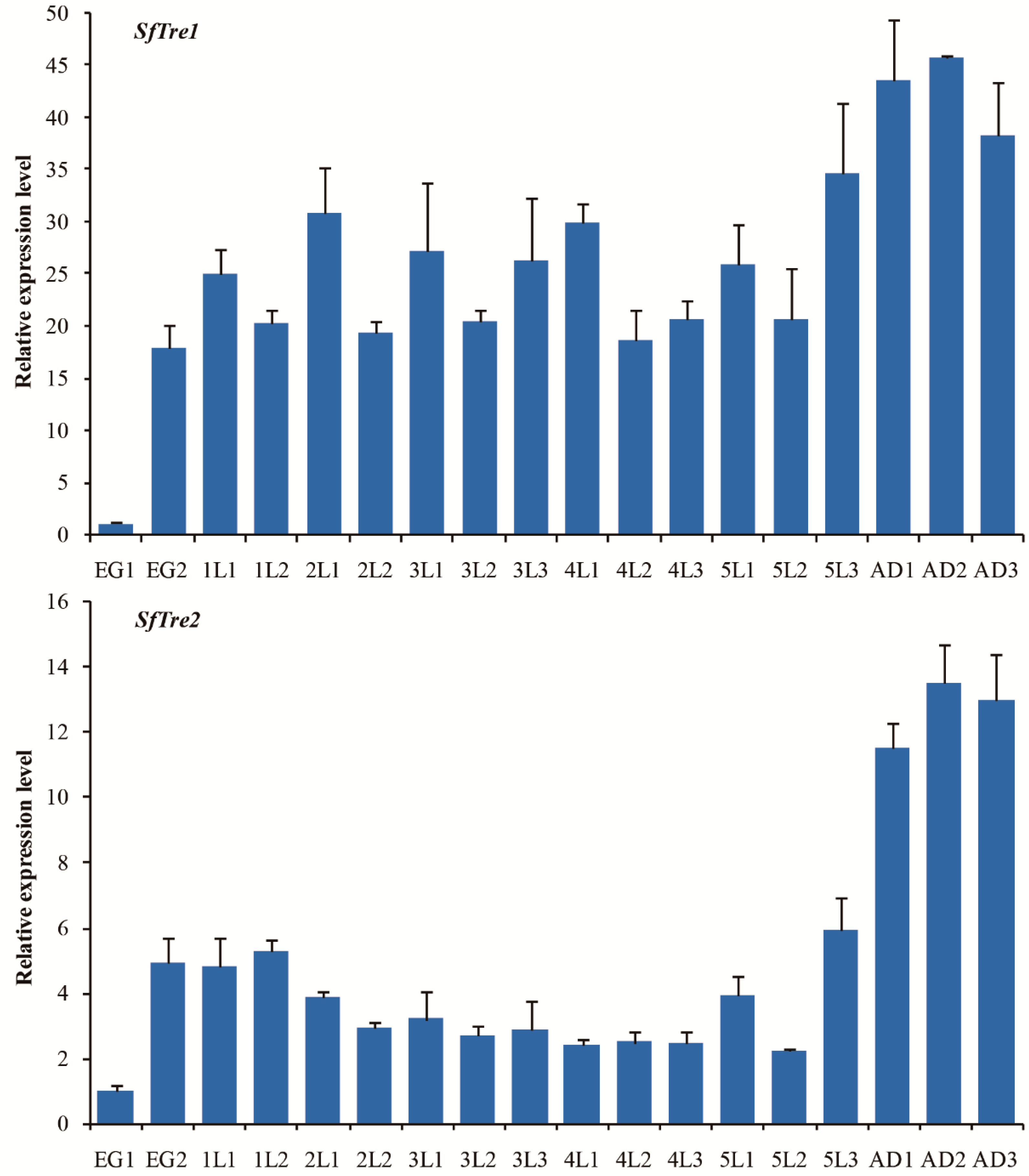
3.2. Spatiotemporal Expression of SfTre1 and SfTre2

The expression profiles of both SfTres at different developmental stages of S. furcifera, including egg, 1–5 instar nymph, and adult, were analyzed; the results are shown in Figure 4. SfTres were continuously expressed at different developmental stages. In detail, the SfTre1 expression level was relatively high after each molting, declined during the interval of the molting phase, and increased before the next molting. However, following eclosion, its expression level significantly increased and reached the maximum in day two adults. Compared with SfTre1, the expression level of SfTre2 initially decreased and then increased. In particular, its expression level sharply increased in S. furcifera adults.
The mRNA expression levels of SfTre1 and SfTre2 in five different tissues of S. furcifera were evaluated by qRT-PCR. SfTre1 was primarily expressed in the integument and ovary, followed by the head, and the lowest expression was observed in the fat body and gut (Figure 5). SfTre2 showed the highest expression in the ovary, followed by the head, epidermis, and fat body, and it was the lowest in the gut (Figure 5).

3.3. Effects of RNAi

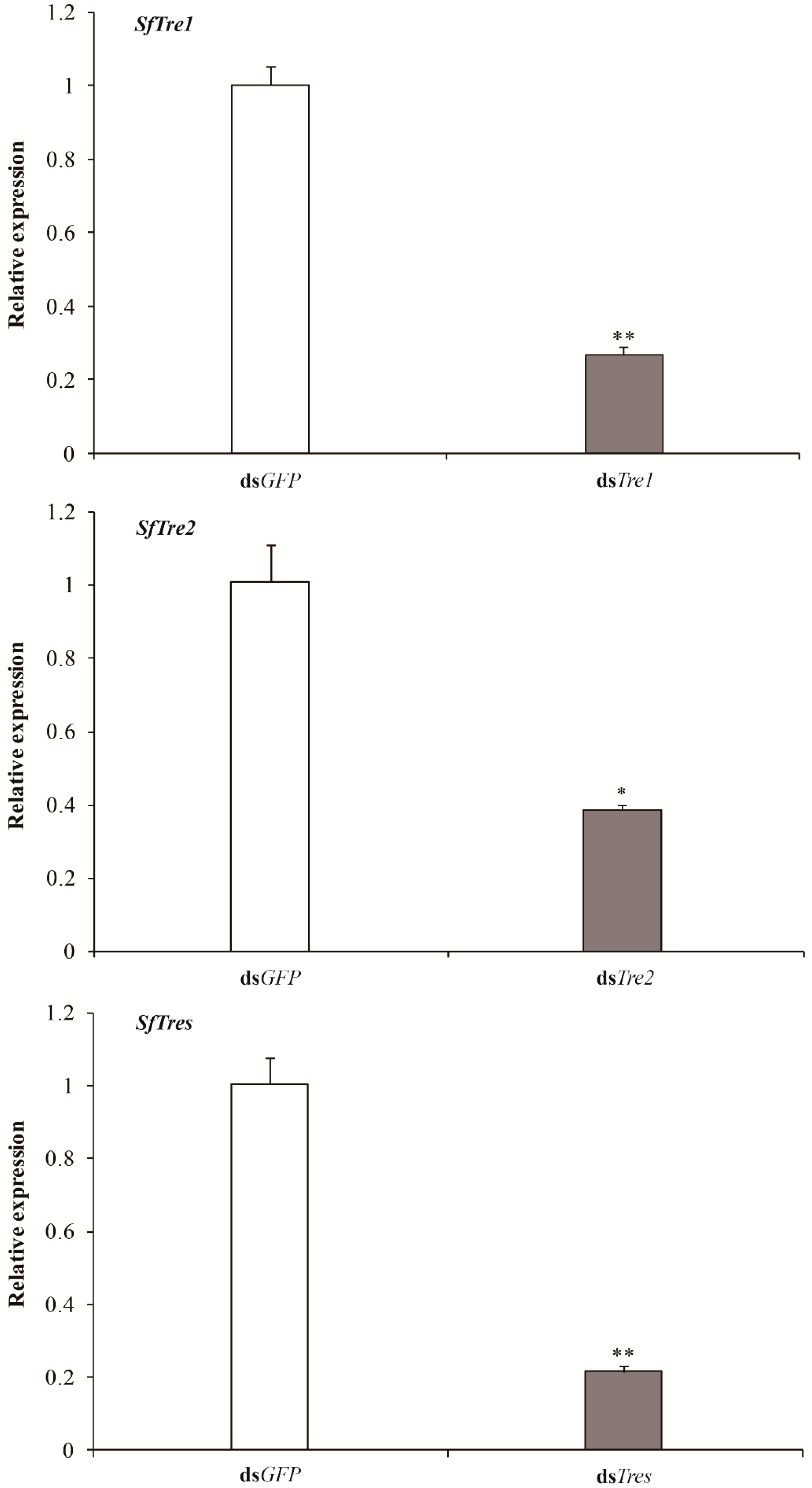
3.3.1. Effects of RNAi on the expression levels of SfTre1 and SfTre2

In RNAi, dsRNAs synthesized in vitro were injected into day one fifth-instar nymphs to verify the functional effect of SfTre1 and SfTre2. The expression levels of the target genes in S. furcifera at 72 h after injection were detected using qRT-PCR. The analysis indicated that all transcripts of the two Tre genes were significantly inhibited. In particular, the mRNA expression level of the dsTre1, dsTre2, and dsTres treatment groups was significantly lower than that of the dsGFP control group, with a reduction of 73%, 62%, and 79%, respectively (Figure 6).

3.3.2. Effects of RNAi on the Survival Rates

Following the successful injection of dsRNAs of the two SfTre genes, the survival of the insects subjected to RNAi was continuously recorded (Figure 7). Compared with the control group, the survival rate of the insects in the treatment group began to decrease significantly on day three after injection and continued to decrease with time. On day five, the survival rate of the insects injected with dsTre1 was 58%, whereas that of insects injected with dsTre2 was 62%. In addition, following the injection of a two gene dsRNA mixture, the survival rate of insects was 48%, which was lower than that following the injection of a single gene dsRNA. Therefore, the interference effect of mixed genes was superior to that of single gene.

3.3.3. Effects of RNAi on Phenotypes

Following the successful injection of dsRNAs of the two SfTre genes, the insects in the nymph and adult stages exhibited various abnormal phenotypes (Figure 8). Four malformed phenotypes were observed in the nymph stage: the cuticle of the injected nymphs was not well hardened and looked transparent (I); the body size of the injected nymphs was significantly smaller than that of the normal ones (II); the old cuticle on the head and thorax slightly split open (III); the injected nymphs partially shed their old cuticle, but the insect body remained encased (IV). All II, III, and IV nymphs with the deformed phenotypes died (Figure 8). Furthermore, during the adult stage, four malformations were observed: the injected nymphs could exuviate the old cuticle to become adults, but the wings were abnormal (V); the body size of the injected adults decreased (VI); injected adults emerged successfully, but they could not extricate their old appendages (VII); the wings were malformed (VIII, Figure 8). These results indicated that the knockdown of both SfTres seriously threatened insect normal growth and development, thereby causing the death of S. furcifera.

4. Discussion

S. furcifera is a serious pest affecting the rice industry in some Asia-Pacific countries. The application of chemical pesticides has extensively been the primary way to control S. furcifera [3,9]. However, the long-term use of insecticides will result in pest resistance, kill natural enemy insects, and pollute the environment [37,38]. Therefore, there is an urgent need to develop green control strategies for S. furcifera management.
Chitin is a major component of the cuticular exoskeleton and peritrophic membrane tissues, and it plays an essential role in insect development and metamorphosis. Insect chitin biosynthesis is a complex, dynamic process regulated by several enzymes [12,39]. Previous studies have suggested that the inhibition of several enzymes in insects could result in molting defects and high mortality [40,41,42,43]. Tre is a pivotal enzyme involved in the hydrolysis of trehalose in almost all tissues in distinct forms. In addition, Tre is commonly found in several organisms, including bacteria, algae, yeast, fungi, plants, nematodes, and insects, but not in mammals [44]. Therefore, Tre is a promising target for pest control. In the present study, we cloned and identified two Tre genes from S. furciferaSfTre1 and SfTre2. Phylogenetic analysis showed that the two Tre genes from S. furcifera shared the highest homology to those from L. striatellus. Furthermore, structural domain analyses demonstrated that these two genes shared some characteristic sequences with little difference, including two conserved signature motifs and a highly conserved glycine-rich region (GGGGEY); these characteristic sequences are unique to Tre and do not exist in other proteins [45,46]. However, we found that SfTre2 has a signal peptide of 29 amino acids at the N-terminus and a transmembrane region at the C-terminus, which is absent from SfTre1. Different results have been observed in Laodelphax striatellus, Spodoptera litura, and Apolygus lucorum, where the Tre1 also contains a signal peptide [30,47,48]. Previously, Tre1 has been purified from the egg homogenates, hemolymph, and goblet cell cavity in the midgut of some insects, whereas Tre2 has been found in the ovarian cells, follicular cells, spermatophore, flight muscles, brain, midgut, and thoracic ganglia [17,20,49,50,51]. Based on these results, we infer that the two Tre genes from S. furcifera could serve different functions.
Based on the expression levels of SfTre1 and SfTre2 genes at different developmental stages, the transcription level of SfTre1 was found to repeat periodically in each molting cycle. In particular, SfTre1 showed the highest expression level after molting, which decreased during the interval of each molting and increased before the next molting. The expression patterns of SfTre1 are quite similar to those of SfCHS1 and SfUAP in S. furcifera [34,35]. Furthermore, the expression levels of SfTre1 and SfTre2 were enriched in the egg and adult stages of S. furcifera, suggesting that their expression was associated with energy demand and chitin biosynthesis during highly metamorphic development. Previous studies have confirmed that the expression of insect Tre genes possesses tissue specific. H. armigera Tres are ubiquitous in detected tissues. In particular, HaTre1 shows high expression levels in the midgut, integument, and head, whereas HaTre2 shows high levels in the head [52]. In A. lucorum, the highest expression level of Tre-1 is observed in the ovary and malpighian tubules, whereas that of Tre-2 is observed in the flight muscle, fat body, and brain [48]. Moreover, Tres are widely distributed in various tissues of G. pyloalis, and GpTre1 and GpTre2 demonstrate the highest transcription levels in the head [32]. Meanwhile, in Diaphorina citri, the highest expression level of Tre1-1 was noted in the head and integument, and that of Tre1-2 was noted in the wing; moreover, the highest expression level of DcTre2 was observed in the integument and fat body [53]. These results indicate that insect Tres play different roles in varying tissues. Our tissue transcription pattern analysis indicated that SfTre1 and SfTre2 showed the highest expression levels in the ovary. Insect ovaries are critical sites for egg cell formation. The high expression levels of SfTre1 and SfTre2 observed in the ovary may be attributed to high energy demand during egg cell development. Moreover, the abundant expression of SfTre1 in the integument indicates that it may supply the nutritional sugar required in chitin biosynthesis. This result is consistent with previous studies on other insects, such as Spodoptera exigua and Spodoptera litura [44,47]. In the present study, SfTre1 and SfTre2 also exhibited high transcription levels in the head. The insect head is the anterior sensory and feeding center [54].
RNAi can induce targeted gene silencing, and it has successfully been used for evaluating functional genes in various insects [55,56,57,58,59,60]. Previous studies have shown that in S. exigua, the knockdown of SeTre1 and SeTre2 increases the mortality and abnormality rates and significantly decreases CHS expression levels and chitin content [44]. In N. lugens, dsNlTres injection represses the expression levels of genes related to the chitin metabolism pathway, including hexokinase (HK), glucose-6-phosphate isomerase (G6PI), chitinase, and CHS, thereby causing molting deformities and resulting in high mortality [61]. Moreover, the silencing of five Tre genes in Tribolium castaneum results in a decrease in the transcription levels of genes involved in the chitin biosynthesis pathway, including HK, G6PI, CHS, TPS, glycogen phosphorylase, glycogen synthase, phosphofructokinase, and fructose-6-phosphate transaminase and an increase in the lethality and malformation rates [62]. These results indicate that Tre plays a vital role during insect metamorphic development and survival. In our study, RNAi was used to investigate the biological function of SfTre1 and SfTre2 during the growth and development of S. furcifera. When fifth-instar nymphs on day one were injected with dsTre1 and dsTre2, qPCR results showed the expression levels of SfTre1 and SfTre2 were significantly hindered. The RNAi-mediated downregulation of SfTre1 and SfTre2 transcription ultimately resulted in various malformed phenotypes and high mortality. Our results were in accordance with previous data obtained in G. pyloalis and A. pisum [32,63]. We infer that the inhibition of Tres may prevent the hydrolysis of trehalose from generating glucose, thereby affecting chitin formation. In summary, our preliminary results may lay a foundation for further exploring the regulatory mechanism of S. furcifera Tre and provide a potential target for planthopper control.

5. Conclusions

In the present study, we successfully identified and characterized two SfTre genes from S. furcifera. Silencing of SfTre1 and SfTre2 via RNAi severely impeded the normal development of the insect. In particular, the knockdown of SfTres (SfTre1 and SfTre2) gene expression by RNAi resulted in a mortality rate of >50%, which indicated the crucial roles played by these genes in the development of S. furcifera. Our findings could provide potential target genes for RNAi-based S. furcifera control.

Author Contributions

Methodology, D.-C.J., Z.W. and G.-Y.L.; resources, D.-C.J. and H.Y.; software, C.Z. and X.-B.Y.; funding acquisition, D.-C.J. and Z.W.; investigation, Z.W., G.-Y.L. and C.Z.; data curation, D.-C.J. and Z.W.; writing–original draft, Z.W. and G.-Y.L. All authors have read and agreed to the published version of the manuscript.

Funding

This research was funded by the Science and Technology Foundation of Guizhou Province (grant numbers 20201Y078 and 201901-2), the Natural Science Research Project of the Education Department of Guizhou Province (grant number KY2020190), the Science and Technology Foundation of Qiandongnan (grant number J2019108), Innovative Talent Team Program from Education Department of Guizhou Province (grant number 201326), and the International Cooperation Base for Insect Evolutionary Biology and Pest Control (grant number 20165802).

Institutional Review Board Statement

Not applicable.

Informed Consent Statement

Not applicable.

Data Availability Statement

The data were deposited in GenBank with accession numbers MG869613 and MG869614.

Conflicts of Interest

The authors declare no conflict of interest.

References

  1. Zhai, B.P. Rice planthoppers: A China problem under the international perspectives. Chin. J. Appl. Entomol. 2011, 48, 1184–1193. [Google Scholar]
  2. Zhu, Z.R.; Cheng, J.A. Sucking rates of the white-backed planthopper Sogatella furcifera (Horv.) (Homoptera: Delphacidae) and yield loss of rice. J. Pest Sci. 2002, 75, 113–117. [Google Scholar] [CrossRef]
  3. Wang, Z.; Zhou, C.; Long, G.Y.; Yang, H.; Jin, D.C. Sublethal effects of buprofezin on development, reproduction, and chitin synthase 1 gene (SfCHS1) expression in the white-backed planthopper, Sogatella furcifera (Hemiptera: Delphacidae). J. Asia-Pac. Entomol. 2018, 21, 585–591. [Google Scholar] [CrossRef]
  4. Huang, S.H.; Cheng, C.H.; Chen, C.N.; Wu, W.J.; Otuka, A. Estimating the immigration source of rice planthoppers, Nilaparvata lugens (Stål) and Sogatella furcifera (Horváth) (Homoptera: Delphacidae), in Taiwan. Appl. Entomol. Zool. 2010, 45, 521–531. [Google Scholar] [CrossRef] [Green Version]
  5. Zheng, D.B.; Hu, G.; Yang, F.; Du, X.D.; Yang, H.B.; Zhang, G.; Qi, G.J.; Liang, Z.L.; Zhang, X.X.; Cheng, X.N.; et al. Ovarian development status and population characteristics of Sogatella furcifera (Horváth) and Nilaparvata lugens (Stål): Implications for pest forecasting. J. Appl. Entomol. 2014, 138, 67–77. [Google Scholar] [CrossRef]
  6. Ali, E.; Liao, X.; Yang, P.; Mao, K.K.; Zhang, X.L.; Shakeel, M.; Salim, A.M.A.; Wan, H.; Li, J.H. Sublethal effects of buprofezin on development and reproduction in the white-backed planthopper, Sogatella furcifera (Hemiptera: Delphacidae). Sci. Rep. 2017, 7, 16913. [Google Scholar] [CrossRef] [Green Version]
  7. Zhou, G.H.; Wen, J.J.; Cai, D.J.; Li, P.; Xu, D.L.; Zhang, S.G. Southern rice black-streaked dwarf virus: A new proposed Fijivirus species in the family Reoviridae. Chin. Sci. Bull. 2008, 53, 3677–3685. [Google Scholar] [CrossRef]
  8. Matsukura, K.; Towata, T.; Yoshida, K.; Sakai, J.; Okuda, M.; Onuki, M.; Matsumura, M. Quantitative analysis of southern rice black-streaked dwarf virus in Sogatella furcifera and virus threshold for transmission. Phytopathology 2015, 105, 550–554. [Google Scholar] [CrossRef] [Green Version]
  9. Mao, K.K.; Ren, Z.J.; Li, W.B.; Liu, C.Y.; Xu, P.F.; He, S.; Li, J.H.; Wan, H. An insecticide resistance diagnostic kit for whitebacked planthopper Sogatella furcifera (Horváth). J. Pest Sci. 2021, 94, 531–540. [Google Scholar] [CrossRef]
  10. Abudulai, M.; Shepard, B.M.; Mitchell, P.L. Parasitism and predation on eggs of Leptoglossus phyllopus (Hemiptera: Coreidae) in cowpea: Impact of endosulfan sprays. Agric. Urban. Entomol. 2001, 18, 105–115. [Google Scholar]
  11. Jin, J.X.; Jin, D.C.; Li, W.H.; Cheng, Y.; Li, F.L.; Ye, Z.C. Monitoring trends in insecticide resistance of field populations of Sogatella furcifera (Hemiptera: Delphacidae) in Guizhou Province, China, 2012–2015. J. Econ. Entomol. 2017, 110, 641–650. [Google Scholar] [CrossRef] [PubMed]
  12. Cohen, E. Chitin synthesis and inhibition: A revisit. Pest Manag. Sci. 2001, 57, 946–950. [Google Scholar] [CrossRef] [PubMed]
  13. Elbein, A.D.; Pan, Y.T.; Pastuszak, I.; Carroll, D. New insights on trehalose: A multifunctional molecule. Glycobiology 2003, 13, 17R–27R. [Google Scholar] [CrossRef] [PubMed]
  14. Shukla, E.; Thorat, L.J.; Nath, B.B.; Gaikwad, S.M. Insect trehalase: Physiological significance and potential applications. Glycobiology 2015, 25, 357–367. [Google Scholar] [CrossRef] [PubMed] [Green Version]
  15. Pan, B.Y.; Xu, K.K.; Luo, Y.J.; Wang, Y.Y.; Zhou, M.; Li, C.; Tang, B. The sequence characteristics and functions on regulating trehalose metabolism of two PTP genes in Tribolium castaneum (Herbst) (Coleoptera: Tenebrionidae). J. Stored Prod. Res. 2020, 89, 101692. [Google Scholar] [CrossRef]
  16. Huang, Q.T.; Zhang, G.J.; Nan, J.L.; Cheng, W.N.; Zhu-Salzman, K. Characterization of trehalose metabolic genes and corresponding enzymatic activities during diapause of Sitodiplosis mosellana. J. Insect Physiol. 2021, 135, 104324. [Google Scholar] [CrossRef]
  17. Wegener, G.; Tschiedel, V.; Schlöder, P.; Ando, O. The toxic and lethal effects of the trehalase inhibitor trehazolin in locusts are caused by hypoglycaemia. J. Exp. Biol. 2003, 206, 1233–1240. [Google Scholar] [CrossRef] [Green Version]
  18. Tatun, N.; Singtripop, T.; Sakurai, S. Dual control of midgut trehalase activity by 20-hydroxyecdysone and an inhibitory factor in the bamboo borer Omphisa fuscidentalis Hampson. J. Insect Physiol. 2008, 54, 351–357. [Google Scholar] [CrossRef]
  19. Ujita, M.; Yamanaka, M.; Maeno, Y.; Yoshida, K.; Ohshio, W.; Ueno, Y.; Banno, Y.; Fujii, H.; Okumura, H. Expression of active and inactive recombinant soluble trehalase using baculovirus-silkworm expression system and their glycan structures. J. Biosci. Bioeng. 2011, 111, 22–25. [Google Scholar] [CrossRef]
  20. Takiguchi, M.; Niimi, T.; Su, Z.H.; Yaginuma, T. Trehalase from male accessory gland of an insect, Tenebrio molitor. cDNA sequencing and developmental profile of the gene expression. Biochem. J. 1992, 288, 19–22. [Google Scholar] [CrossRef] [Green Version]
  21. Parkinson, N.M.; Conyers, C.M.; Keen, J.N.; Macnicoll, A.D.; Smith, I.; Weaver, R.J. cDNAs encoding large venom proteins from the parasitoid wasp Pimpla hypochondriaca identified by random sequence analysis. Comp. Biochem. Phys. C 2003, 134, 513–520. [Google Scholar] [CrossRef]
  22. Lee, J.H.; Saito, S.; Mori, H.; Nishimoto, M.; Okuyama, M.; Kim, D.; Wongchawalit, J.; Kimura, A.; Chiba, S. Molecular cloning of cDNA for trehalase from the European honeybee, Apis mellifera L. and its heterologous expression in Pichia pastoris. Biosci. Biotechnol. Biochem. 2007, 71, 2256–2265. [Google Scholar] [CrossRef] [PubMed]
  23. Mitsumasu, K.; Azuma, M.; Niimi, T.; Yamashita, O.; Yaginuma, T. Membrane-penetrating trehalase from silkworm Bombyx mori. Molecular cloning and localization in larval midgut. Insect Mol. Biol. 2005, 14, 501–508. [Google Scholar] [CrossRef] [PubMed]
  24. Gu, J.H.; Shao, Y.; Zhang, C.W.; Liu, Z.W.; Zhang, Y.J. Characterization of putative soluble and membrane-bound trehalases in a hemipteran insect, Nilaparvata lugens. J. Insect Physiol. 2009, 55, 997–1002. [Google Scholar] [CrossRef]
  25. Tang, B.; Qin, Z.; Shi, Z.K.; Wang, S.; Guo, X.J.; Wang, S.G.; Zhang, F. Trehalase in Harmonia axyridis (Coleoptera: Coccinellidae): Effects on beetle locomotory activity and the correlation with trehalose metabolism under starvation conditions. Appl. Entomol. Zool. 2014, 49, 255–264. [Google Scholar] [CrossRef]
  26. Tan, Y.A.; Xiao, L.B.; Zhao, J.; Xiao, Y.F.; Sun, Y.; Bai, L.X. Ecdysone receptor isoform-B mediates soluble trehalase expression to regulate growth and development in the mirid bug, Apolygus lucorum (Meyer-Dür). Insect Mol. Biol. 2015, 24, 611–623. [Google Scholar] [CrossRef]
  27. Shukla, E.; Thorat, L.J.; Bendre, A.D.; Jadhav, S.; Pal, J.K.; Nath, B.B.; Gaikwad, S.M. Cloning and characterization of trehalase: A conserved glycosidase from oriental midge, Chironomus ramosus. 3 Biotech 2018, 8, 352. [Google Scholar] [CrossRef]
  28. Tevatiya, S.; Kumari, S.; Sharma, P.; Rani, J.; Chauhan, C.; De, T.D.; Pandey, K.C.; Pande, V.; Dixit, R. Molecular and functional characterization of trehalase in the mosquito Anopheles stephensi. Front. Physiol. 2020, 11, 575718. [Google Scholar] [CrossRef]
  29. Santos, R.; Alves-Bezerra, M.; Rosas-Oliveira, R.; Majerowicz, D.; Meyer-Fernandes, J.R.; Gondim, K.C. Gene identification and enzymatic properties of a membrane-bound trehalase from the ovary of Rhodnius prolixus. Arch. Insect Biochem. 2012, 81, 199–213. [Google Scholar] [CrossRef]
  30. Zhang, Q.; Lu, D.H.; Pu, J.; Wu, M.; Han, Z.J. Cloning and RNA interference effects of trehalase genes in Laodelphax striatellus (Homoptera: Delphacidae). Acta Entomol. Sin. 2012, 55, 911–920. [Google Scholar]
  31. Li, Y.; Chen, X.; Wang, S.S.; Pan, B.Y.; Wang, S.G.; Wang, S.; Tang, B. Evaluation of the expression and function of the TRE2-like and TRE2 genes in ecdysis of Harmonia axyridis. Front. Physiol. 2019, 10, 1371. [Google Scholar] [CrossRef] [PubMed]
  32. Shao, Z.M.; Ding, J.H.; Jiang, D.L.; Liu, Z.X.; Li, Y.J.C.; Wang, J.; Wang, J.; Sheng, S.; Wu, F.A. Characterization and functional analysis of trehalase related to chitin metabolism in Glyphodes pyloalis Walker (Lepidoptera: Pyralidae). Insects 2021, 12, 370. [Google Scholar] [CrossRef] [PubMed]
  33. Liu, X.J.; Sun, Y.W.; Cui, M.; Ma, E.B.; Zhang, J.Z. Molecular characteristics and functional analysis of trehalase genes in Locusta migratoria. Sci. Agric. Sin. 2016, 49, 4375–4386. [Google Scholar]
  34. Wang, Z.; Yang, H.; Zhou, C.; Yang, W.J.; Jin, D.C.; Long, G.Y. Molecular cloning, expression, and functional analysis of the chitin synthase 1 gene and its two alternative splicing variants in the white-backed planthopper, Sogatella furcifera (Hemiptera: Delphacidae). Sci. Rep. 2019, 9, 1087. [Google Scholar] [CrossRef] [PubMed]
  35. Wang, Z.; Long, G.Y.; Zhou, C.; Jin, D.C.; Yang, H.; Yang, W.J. Molecular characterization of UDP-N-acetylglucosamine pyrophosphorylase and its role in the growth and development of the white-backed planthopper Sogatella furcifera (Hemiptera: Delphacidae). Genes 2022, 13, 1340. [Google Scholar] [CrossRef] [PubMed]
  36. Livak, K.J.; Schmittgen, T.D. Analysis of relative gene expression data using realtime quantitative PCR and the 2−ΔΔCt method. Methods 2001, 25, 402–408. [Google Scholar] [CrossRef]
  37. Yu, H.Z.; Wen, D.F.; Wang, W.L.; Geng, L.; Zhang, Y.; Xu, J.P. Identification of genes putatively involved in chitin metabolism and insecticide detoxification in the rice leaf folder (Cnaphalocrocis medinalis) larvae through transcriptomic analysis. Int. J. Mol. Sci. 2015, 16, 21873–21896. [Google Scholar] [CrossRef]
  38. Matsumura, M.; Sanada-Morimura, S.; Otuka, A.; Sonoda, S.; Van Thanh, D.; Van Chien, H.; Van Tuong, P.; Loc, P.M.; Liu, Z.W.; Zhu, Z.R.; et al. Insecticide susceptibilities of the two rice planthoppers Nilaparvata lugens and Sogatella furcifera in East Asia, the Red River Delta, and the Mekong Delta. Pest Manag. Sci. 2018, 74, 456–464. [Google Scholar] [CrossRef]
  39. Merzendorfer, H.; Zimoch, L. Chitin metabolism in insects: Structure, function and regulation of chitin synthases and chitinases. J. Exp. Biol. 2003, 206, 4393–4412. [Google Scholar] [CrossRef] [Green Version]
  40. Ullah, F.; Gul, H.; Wang, X.; Ding, Q.; Said, F.; Gao, X.W.; Desneux, N.; Song, D.L. RNAi-mediated knockdown of chitin synthase 1 (CHS1) gene causes mortality and decreased longevity and fecundity in Aphis gossypii. Insects 2020, 11, 22. [Google Scholar] [CrossRef] [Green Version]
  41. Xu, C.D.; Liu, Y.K.; Qiu, L.Y.; Wang, S.S.; Pan, B.Y.; Li, Y.; Wang, S.G.; Tang, B. GFAT and PFK genes show contrasting regulation of chitin metabolism in Nilaparvata lugens. Sci. Rep. 2021, 11, 5246. [Google Scholar] [CrossRef] [PubMed]
  42. Jiang, L.H.; Mu, L.L.; Jin, L.; Anjum, A.A.; Li, G.Q. Silencing uridine diphosphate N-acetylglucosamine pyrophosphorylase gene impairs larval development in Henosepilachna vigintioctopunctata. Pest Manag. Sci. 2022, 78, 3894–3902. [Google Scholar] [CrossRef] [PubMed]
  43. Yang, S.; Zou, Z.W.; Xin, T.R.; Cai, S.Y.; Wang, X.; Zhang, H.J.; Zhong, L.; Xia, B. Knockdown of hexokinase in Diaphorina citri Kuwayama (Hemiptera: Liviidae) by RNAi inhibits chitin synthesis and leads to abnormal phenobypes. Pest Manag. Sci. 2022, 78, 4303–4313. [Google Scholar] [CrossRef] [PubMed]
  44. Chen, J.; Tang, B.; Chen, H.X.; Yao, Q.; Huang, X.F.; Chen, J.; Zhang, D.W.; Zhang, W.Q. Different functions of the insect soluble and membrane-bound trehalase genes in chitin biosynthesis revealed by RNA interference. PLoS ONE 2010, 5, e10133. [Google Scholar] [CrossRef]
  45. Tang, B.; Chen, X.F.; Liu, Y.; Tian, H.G.; Liu, J.; Hu, J.; Xu, W.H.; Zhang, W.Q. Characterization and expression patterns of a membrane-bound trehalase from Spodoptera exigua. BMC Mol. Biol. 2008, 9, 51. [Google Scholar] [CrossRef] [Green Version]
  46. Tatun, N.; Singtripop, T.; Tungjitwitayakul, J.; Sakurai, S. Regulation of soluble and membrane-bound trehalase activity and expression of the enzyme in the larval midgut of the bamboo borer Omphisa fuscidentalis. Insect Biochem. Mol. 2008, 38, 788–795. [Google Scholar] [CrossRef]
  47. Zou, Q.; Wei, P.; Xu, Q.; Zheng, H.Z.; Tang, B.; Wang, S.G. cDNA cloning and characterization of two trehalases from Spodoptera litura (Lepidoptera: Noctuidade). Genet. Mol. Res. 2013, 12, 901–915. [Google Scholar] [CrossRef]
  48. Tan, Y.A.; Xiao, L.B.; Sun, Y.; Zhao, J.; Bai, L.X.; Xiao, Y.F. Molecular characterization of soluble and membrane-bound trehalases in the cotton mirid bug, Apolygus lucorum. Insect Biochem. Physiol. 2014, 86, 107–121. [Google Scholar] [CrossRef]
  49. Huber, R.E.; Lefebvre, Y.A. The Purification and some properties of soluble trehalase and sucrase from Drosophila melanogaster. Can. J. Biochem. 1971, 49, 1155–1164. [Google Scholar] [CrossRef]
  50. Huang, J.; Furusawa, T.; Sadakane, K.; Sugimura, Y. Purification and properties of two types of soluble trehalases from embryonic larvae of the silkworm, Bombyx mori. J. Insect Biotechnol. Sericol. 2006, 75, 1–8. [Google Scholar]
  51. Silva, M.C.P.; Ribeiro, A.F.; Terra, W.R.; Ferreira, C. Sequencing of Spodoptera frugiperda midgut trehalases and demonstration of secretion of soluble trehalase by midgut columnar cells. Insect Mol. Biol. 2009, 18, 769–784. [Google Scholar] [CrossRef] [PubMed]
  52. Ma, L.; Dai, W.; Li, X.C.; Zhang, Y.L.; Zhang, C.N. Molecular cloning and expression analysis of soluble and membrane-bound trehalase genes in the cotton bollworm, Helicoverpa armigera. J. Asia-Pac. Entomol. 2015, 18, 187–195. [Google Scholar] [CrossRef]
  53. Yu, H.Z.; Huang, Y.L.; Lu, Z.J.; Zhang, Q.; Su, H.N.; Du, Y.M.; Yi, L.; Zhong, B.L.; Chen, C.X. Inhibition of trehalase affects the trehalose and chitin metabolism pathways in Diaphorina citri (Hemiptera: Psyllidae). Insect Sci. 2021, 28, 718–734. [Google Scholar] [CrossRef] [PubMed]
  54. Posnien, N.; Schinko, J.B.; Kittelmann, S.; Bucher, G. Genetics, development and composition of the insect head-a beetle’s view. Arthropod Struct. Dev. 2010, 39, 399–410. [Google Scholar] [CrossRef]
  55. Dzitoyeva, S.; Dimitrijevic, N.; Manev, H. Intra-abdominal injection of double-stranded RNA into anesthetized adult Drosophila triggers RNA interference in the central nervous system. Mol. Psychiatr. 2001, 6, 665–670. [Google Scholar] [CrossRef] [PubMed]
  56. Tomoyasu, Y.; Miller, S.C.; Tomita, S.; Schoppmeier, M.; Grossmann, D.; Bucher, G. Exploring systemic RNA interference in insects: A genome-wide survey for RNAi genes in Tribolium. Genome Biol. 2008, 9, R10. [Google Scholar] [CrossRef] [Green Version]
  57. Walshe, D.P.; Lehane, S.M.; Lehane, M.J.; Haines, L.R. Prolonged gene knockdown in the tsetse fly Glossina by feeding double stranded RNA. Insect Mol. Biol. 2009, 18, 11–19. [Google Scholar] [CrossRef]
  58. Upadhyay, S.K.; Chandrashekar, K.; Thakur, N.; Verma, P.C.; Borgio, J.F.; Singh, P.K.; Tuli, R. RNA interference for the control of whiteflies (Bemisia tabaci) by oral route. J. Biosci. 2011, 36, 153–161. [Google Scholar] [CrossRef]
  59. Bhatia, V.; Bhattacharya, R. Host-mediated RNA interference targeting a cuticular protein gene impaired fecundity in the green peach aphid Myzus persicae. Pest Manag. Sci. 2018, 74, 2059–2068. [Google Scholar] [CrossRef]
  60. Wang, Q.T.; Fang, K.; Qi, L.Z.; Wang, X.; Pan, Y.; Li, Y.S.; Xi, J.H.; Zhang, J.H. Purification and functional characterization of a soluble trehalase in Lissorhoptrus oryzophilus (Coleoptera: Curculionidae). Insects 2022, 13, 867. [Google Scholar] [CrossRef]
  61. Zhao, L.N.; Yang, M.M.; Shen, Q.D.; Liu, X.J.; Shi, Z.K.; Wang, S.G.; Tang, B. Functional characterization of three trehalase genes regulating the chitin metabolism pathway in rice brown planthopper using RNA interference. Sci. Rep. 2016, 6, 27841. [Google Scholar] [CrossRef] [PubMed] [Green Version]
  62. Tang, B.; Wei, P.; Zhao, L.N.; Shi, Z.K.; Shen, Q.D.; Yang, M.M.; Xie, G.Q.; Wang, S.G. Knockdown of five trehalase genes using RNA interference regulates the gene expression of the chitin biosynthesis pathway in Tribolium castaneum. BMC Biotechnol. 2016, 16, 67. [Google Scholar] [CrossRef] [PubMed] [Green Version]
  63. Wang, G.; Gou, Y.P.; Guo, S.F.; Zhou, J.J.; Liu, C.Z. RAN interference of trehalose-6-phosphate synthase and trehalase genes regulates chitin metabolism in two color morphs of Acyrthosiphon pisum Harris. Sci. Rep. 2021, 11, 948. [Google Scholar] [CrossRef] [PubMed]
Figure 1. Multiple sequence alignment of insect trehalases was performed using DNAMAN 7. Identical amino acid residues are indicated by the letters below the sequence. The signal peptides are underlined in red, and the transmembrane regions are underlined. The trehalase signature motifs are shadowed in green, and the glycine-rich regions are shadowed in yellow. The N-glycosylation sites are in red. Abbreviations: NlTre1 and NlTre2 from Nilaparvata lugens (ACN85420 and ACV20872), LsTre1 from Laodelphax striatellus (AFL03409), BtTre1 and BtTre2 from Bemisia tabaci (AFV79626 and AFV79627), SfrTre1 and SfrTre2 from Spodoptera frugiperda (ABE27189 and ACF94698), SlTre2 from Spodoptera litura (ADA63845).
Biomolecules 12 01699 g001
Figure 2. Three-dimensional model predictions generated by SWISS-MODEL homology modeling were performed using the crystal structure of periplasmic trehalase (PDB ID: 2wyn.1.A) as the template. (A) SfTre1 model; (B), SfTre2 model. α-helices are indicated in red. β-pleated sheets are indicated in yellow. Random coils are indicated in green. The signature motifs are indicated in blue. The glycine-rich region is indicated in purple.
Biomolecules 12 01699 g002
Figure 3. Phylogenetic tree of Tres in insects and other organisms. Organisms with the associated GenBank accession numbers are as follows: Acyrthosiphon pisum (Ap), Aphis glycines (Ag), Bemisia tabaci (Bt), Laodelphax striatellus (Ls), Nilaparvata lugens (Nl), Bombyx mori (Bm), Helicoverpa armigera (Ha), Omphisa fuscidentalis (Of), Spodoptera exigua (Se), Spodoptera frugiperda (Sfr), Spodoptera litura (Sl), Apis mellifera (Am), Bombus terrestris (Bte), Pimpla hypochondriaca (Ph), Harmonia axyridis (Hax), Tenebrio molitor (Tm), Tribolium castaneum (Tc), Aedes aegypti (Aa), Anopheles gambiae (Aga), Culex quinquefasciatus (Cq), Drosophila melanogaster (Dm), Drosophila simulans (Ds), Locusta migratoria manilensis (Lm), Homo sapiens (Hs), Mus musculus (Mm), Pongo abelii (Pa), and Rattus norvegicus (Rn). GenBank IDs are as follows: ApTre2 (XP_001949459), AgTre1 (AFJ00065), BtTre1 (AFV79626), BtTre2 (AFV79627), LsTre1 (AFL03409), LsTre2 (AFL03410), NlTre1 (ACN85420), NlTre2 (ACV20872), BmTre1 (BAA13042), BmTre2 (BAE45249), HaTre1 (AJK29979), OfTre1 (ABO20846), OfTre2 (ABO20845), SeTre1 (ABY86218), SeTre2 (ABU95354), SfrTre1 (ABE27189), SfrTre2 (ACF94698), SlTre1 (ADA63846), SlTre2 (ADA63845), AmTre1 (XP_393963), AmTre2 (NP_00110614), BteTre1 (XP_003400853), BteTre2 (XP_003393687), PhTre1 (CAD31109), HaxTre1 (ADH94051), TmTre1 (BAA01951), TcTre1 (XP_973919), TcTre1 (XP_972610), AaTre2 (EAT38444), AgaTre2 (XP_320471), CqTre2 (XP_001847934), DmTre2 (ABH06695), DsTre2 (ABH06710), LmTre1 (ACP28173), HsTre (NP_009111), MmTre (NP_067456), PaTre (XP_002822604), and RnTre (NP_001129613).
Biomolecules 12 01699 g003
Figure 4. Expression profiles of SfTre1 and SfTre2 at different developmental stages of S. furcifera. Sf18S was used as an internal reference gene. Relative expression was determined based on the value of the lowest expression level, which was arbitrarily set to 1. Data are represented as means ± standard error of three biological replicates. The age in days of the insects is shown; for example, EG1 indicates the first day of eggs; lL1 indicates the first day of a first-instar nymph; AD1 indicates the first day of adults.
Biomolecules 12 01699 g004
Figure 5. Expression profiles of SfTre1 and SfTre2 in different tissues of S. furcifera. Sf18S was used as an internal reference gene. The relative expression level was calculated based on the value of the lowest expression, which was arbitrarily set to 1. Data are presented as means ± standard error of three biological replicates. Different lowercase letters above the bars indicate significant differences (p < 0.05, Duncan’s multiple range test in one-way analysis of variance).
Biomolecules 12 01699 g005
Figure 6. Relative transcript levels of SfTres following RNAi. Sf18S was used as an internal reference gene. Data are presented as means ± standard error of three biological replicates. * indicates significant differences at the p-value of < 0.05 by independent sample t-test. ** indicates highly significant differences at the p-value of < 0.01. SfTres, SfTre1 + SfTre2; dsTres, dsTre1 + dsTre2.
Biomolecules 12 01699 g006
Figure 7. Survival rates of S. furcifera following RNAi. Data are presented as means ± standard error from three biological replicates with 50 insects in each group. The age in days of the insects is indicated; for example, 5L1 indicates the first day of fifth-instar nymphs; 5L2 and 5L2′ represent the two 12 h in the first day. AD, adults; dsTres, dsTre1 + dsTre2.
Biomolecules 12 01699 g007
Figure 8. Representative phenotypes of S. furcifera caused by RNAi targeting different dsRNAs. (A), dsGFP-injected group; (B), dsTre1-injected group; (C), dsTre2-injected group. (D), dsTres-injected group. All abnormal insects in the two stages (nymph and adult stages) were classified into eight types. The percentage below each type of abnormal insect phenotype indicates the corresponding proportion of abnormal insects in the injected group. dsTres, dsTre1 + dsTre2.
Biomolecules 12 01699 g008
Table 1. Primers used in the study.
GeneApplication TypePrimer NamePrimer Sequence (5′-3′)
SfTre15′ RACETre1-R51CTGCCATTGTGCTGTCTCTTG
Tre1-R52AGTCACCTTCGCAATAGATG
Validation PCRSfTre1-v-FTCAAAATGTCGATTGGCGG
SfTre1-v-RCTCACACTTATCACGTACC
qRT-PCRSfTre1-q-FGACTTCTGCTATGTGATATGC
SfTre1-q-RGCTGTCCACCATCTGAATA
dsRNA synthesisdsSfTre1-FTAATACGACTCACTATAGGGCGGACTTCTGCTATGTGATA
dsSfTre1-RTAATACGACTCACTATAGGGAACGAACCATCTTGAACTGA
SfTre2PCR1SfTre2-F1ATGATGCTGAGGACTGAGA
SfTre2-R1ACTCTTCAACAATGGTCACA
PCR2SfTre2-F2AACTACTACGGAGAACTGAAG
SfTre2-R2TGTGAGATTGTATGCCTTGT
5′ RACETre2-R51TTCCACAGTAGATTGAGTTG
Tre2-R52CATTCTCAGTCCTCAGCATC
Validation PCRSfTre2-v-FTCCATCAAGCATGATAGC
SfTre2-v-RTACCCAGTTGTTCTAATCAT
qRT-PCRSfTre2-q-FGTGGTTGGATGCTGTTACTA
SfTre2-q-RGAGATGTTTGTCGGGTAGAA
dsRNA synthesisdsSfTre2-FTAATACGACTCACTATAGGGTACTGTTGCTGTTGGTGTTA
dsSfTre2-RTAATACGACTCACTATAGGGCGTCTTCACATCATCCTTCA
18S rRNAqRT-PCRSf18S-q-FCGGAAGGATTGACAGATTGAT
Sf18S-q-RCACGATTGCTGATACCACATAC
GFPdsRNA synthesisdsGFP-FTAATACGACTCACTATAGGGAAGGGCGAGGAGCTGTTCACCG
dsGFP-RTAATACGACTCACTATAGGGCAGCAGGACCATGTGATCGCGC
Abbreviations: RACE: Rapid Amplification of cDNA Ends; PCR: Polymerase chain reaction; qRT-PCR: Quantitative real-time PCR; dsRNA: Double-stranded RNA; GFP: Green fluorescent protein.
Table 2. Sequence information of SfTre1 and SfTre2.
GeneLength of cDNA (bp)Number of Amino AcidsMolecular Weight (kDa)Isoelectric PointAccession Number
SfTre1370057567.015.44MG869613
SfTre2275763372.825.65MG869614
Abbreviations: cDNA: complementary DNA.
Publisher’s Note: MDPI stays neutral with regard to jurisdictional claims in published maps and institutional affiliations.

