Article

Why the Ala-His-His Peptide Is an Appropriate Scaffold to Remove and Redox Silence Copper Ions from the Alzheimer’s-Related Aβ Peptide

1
LCC-CNRS, Université de Toulouse, CNRS, 31077 Toulouse, France
2
Laboratory of Biometals and Biological Chemistry, Institut de Chimie (UMR 7177), Université de Strasbourg-CNRS, 4 rue Blaise Pascal, 67000 Strasbourg, France
*
Author to whom correspondence should be addressed.
Biomolecules 2022, 12(10), 1327; https://doi.org/10.3390/biom12101327
Submission received: 20 August 2022 / Revised: 9 September 2022 / Accepted: 13 September 2022 / Published: 20 September 2022
(This article belongs to the Special Issue Development of Peptide-Based Drugs for Alzheimer’s Disease)

Abstract

:
The progressive, neurodegenerative Alzheimer’s disease (AD) is the most widespread dementia. Due to the ageing of the population and the current lack of molecules able to prevent or stop the disease, AD will be even more impactful for society in the future. AD is a multifactorial disease, and, among other factors, metal ions have been regarded as potential therapeutic targets. This is the case for the redox-competent Cu ions involved in the production of reactive oxygen species (ROS) when bound to the Alzheimer-related Aβ peptide, a process that contributes to the overall oxidative stress and inflammation observed in AD. Here, we made use of peptide ligands to stop the Cu(Aβ)-induced ROS production and we showed why the AHH sequence is fully appropriate, while the two parents, AH and AAH, are not. The AHH peptide keeps its beneficial ability against Cu(Aβ)-induced ROS, even in the presence of ZnII-competing ions and other biologically relevant ions. The detailed kinetic mechanism by which AHH could exert its action against Cu(Aβ)-induced ROS is also proposed.

Graphical Abstract

1. Introduction

Alzheimer’s disease (AD) is a progressive, neurodegenerative disorder affecting neurons in specific places in the brain, causing cognitive impairment and disability in daily life activities. It represents more than half of all dementia cases, which are listed as the world’s seventh leading cause of death [1,2,3,4,5]. The number of patients suffering from dementia is 55 million, a number that will dramatically increase in the near future (a double-to-triple increase is expected on the horizon of 2050). This is due, first, to the ageing of the population, but also to the lack of disease-modifying drugs, id est, molecules able not only to relieve symptoms, but also to intervene in the neurodegenerative process.
AD is multifactorial and several therapeutic targets are currently being investigated [6,7,8,9]. This is the case for instance of the amyloid-β (Aβ) peptide [5,10] and the hyperphosphorylated Tau protein [11,12] that can assemble to form extracellular amyloid deposits and intracellular tangles, respectively. In the present article, we focus on the redox-competent copper (Cu) ions [13,14,15,16,17,18]. Cu ions have been shown to modify the assembly properties of Aβ [19]. Beyond that, Cu ions have the ability to generate Reactive Oxygen Species (ROS) when bound to Aβ that contribute to the overall oxidative stress observed in AD in relation with neuro-inflammation [20,21]. Cu(Aβ)-catalyzed incomplete reduction of dioxygen fueled by biological reductants such as ascorbate (Asc) leading to superoxide, [22] hydrogen peroxide [23] and hydroxyl radicals [24,25] have been reported. As a direct consequence, we and others have developed therapeutic approaches targeting Cu ions with the aim to extract them from Aβ and to redox silence them [13,14,15,16,17].
Here, we make use of a short peptide, namely the Ala-His-His-COOH (AHH), to illustrate why such peptide scaffolds are highly suitable to stop the production of ROS induced by Cu(Aβ). The main drawback of peptides, id est, a weak proteolytic stability [26], is currently the subject of intense lines of research aimed at overcoming such issues, either by the use of peptidomimetic molecules [27,28,29], D-isomers, [30] or smart delivery systems [31,32,33,34]. In contrast, peptides have the great advantage of being easily tunable. For instance, cell-penetrating peptides (CPP) [35], including brain-penetrating sequences [36], could easily be appended to improve blood–brain barrier entrance. Another possibility would be the combination with peptide-based scaffolds built on poly(His) [37] and poly(Glu) [38] and recently reported as drug delivery vehicles. Hence, peptides are fully appropriate drug candidates.
A key point to consider is the Cu versus Zn selectivity of the intended peptide sequences. Indeed ZnII is present in the synaptic cleft at concentrations much higher (about 10 to 100-fold) than Cu and can thus interfere with Cu detoxification [39]. Both ions have been reported to bind to the N-terminal part of the Aβ peptide (having the sequence DAEFRGHDSGYEVHHQK, id est, H2N-Asp-Ala-Glu-Phe-Arg-His-Asp-Ser-Gly-Tyr-Glu-Cal-His-His-Gln-Lys-COOH, see Scheme S1, named Aβ1–16, where the full-length peptides are 40- and 42-amino-acids-long: Aβ1–40/42, here, Aβ1–16, is noted Aβ, but with distinct binding sites [39,40,41]. At neutral pH, CuII is bound to the terminal amine, two imidazole rings from two of the three His at positions 6, 13, and 14, and the peptide carbonyl function of Asp1-Ala2, [42] while ZnII is bound by two imidazole rings from the His and two carboxylate (from Asp1, Glu3, Asp7, or Glu11) [43] and CuI by two imidazole rings from the His [44,45]. The Aβ affinity of CuII is about 109 M−1 [46] that of ZnII 105 M−1 [47], while for CuI, this is still under debate, with values ranging from 106 to 1010 M−1 [48,49,50]. Hence, the Cu over Zn selectivity of the intended peptide ligand should overcome that of Aβ, that is, 104 in case of CuII.
CuII binding to the short AHH peptide was previously studied using a set of spectroscopy and it was shown that it has the ability to bind CuII in two different sites at neutral pH (Scheme 1) [51]. The AHH peptide is issued from AH and AAH that were also studied here for comparison purposes. AH is a canonical CuII-binding motif found in the wound-healing factor GHK peptide involved in anti-inflammation process [52,53,54] and binds to CuII via the terminal amine, the His imidazole ring, and the deprotonated amide in between, thus leaving a labile site on the fourth equatorial position (Scheme 1, 3N coordination mode) [55]. AAH contains the ATCUN (Amino-Terminal CU and Ni-binding) motif, which, by definition, has one histidine in the third position and a free terminal amine [56,57,58,59]. In ATCUN peptides, CuII is bound by the terminal amine, the His imidazole ring. and the two deprotonated amides in between (Scheme 1, 4N coordination mode) [55]. In AHH, having His at both positions 2 and 3, the switch between the 3N and 4N motifs can be driven by pH [51] and by the addition of external ligands such as imidazole, in the presence of which the CuII complexes equilibrated between 4N and a 3N + 1N coordination mode [60]. The three peptides may fulfill the selectivity criteria, since CuII affinity lies in the 1012–1014 M−1 range for such motifs [56,57,58,59,61,62], while ZnII affinity is expected to be weaker than the one of Aβ. Indeed, there are fewer than three ZnII binding atoms in the peptides, since ZnII is not able to induce the deprotonation of the peptide bond in contrast to CuII, which is the event leading to the huge affinity observed in the case of CuII [63,64].
In the present article, we report on the ability of the chimera peptide AHH to stop Cu(Aβ)-produced ROS using the ascorbate consumption assay as a leading method, and we tentatively propose a mechanism of action. ATCUN peptides and their derivatives have been used in several biomedical applications [65,66], including against AD [67,68,69,70,71,72,73]. The added value of the AHH peptide is discussed later in the text.

2. Results and Discussion

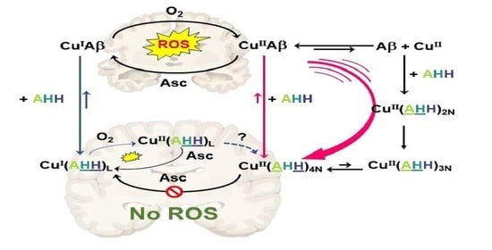
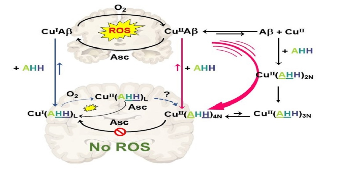
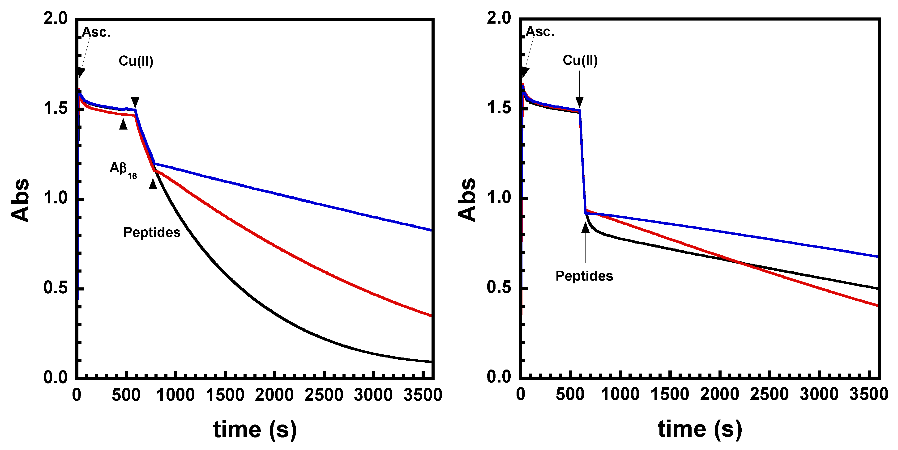
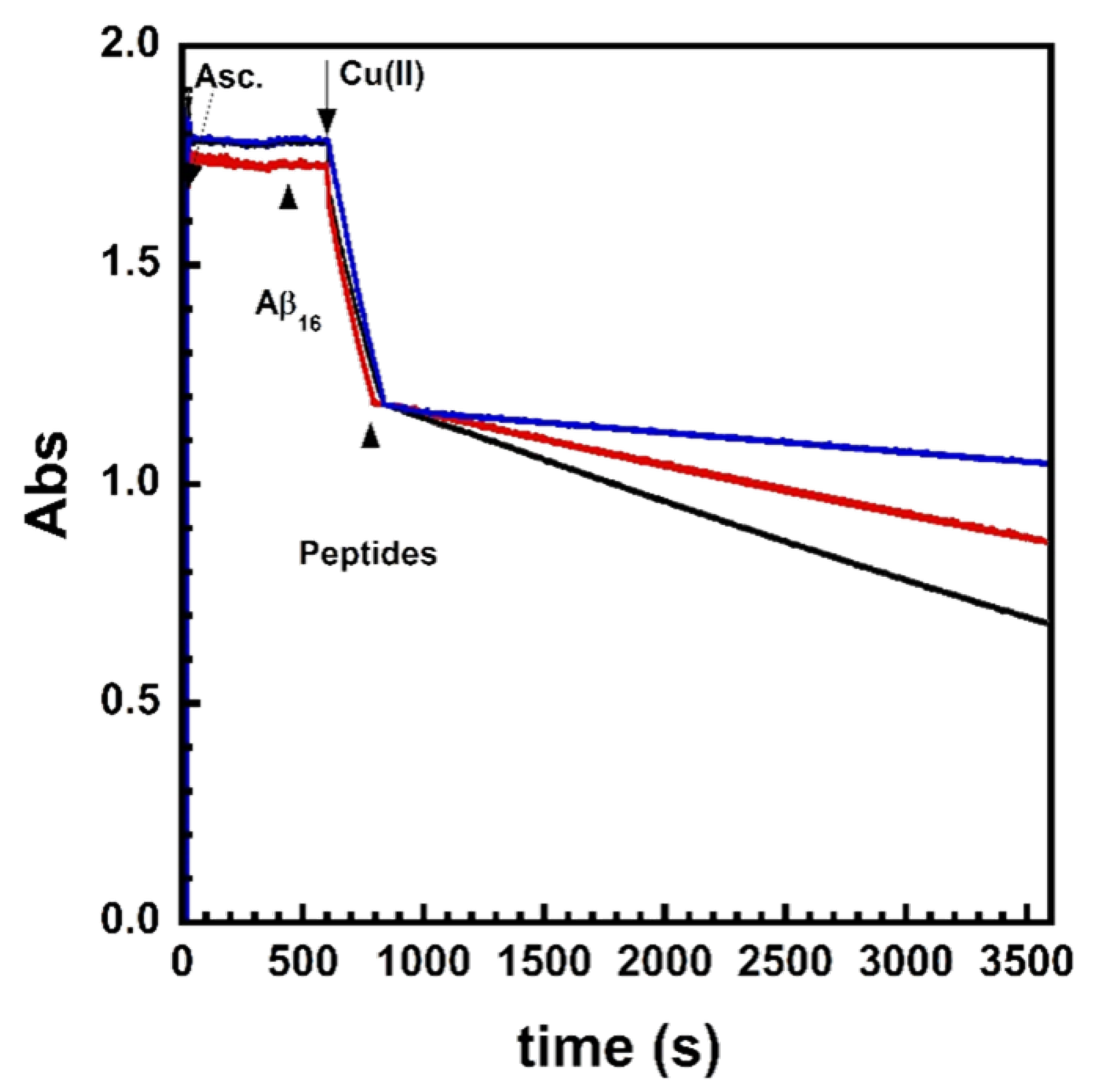
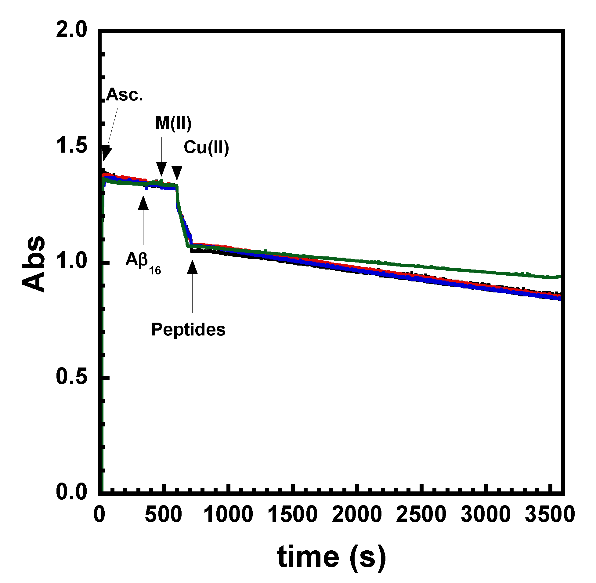
2.1. Effect on Cu- and Cu(Aβ)-Induced ROS Production

The effect of AH, AAH, and AHH on the Cu- and Cu(Aβ)-induced ROS production was investigated by following ascorbate (Asc) consumption by UV–Visible spectroscopy at 265 nm (εAsc = 14,500 M−1.cm−1) (Figure 1 and Figure S1). It was previously shown that, because Asc fuels the production of ROS, following its consumption is a straightforward way to monitor the formation of ROS production, H2O2, or HO° [76,77]. Hence, this is a standard and well-accepted method [16], with less bias than other ROS detection assays [78]. Two kinds of experiments were performed: (i) Asc is added once the peptide (AH, AAH, and AHH, noted pep unless specified) has been mixed with CuII or CuII(Aβ) (Figure S1) or (ii) the peptide is added during the course of Asc consumption triggered by CuII or CuII(Aβ) (Figure 1). With the first experiment (Figure S1), CuII or CuII(Aβ) and the peptides are premixed in solution before the addition of Asc to evidence differences in the redox properties of the CuII(pep) complexes (when added to CuII) and the peptide ability to remove CuII from Aβ (when added to CuII(Aβ)). With the second experiment (Figure 1), the kinetics of CuII complexation are additionally probed; indeed, there is a competition between the reduction of CuII or CuII(Aβ) by Asc and the CuII uptake by the ligand [79,80].
With Cu (in absence of Aβ), AH displays a steeper slope than AAH and AHH, indicating that AH is not able to stop Cu-induced Asc consumption (Figure 1 and Figure S1, red lines, left). This is in accordance with the ability of AH to bind CuII only in a 3N-motif, the resulting complex Cu(AH) being redox-active [55]. AAH is able to prevent or stop Asc consumption in conditions corresponding to pre-mixture or added during Asc consumption, respectively (Figure 1 and Figure S1, black lines, left). This is in line with the formation of a redox-inert Cu(AAH) complex where CuII is bound in a 4N-motif, which is resistant to reduction by Asc, in line with previously reported electrochemical data [51]. As shown in Figure 1, panel A (between t = 600 and 700 s, black curve), the change in the slope to reach a straight line is not immediate (as for AH and AHH) and a delay is thus observed between the addition of AAH and the total slowdown of Cu-induced Asc consumption. This indicates slow kinetics of complex formation. AHH displays the best ability to limit Asc consumption in both experiments (Figure 1 and Figure S1, blue lines, left), which suggests a fast complexation of CuII by the peptide and the subsequent formation of a complex which cannot be reduced by Asc.
The effect of AH, AAH, and AHH was then investigated on Cu(Aβ)-induced ROS production (Figure 1 and Figure S1, right). Similar to the precedent experiments, the ability of AH, AAH, and AHH to limit Asc consumption was tested in pre-mixed (Figure S1, right) or addition during Asc consumption (Figure 1, right) conditions. The results show the ability of AH to remove Cu from Aβ and slow down Asc consumption, but not completely, due to the ability of CuII(AH) to react with Asc, as shown in the experiments without Aβ being present. AAH was able to stop Asc consumption when AAH was first premixed with Cu(Aβ) (Figure S1, right), but not when it was added during Cu(Aβ)-induced Asc consumption (Figure 1, black lines, right). Since binding of CuII by AAH is thermodynamically favored compared to Aβ, these results indicate that the slow kinetics of CuII extraction from Aβ hampered AAH from efficiently halting related Asc consumption. Finally, AHH was able to lessen Asc consumption efficiently regardless of the conditions (Figure 1 and Figure S1, blue lines, right)). The arrest of ROS production is, however, not total.
To gain further insights into the mechanism by which AHH lessens CuII/CuII(Aβ)-induced Asc consumption, the experiments were performed at different pH, with (Figure 2) and without Aβ (Figure S2). The effect of AHH on Cu and Cu(Aβ)-induced Asc consumption is pH-dependent: the higher the pH is, the better the arrest of ROS production. The pH-dependent ability of AHH to limit Asc consumption reflects the pH-driven switch of AHH between the 3N and 4N-motifs, since this equilibrium has a pKa of ~6.5. Hence, at pH 6.8, there is about 30% of the complex Cu(AHH) in a redox-competent 3N-motif, while at pH 8.2, >95% of the formed complex is in the 4N form resistant to Asc reduction.
At this stage, we can hypothesize that AHH first binds CuII in a 3N motif, which would induce kinetics similar to that of AH, and then rearranges in a 4N-redox-inactive motif as AAH. According to this hypothesis, the peptide would combine the two positive effects of the 3N and 4N motifs, which are a fast CuII-binding kinetic, and the formation of a redox-inactive CuII complex, respectively. To question this hypothesis, we investigated the kinetics of CuII captured by the peptides by stopped-flow experiments.

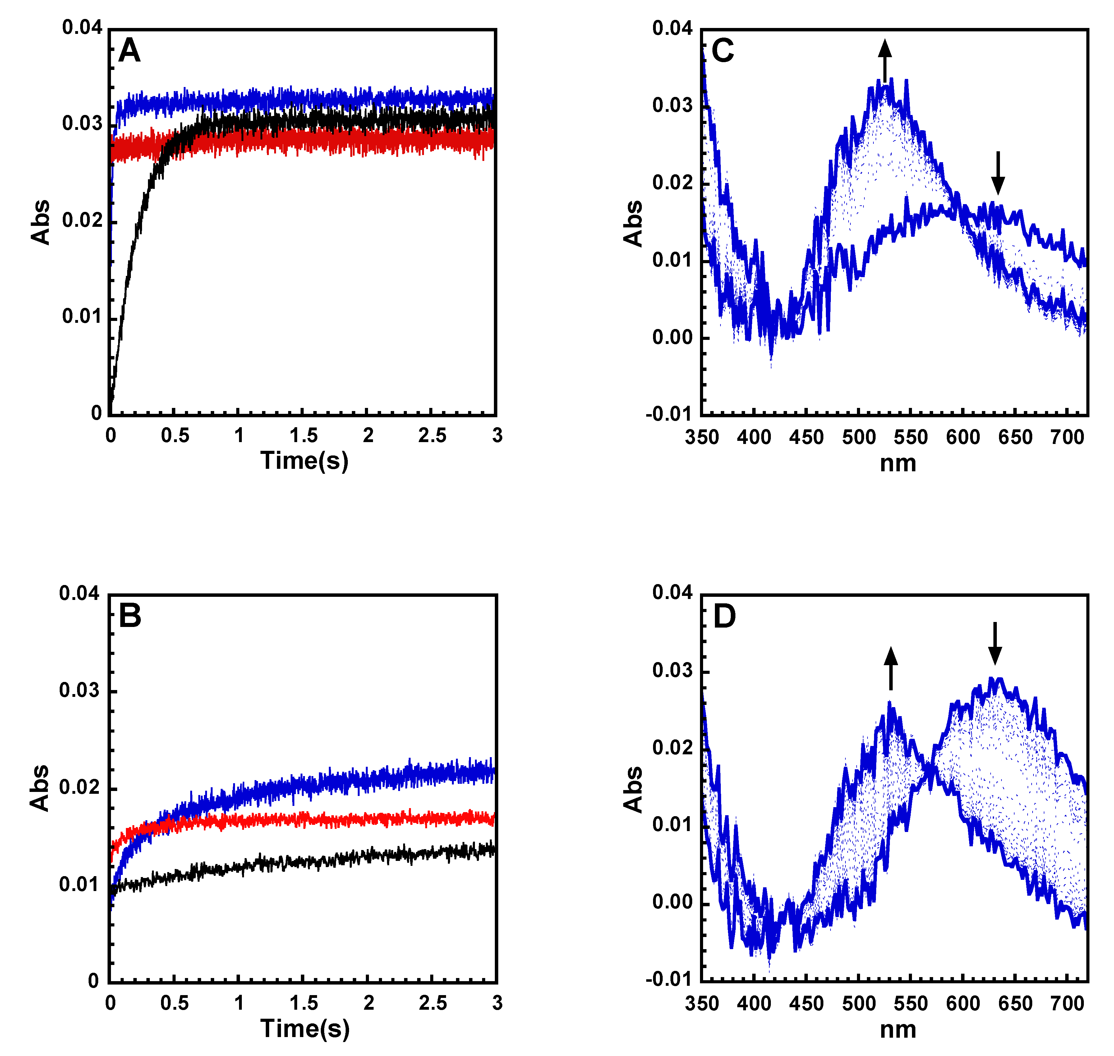
2.2. Kinetics of Cu and Cu(Aβ) Capture by the Peptides

First, stop-flow experiments were performed by mixing CuII with the peptides and recording absorption spectra with an integration time of 1 ms (Figure 3, Figures S3 and S4). The appearance of the Cu(pep) UV–Vis signature was monitored at 530 nm and 590 nm, corresponding, respectively, to the maximum absorption for Cu(AAH) (4N) and Cu(AH) (3N) species [51]. By the naked eye, it can be seen that the rate of Cu(pep) formation is Cu(AH) > Cu(AHH) > Cu(AAH), in line with the previously described ROS results. Cu(AH) formation is too fast to be measured with this technique and is completed in less than 5 ms, which corresponds to the dead time of the UV–Vis stopped-flow spectrometer. Since several steps are at play (see below), the overall kinetic rates of Cu(AAH) and Cu(AHH) formation were evaluated by the t1/2, at which half of the final CuII(pep) complexes are formed at various wavelengths (Table 1). Cu(AHH) formation is about eight times faster (t1/2 = 18 ms) than Cu(AAH) formation (t1/2 = 150 ms).
The formation of Cu(AAH) is at least 20 times slower than that of Cu(AH). To account for this difference, we have to take into consideration very recent reports on the kinetics of formation of ATCUN CuII complexes [81,82,83,84,85,86]. It proceeds via several successive steps: anchoring of the CuII by the imidazole ring of His, then formation of a metallacycle with the anchoring of the N-terminal amine, leading to the 2N intermediate (Scheme S2 for a drawing of the various intermediates described in this work) and rearrangement of the 4N ATCUN motif [81]. The rate-limiting step is the 2N → 4N corresponding to the deprotonation of the peptide bond in between the two anchors. We can thus hypothesize a similar binding pathway for the formation of the Cu(AH) complex, id est, anchoring of the CuII by the imidazole ring of His, then formation of a metallacycle with the anchoring of the N-terminal amine, leading to the 2N intermediate and formation of the 3N motif. In the case of AH, the various rates would be strongly higher than that of AAH because (i) the smaller size of the metallacycle formed is much more favorable to anchor the N-terminal amine and form the 2N intermediates and (ii) there is only one peptide bond to deprotonate (from 2N to 3N, instead of 4N in the case of AAH) (Scheme 2, panels A and B). In the case of AAH, we do observe the very fast formation of the 2N species absorbing near 700 nm (Figure S3, panel C), reminiscent of those thoroughly described in [81], followed by its evolution to the 4N species, which is the rate-limiting step of the whole reaction.
For the hybrid peptide AHH, the rate of complex formation is in between those of AH and AAH (Figure 3, panel A). As can be seen in Figure 3, panel C, a large absorption band is observed at about 600 nm that forms rapidly (within the dead time of the stopped flow) and that can be attributed to a mixture of 2N and 3N intermediates. Then it evolves, during the rate-limiting step, toward the 4N species with a maximum absorption at 530 nm. The formation of the 2N and/or 3N species is so fast that we cannot distinguish between the two possible pathways (paths a and b in Scheme 2, panel C).
The same experiment was performed in the presence of Aβ. Overall, all the previous rates of Cu(pep) formation are slowed down (Figure 3, panel B and Figure S4). This is in line with (i) the presence of the pre-equilibrium of CuII to Aβ complexation, leading to a strongly reduced concentration of unbound CuII in solution (dissociative path, Scheme S3) and/or (ii) the more difficult CuII anchoring when already bound to Aβ (associative path). Based on a recent report [82], we propose that the dissociation path is predominant. Such a contribution of the dissociation path is even more pronounced at the concentration of ROS experiments (50 times lower than in the stopped-flow experiments). For AHH, CuII extraction from Aβ is observed concomitantly to the formation of the Cu(AHH) complex in its 4N form detected at a λmax of 530 nm (Figure 3, panel D) and the formation of the Cu(AHH)4N is about 15 times slower than in the absence of Aβ. No intermediate species is detected, as indicated by the presence of the isosbestic point at 560 nm. This is in line with a slowdown of their formation rate (due to a decreased concentration of free CuII in solution in the presence of Aβ), which thus becomes slower than the conversion to the 4N species. In the presence of Aβ, the formation of the intermediate species thus becomes the rate-limiting step.

2.3. Effect of ZnII on Cu(Aβ)-Induced ROS Production

In a last part, we investigated how ZnII (and other biologically relevant ions) perturb the arrest of Cu(Aβ)-induced ROS production, as ZnII is one of the metal ions whose content is dysregulated in AD. Asc consumption experiments were performed with an increasing amount of ZnII (0 to 10 eq) (Figure 4, Figures S5 and S6). For the three peptides studied here, an increasing amount of ZnII resulted in a slower Asc consumption. This effect was weak in the case of AHH, since the ROS production rate is already slow, but meaningful, since with CaII and MgII, no modification is observed (Figure 4). Thus, for the three peptides, ZnII seems to facilitate CuII binding by the peptides. This is in line with the dissociative pathway we favored previously, where 10 equiv. of ZnII can push CuII out of the Aβ peptide, but not from the AH, AHH, and AAH ones because the ZnII sites are much weaker than that of the CuII. In other words, based on these results, we proposed a push–pull mechanism in which the ZnII bound to Aβ helps to direct CuII into the AH, AAH, and AHH ligands.

3. Concluding Remarks

A tentative picture of the mode of action of the three peptides under focus can thus be drawn (Scheme 3A–C). Addition of peptides during Cu(Aβ)-induced Asc consumption leads to CuII extraction from Aβ. This reaction competes with the reduction of CuII(Aβ) to CuI(Aβ) and is fast enough only in the cases of AH and AHH. Once formed, the resulting Cu(AH)3N is weakly competent in ROS production, while Cu(AHH)4N complexes are resistant to Asc reduction. Only the AHH peptide can both remove CuII from Aβ and form a CuII(AHH) complex resistant to reduction by Asc (and thus is unable to produce ROS). The addition of ZnII improves slightly the effect of the peptides due to an increase in the Cu(Aβ) dissociation pre-equilibrium. This indicates that ZnII selectivity of the AHH peptide is, as explained in the introduction, fully appropriate.
The presence of two His residues could also give to AHH the additional possibility to compete for CuI with Aβ, leading an L-motif (Scheme 1), while the CuI affinity of AH and AAH, containing only one His, is too weak to do so. After oxidation of CuI, the intermediate formed could either rearrange to form the CuII(AHH)4N or be reduced by Asc. To probe the CuI path (blue lines in Scheme 3), the AAHH peptide was tested (Figure S7). This peptide possesses two His residues and is thus able to bind CuI in an L-motif, similarly to AHH, and to compete with Aβ for CuI binding. The results obtained show that AAHH has a weaker ability to arrest Cu(Aβ)-induced ROS formation than AHH. This suggests that the CuI path might not be the one predominant here. We must be very cautious, since two key reactions occurs in the CuI path: (1) CuI extraction from Aβ, which would be similar in case of AHH and AAHH, and (2) the rate of reorganization to the CuII-4N species once the linear CuI intermediate is oxidized, a parameter that could differ between AHH and AAHH (with a fast enough reorganization in case of AHH but not AAHH). To gain further insights into the mechanism at play during ROS production and to discard the CuI path, more systematic studies of the impact of sequence modification (number and position of the His residues) on the level of ROS arrest are needed and are currently under investigation in our group.
In the present study, we have unambiguously shown that the AHH motif is fully appropriate to stop Cu(Aβ)-induced ROS production. AHH combines the two advantageous properties of the two AH and AAH parent ligands, fast CuII binding, and ability to form a redox-incompetent CuII complex, respectively. This result underlines the potential of peptide ligands in the search for Cu-targeting therapeutic approaches in the context of AD. It is also interesting to note that for all the previous ATCUN peptides studied, only experiments where the peptide is added to CuII(Aβ) and then the ROS production is triggered by addition of Asc have been reported [67,68,69,70,71,72,73]. Once formed, CuII(ATCUN) complexes have a very low ability to form ROS, as shown here and previously [55,56,87]. Hence, such studies are incomplete. Indeed, in the brain, which is a quite reductive medium [88,89], with up to 300 µM of Asc being present extracellularly, both redox states of the Cu ions are at play and the Cu(Aβ) may be redox cycling. Hence, targeting both CuI and CuII ions [90], or having fast enough CuII-complexing agents, are crucial [80,91]. In other words, if ATCUN-based peptides are aimed to be used against Cu(Aβ)-induced ROS formation, the rate of CuII extraction from Aβ matters and, as we demonstrated, such a kinetic parameter is highly dependent on the exact sequence of the ATCUN peptide chosen. It is here interesting to note that, among the N-terminally truncated forms of Aβ recently put forward [92,93,94,95,96], several of them possess such H2N-Xxx-His (3N, Aβ5–x) [97,98], H2N-Xxx-Zzz-His (4N, Aβ4–x, [85,86] Aβ11–x [86]), and H2N-Xxx-His-His (hybrid 3N/4N, Aβ12–x [99]) binding sites. Based on our results, the cleavage at position 12 leading to the hybrid 3N/4N binding site may be the most beneficial with respect to Cu(Aβ1–x)-induced ROS formation.
Further works will include modifications of the peptide scaffold to obtain BBB penetration and better metabolic stability. This can be straightforward by appending brain-penetrating sequences and using D-peptide, respectively. To shuttle Cu inside cells is also regarded as an interesting additional property that will overcome the intracellular Cu depletion involved in the disability of secretases to clear Aβ [100,101,102]. This is an exciting follow-up of the present work, which is currently under progress in our team.

4. Experimental Section

4.1. Chemicals

Reagents were commercially available and were used as received. HEPES buffer (sodium salt of 2-[4-(2-hydroxyethyl)piperazin-1-yl]ethanesulfonic acid) was bought from Fluka (bioluminescence grade).
The CuII and ZnII sources were Zn(SO4)(H2O) and CuCl2(H2O)2 and purchased from Sigma. The concentration of CuII was determined by dissolving the Cu salt in Mili-Q water to prepare a 100 mM solution considering an extinction coefficient of ε = 12 M−1cm−1. The concentration of ZnII was determined by weight.
A stock solution (100 mM) of ascorbate was prepared in Milli-Q water at room temperature just before beginning the experiment.

4.2. Peptides

1–16 peptide (sequence DAEFRHDSGYEVHHQK, H2N-Asp-Ala-Glu-Phe-Arg-His-Asp-Ser-Gly-Tyr-Glu-Cal-His-His-Gln-Lys-COOH, with the three-letter code and referred to as Aβ in the text unless specified, see Scheme 1) was bought from GeneCust (Dudelange, Luxembourg) with a purity grade >98%. Stock solutions of the Aβ peptide were prepared by dissolving the powder in Milli-Q water (resulting pH ~2) at approx. 10 mM. Peptide concentration was then determined by UV–Visible absorption of Tyr10 considered as free tyrosine (at pH 2, (ε276–ε296) = 1410 M−1 cm−1). The solutions were diluted down to the appropriate concentration.

4.3. Synthesis of Peptides

The AHH, AAH, and AH peptides were synthesized manually with standard 9-fluorenylmethoxycarbonyl (Fmoc) Chemistry on a Fmoc-L-His(Trt)-Wang resin (0.63 mmol/g from Iris Biotech GMBH), through solid-phase peptide synthesis protocols, as previously reported [51].
Stock solutions of peptides were prepared by dissolving the peptides in Mili-Q water. Concentrations were determined by CuII titration followed by UV–Vis absorption spectroscopy.

4.4. UV–Visible Spectroscopy

UV–Vis experiments were performed on a Cary-60 UV–Vis spectrometer at 25 °C with constant stirring.

4.5. Ascorbate Consumption Assay

Ascorbate consumption was monitored by UV–Vis. Intensity of the Asc absorption band at λ = 265 nm (ε = 14 500 M−1 cm−1) was monitored as a function of time.

4.6. Stopped-Flow Experiments

Rapid-mixing UV–Vis spectroscopy was carried out using an SFM-20 two-syringe stopped-flow from Biologic combined with a diode array spectrometer composed of a TIDAS J&M MMS-UV/VIS 500-3 detector and a light source HAMAMATSU L7893 light source incorporating a deuterium and a tungsten lamp with optic fibers. Data acquisition, extraction, and treatment were realized with Bio-Kine software. The syringes (Hamilton) were mounted on a rigid drive platform ensuring that the flow was stopped precisely and instantaneously. The contents of the two syringes were rapidly mixed in the mixing chamber and the absorbance of the system recorded over time as full spectra at designated time delays. Typically, one syringe was filled with a solution of peptide at 1mM in HEPES buffer (200 mM, pH 7.4), and the other one was filled with a solution of CuSO4 in water at 0.9 mM. An equal quantity of the two solutions were mixed to reach a final concentration of CuII of 450 µM and peptide of 500 µM in HEPES buffer (100 mM, pH 7.4). The optical length of the UV cuvette is 1 cm.

Supplementary Materials

The following supporting information can be downloaded at: https://www.mdpi.com/article/10.3390/biom12101327/s1, Figure S1: Ascorbate consumption of CuII and CuII(Aβ) plus the three AH, AAH and AHH peptides; Figure S2: Ascorbate consumption of CuII(AHH) and CuII/I plus AHH peptide as a function of pH; Figure S3: Stopped-flow kinetic measurements of CuII coordination by AH, AAH and AHH peptides; Figure S4: Stopped-flow kinetic measurements of CuII(Aβ) capture removal by AH, AAH and AHH peptides; Figure S5: Ascorbate consumption of CuII plus the three AH, AAH and AHH peptides in presence of increasing amounts of ZnII; Figure S6: Ascorbate consumption of CuII plus the AH and AAH peptides in presence of various dications; Figure S7: Ascorbate consumption of CuII(Aβ) plus the four AH, AAH, AHH and AAHH peptides; Scheme S1: Proposed coordination sites in the intermediates 2N forms for the AH, AAH and AHH peptides; Scheme S2. Proposed mechanism corresponding to CuII capture out from Aβ by AH (A), AAH (B) and AHH (C); Scheme S3. Proposed coordination sites in the ternary species obtained upon AH and AHH addition to CuII(Aβ) [60,62,82].

Author Contributions

Conceptualization, C.H.; formal analysis, P.G., E.M., L.S., P.F., C.H.; investigation, P.G. and L.S.; data curation, L.S and E.M.; writing—original draft preparation, P.G., E.M. and C.H.; writing—review and editing, C.H.; supervision, P.F. and C.H.; project administration, C.H.; funding acquisition, C.H. All authors have read and agreed to the published version of the manuscript.

Funding

ERC aLzINK-StG 638712 (attributed to C.H.) is acknowledged for funding.

Institutional Review Board Statement

Not applicable.

Informed Consent Statement

Not applicable.

Data Availability Statement

Not applicable.

Acknowledgments

C. Esmieu is acknowledged for fruitful discussions. C.H., L.S., P.G., P.F. thank ERC aLzINK-StG 638712 (attributed to C.H.) for funding.

Conflicts of Interest

The authors declare no conflict of interest.

References

  1. Available online: https://www.alzint.org/about/dementia-facts-figures/dementia-statistics/ (accessed on 18 August 2022).
  2. Available online: https://www.brightfocus.org/alzheimers/article/alzheimers-disease-facts-figures (accessed on 18 August 2022).
  3. Available online: https://aaic.alz.org/releases_2021/global-prevalence.asp (accessed on 18 August 2022).
  4. Available online: https://www.who.int/news-room/fact-sheets/detail/dementia (accessed on 18 August 2022).
  5. Hampel, H.; Hardy, J.; Blennow, K.; Chen, C.; Perry, G.; Kim, S.H.; Villemagne, V.L.; Aisen, P.; Vendruscolo, M.; Iwatsubo, T.; et al. The Amyloid-β Pathway in Alzheimer’s Disease. Mol. Psychiatry 2021, 26, 5481–5503. [Google Scholar] [CrossRef] [PubMed]
  6. Padhi, D.; Govindaraju, T.J. Mechanistic Insights for Drug Repurposing and the Design of Hybrid Drugs for Alzheimer’s Disease. Med. Chem. 2022, 65, 7088–7105. [Google Scholar] [CrossRef] [PubMed]
  7. Gong, C.X.; Dai, C.L.; Liu, F.; Iqbal, K. Multi-Targets: An Unconventional Drug Development Strategy for Alzheimer’s Disease. Front. Aging Neurosci. 2022, 14, 837649. [Google Scholar] [CrossRef] [PubMed]
  8. Cummings, J.; Lee, G.; Nahed, P.; Kambar, M.E.Z.N.; Zhong, K.; Fonseca, J.; Taghva, K. Alzheimer’s disease drug development pipeline: 2022. Alzheimer’s Dement. Transl. Res. Clin. Interv. 2022, 8, e12295. [Google Scholar] [CrossRef] [PubMed]
  9. Athar, T.; Al Balushi, K.; Khan, S.A. Challenges of peptide and protein drug delivery by oral route: Current strategies to improve the bioavailability. Mol. Biol. Rep. 2021, 48, 5629–5645. [Google Scholar] [CrossRef] [PubMed]
  10. Liu, Y.; Cong, L.; Han, C.; Li, B.; Dai, R. Recent Progress in the Drug Development for the Treatment of Alzheimer’s Disease Especially on Inhibition of Amyloid-peptide Aggregation. Mini-Rev. Med. Chem. 2021, 21, 969–990. [Google Scholar] [CrossRef] [PubMed]
  11. Priya, K.; Siddesha, J.M.; Dharini, S.; Shashanka, K.P. Interacting Models of Amyloid-β and Tau Proteins: An Approach to Identify Drug Targets in Alzheimer’s Disease. J. Alzheimer’s Dis. Rep. 2021, 5, 405–411. [Google Scholar] [CrossRef] [PubMed]
  12. West, S.; Bhugra, P. Emerging drug targets for Aβ and tau in Alzheimer’s disease: A systematic review. Br. J. Clin. Pharmacol. 2015, 80, 221–234. [Google Scholar] [CrossRef]
  13. Singh, S.K.; Balendra, V.; Obaid, A.A.; Esposto, J.; Tikhonova, M.A.; Gautam, N.K.; Poeggeler, B. Copper-mediated β-amyloid toxicity and its chelation therapy in Alzheimer’s disease. Metallomics 2022, 14, mfac018. [Google Scholar] [CrossRef]
  14. Fasae, K.D.; Abolaji, A.O.; Faloye, T.R.; Odunsi, A.Y.; Oyetayo, B.O.; Enya, J.I.; Rotimi, J.A.; Akinyemi, R.O.; Whitworth, A.J.; Aschner, M.J. Metallobiology and therapeutic chelation of biometals (copper, zinc and iron) in Alzheimer’s disease: Limitations, and current and future perspectives. Trace Elem. Med. Biol. 2021, 67, 126779. [Google Scholar] [CrossRef]
  15. Ejaz, H.W.; Wang, W.; Lang, M. Copper toxicity links to pathogenesis of Alzheimer’s disease and therapeutics approaches. Int. J. Mol. Sci. 2020, 21, 7660. [Google Scholar] [CrossRef]
  16. Esmieu, C.; Guettas, D.; Conte-Daban, A.; Sabater, L.; Faller, P.; Hureau, C. Copper-Targeting Approaches in Alzheimer’s Disease: How To Improve the Fallouts Obtained from in Vitro Studies. Inorg. Chem. 2019, 58, 13509–13527. [Google Scholar] [CrossRef]
  17. Savelieff, M.G.; Nam, G.; Kang, J.; Lee, H.J.; Lee, M.; Lim, M.H. Development of Multifunctional Molecules as Potential Therapeutic Candidates for Alzheimer’s Disease, Parkinson’s Disease, and Amyotrophic Lateral Sclerosis in the Last Decade. Chem. Rev. 2019, 119, 1221–1322. [Google Scholar] [CrossRef]
  18. Robert, A.; Liu, Y.; Nguyen, M.; Meunier, B. Regulation of copper and iron homeostasis by metal chelators: A possible chemotherapy for Alzheimer’s disease. Acc. Chem. Res. 2015, 48, 1332–1339. [Google Scholar] [CrossRef]
  19. Weibull, M.G.M.; Simonsen, S.; Oksbjerg, C.R.; Tiwari, M.K.; Hemmingsen, L. Effects of Cu(II) on the aggregation of amyloid-β. J. Biol. Inorg. Chem. 2019, 24, 1197–1215. [Google Scholar] [CrossRef]
  20. Cheignon, C.; Tomas, M.; Bonnefont-Rousselot, D.; Faller, P.; Hureau, C.; Collin, F. Oxidative stress and the amyloid beta peptide in Alzheimer’s Disease. Redox Biol. 2018, 14, 450–464. [Google Scholar] [CrossRef]
  21. Huat, T.J.; Camats-Perna, J.; Newcombe, E.A.; Valmas, N.; Kitazawa, M.; Medeiros, R.J. Metal Toxicity Links to Alzheimer’s Disease and Neuroinflammation. Mol. Biol. 2019, 431, 1843–1868. [Google Scholar] [CrossRef]
  22. Reybier, K.; Ayala, S.; Alies, B.; Bustos Rodriguez, S.; Rodrigues, J.; Gomes, C.; La Penna, G.; Collin, F.; Hureau, C.; Faller, P. Free superoxide is an intermediate in the production of H2O2 by Cu(I)-amyloid-β and O2. Angew. Chem. Int. Ed. 2016, 55, 1085–1089. [Google Scholar] [CrossRef]
  23. Nandi, A.; Yan, L.J.; Jana, C.K.; Das, N. Role of Catalase in Oxidative Stress- and Age-Associated Degenerative Diseases. Oxidative Med. Cell. Longev. 2019, 2019, 9613090. [Google Scholar] [CrossRef]
  24. Guilloreau, L.; Combalbert, S.; Sournia-Saquet, A.; Marzaguil, H.; Faller, P. Redox Chemistry of Copper-Amyloid-beta: The Generation of Hydroxyl Radical in the Presence of Ascorbate is Linked to Redox-Potentials and Aggregation State. ChemBioChem 2007, 8, 1317–1325. [Google Scholar] [CrossRef]
  25. Dikalov, S.I.; Vitek, M.P.; Mason, R.P. Cupric-amyloid beta peptide complex stimulates oxidation of ascorbate and generation of hydroxyl radical. Free Radic. Biol. Med. 2004, 36, 340–347. [Google Scholar] [CrossRef]
  26. Bellotti, D.; Remelli, M. Lights and Shadows on the Therapeutic Use of Antimicrobial Peptides. Molecules 2022, 27, 4584. [Google Scholar] [CrossRef]
  27. Li Petri, G.; Di Martino, S.; De Rosa, M.J. Peptidomimetics: An Overview of Recent Medicinal Chemistry Efforts toward the Discovery of Novel Small Molecule Inhibitors. Med. Chem. 2022, 65, 7438–7475. [Google Scholar] [CrossRef]
  28. Lai, X.; Tang, J.; ElSayed, M.E.H. Recent advances in proteolytic stability for peptide, protein, and antibody drug discovery. Exp. Opin. Drug. Discov. 2021, 16, 1467–1482. [Google Scholar] [CrossRef]
  29. Wątły, J.; Miller, A.; Kozłowski, H.; Rowińska-Żyrek, M.J. Peptidomimetics – An infinite reservoir of metal binding motifs in metabolically stable and biologically active molecules. Inorg. Biochem. 2021, 217, 111386. [Google Scholar] [CrossRef]
  30. Aileen Funke, S.; van Groen, T.; Kadish, I.; Bartnik, D.; Nagel-Steger, L.; Brener, O.; Sehl, T.; Batra-Safferling, R.; Moriscot, C.; Schoehn, G.; et al. Oral treatment with the d-enantiomeric peptide D3 improves the pathology and behavior of Alzheimer’s Disease transgenic mice. ACS Chem. Neurosci. 2010, 1, 639–648. [Google Scholar] [CrossRef]
  31. Verma, S.; Goand, U.K.; Husain, A.; Katekar, R.A.; Garg, R.; Gayen, J.R. Challenges of peptide and protein drug delivery by oral route: Current strategies to improve the bioavailability. Drug Dev. Res. 2021, 82, 927–944. [Google Scholar] [CrossRef]
  32. Covarrubias-Zambrano, O.; Yu, J.; Bossmann, H.S. Nano-Inspired Technologies for Peptide Delivery. Curr. Protein Pept. Sci. 2020, 21, 379–400. [Google Scholar] [CrossRef]
  33. Makowski, M.; Silva, Í.C.; Pais do Amaral, C.; Gonçalves, S.; Santos, N.C. Advances in lipid and metal nanoparticles for antimicrobial peptide delivery. Pharmaceutics 2019, 11, 588. [Google Scholar] [CrossRef]
  34. Tang, Y.; Varyambath, A.; Ding, Y.; Chen, B.; Huang, X.; Zhang, Y.; Yu, D.-g.; Kim, I.; Song, W. Porous organic polymers for drug delivery: Hierarchical pore structures, variable morphologies, and biological properties. Biomater. Sci. 2022. [Google Scholar] [CrossRef]
  35. Reissmann, S.; Filatova, M.P. New generation of cell-penetrating peptides: Functionality and potential clinical application. J. Pept. Sci. 2021, 27, e3300. [Google Scholar] [CrossRef] [PubMed]
  36. Zhou, X.; Smith, Q.R.; Liu, X. Brain penetrating peptides and peptide–drug conjugates to overcome the blood–brain barrier and target CNS diseases. WIREs Nanomed. Nanobiotechnology 2021, 13, e1695. [Google Scholar] [CrossRef] [PubMed]
  37. Zhang, Y.; Kim, I.; Lu, Y.; Xu, Y.; Yu, D.-G.; Song, W. Intelligent poly(L-histidine)-based nanovehicles for controlled drug delivery. J. Control. Release 2022, 349, 963–982. [Google Scholar] [CrossRef] [PubMed]
  38. Zhang, Y.; Song, W.; Lu, Y.; Xu, Y.; Wang, C.; Yu, D.-G.; Kim, I. Recent Advances in Poly(L-glutamic acid)-Based Nanomaterials for Drug Delivery. Biomolecules 2022, 12, 636. [Google Scholar] [CrossRef]
  39. Atrian-Blasco, E.; Conte-Daban, A.; Hureau, C. Mutual interference of Cu and Zn ions in Alzheimer’s disease: Perspectives at the molecular level. Dalton Trans. 2017, 46, 12750–12759. [Google Scholar] [CrossRef]
  40. Atrian-Blasco, E.; Gonzalez, P.; Santoro, A.; Alies, B.; Faller, P.; Hureau, C. Cu and Zn coordination to amyloid peptides: From fascinating chemistry to debated pathological relevance. Coord. Chem. Rev. 2018, 375, 38–55. [Google Scholar] [CrossRef]
  41. Rana, M.; Sharma, A.K. Cu and Zn interactions with Aβ peptides: Consequence of coordination on aggregation and formation of neurotoxic soluble Aβ oligomers. Metallomics 2019, 11, 64–84. [Google Scholar] [CrossRef]
  42. Dorlet, P.; Gambarelli, S.; Faller, P.; Hureau, C. Pulse EPR Spectroscopy Reveals the Coordination Sphere of Copper(II) Ions in the 1–16 Amyloid-β Peptide: A Key Role of the First Two N-Terminus Residues. Angew. Chem. Int. Ed. Engl. 2009, 48, 9273–9276. [Google Scholar] [CrossRef]
  43. Alies, B.; Conte-Daban, A.; Sayen, S.; Collin, F.; Kieffer, I.; Guillon, E.; Faller, P.; Hureau, C. Zinc(II) Binding Site to the Amyloid-β Peptide: Insights from Spectroscopic Studies with a Wide Series of Modified Peptides. Inorg. Chem. 2016, 55, 10499–10509. [Google Scholar] [CrossRef]
  44. De Gregorio, G.; Biasotto, F.; Hecel, A.; Luczkowski, M.; Kozlowski, H.; Valensin, D. Structural analysis of copper(I) interaction with amyloid β peptide. J. Inorg. Biochem. 2019, 195, 31–38. [Google Scholar] [CrossRef]
  45. Hureau, C.; Balland, V.; Coppel, Y.; Solari, P.L.; Fonda, E.; Faller, P. Importance of dynamical processes in the coordination chemistry and redox conversion of copper amyloid-β complexes. J. Biol. Inorg. Chem. 2009, 14, 995–1000. [Google Scholar] [CrossRef]
  46. Alies, B.; Renaglia, E.; Rozga, M.; Bal, W.; Faller, P.; Hureau, C. Cu(II) affinity for the Alzheimer’s Peptide: Tyrosine fluorescence studies revisited. Anal. Chem. 2013, 85, 1501–1508. [Google Scholar] [CrossRef]
  47. Zawisza, I.; Rozga, M.; Bal, W. Affinity of peptides (Aβ, APP, α-synuclein, PrP) for metal ions (Cu, Zn). Coord. Chem. Rev. 2012, 256, 2297–2307. [Google Scholar] [CrossRef]
  48. Alies, B.; Badei, B.; Faller, P.; Hureau, C. Reevaluation of Copper(I) affinity for amyloid-β peptides by competition with Ferrozine—An unusual Copper(I) indicator. Chem. Eur. J. 2012, 18, 1161–1167. [Google Scholar] [CrossRef]
  49. Young, T.R.; Kirchner, A.; Wedd, A.G.; Xiao, Z. An integrated study of the affinities of the Aβ16 peptide for Cu(I) and Cu(II): Implications for the catalytic production of reactive oxygen species. Metallomics 2014, 6, 505–517. [Google Scholar] [CrossRef]
  50. Xiao, Z.; Gottschlich, L.; van der Meulen, R.; Udagedara, S.R.; Wedd, A.G. Evaluation of quantitative probes for weaker Cu(i) binding sites completes a set of four capable of detecting Cu(I) affinities from nanomolar to attomolar. Metallomics 2013, 5, 501–513. [Google Scholar] [CrossRef]
  51. Gonzalez, P.; Vileno, B.; Bossak, K.; El Khoury, Y.; Hellwig, P.; Bal, W.; Hureau, C.; Faller, P. Cu(II)-binding to the peptide Ala-His-His, a chimera of the canonical Cu(II)-binding motifs Xxx-His and Xxx-Zzz-His (ATCUN). Inorg. Chem. 2017, 56, 14870–14879. [Google Scholar] [CrossRef]
  52. Pickart, L.; Margolina, A. Regenerative and protective actions of the GHK-Cu peptide in the light of the new gene data. Int J. Mol. Sci. 2018, 19, 1987. [Google Scholar] [CrossRef]
  53. Ma, W.H.; Li, M.; Ma, H.F.; Li, W.; Liu, L.; Yin, Y.; Zhou, X.M.; Hou, G. Protective effects of GHK-Cu in bleomycin-induced pulmonary fibrosis via anti-oxidative stress and anti-inflammation pathways. Life Sci. 2020, 241, 117139. [Google Scholar] [CrossRef]
  54. Wang, X.; Liu, B.; Xu, Q.; Sun, H.; Shi, M.; Wang, D.; Guo, M.; Yu, J.; Zhao, C.; Feng, B. GHK-Cu-liposomes accelerate scald wound healing in mice by promoting cell proliferation and angiogenesis. Wound Repair Regen. 2017, 25, 270–278. [Google Scholar] [CrossRef]
  55. Hureau, C.; Eury, H.; Guillot, R.; Bijani, C.; Sayen, S.; Solari, P.L.; Guillon, E.; Faller, P.; Dorlet, P. X-Ray and solution structures of CuGHK and CuDAHK complexes. Influence on their redox properties. Chem. Eur. J. 2011, 17, 10151–10160. [Google Scholar] [CrossRef]
  56. Gonzalez, P.; Bossak, K.; Stefaniak, E.; Hureau, C.; Raibaut, L.; Bal, W.; Faller, P. N-terminal Cu Binding Motifs Xxx-Zzz-His (ATCUN) and Xxx-His and their derivatives: Chemistry, Biology and Medicinal Applications. Chem. Eur. J. 2018, 24, 8029–8041. [Google Scholar] [CrossRef]
  57. Maiti, B.K.; Govil, N.; Kundu, T.; Moura, J.J.G. Designed Metal-ATCUN Derivatives: Redox- and Non-redox-Based Applications Relevant for Chemistry, Biology, and Medicine. iScience 2020, 23, 101792. [Google Scholar] [CrossRef]
  58. Harford, C.; Sarkar, B. Amino Terminal Cu(II)- and Ni(II)-Binding (ATCUN) Motif of Proteins and Peptides: MetalBindi ng, DNA Cleavage, and Other Properties. Acc. Chem. Res. 1997, 30, 123–130. [Google Scholar] [CrossRef]
  59. Bal, W.; Sokołowska, M.; Kurowska, E.; Faller, P. Binding of transition metal ions to albumin: Sites, affinities and rates. Biochim. Biophys. Acta 2013, 1830, 5444–5455. [Google Scholar] [CrossRef]
  60. Gonzalez, P.; Bossak-Ahmad, K.; Vileno, B.; Wezynfeld, N.E.; El Khoury, Y.; Hellwig, P.; Hureau, C.; Bal, W.; Faller, P. Triggering Cu-coordination change in Cu(ii)-Ala-His-His by external ligands. Chem. Commun. 2019, 55, 8110–8113. [Google Scholar] [CrossRef]
  61. Trapaidze, A.; Hureau, C.; Bal, W.; Winterhalter, M.; Faller, P. Thermodynamic study of Cu2+ binding to the DAHK and GHK peptides by isothermal titration calorimetry (ITC) with the weaker competitor glycine. J. Biol. Inorg. Chem. 2012, 17, 37–47. [Google Scholar] [CrossRef]
  62. Bossak-Ahmad, K.; Wiśniewska, M.D.; Bal, W.; Drew, S.C.; Frączyk, T. Ternary Cu(II) Complex with GHK Peptide and Cis-Urocanic Acid as a Potential Physiologically Functional Copper Chelate. Int. J. Mol. Sci. 2020, 21, 6190. [Google Scholar] [CrossRef]
  63. Sóvágó, I.; Kállay, C.; Várnagy, K. Peptides as complexing agents. Factors influencing the structure and thermodynamic stability of peptide complexes. Coord. Chem. Rev. 2012, 256, 2225–2233. [Google Scholar] [CrossRef]
  64. Hureau, C. Coordination of redox active metal ions to the APP and to the amyloid-β peptides involved in AD. Part 1: An overview. Coord. Chem. Rev. 2012, 256, 2164–2174. [Google Scholar] [CrossRef]
  65. Portelinha, J.; Duay, S.S.; Yu, S.I.; Heilemann, K.; Libardo, M.D.J.; Juliano, S.A.; Klassen, J.L.; Angeles-Boza, A.M. Antimicrobial Peptides and Copper(II) Ions: Novel Therapeutic Opportunities. Chem. Rev. 2021, 121, 2648–2712. [Google Scholar] [CrossRef] [PubMed]
  66. Agbale, C.M.; Cardoso, M.H.; Galyuon, I.K.; Franco, O.L. Designing metallodrugs with nuclease and protease activity. Metallomics 2016, 8, 1159–1169. [Google Scholar] [CrossRef] [PubMed]
  67. Folk, D.S.; Franz, K.J. A prochelator activated by beta-secretase inhibits Aβ aggregation and suppresses copper-induced reactive oxygen species formation. J. Am. Chem. Soc. 2010, 132, 4994–4995. [Google Scholar] [CrossRef] [PubMed]
  68. Caballero, A.B.; Terol-Ordaz, L.; Espargaró, A.; Vázquez, G.; Nicolás, E.; Sabaté, R.; Gamez, P. Histidine-Rich Oligopeptides To Lessen Copper-Mediated Amyloid-β Toxicity. Chem. Eur. J. 2016, 22, 7268–7280. [Google Scholar] [CrossRef]
  69. Jensen, M.; Canning, A.; Chiha, S.; Bouquerel, P.; Pedersen, J.T.; Østergaard, J.; Cuvillier, O.; Sasaki, I.; Hureau, C.; Faller, P. Bi-functional peptide with β-sheet breaker and chelator moieties against Cu- amyloid-β. Chem. Eur. J. 2012, 18, 4836–4839. [Google Scholar] [CrossRef]
  70. Hu, X.; Zhang, Q.; Wang, W.; Yuan, Z.; Zhu, X.; Chen, B.; Chen, X. Tripeptide GGH as the Inhibitor of Copper-Amyloid-β-Mediated Redox Reaction and Toxicity. ACS Chem. Neurosci. 2016, 7, 1255–1263. [Google Scholar] [CrossRef]
  71. Meng, J.; Zhang, H.; Dong, X.; Liu, F.; Sun, Y. RTHLVFFARK-NH2: A potent and selective modulator on Cu2+-mediated amyloid-β protein aggregation and cytotoxicity. J.Inorg. Biochem. 2018, 181, 56–64. [Google Scholar] [CrossRef]
  72. Perrone, L.; Mothes, E.; Vignes, M.; Mockel, A.; Figueroa, C.; Miquel, M.C.; Maddelein, M.L.; Faller, P. Copper transfer from Cu-Abeta to human serum albumin inhibits aggregation, radical production and reduces Aβ toxicity. ChemBioChem 2010, 11, 110–118. [Google Scholar] [CrossRef]
  73. Mital, M.; Wezynfeld, N.E.; Frączyk, T.; Wiloch, M.Z.; Wawrzyniak, U.E.; Bonna, A.; Tumpach, C.; Barnham, K.J.; Haigh, C.L.; Bal, W.; et al. A Functional Role for Aβ in Metal Homeostasis? N-Truncation and High-Affinity Copper Binding. Angew. Chem. Int. Ed. 2015, 54, 10460–10464. [Google Scholar] [CrossRef]
  74. Pushie, M.J.; Stefaniak, E.; Sendzik, M.R.; Sokaras, D.; Kroll, T.; Haas, K.L. Using N-Terminal Coordination of Cu(II) and Ni(II) to Isolate the Coordination Environment of Cu(I) and Cu(II) Bound to His13 and His14 in Amyloid-β(4–16). Inorg. Chem. 2019, 58, 15138–15154. [Google Scholar] [CrossRef]
  75. Streltsov, V.A.; Ekanayake, R.S.K.; Drew, S.C.; Chantler, C.T.; Best, S.P. Structural Insight into Redox Dynamics of Copper Bound N-Truncated Amyloid-β Peptides from In Situ X-ray Absorption Spectroscopy. Inorg. Chem. 2018, 57, 11422–11435. [Google Scholar] [CrossRef]
  76. Alies, B.; Sasaki, I.; Proux, O.; Sayen, S.; Guillon, E.; Faller, P.; Hureau, C. Zn impacts Cu coordination to Amyloid-ß, the Alzheimer’s peptide, but not the ROS production and the associated cell toxicity. Chem. Commun. 2013, 49, 1214–1216. [Google Scholar] [CrossRef]
  77. Cheignon, C.; Collin, F.; Faller, P.; Hureau, C. Is ascorbate Dr Jekyll or Mr Hyde in the Cu(Aβ) mediated oxidative stress linked to Alzheimer’s Disease? Dalton Trans. 2016, 45, 12627–12631. [Google Scholar] [CrossRef]
  78. Chassaing, S.; Collin, F.; Dorlet, P.; Gout, J.; Hureau, C.; Faller, P. Copper and heme-mediated Aβ toxicity: Redox chemistry, Abeta oxidations and anti-ROS compounds. Curr. Top. Med. Chem. 2012, 12, 2573–2595. [Google Scholar] [CrossRef]
  79. Conte-Daban, A.; Day, A.; Faller, P.; Hureau, C. How Zn can impede Cu detoxification by chelating agents in Alzheimer’s disease: A proof-of-concept study. Dalton Trans. 2016, 45, 15671–15678. [Google Scholar] [CrossRef]
  80. Conte-Daban, A.; Beyler, M.; Tripier, R.; Hureau, C. Kinetic is crucial when targeting copper ions to fight Alzheimer’s disease: An illustration with azamacrocyclic ligands. Chem. Eur. J. 2018, 24, 8447–8452. [Google Scholar] [CrossRef]
  81. Kotuniak, R.; Strampraad, M.J.F.; Bossak-Ahmad, K.; Wawrzyniak, U.E.; Ufnalska, I.; Hagedoorn, P.-L.; Bal, W. Key Intermediate Species Reveal the Copper(II)-Exchange Pathway in Biorelevant ATCUN/NTS Complexes. Angew. Chem. Int. Ed. 2020, 59, 11234–11239. [Google Scholar] [CrossRef]
  82. Beuning, C.N.; Zocchi, L.J.; Malikidogo, K.P.; Esmieu, C.; Dorlet, P.; Crans, D.C.; Hureau, C. Measurement of Interpeptidic CuII Exchange Rate Constants of CuII-Amyloid-β Complexes to Small Peptide Motifs by Tryptophan Fluorescence Quenching. Inorg. Chem. 2021, 60, 7650–7659. [Google Scholar] [CrossRef]
  83. Beuning, C.N.; Mestre-Voegtlé, B.; Faller, P.; Hureau, C.; Crans, D.C. Measurement of Interpeptidic Cu(II) Exchange Rate Constants by Static Fluorescence Quenching of Tryptophan. Inorg. Chem. 2018, 57, 4791–4794. [Google Scholar] [CrossRef]
  84. Kotuniak, R.; Bal, W. Kinetics of Cu(ii) complexation by ATCUN/NTS and related peptides: A gold mine of novel ideas for copper biology. Dalton Trans. 2022, 51, 14–26. [Google Scholar] [CrossRef]
  85. Teng, X.; Stefaniak, E.; Girvan, P.; Kotuniak, R.; Płonka, D.; Bal, W.; Ying, L. Hierarchical binding of copperII to N-truncated Aβ4–16 peptide. Metallomics 2020, 12, 470–473. [Google Scholar] [CrossRef]
  86. Esmieu, C.; Ferrand, G.; Borghesani, V.; Hureau, C. Impact of N-Truncated Aβ Peptides on Cu- and Cu(Aβ)-Generated ROS: Cu(I) Matters! Chem. A Eur. J. 2021, 27, 1777–1786. [Google Scholar] [CrossRef]
  87. Santoro, A.; Walke, G.; Vileno, B.; Kulkarni, P.P.; Raibaut, L.; Faller, P. Low catalytic activity of the Cu(II)-binding motif (Xxx-Zzz-His; ATCUN) in reactive oxygen species production and inhibition by the Cu(I)-chelator BCS. Chem. Commun. 2018, 54, 11945–11948. [Google Scholar] [CrossRef]
  88. Harrison, F.E.; May, J.M. Vitamin C function in the brain: Vital role of the ascorbate transporter SVCT2. Free Radic. Biol. Med. 2009, 46, 719–730. [Google Scholar] [CrossRef]
  89. Rice, M.E. Ascorbate regulation and its neuroprotective role in the brain. Trends Neurosci. 2000, 23, 209–216. [Google Scholar] [CrossRef]
  90. Conte-Daban, A.; Boff, B.; Candido Matias, A.; Montes Aparicio, C.N.; Gateau, C.; Lebrun, C.; Cerchiaro, G.; Kieffer, I.; Sayen, S.; Guillon, E.; et al. A trishistidine pseudopeptide with ability to remove both Cu(I) and Cu(II) from the Alzheimer’s peptide and to stop the associated ROS formation. Chem. Eur. J. 2017, 23, 17078–17088. [Google Scholar] [CrossRef]
  91. Conte-Daban, A.; Beyler, M.; Tripier, R.; Hureau, C. Corrigendum: Kinetics Are Crucial When Targeting Copper Ions to Fight Alzheimer’s Disease: An Illustration with Azamacrocyclic Ligands. Chem. Eur. J. 2018, 24, 13058. [Google Scholar] [CrossRef]
  92. Stefaniak, E.; Bal, W. Cu(II) Binding Properties of N-Truncated Aβ Peptides: In Search of Biological Function. Inorg. Chem. 2019, 58, 13561–13577. [Google Scholar] [CrossRef]
  93. Rostagno, A.; Cabrera, E.; Lashley, T.; Ghiso, J. N-terminally truncated Aβ4-x proteoforms and their relevance for Alzheimer’s pathophysiology. Transl. Neurodegener. 2022, 11, 30. [Google Scholar] [CrossRef]
  94. Borghesani, V.; Alies, B.; Hureau, C. Cu(II) binding to various forms of amyloid-β peptides. Are they friends or foes ? Eur. J. Inorg. Chem. 2018, 7–15. [Google Scholar] [CrossRef] [Green Version]
  95. Domingo, G.; Benussi, L.; Saraceno, C.; Bertuzzi, M.; Nicsanu, R.; Longobardi, A.; Bellini, S.; Cagnotto, A.; Salmona, M.; Binetti, G.; et al. N-Terminally Truncated and Pyroglutamate-Modified Aβ Forms Are Measurable in Human Cerebrospinal Fluid and Are Potential Markers of Disease Progression in Alzheimer’s Disease. Front. Neurosci. 2021, 15. [Google Scholar] [CrossRef] [PubMed]
  96. Frączyk, T.; Cieplak, P. Neglected N-Truncated Amyloid-β Peptide and Its Mixed Cu–Zn Complexes. Protein J. 2022, 41, 361–368. [Google Scholar] [CrossRef] [PubMed]
  97. Wezynfeld, N.E.; Tobolska, A.; Mital, M.; Wawrzyniak, U.E.; Wiloch, M.Z.; Płonka, D.; Bossak-Ahmad, K.; Wróblewski, W.; Bal, W. Aβ5–x Peptides: N-Terminal Truncation Yields Tunable Cu(II) Complexes. Inorg. Chem. 2020, 59, 14000–14011. [Google Scholar] [CrossRef] [PubMed]
  98. Tobolska, A.; Wezynfeld, N.E.; Wawrzyniak, U.E.; Bal, W.; Wróblewski, W. Copper(II) complex of N-truncated amyloid-β peptide bearing a His-2 motif as a potential receptor for phosphate anions. Dalton Trans. 2021, 50, 2726–2730. [Google Scholar] [CrossRef]
  99. Bossak-Ahmad, K.; Mital, M.; Płonka, D.; Drew, S.C.; Bal, W. Oligopeptides Generated by Neprilysin Degradation of β-Amyloid Have the Highest Cu(II) Affinity in the Whole Aβ Family. Inorg. Chem. 2019, 58, 932–943. [Google Scholar] [CrossRef]
  100. Crouch, P.J.; Barnham, K.J. Therapeutic redistribution of metal ions to treat Alzheimer’s disease. Acc. Chem. Res. 2012, 45, 1604–1611. [Google Scholar] [CrossRef]
  101. Crouch, P.J.; Savva, M.S.; Hung, L.W.; Donnelly, P.S.; Mot, A.I.; Parker, S.J.; Greenough, M.A.; Volitakis, I.; Adlard, P.A.; Cherny, R.A.; et al. The Alzheimer’s therapeutic PBT2 promotes amyloid-β degradation and GSK3 phosphorylation via a metal chaperone activity. J. Neurochem. 2011, 119, 220–230. [Google Scholar] [CrossRef]
  102. Filiz, G.; Price, K.A.; Caragounis, A.; Du, T.; Crouch, P.J.; White, A.R. The role of metals in modulating metalloprotease activity in the AD brain. Eur. Biophys. J. 2008, 37, 315–321. [Google Scholar] [CrossRef]
Scheme 1. CuII-binding sites in AH, AAH at neutral pH, and pH-dependent equilibrium of Cu(AHH). CuI-binding site in AHH (proposed based on literature data, the participation of the -CO from the peptide bond is still under debate) [48,49,50,74,75].
Biomolecules 12 01327 sch001
Figure 1. Ascorbate consumption (measured at λ = 265 nm) as a function of time. Ascorbate was added first, followed by Aβ1–16 (right panel) after 6 min, and CuII after 10 min, and the peptides (AH in red, AAH in black, and AHH in blue) when an absorbance of 1.0 (left) or 1.2 (right) was reached. Measurements were performed in 50 mM HEPES buffer (pH 7.5) at 25 °C with continuous stirring. Final concentrations: [Asc] = 100 µM, [Aβ] = 12 µM, [Peptide] = 12 µM, CuII = 10 µM.
Biomolecules 12 01327 g001
Figure 2. Ascorbate consumption (measured at λ = 265 nm) as a function of time. Ascorbate was added first, followed by by Aβ1–16 after 6 min and CuII after 10 min, and, finally, the AHH peptides at three pH values: 6.8 (black), 7.5 (red), and 8.2 (blue), when an absorbance of 1.2 was reached. Measurements were performed in 50 mM HEPES buffer (pH 7.5) at 25 °C with continuous stirring. Final concentrations: [Asc] = 100 µM, [AHH] = 12 µM, CuII = 10 µM.
Biomolecules 12 01327 g002
Figure 3. Stopped-flow kinetic traces of CuII coordination by the peptides AH (red), AAH (black), and AHH (blue) (panel (A) (CuII only); panel (B) (CuII(Aβ)) and selection of corresponding UV–Vis spectra (panels (C) and (D), respectively). The absorption was measured at 590 nm for AH and at 530 nm for AAH and AHH. Measurements were performed in 100 mM HEPES buffer (pH 7.4) at 25 °C. Final concentrations: [CuII] = 450 µM, [peptide] = [Aβ] = 500 µM.
Biomolecules 12 01327 g003
Scheme 2. Summary of kinetic data for the CuII binding by AH (A), AAH (B), and AHH (C). The thickness of the arrows mirrors the rate of the reaction (thicker the arrow, faster the reaction is and dotted arrows correspond to the slowest rate). The His (H) residues underlined in (C) correspond to those involved in CuII anchoring.
Biomolecules 12 01327 sch002
Figure 4. Ascorbate consumption (measured at λ = 265 nm) as a function of time. Ascorbate was added first, followed by Aβ at 6 min, ZnII (green), MgII (blue), CaII (red), and no ions (grey) at 10 equiv. versus CuII at 8 min, and CuII at 10 min, and, finally, the AHH peptide when an absorbance of 1.2 was reached. Measurements were performed in 50 mM HEPES buffer (pH 7.5) at 25 °C with continuous stirring. Final concentrations: [Asc] = 100 µM, [Aβ] = [peptide] = 12 µM, [CuII] = 10 µM, [ZnII] = [CaII] = [MgII] = 100 µM.
Biomolecules 12 01327 g004
Scheme 3. Proposed mechanisms by which the Cu(Aβ)-induced ROS production is impacted by the AH (left, panel (A)), AAH (middle, panel (B)), and AHH (right, panel (C)). Dotted lines indicate slow reactions. The size of the star mirrors the level of ROS formed by the redox couple at play.
Biomolecules 12 01327 sch003
Table 1. Parameters for the kinetics of formation of CuII–peptide complex.
Conditions [a]t1/2 [ms]Corresponding Mainly to
AH + Cu<5/
AAH + Cu150 ± 202N to 4N conversion
AHH + Cu18 ± 202N and/or 3N to 4N conversion
AHH + Cu(Aβ)250 ± 50Formation of 2N and/or 3N intermediates
Publisher’s Note: MDPI stays neutral with regard to jurisdictional claims in published maps and institutional affiliations.

