
A Pan-Inhibitor for Protein Arginine Methyltransferase Family Enzymes


Abstract
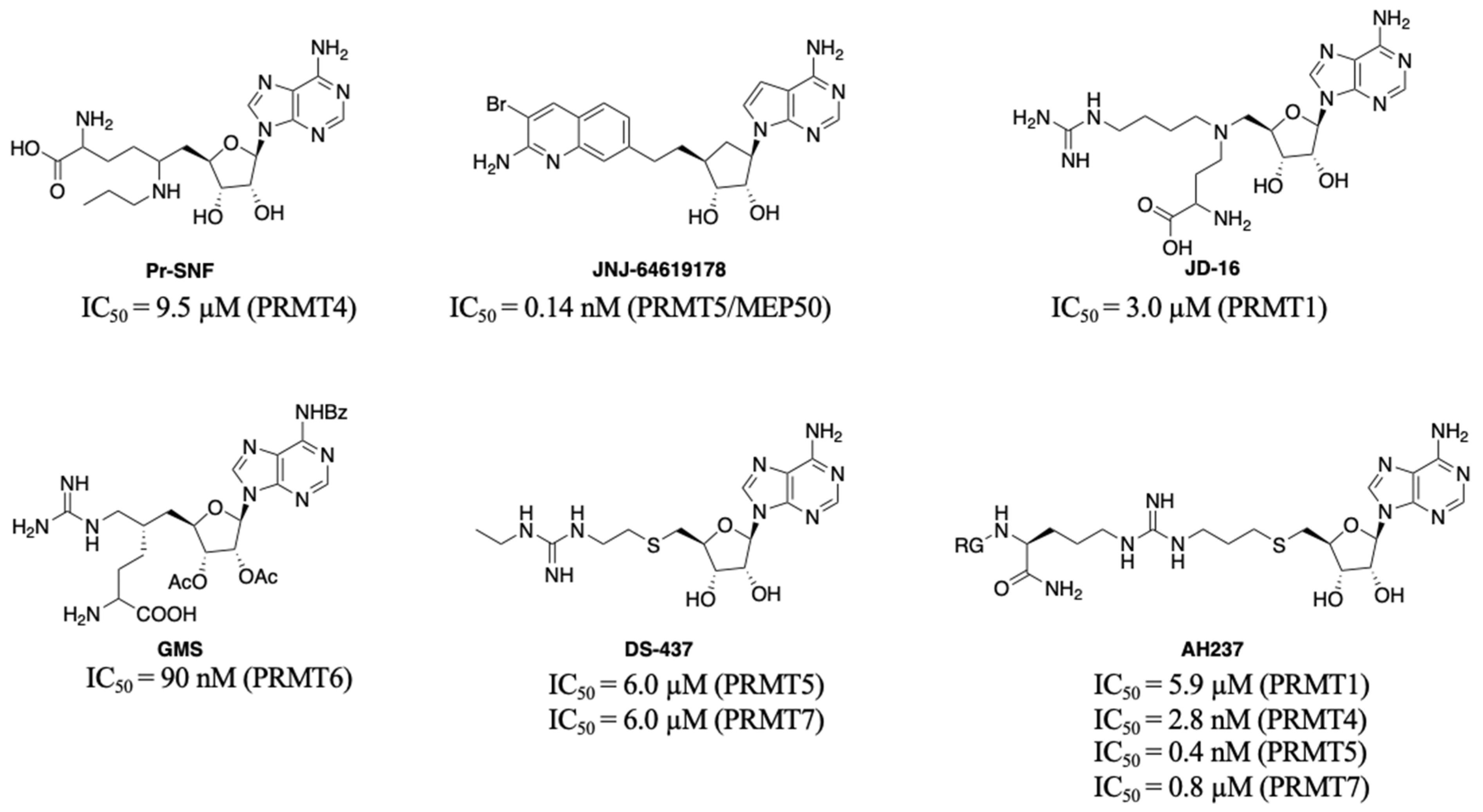
:1. Introduction
2. Design
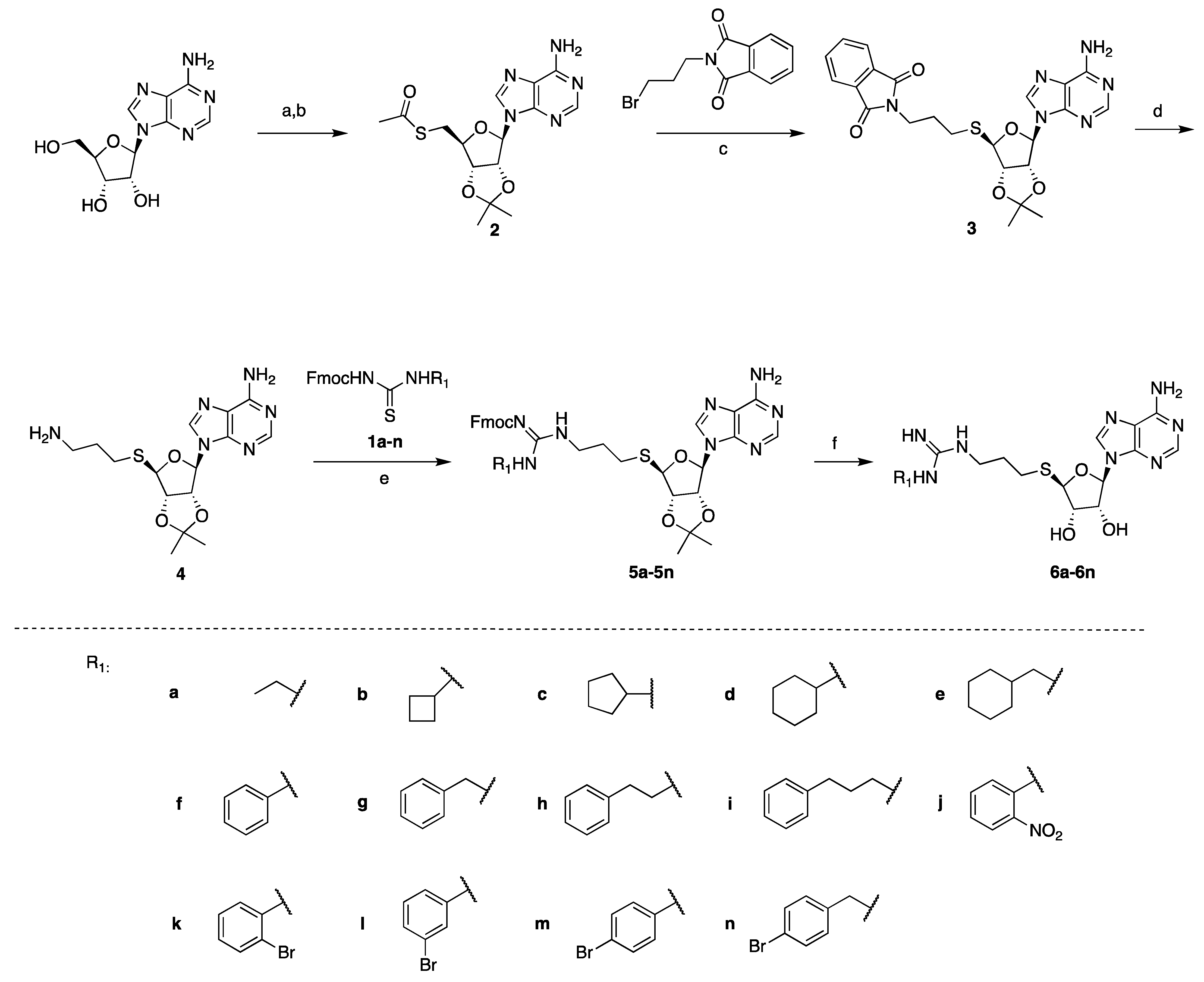
3. Synthesis
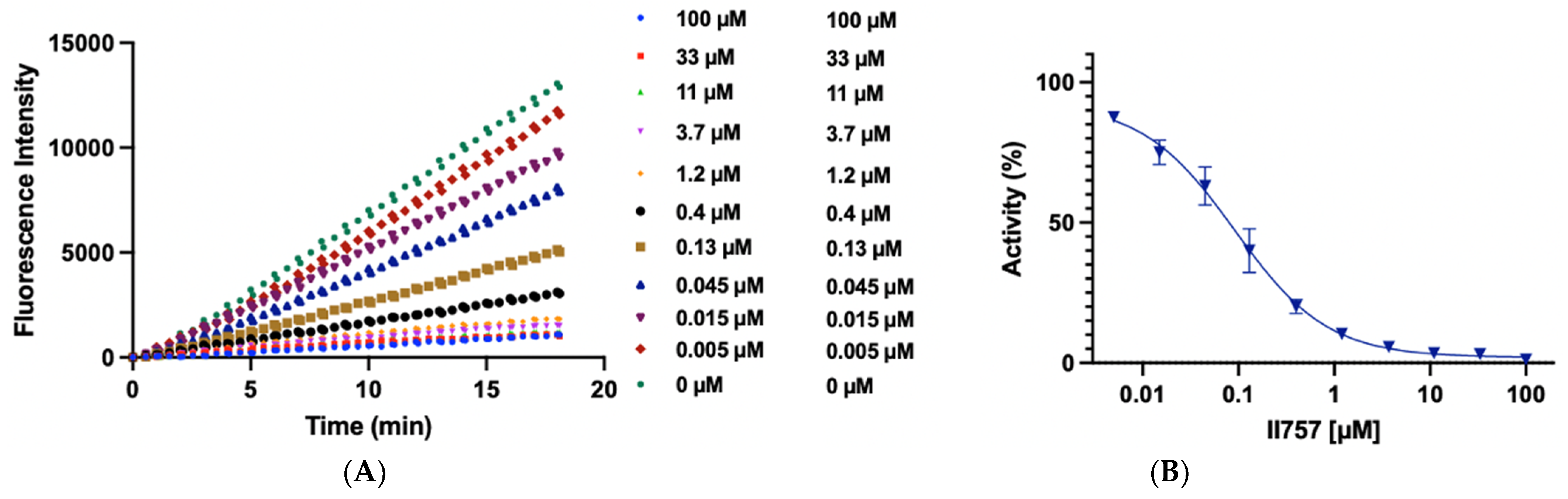
4. Biochemical Characterization
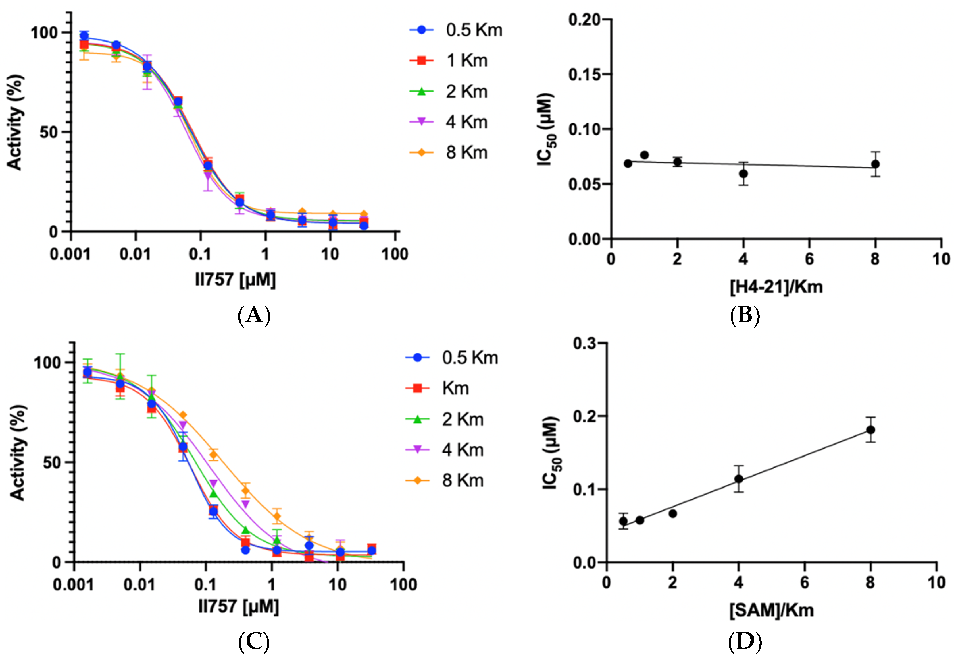
5. Inhibition Mechanism Studies
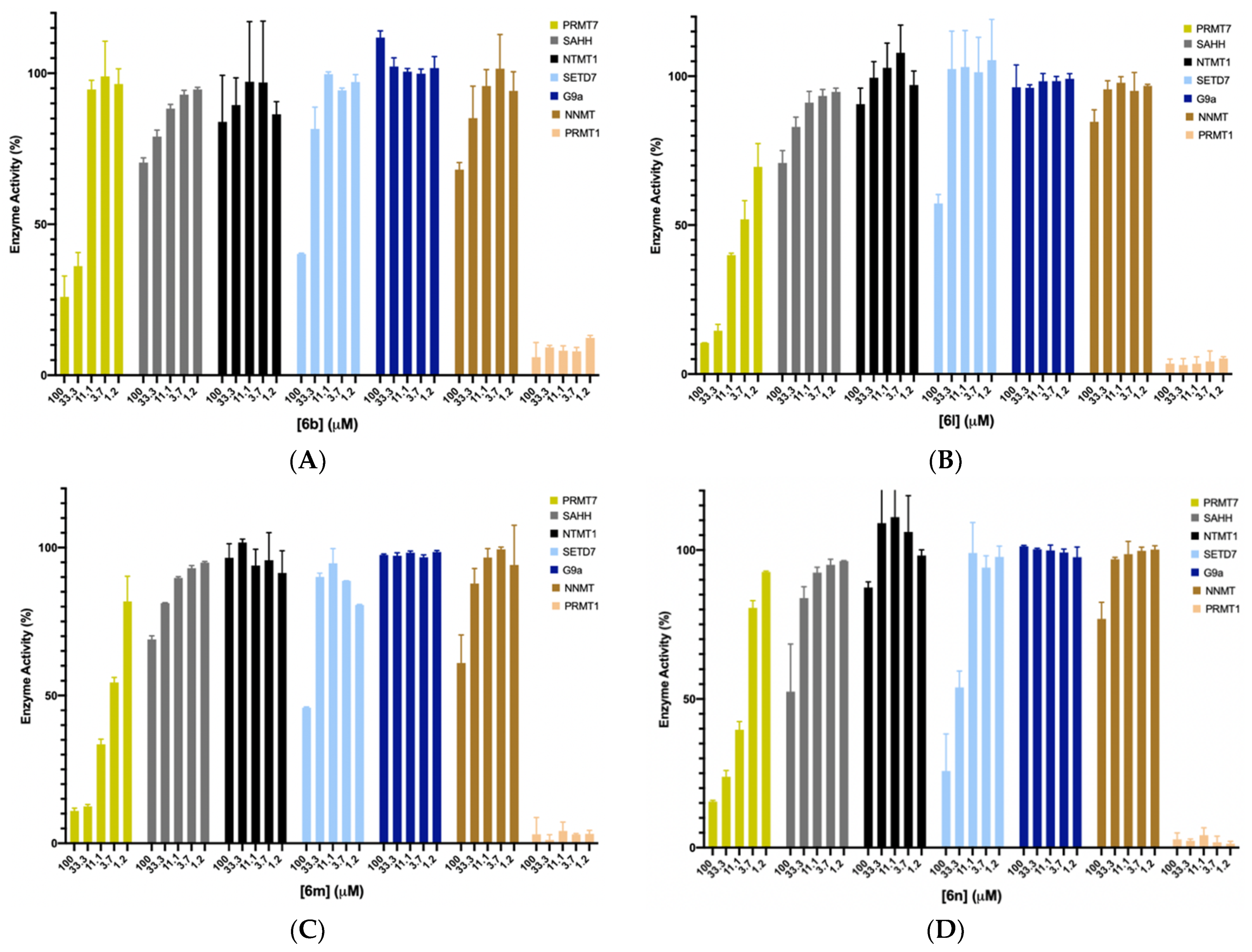
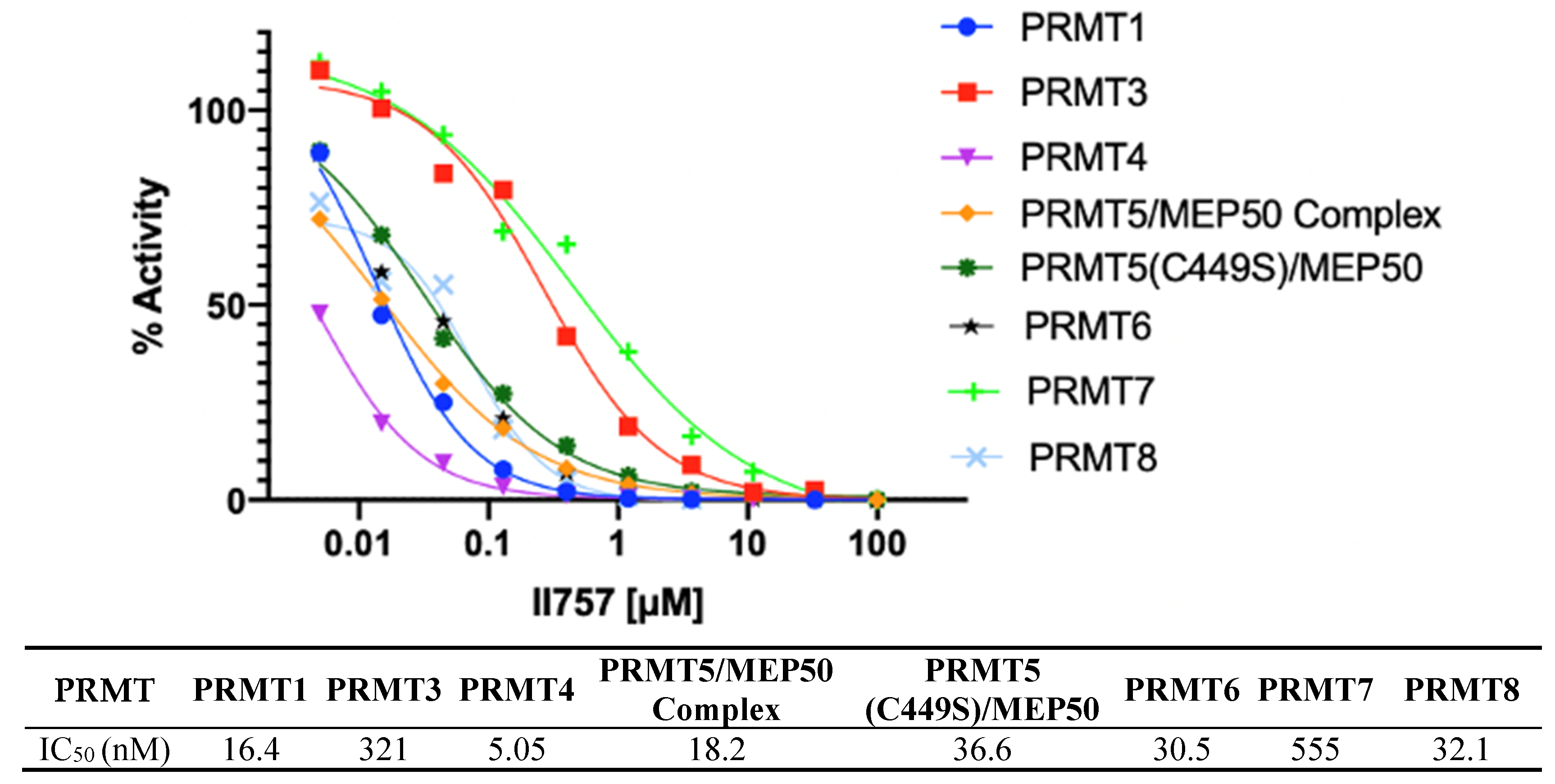
6. Selectivity Studies
7. Pan-PRMT Inhibition
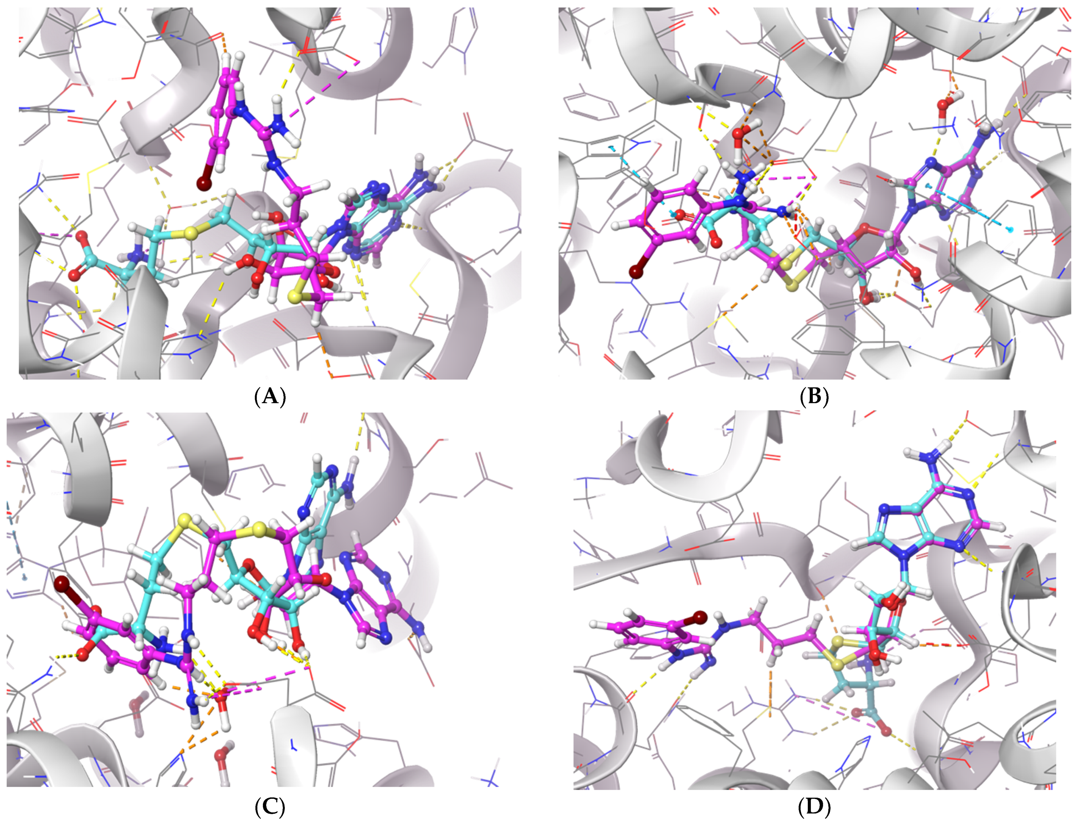
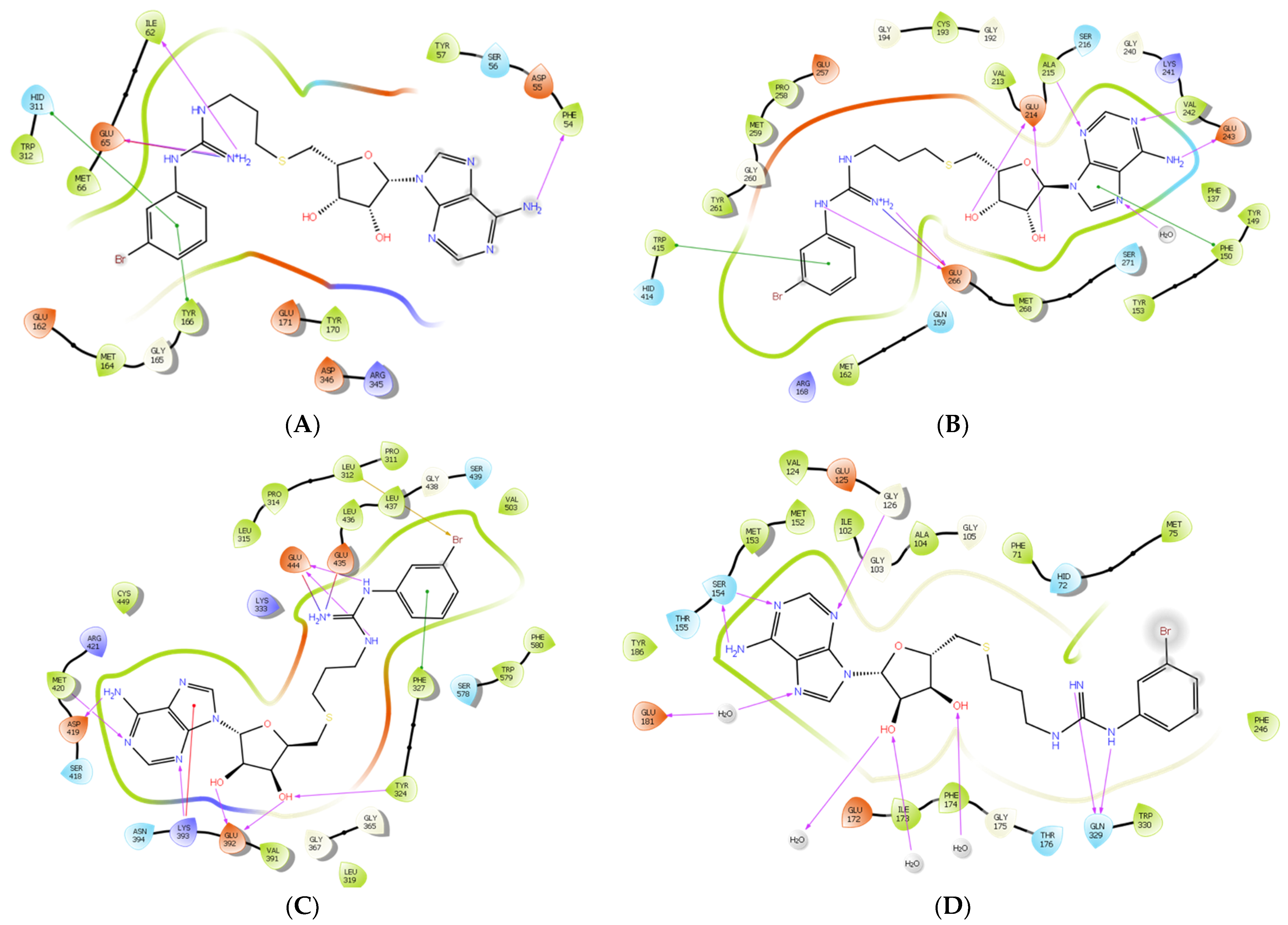
8. Molecular Docking
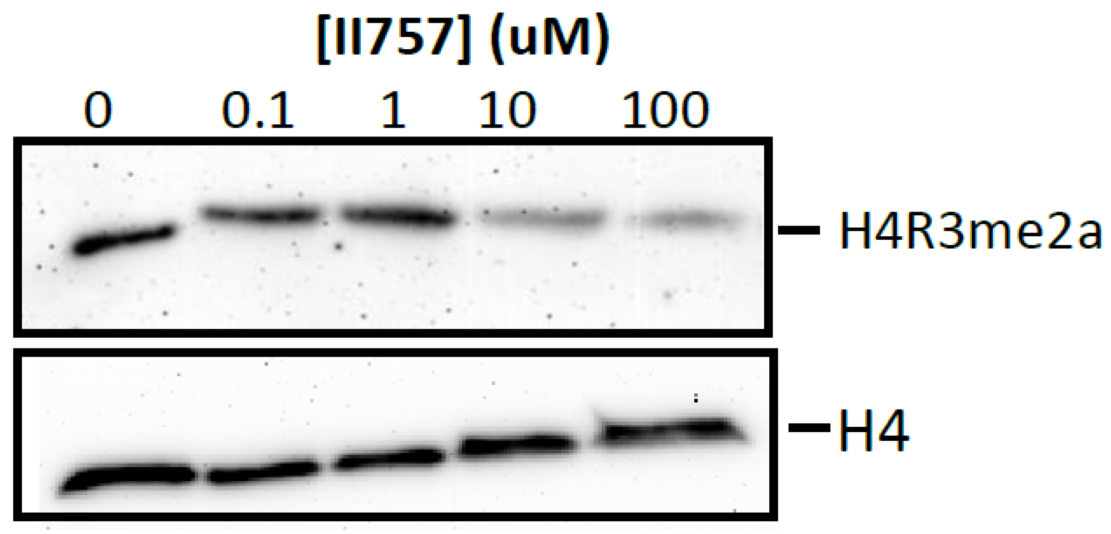
9. Inhibition on Cellular Methylation Level
10. Conclusions
11. Experimental Section
11.1. General Procedure for the Synthesis of Thiourea
11.2. General Procedure for the Synthesis of Guanidine Moiety
11.3. General Procedure for Final Compounds Synthesis
11.4. PRMT1 Biochemical Assays and Enzyme Kinetics Study
11.5. Selectivity Assays
11.6. Inhibition Mechanism Studies
11.7. Molecular Docking
11.8. Cellular Methylation Level
Supplementary Materials
Author Contributions
Funding
Institutional Review Board Statement
Informed Consent Statement
Data Availability Statement
Acknowledgments
Conflicts of Interest
References
- Yang, Y.; Bedford, M.T. Protein Arginine Methyltransferases and Cancer. Nat. Rev. Cancer 2013, 13, 37–50. [Google Scholar] [CrossRef]
- Jahan, S.; Davie, J.R. Protein Arginine Methyltransferases (PRMTs): Role in Chromatin Organization. Adv. Biol. Regul. 2015, 57, 173–184. [Google Scholar] [CrossRef] [PubMed]
- Gayatri, S.; Bedford, M.T. Readers of Histone Methylarginine Marks. Biochim. Biophys. Acta Gene Regul. Mech. 2014, 1839, 702–710. [Google Scholar] [CrossRef] [PubMed] [Green Version]
- Fuhrmann, J.; Clancy, K.W.; Thompson, P.R. Chemical Biology of Protein Arginine Modifications in Epigenetic Regulation. Chem. Rev. 2015, 115, 5413–5461. [Google Scholar] [CrossRef] [PubMed] [Green Version]
- Krause, C.D.; Yang, Z.H.; Kim, Y.S.; Lee, J.H.; Cook, J.R.; Pestka, S. Protein Arginine Methyltransferases: Evolution and Assessment of Their Pharmacological and Therapeutic Potential. Pharmacol. Ther. 2007, 113, 50–87. [Google Scholar] [CrossRef] [PubMed]
- Bedford, M.T.; Clarke, S.G. Protein Arginine Methylation in Mammals: Who, What, and Why. Mol. Cell 2009, 33, 1–13. [Google Scholar] [CrossRef] [PubMed] [Green Version]
- Blanc, R.S.; Richard, S. Arginine Methylation: The Coming of Age. Mol. Cell 2017, 65, 8–24. [Google Scholar] [CrossRef] [Green Version]
- Sun, Q.; Liu, L.; Roth, M.; Tian, J.; He, Q.; Zhong, B.; Bao, R.; Lan, X.; Jiang, C.; Sun, J.; et al. PRMT1 Upregulated by Epithelial Proinflammatory Cytokines Participates in COX2 Expression in Fibroblasts and Chronic Antigen-Induced Pulmonary Inflammation. J. Immunol. 2015, 195, 298–306. [Google Scholar] [CrossRef]
- Liu, F.; Li, F.; Ma, A.; Dobrovetsky, E.; Dong, A.; Gao, C.; Korboukh, I.; Liu, J.; Smil, D.; Brown, P.J.; et al. Exploiting an Allosteric Binding Site of PRMT3 Yields Potent and Selective Inhibitors. J. Med. Chem. 2013, 56, 2110–2124. [Google Scholar] [CrossRef] [PubMed] [Green Version]
- Palte, R.L.; Schneider, S.E.; Altman, M.D.; Hayes, R.P.; Kawamura, S.; Lacey, B.M.; Mansueto, M.S.; Reutershan, M.; Siliphaivanh, P.; Sondey, C.; et al. Allosteric Modulation of Protein Arginine Methyltransferase 5 (PRMT5). ACS Med. Chem. Lett. 2020, 11, 1688–1693. [Google Scholar] [CrossRef]
- Li, A.S.M.; Li, F.; Eram, M.S.; Bolotokova, A.; dela Seña, C.C.; Vedadi, M. Chemical Probes for Protein Arginine Methyltransferases. Methods 2020, 175, 30–43. [Google Scholar] [CrossRef]
- Chan-Penebre, E.; Kuplast, K.G.; Majer, C.R.; Boriack-Sjodin, P.A.; Wigle, T.J.; Johnston, L.D.; Rioux, N.; Munchhof, M.J.; Jin, L.; Jacques, S.L.; et al. A Selective Inhibitor of PRMT5 with in Vivo and in Vitro Potency in MCL Models. Nat. Chem. Biol. 2015, 11, 432–437. [Google Scholar] [CrossRef]
- Kaniskan, H.Ü.; Eram, M.S.; Zhao, K.; Szewczyk, M.M.; Yang, X.; Schmidt, K.; Luo, X.; Xiao, S.; Dai, M.; He, F.; et al. Discovery of Potent and Selective Allosteric Inhibitors of Protein Arginine Methyltransferase 3 (PRMT3). J. Med. Chem. 2018, 61, 1204–1217. [Google Scholar] [CrossRef] [Green Version]
- Mitchell, L.H.; Drew, A.E.; Ribich, S.A.; Rioux, N.; Swinger, K.K.; Jacques, S.L.; Lingaraj, T.; Boriack-Sjodin, P.A.; Waters, N.J.; Wigle, T.J.; et al. Aryl Pyrazoles as Potent Inhibitors of Arginine Methyltransferases: Identification of the First PRMT6 Tool Compound. ACS Med. Chem. Lett. 2015, 6, 655–659. [Google Scholar] [CrossRef] [PubMed] [Green Version]
- Zheng, W.; Ibáñez, G.; Wu, H.; Blum, G.; Zeng, H.; Dong, A.; Li, F.; Hajian, T.; Allali-Hassani, A.; Amaya, M.F.; et al. Sinefungin Derivatives as Inhibitors and Structure Probes of Protein Lysine Methyltransferase SETD2. J. Am. Chem. Soc. 2012, 134, 18004–18014. [Google Scholar] [CrossRef] [Green Version]
- Wu, T.; Millar, H.; Gaffney, D.; Beke, L.; Mannens, G.; Vinken, P.; Sommers, I.; Thuring, J.-W.; Sun, W.; Moy, C.; et al. Abstract 4859: JNJ-64619178, a Selective and Pseudo-Irreversible PRMT5 Inhibitor with Potent in vitro and in vivo Activity, Demonstrated in Several Lung Cancer Models. Cancer Res. 2018, 78 (Suppl. 13), 4859. [Google Scholar] [CrossRef]
- Osborne, T.; Weller Roska, R.L.; Rajski, S.R.; Thompson, P.R. In Situ Generation of a Bisubstrate Analogue for Protein Arginine Methyltransferase 1. J. Am. Chem. Soc. 2008, 130, 4574–4575. [Google Scholar] [CrossRef]
- Weller, R.L.; Rajski, S.R. Design, Synthesis, and Preliminary Biological Evaluation of a DNA Methyltransferase-Directed Alkylating Agent. ChemBioChem 2006, 7, 243–245. [Google Scholar] [CrossRef]
- Dowden, J.; Hong, W.; Parry, R.V.; Pike, R.A.; Ward, S.G. Toward the Development of Potent and Selective Bisubstrate Inhibitors of Protein Arginine Methyltransferases. Bioorg. Med. Chem. Lett. 2010, 20, 2103–2105. [Google Scholar] [CrossRef] [PubMed]
- Dowden, J.; Pike, R.A.; Parry, R.V.; Hong, W.; Muhsen, U.A.; Ward, S.G. Small Molecule Inhibitors That Discriminate between Protein Arginine N-Methyltransferases PRMT1 and CARM1. Org. Biomol. Chem. 2011, 9, 7814–7821. [Google Scholar] [CrossRef]
- Wu, H.; Zheng, W.; Eram, M.S.; Vhuiyan, M.C.; Dong, A.; Zeng, H.; He, H.; Brown, P.; Frankel, A.; Vedadi, M.; et al. Structural Basis of Arginine Asymmetrical Dimethylation by PRMT6. Biochem. J. 2016, 473, 3049–3063. [Google Scholar] [CrossRef] [PubMed] [Green Version]
- Smil, D.; Eram, M.S.; Li, F.; Kennedy, S.; Szewczyk, M.M.; Brown, P.J.; Barsyte-Lovejoy, D.; Arrowsmith, C.H.; Vedadi, M.; Schapira, M. Discovery of a Dual PRMT5-PRMT7 Inhibitor. ACS Med. Chem. Lett. 2015, 6, 408–412. [Google Scholar] [CrossRef] [Green Version]
- Al-Hamashi, A.A.; Chen, D.; Deng, Y.; Dong, G.; Huang, R. Discovery of a Potent and Dual-Selective Bisubstrate Inhibitor for Protein Arginine Methyltransferase 4/5. Acta Pharm. Sin. B 2020. [Google Scholar] [CrossRef]
- Lin, H.; Luengo, J.I. Nucleoside Protein Arginine Methyltransferase 5 (PRMT5) Inhibitors. Bioorg. Med. Chem. Lett. 2019, 29, 1264–1269. [Google Scholar] [CrossRef] [PubMed]
- van Haren, M.J.; Marechal, N.; Troffer-Charlier, N.; Cianciulli, A.; Sbardella, G.; Cavarelli, J.; Martin, N.I. Transition State Mimics Are Valuable Mechanistic Probes for Structural Studies with the Arginine Methyltransferase CARM1. Proc. Natl. Acad. Sci. USA 2017, 114, 3625–3630. [Google Scholar] [CrossRef] [PubMed] [Green Version]


 | |||
|---|---|---|---|
| Compound | R1 | IC50 (µM) * | Ki, app (µM) b |
| 6a |  | 0.62 ± 0.08 | 0.31 ± 0.04 |
| 6b |  | 0.23 ± 0.02 a | 0.045 ± 0.005 |
| 6c |  | 0.22 ± 0.016 | 0.11 ± 0.008 |
| 6d |  | 0.11 ± 0.048 | 0.055 ± 0.024 |
| 6e |  | 0.12 ± 0.026 | 0.06 ± 0.013 |
| 6f |  | 0.29 ± 0.01 | 0.15 ± 0.005 |
| 6g |  | 0.72 ± 0.15 | 0.36 ± 0.075 |
| 6h |  | 0.21 ± 0.06 | 0.11 ± 0.03 |
| 6i |  | 0.33 ±0.11 | 0.17 ± 0.055 |
| 6j |  | 0.21 ± 0.01 | 0.11 ± 0.005 |
| 6k |  | 0.44 ± 0.03 | 0.22 ± 0.015 |
| 6l (II757) |  | 0.09 ± 0.04 a | 0.018 ± 0.008 |
| 6m |  | 0.29 ± 0.04 a | 0.058 ± 0.007 |
| 6n |  | 0.26 ± 0.07 a | 0.052 ± 0.014 |
Publisher’s Note: MDPI stays neutral with regard to jurisdictional claims in published maps and institutional affiliations. |
© 2021 by the authors. Licensee MDPI, Basel, Switzerland. This article is an open access article distributed under the terms and conditions of the Creative Commons Attribution (CC BY) license (https://creativecommons.org/licenses/by/4.0/).
Share and Cite
Iyamu, I.D.; Al-Hamashi, A.A.; Huang, R. A Pan-Inhibitor for Protein Arginine Methyltransferase Family Enzymes. Biomolecules 2021, 11, 854. https://doi.org/10.3390/biom11060854
Iyamu ID, Al-Hamashi AA, Huang R. A Pan-Inhibitor for Protein Arginine Methyltransferase Family Enzymes. Biomolecules. 2021; 11(6):854. https://doi.org/10.3390/biom11060854
Chicago/Turabian StyleIyamu, Iredia D., Ayad A. Al-Hamashi, and Rong Huang. 2021. "A Pan-Inhibitor for Protein Arginine Methyltransferase Family Enzymes" Biomolecules 11, no. 6: 854. https://doi.org/10.3390/biom11060854