Review

The Role of Stress Proteins in Haloarchaea and Their Adaptive Response to Environmental Shifts

Agrochemistry and Biochemistry Department, Biochemistry and Molecular Biology Area, Faculty of Science, University of Alicante, Ap 99, 03080 Alicante, Spain
*
Author to whom correspondence should be addressed.
Biomolecules 2020, 10(10), 1390; https://doi.org/10.3390/biom10101390
Submission received: 31 July 2020 / Revised: 18 September 2020 / Accepted: 24 September 2020 / Published: 29 September 2020
(This article belongs to the Collection Archaea: Diversity, Metabolism and Molecular Biology)

Abstract

:
Over the years, in order to survive in their natural environment, microbial communities have acquired adaptations to nonoptimal growth conditions. These shifts are usually related to stress conditions such as low/high solar radiation, extreme temperatures, oxidative stress, pH variations, changes in salinity, or a high concentration of heavy metals. In addition, climate change is resulting in these stress conditions becoming more significant due to the frequency and intensity of extreme weather events. The most relevant damaging effect of these stressors is protein denaturation. To cope with this effect, organisms have developed different mechanisms, wherein the stress genes play an important role in deciding which of them survive. Each organism has different responses that involve the activation of many genes and molecules as well as downregulation of other genes and pathways. Focused on salinity stress, the archaeal domain encompasses the most significant extremophiles living in high-salinity environments. To have the capacity to withstand this high salinity without losing protein structure and function, the microorganisms have distinct adaptations. The haloarchaeal stress response protects cells against abiotic stressors through the synthesis of stress proteins. This includes other heat shock stress proteins (Hsp), thermoprotectants, survival proteins, universal stress proteins, and multicellular structures. Gene and family stress proteins are highly conserved among members of the halophilic archaea and their study should continue in order to develop means to improve for biotechnological purposes. In this review, all the mechanisms to cope with stress response by haloarchaea are discussed from a global perspective, specifically focusing on the role played by universal stress proteins.

1. Introduction

Over the years, organisms have survived a wide range of changing conditions such as temperature, salinity, oxygen levels, pH, nutrients, radiation, pressure, and toxic chemicals [1]. These agents, known as stressors, alter the status and homeostasis of the cells in all three domains of life [2]. Cell stressors can be classified by their nature: physical (heat, several types of irradiation, pressure, high salinity, sound), oxidative (H2O2, reactive oxygen species (ROS), anaerobiosis to aerobiosis shift), pH (alkalinity and acidity), osmotic (changes in the concentration of osmolytes), nutritional (starvation of carbon, nitrogen or phosphate), antibiotics (tetracycline, puromycin), alcohols (ethanol, methanol, butanol, propanol, octanol), metals (cadmium, copper, zinc, lead, nickel, tin, aluminum, chromium), insecticides (lindane, diazinon, paraquat), mechanical (compression, shearing), and others (benzene, phenol, mutagens, ammonia, arsenite, arsenate, amino acids, desiccation, anesthetics) [3]. Some organisms have been able to develop different strategies that have allowed them to adapt to these stress conditions. This has led to a great variety of habitats spread over the world existing today that are optimal for a wide range of organisms, but uninhabitable for others organisms [1]; for example, high salinity acts as a stressor for a given organism but at the same time can act as an optimal growth condition for another organism [2]. It is well known that the central event caused by a stressor on the cell is protein denaturation [4]. However, microorganisms have developed other molecular adaptation mechanisms to each stressful condition such as overexpression of stress genes or downregulation of nonstress genes.
All three domains of life are exposed to environmental stresses that usually cause a modification in their growth. Today, the effects of abiotic stresses over cells are studied under controlled growth conditions in the laboratory using not only standard methods, but also omics techniques [4,5]. However, it is difficult to perform this type of study under natural conditions because organisms found in their natural ecosystems may grow under different simultaneous stresses, e.g., salinity and heat or drought and salinity, consequently, stress responses are usually multifactorial and integrated. Over the years, biological systems have developed mechanisms to respond in an appropriate way to natural environmental stresses that can end up damaging the DNA and proteins in cells. In order to study the response to a defined experimental perturbation, system approaches allow the formulation of a hypothesis that simulates cycles of perturbations and analysis to verify them [6].
In ecological niches and habitats, microbial life has started to be affected due to increased anthropogenic activity as well as extreme changes in the ecosystems as a result of global warming. In fact, the majority of organisms are not able to deal with rapid changes because they need optimal conditions to maintain their growth, while prokaryotes like extremophiles are able to adapt and survive under extreme conditions [7]. An example of extremophiles are halophilic organisms like Haloarchaea that belong to the Archaea domain and require at least 1.5 mol/L of sodium chloride to survive and grow [8]. These microorganisms mainly inhabit saline and hypersaline ecosystems and therefore may be exposed to changes in salinity due to rain and water runoff that would have the effect of diluting the normal salt content. It should be noted that these saline environments are strongly influenced by climate variability and extreme weather events. For the time being, there is no accurate data on their whole extension in the world, but the incidence and extent of these hypersaline areas are increasing due to the effects of climate change and anthropogenic activities such as desertification [9]. Not only are halophilic Archaea exposed to salinity stress, but also to ultraviolet (UV) radiations, high ionic stress, high temperature, and alkaline pH [7]. Needless to say, climate change is predicted to lead to more frequent and severe stressful events such as heavy rainfall or an increase in temperature [10,11,12]. Therefore, it is essential to analyze the effects of climate change conditions on microbial communities and understand how these communities recover cell homeostasis after their exposure to different stress conditions [13].
Focusing on the Haloarchaea, this review provides an overview of the proteins and biomolecules involved in their environmental stress responses. Knowledge about how and when haloarchaeal stress genes are expressed and how they respond to these adverse stimuli is less advanced than in other group of prokaryotes. Transcription stress genes and their regulation mechanisms are still unclear and, in general, very little is known about these genes in comparison with those from bacteria and eukaryotes. Thus, the present work represents an excellent basis to further advance not only the study of the Archaea domain under stress conditions, but also in the field of biotechnology, as it could find applications of industrial (protein production or design of stress resistant microorganisms) or environmental interest (global warming).

2. Stress in Halophilic Archaea

The central characteristic of hypersaline environments (soda lakes, lagoons, saltern ponds, salt deposits, artificial solar salterns, submarine salt domes, surface salt deposits and salt marshes) is the high concentration of total salt found in water and soil higher than 3.5% (w/v) of seawater [14,15,16,17]. These saline and hypersaline environments are found throughout the world; where both saline/hypersaline environments and semi-arid areas are concomitant, drylands cover about 41% of the Earth’s total land surface [18]. Extreme halophiles (salt-loving organisms) found in all three branches of life (Bacteria, Archaea, and Eukarya) inhabit these natural environments. Within the Archaea domain, the most salt-requiring microorganisms are extreme halophiles belonging to the Haloarchaea class [19]. Members of this class stand out for being obligate halophiles, but also for being facultative aerobes, heterotrophic, slightly thermophilic, and phototrophic [17]. Moreover, haloarchaea require a salt concentration of 10–35% (w/v) for optimal growth and constitute the dominant microbial population when the salt concentration exceeds 16% (w/v) [15,16]. In order to withstand this high salinity without losing protein structure and function, these microorganisms have developed different molecular adaptations, such as a high density of acidic residues on the surface of haloarchaeal proteins and a decrease in exposed hydrophobic residues through loss of surface lysine [20,21,22]. These negative charges on the surface form clusters that bind networks of hydrated ions, which prevent protein precipitation in the presence of a high cytoplasmic KCl concentration. In contrast, an absence of salt promotes unfolding of halophilic proteins due to repulsive forces between negative charges of acidic amino acids [14,15,16,20]
From an ecological point of view, there are well known effects due to the high content of salts, whereby the overall biodiversity decreases as halotolerant and halophilic species develop and replace halosensitive biota [23]. The properties of the salterns are stable from year to year, so the gradient of salinity remains relatively constant, which allows communities of halophilic and halotolerant organisms to adapt to local salinity [24]. Nonetheless, many haloarchaea species thrive despite a wide variety of environmental stresses (Table 1), to which they exhibit considerable tolerance, making them interesting from an ecological perspective. As they have developed strategies enabling them to grow and proliferate under a multitude of extreme conditions, some haloarchaea species are considered “polyextremophiles” [17]. At the time of this review, Halobacterium sp. NRC-1 is being used as a model organism for the study of tolerance mechanisms to environmental stress agents due to its extremophilic character as well as it also being widely distributed in nature.
Since members of Haloarchaea thrive in extremely saline environments, one of the potential problems to address is osmotic stress due to desiccation and dilution. For instance, rainfall and evaporation may cause major changes in salinity, and halophilic Archaea have to adapt their metabolism in order to survive. Consequently, they have mastered the art of osmoadaptation to salt at the gene and protein levels [49]. These natural hypersaline environments are characterized by low water activity due to high NaCl concentrations, so microorganisms living there must develop mechanisms to prevent water loss by osmosis to avoid protein inactivation, inhibition, and even denaturation. Basically, there are two broad strategies, “salt in” and “low salt in” used to resist osmotic stress and achieve osmotic stability by halophilic microorganisms in hypersaline environments [50]. The “salt in” effect is commonly used by Haloarchaea and consists of compensating ion levels: KCl usually provides an osmotic balance. This accumulation of dominant ion K+ and also of Na+ within cells is maintained by ion pumps and protein transport [50,51]. By contrast, the most used strategy in the Bacteria domain, “low salt in” is used to produce or accumulate compatible osmotic solutes at low concentration in the cytoplasmic medium, such as sugar derivates, polyols, betaines, thetines, amino acids, glutamine amide derivates, and ectoines that have osmotic potential. Unusually, the genera Natronobacterium and Natronococcus accumulate as solutes 2-sulphotrehalose and glycine, respectively [50,52,53]. This is one of the few examples of Haloarchaea that use the “low salt in” strategy, with the accumulation of intracellular KCl being the most common strategy in the Archaea domain. Halobacterium sp. NRC-1 is one of the most well-known systems. It has several genes that code for multiple active K+ transporters and an active Na+ efflux system involved in the maintenance of the intracellular ionic concentrations appropriate for growth [54,55].
Temperature in high-salinity environments can vary widely, both daily and seasonally, and is considered another relevant limiting factor in the survival of many halophilic microorganisms [44,56]. In order to maintain physiological balance under challenging temperature conditions, Haloarchaea must withstand these changes. In the case of the haloarchaeon Halobacterium sp. NRC-1, its exposure to extreme temperatures elicits specific responses to promote the protein folding and limiting factors responsible for growth inhibition [25]. Indeed, this haloarchaeon showed induction of molecular chaperones, which are a diverse group of proteins that assists in the folding/unfolding and assembly/disassembly of other macromolecular structures [25,57,58]. On the other hand, in response to cold temperatures, Halobacterium sp. NRC-1 changes the expression of cold shock proteins, alters polar lipids biosynthesis, and increases the expression gas vesicles [25]. These mechanisms that adjust differential gene expression are used to maintain homeostasis as environmental conditions change.
Haloarchaea have been isolated from saline environments with a broad pH range, from extremely acidic lakes such as Lake Afrera, Ethiopia [59] to environments with a very high pH, such as Lake Magadi, Africa and Mono Lake, California [60,61]. However, little is known about the molecular adaptations associated with the growth of haloarchaea at extreme pH values. One of the few studies performed on haloarchaea described the transcriptomic profile differences of Halorubrum lacusprofundi, Haloferax volcanii, and Halobacterium sp. NRC-1 linked to extreme pH values. This work concludes that alkaline conditions promote, in the three species analyzed, the overexpression of genes involved in stress, such as the hsp20 family, uspA, or groEL chaperone. This response is quite similar to that described in other prokaryotes belonging to the Bacteria domain. However, under acidic conditions, Hrr. lacusprofundi presents a different transcriptomic profile to Hfx. volcanii and Halobacterium sp. NRC1. In particular, transcripts related to spore formation and dormancy are up-regulated in Hrr. lacusprofundi, which suggests that this microorganism has a species-specific response and becomes dormant at an acidic pH [34].
Some halophilic habitats contain a high concentration of metals, since various anthropogenic activities like urbanization and industrialization contribute to heavy metal pollution of these sites [62]. Such heavy metal pollution is an important environmental issue due to the toxic effects of metals, and their accumulation leads to health and ecological problems [63]. Therefore, haloarchaea have developed different metal-resistance mechanisms (extracellular metal sequestration by biopolymers, metal efflux mediated by specific transporters and enzymatic detoxification) to withstand stress from transition metals such as arsenic, cadmium, copper, cobalt, zinc, and iron [39,64], which allow them to tolerate a broad range of these metal concentrations [42,43,64]. For instance, in Halobacterium sp. NRC-1, has developed two different arsenic resistant mechanisms. In the first one, arsenic is transported out of the cell by promoting the expression of a cluster of genes (arsADRC) encoding enzymes that alter the metal oxidation state and, in the other, arsenic is converted to methylated species like dimethylarsinate (DMA), trimethylarsine oxide (TMAO), or trimethylarsine (TMA) gas thanks to arsR2M operon [65,66].
Although temperature, pH, and salinity, as previously mentioned, are potential stressors, oxygen is also considered a relevant stressor. Halophilic archaea generally grow under aerobic conditions in hypersaline environments, despite possessing anaerobic capabilities. The poor concentration of oxygen in hypersaline environments strictly regulates anaerobic metabolism in order to allow organisms to thrive in their natural environment. Under low oxygen conditions, some members of halophilic archaea such as Halobacterium salinarum and Haloferax mediterranei produce gas vesicles to enable cells to float vertically and reach the oxygen-rich surface layers in the water column, where the oxygen concentration is higher to prevent oxygen limitation and sustain aerobic growth [45,67,68]. However, this strategy is highly limited, depending on external factors such as light, salt concentration, temperature, and oxygen availability. However, under strict anaerobic conditions, some halophilic archaea species like Hfx. mediterranei are able to use alternative electron acceptors, such as nitrate, dimethyl sulfoxide (DMSO), trimethylamine N-oxide (TMAO), chlorate, and perchlorate, as a strategy to maintain a respiratory metabolism that supports cell growth [46,47,48,69]. On the other hand, Halobacterium sp. NRC-1 was found to grow on either DMSO or TMAO, and, to some extent, fumarate, as the terminal electron acceptor, but with nitrate no significant growth was observed [70,71]; no growth was detected in others like Haloquadratum walsbyi due to anaerobic respiration [72].
Recent studies have explored the tolerance of extremely halophilic archaea to high levels of sunlight in the natural environment. As halophilic archaea are found in saltern crystallizer ponds, they are exposed to intense solar radiations, which damage their DNA. In fact, haloarchaea are considered the second most resistant microorganism worldwide, only slightly less resistant than Denicoccus radiodurans [73]. Under these conditions, halophilic archaea, apart from the mechanism for repairing DNA damage (photoreactivation, nucleotide excision repair, base excision repair, and homologous recombination), have developed different strategies for the tolerance, survival, and maintenance of their growth that provide protection from the consequences of habitual exposure to intense UV. These strategies include the synthesis of carotenoids, oxidative damage avoidance, polyploidy, and a genome with high CG content [74]. Photoreactivation is likely to be the most important strategy for dealing with UV irradiation [75]. Halobacterium sp. NRC-1 is highly resistant to the effects of UV because it undergoes efficient photoreactivation of DNA damage; for that reason, it has been used as a model organism in a wide variety of studies on tolerance to UV and ionizing radiation tolerance [74,75,76,77]. It is remarkable that Halobacterium sp. NRC-1 is resistant to high UV radiation withstanding up to 110 J/m2 without losing its viability [6]. Not only can Halobacterium sp. NRC-1 tolerate UV radiation; it can also resist gamma radiation due to its ability to repair double-strand DNA breaks [73,78].

3. Response to Stress in Halophilic Archaea

Microorganisms achieve their physiological functions and survive only if they have proteins that are synthesized in optimal concentrations and folded in a correct three-dimensional configuration, that is to say, a native configuration [4]. By contrast, there are some occasions when proteins lose their functional configuration and become denatured. This process may in some cases be reversible, as mentioned above, with partial protein unfolding and loss of function being the major effects of a stressor [79,80]. Thus, the stress response counteracts the effects caused by denatured proteins or the process of avoiding protein denaturation.
The stress response manifests itself in several ways: down-regulation of many housekeeping genes; activation of stress genes; partial protein unfolding; housekeeping gene repression and down-regulation; variations in cell mobility; changes in the permeability of the cell membrane to some compound; cell aggregation which results in multicellular structures of different complexity; and other phenomena in terms of changes in electrolytes or other components [4,80]. Some of the above mentioned events may occur in response to stressful conditions; however, the down-regulation of many housekeeping genes and the activation of stress genes are the most important strategies for cells under stress conditions [79].
There are several strategies to protect the cell against stressors in the domain Archaea: stress proteins, chaperones, thermoprotectants, the proteasome, multicellular structures, and other molecules whose function is not fully understood [80]. All these components are useful in order to ensure tolerance, macromolecular repair, and cell homeostasis maintenance. Indeed, the knowledge of the molecular mechanisms involved in the regulation stress genes of halophilic archaea that thrive under stressful conditions is fundamental. These studies are relevant for the exploration of ecosystems that are not habitable to humans, mammals, or plants. In addition, some recent research suggests that haloarchaeal species like Hfx. volcanii and Halobacterium sp. NRC-1 may be a candidate species for life on Mars as well. This possibility is being considered since haloarchaea are resistant to extreme conditions, and evidence suggests that chloride and sulfate salts are abundant on Mars [81].

3.1. Heat Shock and Stress Proteins

In general terms, an important component of the stress response is the activation of stress genes, increasing their transcriptional activity in order to protect the cell. However, not only are stress genes actively transcribed under non-physiological normal conditions, but also they have transcriptional activity in the absence of stress [80]. The products of stress genes against temperature are called Heat-shock response (Hsp), which can be classified according to mass. Hsp refers to a protein encoded by a gene that is induced by heat shock. Nowadays, considering the information available on stress proteins, the overwhelming majority comes from bacteria and eukaryotes, not from Archaea. Thus, it is crucial to identify the genes involved in cell survival under stress conditions in halophilic organisms, in order to improve the knowledge and use of these genes for biotechnological purposes. Furthermore, understanding stress proteins will help us to design several more efficient gene engineering strategies.
To sum up, the main known functions of stress proteins are to assist other proteins in the cell with their folding, translocation, and assembly, but also to contribute to the elimination of proteins that may endanger cell viability and have lost their usefulness [82,83,84,85]. They are also involved in some other functions, such as binding to denatured proteins [86], preventing enzymes from denaturation and aggregation [87], participation in signal transduction pathways [88], rRNA processing [89], etc. Therefore, stress proteins are considered multifunctional, ubiquitous, and promiscuous [80]. The motifs identification of these proteins helps to highlight their diversity of function.
Hsps are classified into six major conserved families according to their molecular weight (MW): Hsp100, Hsp90, Hsp70, Hsp60, Hsp40, and sHsp (small heat shock proteins). The two most studied families are Hsp70 and Hsp60, which contain 70 kDa and 60 kDa, respectively. These have mostly been studied in many bacterial and eukaryotic species. The Hsp60 family is also called chaperonins and is included within the molecular chaperones. Hsp70 (DnaK) carries out its chaperone functions by teaming up with Hsp40 (DnaJ) and GrpE in the cytosol [90,91]. This archaeal machinery is much closer to bacterial equivalents due to previous events of lateral transfer from bacteria [92], whereas, chaperonins are closely related to eukaryotic counterparts [57,58]. In addition, the chaperone machinery (DnaK, DnaJ, GrpE) is usually present in bacteria and eukaryotes, while some species of archaea lack it or do not have all the components of this machinery [5,92,93].
To have an idea of the representation of all Hsp families included in haloarchaea, in Table 2, it is shown that not all the families of Hsp are present in the halophilic archaea. The heavy heat shock protein family, Hsp100, is present neither in halophilic archaea nor in any archaeal species. The most common families in haloarchaea are Hsp70 and Hsp60. These families have been studied for years in Bacteria and Eukarya.

3.1.1. Molecular Chaperones

When the cell has to respond to difficult environmental situations, stress proteins play the most important role; among them are the molecular chaperones [5]. In the domain Archaea, systems like the chaperone machine and chaperonins are better known than stress genes and proteins; in fact, the expression and regulation of the chaperonin machine have also been studied. There are extensive studies about the impact on the chaperonin gene expression under salt stress conditions. These studies conclude that chaperonins are likely to play important roles in salt tolerance, for example, in the case of the halophilic archaeon Methanohalophilus portucalensis, the expression profile changes in response to hyper-salt stress and hypo-salt stress [94].
Some stress proteins are chaperones (Table 2) whose main function is to help fold newly synthesized cytosolic proteins into their functional conformation, preventing misfolded protein structures during and after translation, refolding denatured proteins due to a stress in the cytosol, assisting with the modulation of the oligomeric state of protein complexes, facilitating protein degradation and translocations across biological membranes, and guiding proteins in the migration to the cells where they will reside and function [95,96,97,98,99]. It is important to keep in mind that not all stress proteins are chaperones, and not all molecular chaperones are stress proteins, as some chaperone genes are not stress inducible. Chaperones and chaperonins play an important role in the maintenance of cellular proteins at the physiological level; when there is an alteration in the native conformation of a protein, the stress response will be induced and Hsp will be activated including the chaperones and chaperonins. The halophilic archaea families that contain molecular chaperone machinery genes are summarized in Table 3. These data may change as the number of sequenced archaeal genomes increases. It should be taken into account that the components of the molecular chaperone machine are highly conserved in bacteria and also in eukaryotes, but not in archaea since many archaeal species do not have them [92,100]. Despite this, all haloarchaea families have the chaperone machinery. According to the sequenced genome of the species included in haloarchaeal families, the largest number of chaperone proteins are found in Halorubraceae, Natrialbaceae, and Haloferacaceae, which contain more than 60 proteins.

3.1.2. Peptidyl-Propyl Cis–Trans Isomerases

Another relevant family within the Hsp is sHsp, which has a molecular mass lower than 34 kDa (Table 2). Peptidyl-propyl cis–trans isomerase (PPIase) is one of the members of this family. PPIase is an enzyme found in various organisms belonging to the three phylogenetic domains of life. Its function is to mediate the peptidyl-prolyl isomerization; it is a step in the folding of proteins, so it catalyzes the isomerization between the cis and trans forms of the peptide bonds [79,101]. In other words, protein folding requires rotation of the peptidyl-propyl bonds and this enzyme catalyzes this rotation making it faster [2]. PPIases are divided into three unrelated structural families: cyclophilins (Cyp), FK506 binding proteins (FKBPs), and the parvulin family [102,103]. This enzyme has been found in the genomes of all six families of haloarchaea, but there have still been no characterization studies of PPIases in any species. In archaea, there has been research into the hyperthermophilic methanarchaeon Methanococcus jannaschii in response to heat and cold shock, where a PPIase belonging to the FKBP family is characterized in some detail [104]. Few PPIases are found in archaeal microorganisms. Future studies will deepen our understanding of PPIases in halophilic archaea to find out how the folding machinery works in response to stress agents to maintain balanced concentrations of proteins for survival and growth.

3.2. Thermoprotectants

Thermoprotectants are nonprotein molecules that have the ability to protect cells against stress agents. In some organisms, trehalose plays an important role in stress tolerance and protein folding. Not only has it been shown that this disaccharide protects DNA from radiation damage, but it also protects proteins and cellular membranes from the denaturation caused by stressors [105,106]; it also helps ensure genomic integrity, as the cell fragmentation is lower. In the halophilic archaeon Halococcus hameliensis, trehalose led to a great improvement in solar radiation resistance. Maybe this archaeon accumulates this solute as a strategy to survive under osmotic pressure [107]; however, it is clear that in this microorganism trehalose is not directly related to DNA protection [107].

3.3. Multicellular Structures

Another way to deal with a stressor is by generating multicellular structures. However, this line of defense is only used by some archaea to fight against stressors (mechanical, physical, and chemical). Biofilms are multicellular structures that are formed in extreme and moderate environments [108]. Biofilms constitute several benefits against stress such as a source of energy and nutrients or desiccation. Biofilm-forming archaea have been observed in single archaea species or in mixed communities, with bacteria in a broad range of habitats from hydrothermal vents to Antarctic sea water [109,110,111]. As there is a wide variety of archaeal species identified in mixed biofilms, this suggests a significant ecological impact on the biogeochemical cycles of carbon, iron, sulfur, and nitrogen [112,113,114]. Biofilms are now recognized as dynamic communities in which phenotypic diversification allows microorganisms to adapt to different environments.
This multicellular structure is found in a wide variety of haloarchaeal species from Halobacterium, Haloferax, and Halorubrum, showing two different types of biofilm structures. However, the biofilm architecture differs between the genera. The first is a carpet-like, multi-layered, biofilm containing macro- and microcolonies, present in Halobacterium salinarum DSM 3754T, strain R1, and a novel Antarctic isolate, t-ADL DL24. The second type is characterized by large aggregates of cells that adhere to surfaces, present in Haloferax volcanii and Halorubrum lacusprofundi. The presence of extracellular polymers was also revealed to stabilize them [115]. Since there are various biofilm architectures in halophilic archaea, it is questionable if these versatile architectures under artificial conditions could be present in their natural environment when there are not only single species but also mixed populations [108].

3.4. Other Stress Proteins

In addition to Hsps proteins, there are other genes and proteins that should be considered pertinent to the stress response because of: the increasing amount of protein and/or mRNA during or after stress, the high concentration of the protein during stress, assistance with protein folding, and a similar sequence to a well-known stress gene or protein [80]. These other stress proteins found in archaea species are exemplified by DNA repair proteins, proteasome, or survival proteins as universal stress proteins (USP) (Table 4). These proteins have been identified by bioinformatic analysis in sequenced haloarchaeal genomes in the six families of haloarchaea that is to say, they are present in Halococcaceae, Halorubraceae, Natrialbaceae, Haloferacaceae, Haloarculaceae, and Halobacteriaceae. A list of proteins identified under stress conditions in the haloarchaea group is given in Table 4.

3.4.1. DNA Repair Proteins

In halophilic archaea, DNA repair mechanisms of UV-induced damage include photoreactivation, nucleotide excision repair, base excision repair, and homologous recombination [121]. Within the group of proteins that helps in DNA repair, DNA mismatch proteins have been found to have the function of correcting the mispaired or unpaired bases from replication errors, physical damage to bases, and homologous recombination, increasing the overall fidelity of DNA synthesis [122]. By studying the response of the halophilic archaeon Methanohalophilus portucalensi after long exposure to salt stress, the expression of MutS, a mismatched DNA repair protein, was detected [94]. Otherwise, the UvrABC system has the function of catalyzing the recognition and processing of DNA lesions. In haloarchaea, it was found that the genes uvrA, uvrB, and uvrC are required to repair UV photoproducts in Halobacterium sp. NRC-1 and that this pathway is exclusively responsible for the excision repair of UV lesions from its genome [116]. To sum up, these are some examples, but there are a great number of DNA repair proteins used by halophilic archaea to protect against external environmental stresses. These stresses involving DNA repair not only occur in UV radiation but also in osmotic stress, low water activity, and desiccation.

3.4.2. Proteasome

There is still some doubt about the biological roles of the archaeal proteasome, but it should be considered under the umbrella of stress. The proteasome is considered to be the main molecular machine for regulating the degradation of intracellular proteins. Selective protein degradation plays an important role in the cellular physiology [123]. Therefore, the elimination of molecules that are abnormal because they tend to aggregate, or precipitate is carried out by proteases. This degradation of proteins includes those that have been mistranslated, misfolded, damaged or are malfunctioning. Meanwhile, there are two key pathways in eukaryotes for the selective removal of undesirable proteins. These pathways are the ubiquitin-proteasome system and autophagy-lysosome system [124,125]. In contrast, the existence of any pathway has not been confirmed in either bacteria or archaea. Proteasomes and small ubiquitin-like modifier proteins have been reported, but their functions and induction mechanisms have not yet been elucidated [123]. As a result, the complexity of the eukaryotic proteasome is contrasted with the relative simplicity of its archaeal counterpart; in archaea there are only two types of α and β-subunits, while in eukaryotes there are at least 14 unique subunits [123]. A proteasome is a multi-subunit protease complex synthesized in cells whose function is involved in the degradation of proteins that are partially or fully unfolded due to stress or an alteration like a mutation [79]; its catalytic activity is to cleave the peptide bonds with high specificity. A proteasome is required for cell growth due to its role in protein degradation.
There is little information about the proteasome concerning halophilic archaea; but the proteasome system in Hfx. volcanii has been widely studied in the presence of some stressors (high temperature, low salt, and the addition of l-canavanine), making it the best studied organism in haloarchaea [126,127]. This research concludes that psmA and panA are required to withstand low salt stress and l-canavanine, while psmA helps to overcome the hypersensitive responses of high temperature stress. psmA and panA genes encode α1 and PanA proteasomal proteins, which are predominant during the growth of this haloarchaeon [119,120].

3.4.3. Survival Proteins

Another important family of proteins, which are significantly overexpressed under unfavorable environmental stresses, is the universal stress protein (USP). These proteins are induced under heat/cold shock, nutrient starvation, oxidative stress, heavy metal toxicity, exposure to cycloserine, ethanol, antibiotics, etc. [128]. This family of stress proteins is clarified in the following section.

3.4.4. Transcriptional Factors

The growth and survival of archaeal organisms depend on their ability to deal with environmental variations, unexpected stress conditions, and metabolic changes. An important mechanism underlying this response to stress is the gene regulation mediated by transcriptional factors (TFs). Archaeal TFs are classified into families based on their structural similarity. The most abundant families in archaea are Lrp/AsnC, MarR, ArsR, and TrmB, according to their members per genome [129]. As TFs play an important role in the adaptation to environmental and nutritional changes to preserve the survival of organisms, this section will show some examples of TFs studied in haloarchaea involved in the stress response. It is clear that not all the TFs play a role in stress conditions.
In general, the Lrp/AsnC family may be involved in several functions: the regulation of amino acid metabolism and transport, the maintenance of processes (translation and DNA repair), and the regulation of central and energy metabolism. In the case of the haloarchaeon Hbt. salinarum, it has been shown that transcriptional factors Lrp/AsnC are regulated in response to oxidative stress conditions [130]. Otherwise, the ArsR/SmtB TFs family has different functions, including leading under stressful conditions. In bacteria, these ArsR-like TFs, which have been previously characterized, have been shown to be linked to stress-inducing concentrations of heavy metals by repressing the expression of operons [131]. Unlike TFs in bacteria, archaeal ArsR-like TFs have also developed several physiological roles and signal responses [132]; an example of this is found in the Methanothermobacter thermautotrophicus as it has been demonstrated that an ArsR-like MsvR regulates the expression of genes involved in oxidative stress response; this suggests that it may play a role in detecting the redox state of the cell [133].

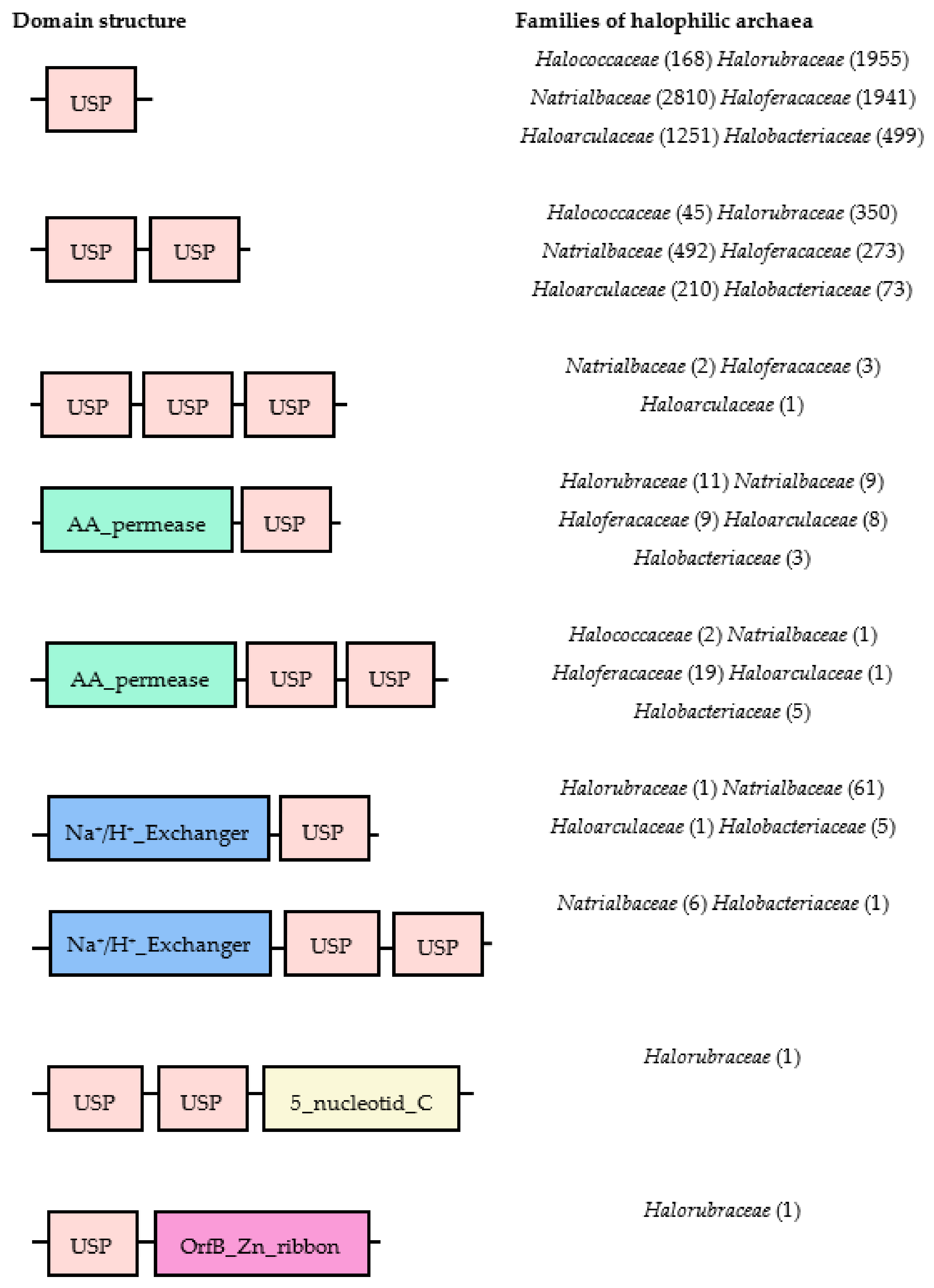
4. Universal Stress Proteins among Three Domains of Life

The Universal Stress Proteins (USP) superfamily was discovered in Escherichia coli; since then, a large number of USPs have been identified in bacteria, plants, fungi, protozoa, and archaea, but not in humans, making USPs potential drug targets [134,135]. The induction of these proteins is due to the growth arrest caused by a wide range of biotic and abiotic stresses. Therefore, these proteins are expressed in a large variety of conditions, such as heat shock, genotoxicity, membrane damage, and starvation of carbon, nitrogen, sulphate, phosphate, and amino acids [128,136,137,138]. Under stressful conditions, USPs are overexpressed by different mechanisms that help the organism to survive [139]. USPs are also known to help pathogens with the invasion of host organisms [140]. USPs cause survival defects under a variety of growth-arrested conditions in Mycobacterium tuberculosis [135], Salmonella typhimurium [141], and Burkholderia pseudomallei [142], making an important contribution to virulence and highlighting the importance of stress resistance regulation in pathogenicity, and survival within the host.
This group of proteins is directly or indirectly involved in transport functions, protein scaffolding, and signaling [143]. Furthermore, USPs appear to be linked to resistance to DNA-damaging agents and thus protect their nucleic acids from external stresses [128].
Multiple paralogs of USPs have been found; the number of copies depends on the organism. E. coli harbors six different proteins, USPA, USPC, USPD, USPE, USPF, and USPG, which have shown undefined functions ranging from oxidative stress resistance to motility [144]. These six E. coli USPs are classified into four subclasses based on the amino acid sequence homology and structural similarity: Class I includes USPA, USPC, and USPD without ATP binding motif; Class II comprises USPF and USPG with ATP binding motif; and USPE has two tandem-repeated USP domains in a polypeptide, designated as E1 and E2, which are grouped into Class III and Class IV, respectively [145]. Each subclass has its own specific function to deal with a particular environmental stress. Both USPA and USPD play an important role in the defense against oxidative stress, and USPD also participates in iron metabolism. As well as their anti-oxidative function, USPC and USPE are involved in cellular motility, cell adhesion, and cell aggregation. In addition to the functions of USPC and USPE, USPF and USPG play different roles in cell migration, negatively regulating mobility, and positively controlling cell agglomeration. These results clearly suggest that bacterial USPs have developed distinct physiological functions in order to act in a coordinated way, defending the cell from an external stress. Although their functions have been demonstrated by their structural diversity, the biochemical and molecular mechanism remains largely unknown [144,145]. Focusing on the molecular structures of the diverse bacterial USPs, the USP domain is always present, followed by another catalytic motif in some cases. These motifs include amino acid permease, protein kinase, Na+/H+ antiporter, and a voltage gated Cl channel [128,139].
Similar to bacterial USPs, all the USPs identified in plant species contain at least one USP domain and one other catalytic motif. Hence, plant USPs play a distinctive role in specific tissues and developmental stages under diverse stress conditions [145]. The other catalytic motifs found in plant USPs are the cation exchanger, the C1 motif of Insensitive to Killer toxin3 (IKI3), the HomeoDomain leucine zipper (HDzip), SWI2/snf2 and Mudr (SWIM)-zinc finger, tyrosine kinase, and serine/threonine kinase [145]. As in bacterial USPs, the physiological and biochemical functions are still unknown but are essential to plant development and growth.
Like the bacterial and plant USPs, archaeal USPs have different functional motifs. The USPs in the six families of halophilic archaea Halococcaceae, Halorubraceae, Natrialbaceae, Haloferacaceae, Haloarculaceae, and Halobacteriaceae have been analyzed using different bioinformatic tools (Figure 1). The molecular structures of the diverse halophilic archaeal USPs only contain a USP domain or USP domain fused with other catalytic motifs, such as amino acid permease or a Na+/H+ exchanger. A single USP motif is the most common domain structure in all families and contains around 140–160 amino acids. These motifs are not conserved in all halophilic archaea, so the combination of the USP domain with other highly divergent functional motifs might produce multiple functions to protect the organism against harsh conditions.
The genome analysis of the USPs found in all available genera of halophilic archaea is summarized in Table 5, but the rest that do not appear in this table, Halomicroarcula, Halosiccatus, Halocalculus, Halomicrococcus, Halorubellus, Halorussus, Halostella, Salinirubrum, Salinirussus, Halegenticoccus, Halopelagius, Haloparvum, Halovarius, Natribaculum, Natronobiforma, and Saliphagus, had no USPs in their genome. Given the large number of halophilic archaeal USPs, it is reasonable to think that proteins play crucial roles in diverse aspects of metabolism to maintain cell homeostasis. Some genera comprise a large number of USPs, such as Halorubrum, Haloferax, and Haloarcula, which present a total of 1939, 1301, and 970, respectively. The analysis of protein sequences revealed that all these USPs proteins contain at least one USP domain. As previously explained, in the case of plants and bacterial USPs, halophilic archaeal USPs have several catalytic motifs that will determine numerous functions derived from their variety of structural characteristics. It is strongly suggested that halophilic archaeal USPs have a great functional diversity. During the evolutionary process, the pressure against stress may lead to the fusion of the USP domain with other catalytic motifs, providing the archaea with the ability to protect them from external environmental stress. This is exemplified by the fusion of the USP domain with the amino acid permease, which allows the transport of amino acids into the cell. The Na+/H+ exchange motif, fused with the USP domain, sends sodium out of the cell, preventing its accumulation. Therefore, under stressful conditions, these proteins are involved in multiple reactions and diverse cellular processes. Not only is it necessary to determine their functional specificity, but also to discover their physiological and biochemical functions for archaeal metabolism. Unravelling these functions may unlock new biotechnological applications of these proteins.
Furthermore, throughout history, these climatic changes have occurred many times and they are an evolutionary engine to adapt to stress in order to colonize new habitats.

5. Conclusions

This review highlights critical factors that influence the survival of microorganisms, in general, and of halophilic archaea, in particular, under different stress conditions that remain largely unknown. Interactions among haloarchaea in saline/hypersaline environments can be influenced by temperature, pH, UV radiation, concentration of heavy metals, etc. These stressful conditions are increasing daily due to climate change, and microorganisms have had to adapt to them throughout history to colonize new habitats. Thus, new biotechnological and industrial applications may be unlocked after discovering the functions of stress proteins, focusing on USPs. USPs are emerging as important players in stress resistance. Hence, the results of this review should serve as the basis for future investigations to elucidate the role of stress proteins in resistance against stressors from a molecular and biochemical point of view. Within this context, new questions arise: How and when do stress genes respond to a given stress? How are these responses regulated? How many molecules are involved in this response? Can we define gene regulatory networks? How will microorganisms adapt to the effects of climate change in their ecosystems? How do antistress mechanisms work at the molecular and genetic levels? Many questions remain unanswered, but this review is a good starting point. Undoubtedly, more work could result in interesting biotechnological applications in order to improve the capacity of microorganisms to withstand environmental shifts.

Author Contributions

Conceptualization, L.M., M.-J.B., and J.E.; Investigation, L.M. and J.E.; Writing—original draft preparation, L.M.; Writing—review and editing, L.M., M.C., B.Z., M.-J.B., and J.E.; Supervision, M.-J.B. and J.E.; Project administration, M.-J.B. All authors have read and agreed to the published version of the manuscript.

Funding

This research received no external funding.

Conflicts of Interest

The authors declare no conflict of interest.

References

  1. Madigan, M.; Bender, K.S.; Buckley, D.H.; Sattley, W.M.; Stahl, D.A. Brock Biology of Microorganisms, 15th ed.; Pearson: London, UK, 2017; ISBN 9780134261928. [Google Scholar]
  2. Conway de Macario, E.; Macario, A.J. Stressors, stress and survival: Overview. Front. Biosci. 2000, 5, D780–D786. [Google Scholar] [CrossRef] [PubMed] [Green Version]
  3. Macario, A.J.L.; Conway de Macario, E. Stress and molecular chaperones in disease. Int. J. Clin. Lab. Res. 2000, 30, 49–66. [Google Scholar] [CrossRef] [PubMed]
  4. Macario, A.J.L.; Conway de Macario, E. Heat Shock Response, Overview. In The Encyclopedia of Stress; Academic Press: San Diego, CA, USA, 2000; p. 350. ISBN 9780123739476. [Google Scholar]
  5. Macario, A.J.; Conway De Macario, E. The molecular chaperone system and other anti-stress mechanisms in archaea. Front. Biosci. 2001, 6. [Google Scholar] [CrossRef] [PubMed] [Green Version]
  6. Baliga, N.S.; Bjork, S.J.; Bonneau, R.; Pan, M.; Iloanusi, C.; Kottemann, M.C.H.; Hood, L.; DiRuggiero, J. Systems level insights into the stress response to UV radiation in the halophilic archaeon Halobacterium NRC-1. Genome Res. 2004, 14, 1025–1035. [Google Scholar] [CrossRef] [Green Version]
  7. Thombre, R.S.; Shinde, V.D.; Oke, R.S.; Dhar, S.K.; Shouche, Y.S. Biology and survival of extremely halophilic archaeon Haloarcula marismortui RR12 isolated from Mumbai salterns, India in response to salinity stress. Sci. Rep. 2016, 6, 1–10. [Google Scholar] [CrossRef]
  8. Grant, W.D.; Kamekura, M.; McGenity, T.J.; Ventosa, A.; Holt, J.G. The History of Bergey’s ManualClass III. Halobacteria class. nov. In Bergey’s Manual of Systematic Bacteriology; Springer: New York, NY, USA, 2001; p. 294. [Google Scholar]
  9. Torregrosa-Crespo, J.; Bergaust, L.; Pire, C.; Martínez-Espinosa, R.M. Denitrifying haloarchaea: Sources and sinks of nitrogenous gases. FEMS Microbiol. Lett. 2018, 365. [Google Scholar] [CrossRef]
  10. Sánchez, E.; Domínguez, M.; Romera, R.; de la Franca, N.L.; Gaertner, M.A.; Gallardo, C.; Castro, M. Regional modeling of dry spells over the Iberian Peninsula for present climate and climate change conditions. Clim. Chang. 2011, 107, 625–634. [Google Scholar] [CrossRef]
  11. Field, C.B.; Barros, V.; Stocker, T.F.; Dahe, Q.; Dokken, D.J.; Plattner, G.-K.; Ebi, K.L.; Allen, S.K.; Mastrandrea, M.D.; Tignor, M.; et al. Managing the Risks of Extreme Events and Disasters to Advance Climate Change Adaptation. A Special Report of Working Groups I and II of the Intergovernmental Panel on Climate Change; Cambridge University: Cambridge, UK, 2012. [Google Scholar]
  12. Gornall, J.; Betts, R.; Burke, E.; Clark, R.; Camp, J.; Willett, K.; Wiltshire, A. Implications of climate change for agricultural productivity in the early twenty-first century. Phil. Trans. R. Soc. B 2010, 365, 2973–2989. [Google Scholar] [CrossRef]
  13. Griffiths, B.S.; Philippot, L. Insights into the resistance and resilience of the soil microbial community. FEMS Microbiol. Rev. 2013, 37, 112–129. [Google Scholar] [CrossRef] [Green Version]
  14. Zafrilla, B.; Martínez-Espinosa, R.M.; Alonso, M.A.; Bonete, M.J. Biodiversity of Archaea and floral of two inland saltern ecosystems in the Alto Vinalopó Valley, Spain. Saline Syst. 2010, 6, 10. [Google Scholar] [CrossRef] [Green Version]
  15. Oren, A. Molecular ecology of extremely halophilic Archaea and Bacteria. FEMS Microbiol. Ecol. 2002, 39, 1–7. [Google Scholar] [CrossRef] [PubMed]
  16. Andrei, A.Ş.; Banciu, H.L.; Oren, A. Living with salt: Metabolic and phylogenetic diversity of archaea inhabiting saline ecosystems. FEMS Microbiol. Lett. 2012, 330, 1–9. [Google Scholar] [CrossRef] [PubMed] [Green Version]
  17. DasSarma, P.; Capes, M.D.; DasSarma, S. Comparative Genomics of Halobacterium Strains From Diverse Locations. In Microbial Diversity in the Genomic Era; Elsevier: Cambridge, MA, USA, 2019; pp. 285–322. [Google Scholar]
  18. Delgado-Baquerizo, M.; Maestre, F.T.; Gallardo, A.; Bowker, M.A.; Wallenstein, M.D.; Quero, J.L.; Ochoa, V.; Gozalo, B.; García-Gómez, M.; Soliveres, S.; et al. Decoupling of soil nutrient cycles as a function of aridity in global drylands. Nature 2013, 502, 672–676. [Google Scholar] [CrossRef]
  19. Oren, A. Microbial life at high salt concentrations: Phylogenetic and metabolic diversity. Saline Syst. 2008, 4, 2. [Google Scholar] [CrossRef] [Green Version]
  20. Kennedy, S.P.; Ng, W.V.; Salzberg, S.L.; Hood, L.; DasSarma, S. Understanding the adaptation of Halobacterium species NRC-1 to its extreme environment through computational analysis of its genome sequence. Genome Res. 2001, 11, 1641–1650. [Google Scholar] [CrossRef] [PubMed] [Green Version]
  21. Britton, K.L.; Baker, P.J.; Fisher, M.; Ruzheinikov, S.; Gilmour, D.J.; Bonete, M.J.; Ferrer, J.; Pire, C.; Esclapez, J.; Rice, D.W. Analysis of protein solvent interactions in glucose dehydrogenase from the extreme halophile Haloferax mediterranei. Proc. Natl. Acad. Sci. USA 2006, 103, 4846–4851. [Google Scholar] [CrossRef] [Green Version]
  22. Esclapez, J.; Pire, C.; Bautista, V.; Martínez-Espinosa, R.M.; Ferrer, J.; Bonete, M.J. Analysis of acidic surface of Haloferax mediterranei glucose dehydrogenase by site-directed mutagenesis. FEBS Lett. 2007, 581, 837–842. [Google Scholar] [CrossRef] [Green Version]
  23. Williams, W.D. Anthropogenic salinisation of inland waters. In Saline Lakes; Springer: Dordrecht, The Netherlands, 2001; pp. 329–337. [Google Scholar]
  24. Oren, A. Solar salterns as model systems for the study of halophilic microorganisms in their natural environments. In Model Ecosystems in Extreme Environments; Elsevier: Cambridge, MA, USA, 2019; pp. 41–56. [Google Scholar]
  25. Coker, J.; DasSarma, P.; Kumar, J.; Müller, J.A.; DasSarma, S. Transcriptional profiling of the model Archaeon Halobacterium sp. NRC-1: responses to changes in salinity and temperature. Saline Syst. 2007, 3, 6. [Google Scholar] [CrossRef] [Green Version]
  26. Leuko, S.; Raftery, M.J.; Burns, B.P.; Walter, M.R.; Neilan, B.A. Global Protein-Level Responses of Halobacterium salinarum NRC-1 to Prolonged Changes in External Sodium Chloride Concentrations. J. Proteome Res. 2009, 8, 2218–2225. [Google Scholar] [CrossRef]
  27. Mojica, F.J.; Juez, G.; Rodriguez-Valera, F. Transcription at different salinities of Haloferax mediterranei sequences adjacent to partially modified PstI sites. Mol. Microbiol. 1993, 9, 613–621. [Google Scholar] [CrossRef]
  28. Ferrer, C.; Mojica, F.J.; Juez, G.; Rodriguez-Valera, F. Differentially transcribed regions of Haloferax volcanii genome depending on the medium salinity. J. Bacteriol. 1996, 178, 309–313. [Google Scholar] [CrossRef] [PubMed] [Green Version]
  29. Kurt-Kızıldoğan, A.; Abanoz, B.; Okay, S. Global transcriptome analysis of Halolamina sp. to decipher the salt tolerance in extremely halophilic archaea. Gene 2017, 601, 56–64. [Google Scholar] [CrossRef] [PubMed]
  30. Jevtic, Z.; Stoll, B.; Pfeiffer, F.; Sharma, K.; Urlaub, H.; Marchfelder, A.; Lenz, C. The Response of Haloferax volcanii to Salt and Temperature Stress: A Proteome Study by Label-Free Mass Spectrometry. Proteom. 2019, 19, e1800491. [Google Scholar] [CrossRef] [PubMed] [Green Version]
  31. Bidle, K.A.; Kirkland, P.A.; Nannen, J.L.; Maupin-Furlow, J.A. Proteomic analysis of Haloferax volcanii reveals salinity-mediated regulation of the stress response protein PspA. Microbiology 2008, 154, 1436–1443. [Google Scholar] [CrossRef] [PubMed] [Green Version]
  32. Leuko, S.; Neilan, B.A.; Burns, B.P.; Walter, M.R.; Rothschild, L.J. Molecular assessment of UVC radiation-induced DNA damage repair in the stromatolitic halophilic archaeon, Halococcus hamelinensis. J. Photochem. Photobiol. B: Biol. 2011, 102, 140–145. [Google Scholar] [CrossRef] [PubMed]
  33. Stantial, N.; Dumpe, J.; Pietrosimone, K.; Baltazar, F.; Crowley, D.J. Transcription-coupled repair of UV damage in the halophilic archaea. DNA Repair 2016, 41, 63–68. [Google Scholar] [CrossRef] [Green Version]
  34. Moran-Reyna, A.; A Coker, J. The effects of extremes of pH on the growth and transcriptomic profiles of three haloarchaea. F1000Research 2014, 3, 168. [Google Scholar] [CrossRef] [Green Version]
  35. Sharma, K.; Gillum, N.; Boyd, J.L.; Schmid, A.K. The RosR transcription factor is required for gene expression dynamics in response to extreme oxidative stress in a hypersaline-adapted archaeon. BMC Genom. 2012, 13, 351. [Google Scholar] [CrossRef] [Green Version]
  36. Gelsinger, D.R.; DiRuggiero, J. Transcriptional Landscape and Regulatory Roles of Small Noncoding RNAs in the Oxidative Stress Response of the Haloarchaeon Haloferax volcanii. J. Bacteriol. 2018, 200, e00779-17. [Google Scholar] [CrossRef] [Green Version]
  37. McMillan, L.J.; Hwang, S.; Farah, R.E.; Koh, J.; Chen, S.; Maupin-Furlow, J.A. Multiplex quantitative SILAC for analysis of archaeal proteomes: a case study of oxidative stress responses. Environ. Microbiol. 2017, 20, 385–401. [Google Scholar] [CrossRef]
  38. Gomez, M.; Leung, W.; Dantuluri, S.; Pillai, A.; Gani, Z.; Hwang, S.; McMillan, L.J.; Kiljunen, S.; Savilahti, H.; Maupin-Furlow, J.A. Molecular Factors of Hypochlorite Tolerance in the Hypersaline Archaeon Haloferax volcanii. Genes 2018, 9, 562. [Google Scholar] [CrossRef] [PubMed] [Green Version]
  39. Wang, G.; Kennedy, S.; Fasiludeen, S.; Rensing, C.; DasSarma, S. Arsenic Resistance in Halobacterium sp. Strain NRC-1 Examined by Using an Improved Gene Knockout System. J. Bacteriol. 2004, 186, 3187–3194. [Google Scholar] [CrossRef] [PubMed] [Green Version]
  40. Buda, D.-M.; Bulzu, P.-A.; Barbu-Tudoran, L.; Porfire, A.; Pătraș, L.; Sesărman, A.; Tripon, S.; Șenilă, M.; Ionescu, M.I.; Banciu, H.L.; et al. Physiological response to silver toxicity in the extremely halophilic archaeon Halomicrobium mukohataei. FEMS Microbiol. Lett. 2019, 366. [Google Scholar] [CrossRef] [PubMed]
  41. Chaudhary, A.; Imran Pasha, M.; Salgaonkar, B.B.; Bragança, J.M. Cadmium Tolerance by Haloarchaeal Strains Isolated from Solar Salterns of Goa, India. Int. J. Biosci. Biochem. Bioinform. 2014, 1–6. [Google Scholar] [CrossRef]
  42. Sehgal, S.N.; Gibbons, N.E. Effect of some metal ions on the growth of Halobacterium cutirubrum. Can. J. Microbiol. 1960, 6, 165–169. [Google Scholar] [CrossRef]
  43. Nieto, J.J.; Ventosa, A.; Ruiz-Berraquero, F. Susceptibility of Halobacteria to Heavy Metals. Appl. Environ. Microbiol. 1987, 53, 1199–1202. [Google Scholar] [CrossRef] [Green Version]
  44. Weng, R.R.; Shu, H.-W.; Chin, S.-W.; Kao, Y.; Chen, T.-W.; Liao, C.-C.; Tsay, Y.-G.; Ng, W.V. OMICS in Ecology: Systems Level Analyses of Halobacterium salinarum Reveal Large-scale Temperature-Mediated Changes and a Requirement of CctA for Thermotolerance. OMICS J. Integr. Biol. 2014, 18, 65–80. [Google Scholar] [CrossRef] [Green Version]
  45. Gruber, C.; Legat, A.; Pfaffenhuemer, M.; Radax, C.; Weidler, G.; Stan-Lotter, H. Halobacterium noricense sp. nov., an archaeal isolate from a bore core of an alpine Permian salt deposit, classification of Halobacterium sp. NRC-1 as a strain of H. salinarum and emended description of H. salinarum. Extremophiles 2004, 8, 431–439. [Google Scholar] [CrossRef]
  46. Lledó, B.; Martı́nez-Espinosa, R.; Marhuenda-Egea, F.; Bonete, M.-J.; Marhuenda-Egea, F.C. Respiratory nitrate reductase from haloarchaeon Haloferax mediterranei: biochemical and genetic analysis. Biochim. Biophys. Acta (BBA) - Gen. Subj. 2004, 1674, 50–59. [Google Scholar] [CrossRef]
  47. Martínez-Espinosa, R.M.; Lledó, B.; Marhuenda-Egea, F.C.; Bonete, M.-J. The effect of ammonium on assimilatory nitrate reduction in the haloarchaeon Haloferax mediterranei. Extremophiles 2007, 11, 759–767. [Google Scholar] [CrossRef]
  48. Oren, A.; Bardavid, R.E.; Mana, L. Perchlorate and halophilic prokaryotes: implications for possible halophilic life on Mars. Extremophiles 2013, 18, 75–80. [Google Scholar] [CrossRef] [PubMed]
  49. Becker, E.; Seitzer, P.M.; Tritt, A.; Larsen, D.; Krusor, M.; Yao, A.I.; Wu, N.; Madern, M.; Eisen, J.A.; E Darling, A.; et al. Phylogenetically Driven Sequencing of Extremely Halophilic Archaea Reveals Strategies for Static and Dynamic Osmo-response. PLoS Genet. 2014, 10, e1004784. [Google Scholar] [CrossRef] [PubMed]
  50. Oren, A. Life at High Salt Concentrations. In The Prokaryotes; Springer Science and Business Media LLC: New York, NY, USA, 2013; Volume 9783642301230, pp. 421–440. [Google Scholar]
  51. Schäfer, G.; Purschke, W.; Schmidt, C.L. On the origin of respiration: electron transport proteins from archaea to man. FEMS Microbiol. Rev. 1996, 18, 173–188. [Google Scholar] [CrossRef]
  52. Grant, W.D. Life at low water activity. Philos. Trans. R. Soc. B Biol. Sci. 2004, 359, 1249–1267. [Google Scholar] [CrossRef] [PubMed] [Green Version]
  53. Ali, I.; Prasongsuk, S.; Akbar, A.; Aslam, M.; Lotrakul, P.; Punnapayak, H.; Rakshit, S.K. Hypersaline habitats and halophilic microorganisms. Maejo Int. J. Sci. Technol 2015, 10, 330–345. [Google Scholar]
  54. Ng, W.V.; Kennedy, S.; Mahairas, G.G.; Berquist, B.; Pan, M.; Shukla, H.D.; Lasky, S.R.; Baliga, N.S.; Thorsson, V.; Sbrogna, J.; et al. Genome sequence of Halobacterium species NRC-1. Proc. Natl. Acad. Sci. USA 2000, 97, 12176–12181. [Google Scholar] [CrossRef] [Green Version]
  55. DasSarma, S. Genome sequence of an extremely halophilic archaeon. In Microbial Genomes; Fraser, C.M., Ed.; Humana Press Inc.: Totowa, NJ, USA, 2004; pp. 383–399. [Google Scholar]
  56. Bowers, K.J.; Wiegel, J. Temperature and pH optima of extremely halophilic archaea: a mini-review. Extremophiles 2011, 15, 119–128. [Google Scholar] [CrossRef]
  57. Ellis, R.J. Chaperone Function: The Orthodox View. In Molecular Chaperones and Cell Signalling; Cambridge University Press (CUP): Cambridge, MA, USA, 2005; pp. 3–21. [Google Scholar]
  58. Shukla, H.D. Proteomic analysis of acidic chaperones, and stress proteins in extreme halophile Halobacterium NRC-1: a comparative proteomic approach to study heat shock response. Proteome Sci. 2006, 4, 6. [Google Scholar] [CrossRef] [Green Version]
  59. Mormile, M.R.; Hong, B.-Y.; Benison, K.C. Molecular Analysis of the Microbial Communities of Mars Analog Lakes in Western Australia. Astrobiology 2009, 9, 919–930. [Google Scholar] [CrossRef]
  60. Kulp, T.R.; Han, S.; Saltikov, C.W.; Lanoil, B.D.; Zargar, K.; Oremland, R.S. Effects of Imposed Salinity Gradients on Dissimilatory Arsenate Reduction, Sulfate Reduction, and Other Microbial Processes in Sediments from Two California Soda Lakes. Appl. Environ. Microbiol. 2007, 73, 5130–5137. [Google Scholar] [CrossRef] [Green Version]
  61. Soliman, G.S.; Trüper, H.G. Halobacterium pharaonis sp. nov., a New, Extremely Haloalkaliphilic Archaebacterium with Low Magnesium Requirement. Zentralblatt für Bakteriologie Mikrobiologie und Hygiene: I. Abt. Originale C: Allgemeine, angewandte und ökologische Mikrobiologie 1982, 3, 318–329. [Google Scholar] [CrossRef]
  62. Tabak, H.H.; Lens, P.N.L.; Van Hullebusch, E.D.; Dejonghe, W. Developments in Bioremediation of Soils and Sediments Polluted with Metals and Radionuclides. Microbial Processes and Mechanisms Affecting Bioremediation of Metal Contamination and Influencing Metal Toxicity and Transport. Rev. Environ. Sci. Bio/Technology 2005, 4, 115–156. [Google Scholar] [CrossRef]
  63. Popescu, G.; Dumitru, L. Biosorption of Some Heavy Metals from Media with High Salt Concentrations by Halophilic Archaea. Biotechnol. Biotechnol. Equip. 2009, 23, 791–795. [Google Scholar] [CrossRef]
  64. Kaur, A.; Pan, M.; Meislin, M.; Facciotti, M.T.; El-Gewely, R.; Baliga, N.S. A systems view of haloarchaeal strategies to withstand stress from transition metals. Genome Res. 2006, 16, 841–854. [Google Scholar] [CrossRef] [PubMed] [Green Version]
  65. DasSarma, S. Extreme Microbes. Am. Sci. 2007, 95, 224–231. [Google Scholar] [CrossRef]
  66. Srivastava, P.; Kowshik, M. Mechanisms of Metal Resistance and Homeostasis in Haloarchaea. Archaea 2013, 2013, 1–16. [Google Scholar] [CrossRef] [Green Version]
  67. Oren, A. The Function of Gas Vesicles in Halophilic Archaea and Bacteria: Theories and Experimental Evidence. Life 2012, 3, 1–20. [Google Scholar] [CrossRef] [Green Version]
  68. Pfeifer, F. Haloarchaea and the Formation of Gas Vesicles. Life 2015, 5, 385–402. [Google Scholar] [CrossRef]
  69. Oren, A.; Trüper, H.G. Anaerobic growth of halophilic archaeobacteria by reduction of dimethysulfoxide and trimethylamineN-oxide. FEMS Microbiol. Lett. 1990, 70, 33–36. [Google Scholar] [CrossRef]
  70. Oren, A. Anaerobic growth of halophilic archaeobacteria by reduction of fumarate. J. Gen. Microbiol. 1991, 137, 1387–1390. [Google Scholar] [CrossRef] [Green Version]
  71. Müller, J.A.; DasSarma, S. Genomic Analysis of Anaerobic Respiration in the Archaeon Halobacterium sp. Strain NRC-1: Dimethyl Sulfoxide and Trimethylamine N-Oxide as Terminal Electron Acceptors. J. Bacteriol. 2005, 187, 1659–1667. [Google Scholar] [CrossRef] [PubMed] [Green Version]
  72. Burns, D.G.; Janssen, P.H.; Itoh, T.; Kamekura, M.; Li, Z.; Jensen, G.; Rodriguez-Valera, F.; Bolhuis, H.; Dyall-Smith, M.L. Haloquadratum walsbyi gen. nov., sp. nov., the square haloarchaeon of Walsby, isolated from saltern crystallizers in Australia and Spain. Int. J. Syst. Evol. Microbiol. 2007, 57, 387–392. [Google Scholar] [CrossRef] [PubMed]
  73. Deveaux, L.C.; Müller, J.A.; Smith, J.; Petrisko, J.; Wells, D.P.; DasSarma, S. Extremely Radiation-Resistant Mutants of a Halophilic Archaeon with Increased Single-Stranded DNA-Binding Protein (RPA) Gene Expression. Radiat. Res. 2007, 168, 507–514. [Google Scholar] [CrossRef] [PubMed]
  74. McCready, S.; Müller, J.A.; Boubriak, I.; Berquist, B.R.; Ng, W.L.; DasSarma, S. UV irradiation induces homologous recombination genes in the model archaeon, Halobacterium sp. NRC-1. Saline Syst. 2005, 1, 3. [Google Scholar] [CrossRef] [Green Version]
  75. McCready, S.; Marcello, L. Repair of UV damage in Halobacterium salinarum. Biochem. Soc. Trans. 2003, 31, 694–698. [Google Scholar] [CrossRef] [Green Version]
  76. Hescox, M.A.; Carlberg, D.M. Photoreactivation in Halobacterium cutirubrum. Can. J. Microbiol. 1972, 18, 981–985. [Google Scholar] [CrossRef]
  77. Whitehead, K.; Kish, A.; Pan, M.; Kaur, A.; Reiss, D.J.; King, N.; Hohmann, L.; DiRuggiero, J.; Baliga, N.S. An integrated systems approach for understanding cellular responses to gamma radiation. Mol. Syst. Biol. 2006, 2, 47. [Google Scholar] [CrossRef] [Green Version]
  78. Kottemann, M.; Kish, A.; Iloanusi, C.; Bjork, S.; DiRuggiero, J. Physiological responses of the halophilic archaeon Halobacterium sp. strain NRC1 to desiccation and gamma irradiation. Extremophiles 2005, 9, 219–227. [Google Scholar] [CrossRef]
  79. De Macario, E.C.; Macario, A.J.L. Molecular Biology of Stress Genes in Methanogens: Potential for Bioreactor Technology. Adv. Biochem. Eng. Biotechnol. 2003, 81, 95–150. [Google Scholar] [CrossRef]
  80. Macario, A.J.; Lange, M.; Ahring, B.K.; de Macario, E.C. Stress Genes and Proteins in the Archaea. Microbiol Mol. Biol Rev. 1999, 64, 923–967. [Google Scholar] [CrossRef] [Green Version]
  81. Oren, A. Halophilic archaea on Earth and in space: Growth and survival under extreme conditions. Philos. Trans. R. Soc. A Math. Phys. Eng. Sci. 2014, 372, 20140194. [Google Scholar] [CrossRef] [PubMed]
  82. Gottesman, S.; Roche, E.; Zhou, Y.; Sauer, R.T. The ClpXP and ClpAP proteases degrade proteins with carboxy-terminal peptide tails added by the SsrA-tagging system. Genes Dev. 1998, 12, 1338–1347. [Google Scholar] [CrossRef] [PubMed]
  83. Herman, C.; D’Ari, R. Proteolysis and chaperones: the destruction/reconstruction dilemma. Curr. Opin. Microbiol. 1998, 1, 204–209. [Google Scholar] [CrossRef]
  84. Martin, J.; Hartl, F.U. Chaperone-assisted protein folding. Curr. Opin. Struct. Biol. 1997, 7, 41–52. [Google Scholar] [CrossRef]
  85. Netzer, W.J.; Hartl, F.U. Protein folding in the cytosol: chaperonin-dependent and -independent mechanisms. Trends Biochem. Sci. 1998, 23, 68–73. [Google Scholar] [CrossRef]
  86. Trent, J.D.; Nimmesgern, E.; Wall, J.S.; Hartl, F.-U.; Horwich, A.L. A molecular chaperone from a thermophilic archaebacterium is related to the eukaryotic protein t-complex polypeptide-1. Nature 1991, 354, 490–493. [Google Scholar] [CrossRef]
  87. Condò, I.; Ruggero, D.; Reinhardt, R.; Londei, P. A novel aminopeptidase associated with the 60 kDa chaperonin in the thermophilic archaeon Sulfolobus solfataricus. Mol. Microbiol. 1998, 29, 775–785. [Google Scholar] [CrossRef] [Green Version]
  88. Louvion, J.-F.; Abbas-Terki, T.; Picard, D. Hsp90 Is Required for Pheromone Signaling in Yeast. Mol. Biol. Cell 1998, 9, 3071–3083. [Google Scholar] [CrossRef] [Green Version]
  89. Ruggero, D.; Ciammaruconi, A.; Londei, P. The chaperonin of the archaeon Sulfolobus solfataricus is an RNA-binding protein that participates in ribosomal RNA processing. EMBO J. 1998, 17, 3471–3477. [Google Scholar] [CrossRef] [Green Version]
  90. Hartl, F.-U.; Martin, J. Molecular chaperones in cellular protein folding. BioEssays 1994, 16, 689–692. [Google Scholar] [CrossRef]
  91. Callahan, M.K.; Chaillot, D.; Jacquin, C.; Clark, P.R.; Menoret, A. Differential Acquisition of Antigenic Peptides by Hsp70 and Hsc70 under Oxidative Conditions. J. Biol. Chem. 2002, 277, 33604–33609. [Google Scholar] [CrossRef] [PubMed] [Green Version]
  92. Zmijewski, M.A.; Macario, A.J.; Lipińska, B. Functional Similarities and Differences of an Archaeal Hsp70 (DnaK) Stress Protein Compared with its Homologue from the Bacterium Escherichia coli. J. Mol. Biol. 2004, 336, 539–549. [Google Scholar] [CrossRef] [PubMed]
  93. Petitjean, C.; Moreira, D.; López-García, P.; Brochier-Armanet, C. Horizontal gene transfer of a chloroplast DnaJ-Fer protein to Thaumarchaeota and the evolutionary history of the DnaK chaperone system in Archaea. BMC Evol. Biol. 2012, 12, 226. [Google Scholar] [CrossRef] [PubMed] [Green Version]
  94. Shih, C.-J.; Lai, M.-C. Differentially expressed genes after hyper- and hypo-salt stress in the halophilic archaeon Methanohalophilus portucalensis. Can. J. Microbiol. 2010, 56, 295–307. [Google Scholar] [CrossRef]
  95. Bukau, B.; Deuerling, E.; Pfund, C.; A Craig, E. Getting Newly Synthesized Proteins into Shape. Cell 2000, 101, 119–122. [Google Scholar] [CrossRef] [Green Version]
  96. Ellis, R.J.; Hartl, F.U. Principles of protein folding in the cellular environment. Curr. Opin. Struct. Biol. 1999, 9, 102–110. [Google Scholar] [CrossRef]
  97. Frydman, J. Chaperones get in touch: the Hip-Hop connection. Trends Biochem. Sci. 1997, 22, 87–92. [Google Scholar] [CrossRef]
  98. Hartl, F.U. Molecular chaperones in cellular protein folding. Nature 1996, 381, 571–580. [Google Scholar] [CrossRef]
  99. Pfanner, N. Protein folding: Who chaperones nascent chains in bacteria? Curr. Biol. 1999, 9. [Google Scholar] [CrossRef] [Green Version]
  100. De Macario, E.C.; Macario, A.J. Heat-shock response in Archaea. Trends Biotechnol. 1994, 12, 512–518. [Google Scholar] [CrossRef]
  101. Lin, W.; Bonin, M.; Boden, A.; Wieduwild, R.; Murawala, P.; Wermke, M.; Andrade, H.; Bornhäuser, M.; Zhang, Y. Peptidyl prolyl cis/trans isomerase activity on the cell surface correlates with extracellular matrix development. Commun. Biol. 2019, 2, 1–11. [Google Scholar] [CrossRef] [Green Version]
  102. Fischer, G. Peptidyl-Prolyl cis/trans Isomerases and Their Effectors. Angew. Chem. Int. Ed. Engl. 1994, 33, 1415–1436. [Google Scholar] [CrossRef]
  103. Rudd, K.E.; Sofia, H.J.; Koonin, E.V.; Plunkett, G.; Lazar, S.; Rouvière, P.E. A new family of peptidyl-prolyl isomerases. Trends Biochem. Sci. 1995, 20, 12–14. [Google Scholar] [CrossRef]
  104. Boonyaratanakornkit, B.B.; Simpson, A.J.; Whitehead, T.A.; Fraser, C.; El-Sayed, N.M.; Clark, D.S. Transcriptional profiling of the hyperthermophilic methanarchaeon Methanococcus jannaschii in response to lethal heat and non-lethal cold shock. Environ. Microbiol. 2005, 7, 789–797. [Google Scholar] [CrossRef]
  105. Yoshinaga, K.; Yoshioka, H.; Kurosaki, H.; Hirasawa, M.; Uritani, M.; Hasegawa, K. Protection by Trehalose of DNA from Radiation Damage. Biosci. Biotechnol. Biochem. 1997, 61, 160–161. [Google Scholar] [CrossRef] [PubMed]
  106. Elbein, A.D.; Pan, Y.; Pastuszak, I.; Carroll, D. New insights on trehalose: a multifunctional molecule. Glycobiol. 2003, 13, 17R–27. [Google Scholar] [CrossRef] [PubMed]
  107. Leuko, S.; Domingos, C.; Parpart, A.; Reitz, G.; Rettberg, P. The Survival and Resistance of Halobacterium salinarum NRC-1, Halococcus hamelinensis, and Halococcus morrhuae to Simulated Outer Space Solar Radiation. Astrobiol. 2015, 15, 987–997. [Google Scholar] [CrossRef]
  108. Fröls, S. Archaeal biofilms: widespread and complex. Biochem. Soc. Trans. 2013, 41, 393–398. [Google Scholar] [CrossRef]
  109. Mayerhofer, L.E.; Macario, A.J.; De Macario, E.C. Lamina, a novel multicellular form of Methanosarcina mazei S-6. J. Bacteriol. 1992, 174, 309–314. [Google Scholar] [CrossRef] [Green Version]
  110. Pecl, G.T.; Araújo, M.B.; Bell, J.D.; Blanchard, J.L.; Bonebrake, T.C.; Chen, I.-C.; Clark, T.D.; Colwell, R.K.; Danielsen, F.; Evengård, B.; et al. Biodiversity redistribution under climate change: Impacts on ecosystems and human well-being. Science 2017, 355, eaai9214. [Google Scholar] [CrossRef]
  111. Webster, N.S.; Negri, A.P. Site-specific variation in Antarctic marine biofilms established on artificial surfaces. Environ. Microbiol. 2006, 8, 1177–1190. [Google Scholar] [CrossRef]
  112. Justice, N.B.; Pan, C.; Mueller, R.; Spaulding, S.E.; Shah, V.; Sun, C.L.; Yelton, A.P.; Miller, C.S.; Thomas, B.C.; Shah, M.; et al. Heterotrophic Archaea Contribute to Carbon Cycling in Low-pH, Suboxic Biofilm Communities. Appl. Environ. Microbiol. 2012, 78, 8321–8330. [Google Scholar] [CrossRef] [PubMed] [Green Version]
  113. Edwards, K.J. An Archaeal Iron-Oxidizing Extreme Acidophile Important in Acid Mine Drainage. Science 2000, 287, 1796–1799. [Google Scholar] [CrossRef] [PubMed] [Green Version]
  114. Weidler, G.W.; Gerbl, F.W.; Stan-Lotter, H. Crenarchaeota and Their Role in the Nitrogen Cycle in a Subsurface Radioactive Thermal Spring in the Austrian Central Alps. Appl. Environ. Microbiol. 2008, 74, 5934–5942. [Google Scholar] [CrossRef] [Green Version]
  115. Fröls, S.; Dyall-Smith, M.; Pfeifer, F. Biofilm formation by haloarchaea. Environ. Microbiol. 2012, 14, 3159–3174. [Google Scholar] [CrossRef]
  116. Crowley, D.J.; Boubriak, I.; Berquist, B.R.; Clark, M.; Richard, E.; Sullivan, L.; DasSarma, S.; McCready, S. The uvrA, uvrB and uvrC genes are required for repair of ultraviolet light induced DNA photoproducts in Halobacterium sp. NRC-1. Saline Syst. 2006, 2, 11. [Google Scholar] [CrossRef] [Green Version]
  117. Delmas, S.; Duggin, I.G.; Allers, T. DNA damage induces nucleoid compaction via the Mre11-Rad50 complex in the archaeon Haloferax volcanii. Mol. Microbiol. 2012, 87, 168–179. [Google Scholar] [CrossRef] [Green Version]
  118. Zhao, A.; Gray, F.C.; MacNeill, S.A. ATP- and NAD+-dependent DNA ligases share an essential function in the halophilic archaeon Haloferax volcanii. Mol. Microbiol. 2006, 59, 743–752. [Google Scholar] [CrossRef]
  119. Reuter, C.J.; Kaczowka, S.J.; Maupin-Furlow, J.A. Differential Regulation of the PanA and PanB Proteasome-Activating Nucleotidase and 20S Proteasomal Proteins of the Haloarchaeon Haloferax volcanii. J. Bacteriol. 2004, 186, 7763–7772. [Google Scholar] [CrossRef] [PubMed] [Green Version]
  120. Zhou, G.; Kowalczyk, D.; Humbard, M.A.; Rohatgi, S.; Maupin-Furlow, J.A. Proteasomal Components Required for Cell Growth and Stress Responses in the Haloarchaeon Haloferax volcanii. J. Bacteriol. 2008, 190, 8096–8105. [Google Scholar] [CrossRef] [PubMed] [Green Version]
  121. Jones, D.L.; Baxter, B.K. DNA Repair and Photoprotection: Mechanisms of Overcoming Environmental Ultraviolet Radiation Exposure in Halophilic Archaea. Front. Microbiol. 2017, 8, 1882. [Google Scholar] [CrossRef] [PubMed]
  122. Jun, S.-H.; Kim, T.G.; Ban, C. DNA mismatch repair system. Classical and fresh roles. FEBS J. 2006, 273, 1609–1619. [Google Scholar] [CrossRef] [PubMed]
  123. Majumder, P.; Baumeister, W. Proteasomes: unfoldase-assisted protein degradation machines. Biol. Chem. 2019, 401, 183–199. [Google Scholar] [CrossRef] [PubMed]
  124. Ciechanover, A. Proteolysis: from the lysosome to ubiquitin and the proteasome. Nat. Rev. Mol. Cell Biol. 2005, 6, 79–87. [Google Scholar] [CrossRef]
  125. Dikic, I. Proteasomal and Autophagic Degradation Systems. Annu. Rev. Biochem. 2017, 86, 193–224. [Google Scholar] [CrossRef]
  126. Fu, X.; Adams, Z.; Maupin-Furlow, J.A. Assays for ubiquitin-like protein ligation and proteasome function in archaea. Methods Enzymol. 2019, 619, 161–178. [Google Scholar] [CrossRef]
  127. Prunetti, L.; Reuter, C.J.; Hepowit, N.L.; Wu, Y.; Barrueto, L.; Miranda, H.V.; Kelly, K.; Maupin-Furlow, J.A. Structural and biochemical properties of an extreme ’salt-loving’ proteasome activating nucleotidase from the archaeon Haloferax volcanii. Extremophiles 2013, 18, 283–293. [Google Scholar] [CrossRef]
  128. Kvint, K.; Nachin, L.; A Diez, A.; Nyström, T. The bacterial universal stress protein: function and regulation. Curr. Opin. Microbiol. 2003, 6, 140–145. [Google Scholar] [CrossRef]
  129. Perez-Rueda, E.; Hernandez-Guerrero, R.; Martínez-Núñez, M.A.; Armenta-Medina, D.; Sanchez, I.; Ibarra, J.A. Abundance, diversity and domain architecture variability in prokaryotic DNA-binding transcription factors. PLoS ONE 2018, 13, e0195332. [Google Scholar] [CrossRef] [Green Version]
  130. Plaisier, C.L.; Lo, F.-Y.; Ashworth, J.; Brooks, A.N.; Beer, K.D.; Kaur, A.; Pan, M.; Reiss, D.J.; Facciotti, M.T.; Baliga, N.S. Evolution of context dependent regulation by expansion of feast/famine regulatory proteins. BMC Syst. Biol. 2014, 8, 122. [Google Scholar] [CrossRef] [Green Version]
  131. Busenlehner, L.S.; Pennella, M.A.; Giedroc, D.P. The SmtB/ArsR family of metalloregulatory transcriptional repressors: structural insights into prokaryotic metal resistance. FEMS Microbiol. Rev. 2003, 27, 131–143. [Google Scholar] [CrossRef] [Green Version]
  132. Lemmens, L.; Maklad, H.R.; Bervoets, I.; Peeters, E. Transcription Regulators in Archaea: Homologies and Differences with Bacterial Regulators. J. Mol. Biol. 2019, 431, 4132–4146. [Google Scholar] [CrossRef] [PubMed]
  133. Karr, E.A. The Methanogen-Specific Transcription Factor MsvR Regulates the fpaA-rlp-rub Oxidative Stress Operon Adjacent to msvR in Methanothermobacter thermautotrophicus. J. Bacteriol. 2010, 192, 5914–5922. [Google Scholar] [CrossRef] [PubMed] [Green Version]
  134. Siegele, D.A. Universal Stress Proteins in Escherichia coli. J. Bacteriol. 2005, 187, 6253–6254. [Google Scholar] [CrossRef] [PubMed] [Green Version]
  135. Hingley-Wilson, S.; Lougheed, K.; Ferguson, K.; Leiva, S.; Williams, H.D. Individual Mycobacterium tuberculosis universal stress protein homologues are dispensable in vitro. Tuberc. 2010, 90, 236–244. [Google Scholar] [CrossRef] [Green Version]
  136. Nyström, T.; Neidhardt, F.C. Cloning, mapping and nucleotide sequencing of a gene encoding a universal stress protein in Eschericha coli. Mol. Microbiol. 1992, 6, 3187–3198. [Google Scholar] [CrossRef] [Green Version]
  137. Gustavsson, N.; A Diez, A.; Nyström, T. The universal stress protein paralogues of Escherichia coli are co-ordinately regulated and co-operate in the defence against DNA damage. Mol. Microbiol. 2002, 43, 107–117. [Google Scholar] [CrossRef]
  138. Nyström, T.; Neidhardt, F.C. Expression and role of the universal stress protein, UspA, of Escherichia coli during growth arrest. Mol. Microbiol. 1994, 11, 537–544. [Google Scholar] [CrossRef] [Green Version]
  139. Tkaczuk, K.L.; Shumilin, I.A.; Chruszcz, M.; Evdokimova, E.; Savchenko, A.; Minor, W. Structural and functional insight into the universal stress protein family. Evol. Appl. 2013, 6, 434–449. [Google Scholar] [CrossRef]
  140. Hensel, M. Secreted proteins and virulence in Salmonella enterica. In Bacterial Secreted Proteins: Secretory Mechanisms and Role in Pathogenesis; Caister Academic Press: Nottingham, UK, 2009. [Google Scholar]
  141. Liu, W.-T.; Karavolos, M.H.; Bulmer, D.M.; Allaoui, A.; Hormaeche, R.D.C.E.; Lee, J.J.; Khan, C.A. Role of the universal stress protein UspA of Salmonella in growth arrest, stress and virulence. Microb. Pathog. 2007, 42, 2–10. [Google Scholar] [CrossRef]
  142. Al-Maleki, A.R.; Mariappan, V.; Vellasamy, K.M.; Shankar, E.M.; Tay, S.T.; Vadivelu, J. Enhanced intracellular survival and epithelial cell adherence abilities of Burkholderia pseudomallei morphotypes are dependent on differential expression of virulence-associated proteins during mid-logarithmic growth phase. J. Proteom. 2014, 106, 205–220. [Google Scholar] [CrossRef] [PubMed]
  143. Vollmer, A.C.; Bark, S. Twenty-Five Years of Investigating the Universal Stress Protein: Function, Structure, and Applications. In Advances in Applied Microbiology; Elsevier BV: Amsterdam, The Netherlands, 2018; Volume 102, pp. 1–36. [Google Scholar]
  144. Nachin, L.; Nannmark, U.; Nyström, T. Differential Roles of the Universal Stress Proteins of Escherichia coli in Oxidative Stress Resistance, Adhesion, and Motility. J. Bacteriol. 2005, 187, 6265–6272. [Google Scholar] [CrossRef] [PubMed] [Green Version]
  145. Chi, Y.H.; Koo, S.S.; Oh, H.T.; Lee, E.S.; Park, J.H.; Phan, K.A.T.; Wi, S.D.; Bin Bae, S.; Paeng, S.K.; Chae, H.B.; et al. The Physiological Functions of Universal Stress Proteins and Their Molecular Mechanism to Protect Plants From Environmental Stresses. Front. Plant Sci. 2019, 10. [Google Scholar] [CrossRef] [PubMed] [Green Version]
Figure 1. Domain structures of halophilic archaeal Universal Stress Proteins (USPs). The diverse domains were obtained from the UniProt database (https://www.uniprot.org/). The numbers in parenthesis indicate the numbers of USPs having this specific structure in each haloarchaea family. Each type of domain is in different color-boxes. AA: amino acid.
Biomolecules 10 01390 g001
Table 1. Examples of the most studied stressors tested in halophilic archaeal cells.
StressorSpeciesReference(s)
Hypo-osmolarityHalobacterium sp. NRC-1,
Haloferax mediterranei,
Haloferax volcanii, Halolamina sp
[25,26,27,28,29,30]
Hyper-osmolarityHalobacterium sp. NRC-1,
Haloferax mediterranei,
Haloferax volcanii, Halolamina sp.
[25,26,27,28,29,30,31]
Ultraviolet radiationHalococcus hamelinensis,
Halobacterium sp. NRC-1, Haloferax volcanii
[6,32,33]
pHHaloferax volcanii, Halorubrum lacusprofundi,
Halobacterium sp. NRC-1
[34]
H2O2Halobacterium sp. NRC-1, Haloferax volcanii[35,36,37,38]
Heavy metalsHalobacterium sp. NRC-1, Halomicrobium mukohataei, Halococcus salifodinae,
Haloferax volcanii, Haloferax mediterranei,
Haloarcula japónica, Halorubrum sp.,
Halobacterium cutirubrum
[39,40,41,42,43]
TemperatureHalobacterium sp. NRC-1
Haloferax volcanii
[25,30,44]
OxygenHalobacterium sp. NRC-1,
Haloferax mediterranei,
Haloferax denitrificans, Haloferax gibbonsii,
Haloarcula marismortui, Haloarcula vallismortis
[45,46,47,48]
Table 2. Classification of Heat-shock response (Hsp) according to molecular mass.*.
Family NameSubunit (kDa)Families of Haloarchaea
Hsp100
Heavy Hsp, high MW
≥100-
Hsp9081–99Halorubraceae, Natrialbaceae, Haloferacaceae, Haloarculaceae,
Halobacteriaceae
Hsp70
Chaperones
65–80Halococcaceae, Halorubraceae,
Natrialbaceae, Haloferacaceae,
Haloarculaceae, Halobacteriaceae
Hsp60
Chaperonins
55–64Halococcaceae, Halorubraceae,
Natrialbaceae, Haloferacaceae,
Haloarculaceae, Halobacteriaceae
Hsp40
DnaJ
35–54Halococcaceae, Halorubraceae,
Natrialbaceae, Haloferacaceae,
Haloarculaceae, Halobacteriaceae
sHsp
Small Hsp
≤34Halococcaceae, Halorubraceae,
Natrialbaceae, Haloferacaceae,
Haloarculaceae, Halobacteriaceae
Others (Proteases, etc.)variousHalococcaceae, Halorubraceae,
Natrialbaceae, Haloferacaceae,
Haloarculaceae, Halobacteriaceae
* Adapted from [80]. Protein numbers were obtained from the UniProt database (https://www.uniprot.org/).
Table 3. Molecular machinery (Hsp70, Hsp 40 and GrpE proteins) found in some halophilic archaea *.
Number of Proteins
Haloarchaea FamiliesHsp70(DnaK)Hsp40(DnaJ)GrpE
Halococcaceae656
Halorubraceae666369
Natrialbaceae676264
Haloferacaceae626062
Haloarculaceae464448
Halobacteriaceae242223
* Protein numbers were obtained from the UniProt database (https://www.uniprot.org/). They were only considered proteins whose genes were identified as dnaK, dnaJ, and grpE, and not those where the protein names are prefoldin alpha and prefoldin beta classified as pfdA and pfdB, respectively.
Table 4. Examples of stress genes involved in protecting the cell from unfavorable external conditions, identified in halophilic archaea.
Gene(s)ProteinTheoretical FunctionSpeciesRef.
mutSDNA
mismatch
This protein is involved in the repair of mismatches in DNA.Methanohalophilus portucalensis[94]
uvrA
uvrB
uvrC
UvrABC
system
The UvrABC system catalyzes the recognition and processing of DNA lesions.Halobacterium sp. NRC-1[116]
radADNA repairInvolved in DNA repair and in homologous recombination.Haloferax volcanii[117]
ligA
ligN
DNA ligaseEssential for DNA replication and repair of damaged DNA.Haloferax volcanii[118]
psmA
psmB
psmC
panA
panB
Proteasome subunitInvolved in protein degradation.Haloferax volcanii[119,120]
Table 5. Number of Universal Stress Proteins (USPs) found in halophilic archaea *.
GenusNumber of USPsGenusNumber of USPs
Halapricum26Haloplanus244
Haloarcula970Haloprofundus51
Halomicrobium118Haloquadratum106
Halorhabdus40Halobaculum21
Halorientalis139Halobium22
Halosimplex32Halohasta38
Natronomonas197Halolamina88
Haladaptatus148Halonotius105
Halalkalicoccus83Halopenitus59
Halanaeroarchaeum25Halorubrum1939
Halarchaeum47Salinigranum52
Haloarchaeobius31Halobiforma184
Halobacterium412Halopiger165
Halodesulfurarchaeum21Halostagnicola107
Halomarina21Haloterrigena428
Halovenus28Halovivax60
Natronoarchaeum30Natrarchaeobius174
Salarchaeum17Natrialba352
Halococcus278Natrinema571
Halobellus165Natronobacterium289
Haloferax1301Natronococcus205
Halogeometricum194Natronolimnobius362
Halogranum160Natronorubrum557
Halopelagius70Salinarchaeum32
* The number of USPs was obtained from UniProt database (https://www.uniprot.org/).

