Review

Development of Topical/Transdermal Self-Emulsifying Drug Delivery Systems, Not as Simple as Expected

Faculty of Health Sciences, Centre of Excellence for Pharmaceutical Sciences (PharmacenTM), Building G16, North-West University, 11 Hoffman Street, Potchefstroom 2520, North-West Province, South Africa
*
Author to whom correspondence should be addressed.
Sci. Pharm. 2020, 88(2), 17; https://doi.org/10.3390/scipharm88020017
Submission received: 25 February 2020 / Revised: 15 March 2020 / Accepted: 18 March 2020 / Published: 27 March 2020
(This article belongs to the Special Issue Functional Ingredients in Topical and Transdermal Delivery Systems)

Abstract

:
Self-emulsifying drug delivery systems (SEDDSs) originated as an oral lipid-based drug delivery system with the sole purpose of improving delivery of highly lipophilic drugs. However, the revolutionary drug delivery possibilities presented by these uniquely simplified systems in terms of muco-adhesiveness and zeta-potential changing capacity lead the way forward to ground-breaking research. Contrarily, SEDDSs destined for topical/transdermal drug delivery have received limited attention. Therefore, this review is focused at utilising principles, established during development of oral SEDDSs, and tailoring them to fit evaluation strategies for an optimised topical/transdermal drug delivery vehicle. This includes a detailed discussion of how the authentic pseudo-ternary phase diagram is employed to predict phase behaviour to find the self-emulsification region most suitable for formulating topical/transdermal SEDDSs. Additionally, special attention is given to the manner of characterising oral SEDDSs compared to topical/transdermal SEDDSs, since absorption within the gastrointestinal tract and the multi-layered nature of the skin are two completely diverse drug delivery territories. Despite the advantages of the topical/transdermal drug administration route, certain challenges such as the relatively undiscovered field of skin metabolomics as well as the obstacles of choosing excipients wisely to establish skin penetration enhancement might prevail. Therefore, development of topical/transdermal SEDDSs might be more complicated than expected.

Graphical Abstract

1. Introduction

The concept of spontaneous emulsification was initially employed in the commercial sector to ensure efficacious transport of hydrophobic herbicides and/or pesticides that could easily be prepared by means of water addition as performed by consumers themselves [1]. The same principle was later utilised in the pharmaceutical industry to improve solubility and oral delivery of Biopharmaceutical Classification System (BCS) class II drugs, as well as similarly enhancing the effective potential delivery of BCS class III, BCS class IV, and drugs susceptible to hydrolysis [1,2].
Self-emulsifying drug delivery systems (SEDDSs) are isotropic blends of an active compound with a mixture of lipids, surfactants, and co-surfactants that produce spontaneous oil-in-water emulsions (dispersions) during moderate agitation in an aqueous phase, such as the upper gastrointestinal tract. A highly solubilised, thermodynamically stable phase of drug for improved drug absorption is subsequently formed [3,4,5,6,7,8]. SEDDS, however, is a comprehensive term for selected lipid-based drug delivery systems and can rather be differentiated into three categories in terms of droplet size. The first being the typical SEDDSs that produce milky emulsions with a droplet size >300 nm. Then, there is also self-micro-emulsifying drug delivery systems (SMEDDSs) and self-nanoemulsifying drug delivery systems (SNEDDSs), which are considered transparent and fall within a droplet size range of 100–250 nm and <100 nm, respectively [6,9,10]. These drug delivery systems differ in composition as well. SEDDSs normally comprise an oil concentration of 40–80% and are prepared utilising hydrophobic surfactants with hydrophilic-lipophilic balance (HLB) values <12, whereas SMEDDSs and SNEDDSs usually contain an oil phase of less than 20%, and these systems are formulated incorporating hydrophilic surfactants with HLB values >12. A distinction can furthermore be made regarding the mixing process; i.e., SNEDDSs will only form when the surfactant and oil phases are mixed first—after which, water is added. With SMEDDS, the order in which the ingredients are mixed is not a crucial factor [6,11]. The input of energy required to form an emulsion is furthermore a differentiating factor between SNEDDS and SMEDDS. Typically, SNEDDSs require an input of energy, either by mechanical interference or the chemical potential found within the components. SMEDDSs and SNEDDSs, however, have some disadvantages compared to SEDDS that include higher production costs, lower drug loading, and often, irreversible drug/excipient precipitation that may also be challenging. More significantly, the high amounts of surfactants included in these formulations may induce gastrointestinal irritation, which may also be problematic if topical/transdermal drug delivery is to be deliberated [3,6,11]. On the other hand, the smaller average oil droplet size enhances drug bioavailability, which is due to an increased surface area [12,13]. SNEDDS have also been found to naturally circumvent first-pass metabolism due to lymphatic absorption [6].
Pouton [9,10,14] introduced the Lipid Formulation Classification System (LFCS) to better explain and divide different types of self-emulsifying formulations in a very simple way. This system divides them into four groups (I–IV) based on composition as well as the potential influence of dilution and digestion on their ability to avert drug precipitation. Class I systems consist of simple oil solutions (100% pure oil) that are surfactant free, containing only mono-, di-, and/or tri-glycerides. Class II systems comprise an oil phase (60–80%) where lipophilic surfactant(s) are added to enhance the solubilisation capacity for the incorporated drugs and to aid the stability of the emulsion formed upon dilution. These formulations are typically recognised as SEDDSs. The next class represents both SMEDDS and SNEDDS, as these terms are still used interchangeably here. More hydrophilic surfactants and/or co-solvents are included in class III systems, and these systems are further divided into two types: namely, class IIIA and class IIIB. This grouping is used to distinguish between the hydrophilic and lipophilic character of the formulations. Class IIIA systems contain approximately 40–60% oil and are more hydrophobic in nature, whereas class IIIB comprises only 20–50% oil and utilises more hydrophilic surfactants and/or co-surfactants [10,15,16]. Comparatively, class IIIB displays higher dispersion rates; however, the risk of premature drug precipitation upon dispersion is higher due to its low lipid content [17]. Finally, class IV systems form the most hydrophilic group, representing systems that only comprise hydrophilic surfactants as well as co-solvents, which form colloidal micellar dispersions upon dilution with aqueous media [14,15]. The LFCS is thus considered an easy easy way to distinguish between SEDDS, SMEDDS, and SNEDDS.
Traditionally, liquid state SEDDSs are utilised to fill either soft or hard capsules. However, numerous solidification techniques have been explored, such as transforming liquid SEDDSs into pellets, dispersible powders, and granules, in order to improve stability of these uniquely simplified systems [2,15,18]. Moreover, apart from rendering enhanced oral drug delivery, SEDDSs have demonstrated the potential for improving drug delivery via diverse pathways, including the ocular-, vaginal-, rectal-, and nasal routes of administration [19,20,21,22,23,24,25]. For example, a recent study was published as the first proof of concept for employing SEDDSs as an oral vaccination vehicle. This is considered a ground-breaking approach due to the simplified methods of upscaling as well as commercial benefits that accompany the ease of SEDDSs production [26]. Contrarily, a limited investigation of SEDDSs destined for topical/transdermal drug delivery has been reported.
The skin presents a highly accessible route for drug administration, especially due to hepatic metabolism being circumvented, enhanced drug bioavailability, reduced adverse effects of drugs, allowing immediate drug withdrawal, and improved patient compliance [27]. However, despite the ease of administration in addition to accessibility of this multidimensional organ, the multi-layered nature of the skin presents drug delivery challenges due to the lipophilicity of the outermost skin layer, followed by the hydrophilic underlying skin layers [28]. Hence, exploiting the mechanism of spontaneous emulsification as a potential topical/transdermal drug delivery system might not be as simple as expected.

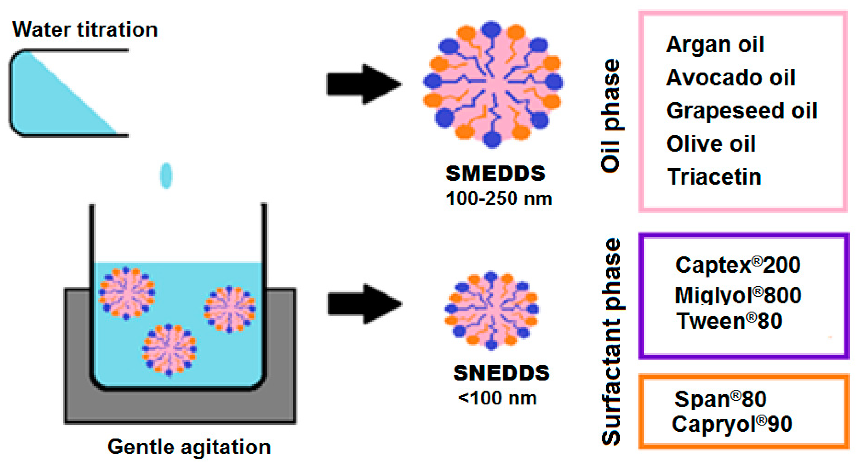
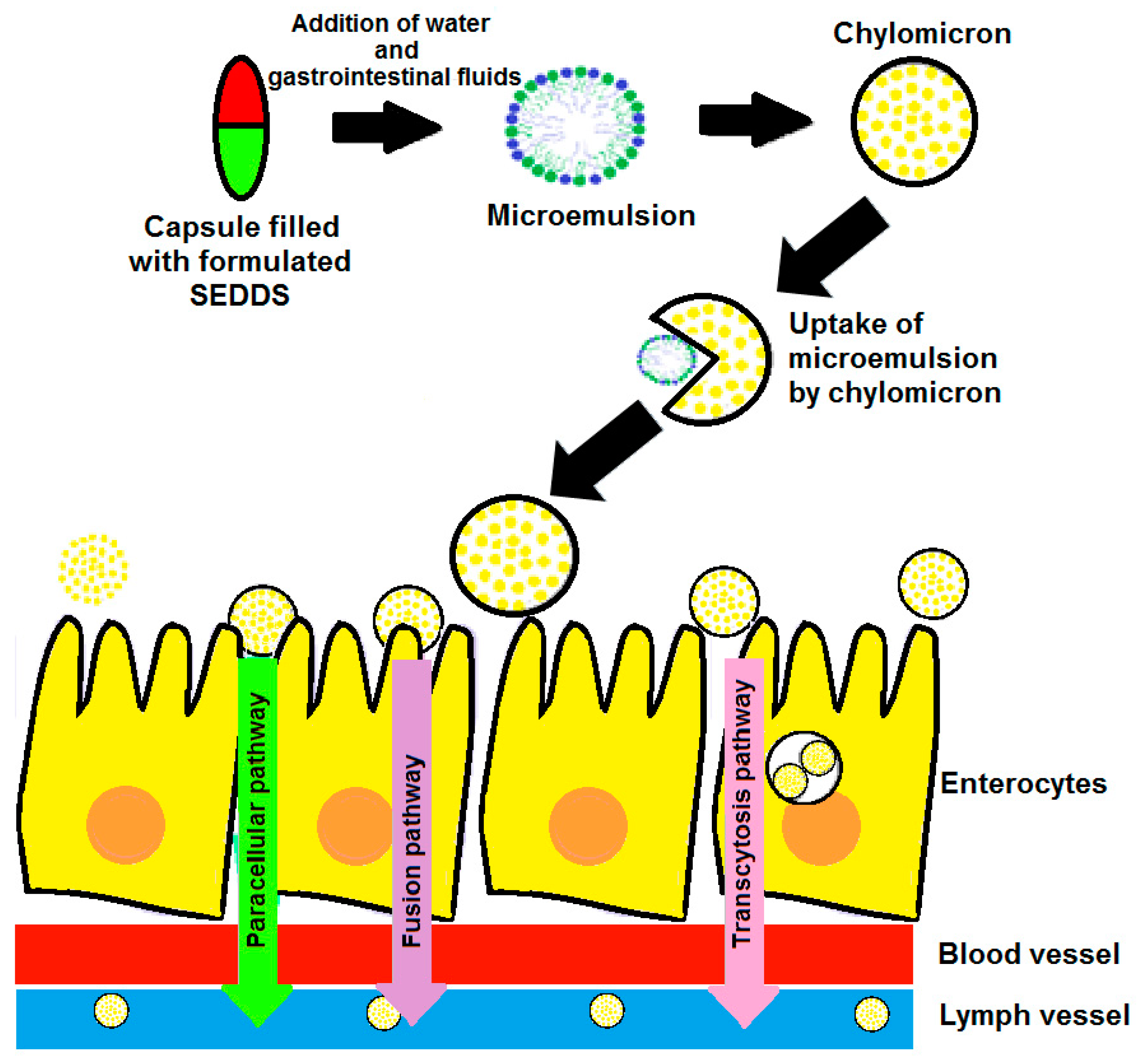
2. Mechanism of Spontaneous Emulsification

Generating a system prone towards spontaneous emulsification is a challenging process [29]. It is suggested by literature that spontaneous emulsification is facilitated by a system that favours dispersion formation instead of increasing the surface area of the dispersion upon exposure to a change in entropy. Thus, the free energy present between SEDDS components is directly related to the energy required by the system to generate a new surface between the two immiscible phases [5,30]. This relationship between the free energy required to establish a self-emulsifying system and the energy at work at the interface is described by the following equation:
Δ G = N π r 2 σ
where G indicates the free energy of the self-emulsification process (excluding the free energy facilitated by mixing), N represents the quantity of droplets within the radius (r), and σ is the energy at the interface [30].
Theoretically, the process of spontaneous emulsification should only include slight stirring of the system so as to mimic peristaltic movements of the gastrointestinal tract, since the chemical potential energy gradient between the two immiscible phases is of a non-equilibrated nature and is increased enough to suffice in establishing self-emulsification [31]. Contrarily, in practice, temperature-sensitive systems with the potential of spontaneous emulsification are occasionally heated while subjected to mild agitation to ensure phase inversion. Likewise, some systems have portrayed self-emulsification during cooling after exposure to increased temperatures [32]. Additionally, emulsions established by spontaneous emulsification can be considered time-sensitive, as these systems have a time-dependant tendency to fall back to phase separation [30]. Therefore, surface-active agents are added to these emulsions, especially in the pharmaceutical industry, in order to ensure stability of spontaneous emulsions [29]. However, systems established by spontaneous emulsification are still not fully understood, despite considerable research efforts and excipient combinations [30,32]. Some studies investigated the mechanism of spontaneous emulsification by removing surfactants from potential self-emulsifying systems in order to obtain insight into this mysterious mechanism. These studies discovered that interfacial turbulence might not be individually responsible for establishing self-emulsification, since surfactants can completely suppress the flow of turbulence and spontaneous emulsions form within the presence, as well as absence of surfactants. The concept of having a continuously low (negative) interfacial energy within a system is not necessary compulsory for the purpose of achieving self-emulsification, since surfactant-free systems exclusively displayed self-emulsification when nearing a critical point and, sometimes, even beyond the critical point. Lastly, the ability of self-emulsifying systems to render both micro- and nanosized droplets indicates that spontaneous emulsification might even be a diffusion-driven process [32]. Nonetheless, manufacturing as well as the up-scaled production of SEDDSs are simple, yet cost-effective, processes [33,34]. Therefore, the multifaceted potential of SEDDSs can possibly provide excellent topical/transdermal drug delivery vehicles by considering the multi-layered nature of skin.

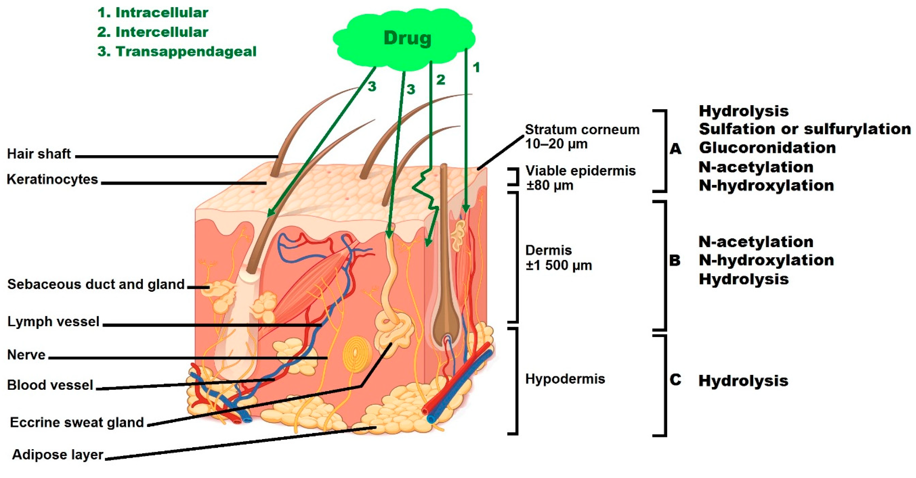
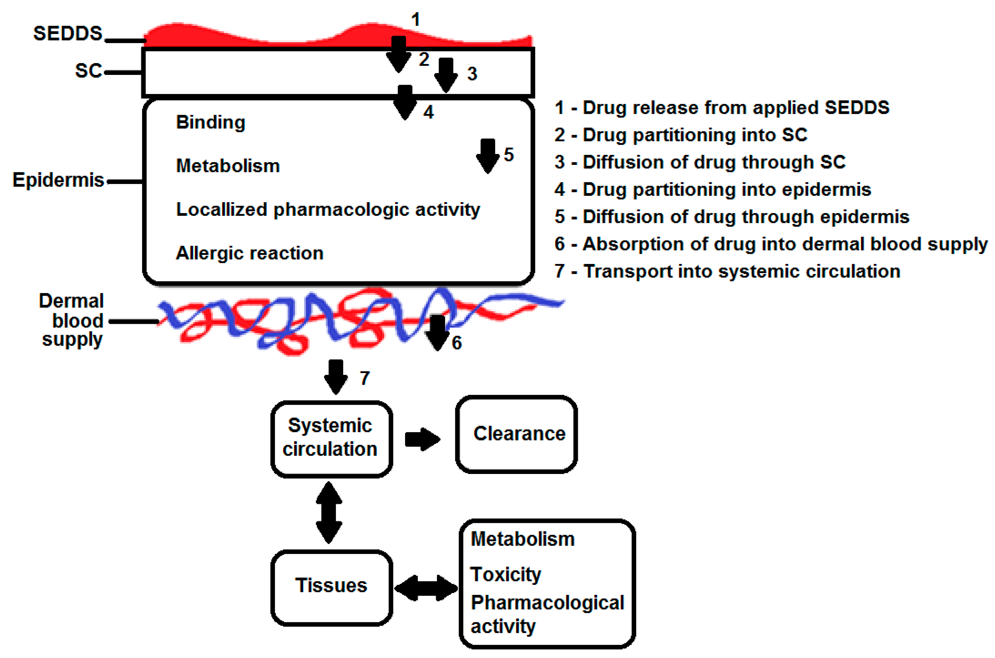
3. Skin, a Multi-Layered Organ

Skin represents the first barrier between the body and exogenous substances [35,36,37]. Furthermore, the multi-layered structure of skin allows this organ to be a regulator of topical signals by means of active metabolic pathways in order to influence physiological activities as well as regulate internal homeostasis [35,36]. Hence, skin should be considered a metabolically active part of the human body, as the enzymatic profile of the skin is quite similar to the scientifically generated enzyme profile of the liver. Enzymatic activity of skin intensifies within the deeper skin layers, such as the epidermis and dermis, compared to the outermost stratum corneum (SC). Metabolomic studies have displayed that the enzymatic activity of the skin can add up to approximately 10% of total liver metabolism [36]. The process of skin metabolism is defined as a two-phase reaction, where phase I consists of reactions for instance oxidation, hydrolysis, and reduction, and phase II refers to conjugated reactions [36,38]. Enzymes responsible for establishing skin metabolism include cytochrome P450, nonspecific esterases, the enzyme family of flavin monooxygenase, as well as transferases [36,37,39]. Hence, drugs that are able to cross the highly lipophilic SC must still withstand metabolic degradation after entry into the underlying skin layers, as illustrated in Figure 1.
As indicated in Figure 1, there are three different skin penetration pathways by which drugs can cross the SC, namely, the transcellular-, transappengeal-, and intercellular pathways [36]. The highly hydrated keratins serve as building blocks of corneocytes, which are known as the main components of the epidermis skin layer [40,41]. Hence, hydrophilic drugs tend to pass through the transcellular pathway provided by highly hydrated keratins [40]. Likewise, intercellular permeation creates a favourable route for lipophilic drugs, as diffusion of these compounds is guided by the lipid matrix. Modest dermal transport of drugs occurs via the transappengeal pathway, where a mere 0.1% of the total skin surface is covered by hair follicles and sebaceous glands [41]. However, the lipid rich sebum that fills sebaceous glands could provide a potential route favoured by the lipophilic nature of SEDDSs as well as the fine-oil droplets generated by these simplified drug delivery systems [42]. Contrarily, sweat could provide a minute obstacle in terms of lipophilic drug diffusion due to its hydrophilic character. However, if topically applied SEDDSs can portray robustness to dilution, this additional barrier provided by sweat might be overruled as an agent slowing dermal permeation instead of a mediator-blocking dermal permeation [41]. In addition, it is a well-known fact that drugs suitable for topical/transdermal drug delivery should own lipophilic as well as hydrophilic properties to enable crossing of the lipophilic outermost skin layer into the hydrophilic epidermis and dermis [43]. Interestingly, another potential topical/transdermal drug delivery angle can be to utilise the lipophilic nature of SEDDSs to reach lymph nodes located within the epidermis and dermis in order to further refine targeting of dermal drug delivery systems [44,45]. Lymphatic uptake of oral SEDDSs demonstrated successful avoidance of hepatic metabolism as well as increased drug bioavailability [33,46,47]. Therefore, similar advantages can potentially be created by dermal lymphatic uptake to further protect drugs from dermal metabolism, as research confirms intensified dermal metabolism within deeper skin layers such as the dermis [36]. Consequently, special consideration must be taken when choosing excipients included in topical/transdermal SEDDSs, as these compounds will be the decisive factor in terms of optimised topical/transdermal drug delivery across the multiple layers of the skin.

4. Excipients Fulfilling Different Roles Depending on the Route of Delivery

4.1. Active Compounds Incorporated in SEDDSs

SEDDSs were created as an alternative vehicle for the purpose of improving lipophilic drug delivery [1,2,33]. Lipid-based drug delivery systems have received substantial attention during the last decade, since 30% of marketed drugs are poorly water soluble and where approximately 50% of all new drug entities are considered highly lipophilic [17,48,49,50]. The ideal lipophilicity for drugs included in oral SEDDSs have been established as depicting a log p value ≥ 2. Interestingly, it was found that, due to increased solubility of highly lipophilic drugs in SEDDS formulations, less of the said drug is essential in order to obtain equivalent therapeutic effects [33]. Moreover, hepatic metabolism, which normally portrays a high affinity for lipophilic drug entities, is circumvented, since SEDDSs disperse into fine droplets within the aqueous environment of the gastrointestinal tract and are also lymphatically absorbed prior to hepatic exposure [33,46,47]. Thus, decreased drug concentrations in these delivery systems still render an appropriate therapeutic response due to the solubilisation as well as protection provided by the SEDDS vehicle [33].
In terms of topical or transdermal drug delivery, the ideal log p value ranges between a value of 1 and 3 [51]. Moreover, a potential aim of topical and transdermal drug delivery, such as decreasing dosing intervals (the frequency of administered doses) by delivering decreased concentrations of drugs with longer elimination half-lives, can be met [52]. Additionally, topical/transdermal drug delivery enables transport of sufficient concentrations of drugs prone towards hepatic metabolism, since liver metabolism is bypassed during dermal drug delivery [28]. Therefore, lower drug concentrations incorporated into topical/transdermal SEDDSs might not be problematic [28,33]. On the other hand, supersaturated vehicles destined for topical/transdermal drug delivery have high success rates due to enhanced flux values as the high drug concentration of the vehicle establishes a driving force for drug partitioning into the skin due to maintained saturated drug concentrations at the skin surface [33,53]. Remarkably, supersaturated SEDDSs evidently rendered the inhibition of drug precipitation in a kinetic and thermodynamic fashion by slowing crystal growth together with nucleation [33]. Therefore, it might be more advantageous to present supersaturated SEDDSs at the skin surface compared to decreased drug concentrations [53].

4.2. The Oil Phase

Synthetic or natural oils are included as the lipophilic component of SEDDSs for the purpose of enhanced solubilisation of lipophilic drugs [33]. Improved solubilisation of orally administered drugs establishes increased bioavailability, which in turn improves drug absorption in the gastrointestinal tract due to involvement of the intestinal lymphatic system. Certain characteristics of the oil phase are indicated by the melting point, physical characteristics, as well as HLB profiles of glycerides contained by the oil itself. These characterisation profiles are also influenced by the extent of etherification together with the type of fatty acid(s) present within the oil phase. Some studies noted that medium-chain in addition to long-chain triglycerides, owning different levels of saturation, can be successfully utilised during formulation of oral SEDDSs [33,54,55]. This is confirmed by research indicating that medium-chain triglycerides with carbon backbones of 6–12 carbons are easily transported into the systemic circulation via portal blood supply; where, sequentially, long-chain triglycerides, exceeding 12 carbon chains, can be lymphatically transported [33]. However, the enhanced fluidity of medium-chain triglycerides, followed by improved solubility properties and anti-oxidative effects, deem medium-chain triglycerides the most favourable components of the oil phase for oral SEDDSs [33,54,55].
Regarding topical/transdermal drug delivery, the extent of glyceride saturation is considered a more determining drug delivery factor, as numerous studies have indicated that unsaturated fatty acids (UFAs) portray more powerful skin penetration-enhancing effects compared to saturated fatty acids (SFAs) [56,57,58,59]. This finding can be explained by the decreased capability of SFAs to dissolve within the natural lipids of the SC, which leads to reduced SC lipid disruption and, subsequently, less effective skin penetration enhancement [60,61,62]. Therefore, considering topical/transdermal drug delivery, the oil phase is not only vital for solubilising lipophilic drug(s), but it is also crucial in facilitating skin penetration enhancement, as established by fatty acids that form part of the oil phase [33,58]. Skin penetration enhancement can be established through different mechanisms, including fluidisation or disruption of lipids present within the outermost skin layer [56]. However, the fatty acid concentration, as well as the specific types of fatty acids included in the oil phase, also have a significant influence on skin penetration-enhancing effects [58,63]. Hence, an oil phase must be chosen with optimum drug solubility together with a favourable fatty acid composition for the purpose of creating an optimised topical/transdermal drug delivery vehicle.
Oleic acid is the most-recognised skin penetration enhancer of all time [56,58,64,65]. It is a trademark component in skin penetration-enhancing natural oils, such as olive-, avocado-, and pequi oil [66]. The mechanism of skin penetration enhancement, as achieved by oleic acid, is partially established by the disordering effects on SC lipids [56,58]. Moreover, oleic acid has portrayed an increased capacity to facilitate fluidisation of SC lipids, which leads to enhanced drug permeation in addition to increased flux values [56]. The fluidisation theory is confirmed by studies that observed oleic acid in separate pools of SC lipid domains [65]. However, it is mentioned in literature that only the cis form of oleic acid can establish an enhanced flux of topically applied drugs due to the unsaturated structure of this form of oleic acid [56]. Although both the cis and trans forms of oleic acid are deemed unsaturated, it is vital to understand that the inclusion of a trans double bond in a fatty acyl chain inaugurates an abridged bonding angle relative to a cis double bond, leading to a fatty acid acyl chain conformation that bears a significant similarity to a saturated fatty acid structure rather than an unsaturated fatty acid structure, regardless of the overall unsaturation [67]. Additionally, this knowledge can assist in explaining fluidised skin lipid domains, as the cis formation predominantly condenses with itself instead of facilitating even distribution throughout SC lipids [65]. This non-homogenous distribution of oleic acid may suggest that reversible, together with permeable, skin defects are created to further improve dermal drug delivery [56,58,65]. However, not all natural oils are rich sources of oleic acid; for example, grapeseed- and argan oil [66,68]. Interestingly, linoleic acid rich oils have portrayed intensified skin repair effects, which are achieved by preventing SC water loss [69]. These increased hydration properties, facilitated by linoleic acid, can similarly be enabled by other fatty acids, especially those of a saturated nature [63,70]. For instance, stearic acid is known for its high melting point and subsequent film formation on the skin surface. The concentration of undissolved stearic acid applied onto skin cannot partition into the SC to achieve a lipid-disordering effect. Even so, film formation can contribute towards improved dermal permeation due to occlusive effects that provide increased hydration followed by loosening of the strict SC structure that allows a permeation of drugs into the underlying skin layers [70,71]. Therefore, indicating the crucial contribution of different fatty acids on the skin and, particularly, the potential influence of different fatty acids utilised in combination in order to achieve optimised topical/transdermal drug delivery. Furthermore, it has been found that polyunsaturated fatty acids (PUFAs) tend to participate in oxidative reactions to a higher degree than monounsaturated fatty acids (MUFAs) [69]. Hence, fatty acid composition can correspondingly influence shelf life of topical/transdermal SEDDSs.

4.3. Surfactants

The interfacial tension between the lipophilic and hydrophobic components of SEDDSs are reduced by surfactants. They create an interfacial film between the two immiscible phases for the purpose of generating a dispersion [3,4,5,6,7,8,72]. During formulation of SEDDSs, higher emulsification properties are achieved by including surfactants with HLB values exceeding a value of 12. This allows fine-oil droplet formation within the water that establishes rapid spreading of the formulation in the gastrointestinal environment. Overall, non-ionic surfactants are mostly employed during the formulation of oral SEDDSs, as these surfactants have decreased toxic profiles compared to anionic- and cationic surfactants. However, even non-ionic surfactants can establish irreversible, moderate alteration to the permeability of the gastrointestinal wall. Nonetheless, it has been found that a surfactant concentration of approximately 30–60% w/w of an oral SEDDS formulation renders enhanced spontaneous emulsification within the gastrointestinal environment [33].
Additionally, similar challenges, concerning surfactant concentrations, exist while developing SEDDSs destined for topical/transdermal drug delivery, as skin is also sensitive towards high surfactant concentrations [73,74]. Skin irritation reactions associated with surfactant exposure is linked to the capacity of surfactants to solubilise lipid membranes [75]. However, this property of surfactants can contribute towards enhanced dermal drug delivery, as the SC is comprised of lipids that form a formidable barrier to drug delivery systems [58]. Hence, when deciding on surfactant concentrations of dermal SEDDSs, the choice must be based on the principle of finding an area of compromise so as to not only include low enough surfactant concentrations to avoid skin irritation, but these concentrations must be high enough to achieve skin penetration enhancement.
Co-surfactants are included in SEDDS formulations to attain an even smaller transient negative value by further decreasing interfacial tension between the two immiscible phases. This enables increased flexibility of the interfacial film. By imparting enhanced flexibility to the interfacial film, different curvatures can lead to the formation of different concentrations of microemulsions. Furthermore, inclusion of co-surfactants can assist in incorporating decreased surfactant concentrations due to enhanced interfacial flexibility [33,76]. At this stage, an expanded interface enables fine droplet formations that are capable of adsorbing more surfactants together with co-surfactants until the film is diminished to shift back towards a positive interfacial tension that renders spontaneous emulsification [33]. As a result, by including co-surfactants, the ease of producing self-emulsification is enhanced while surfactant concentrations associated with skin irritation, as well as irreversible gastrointestinal alterations, are reduced. Substances most widely incorporated as co-surfactants are medium-chain-length alcohols (C3–C8) [33,76]. Examples of potential surfactant phases are displayed in Figure 2.

4.4. Water

As the hydrophilic component of both oral and topical/transdermal SEDDSs, water is the last- but not least-important component of these uniquely formulated drug delivery systems. With orally administered SEDDSs, water may be included as part of the formulation itself, for example, contained in filled capsules or dispersible granules, or SEDDSs may be exposed to water within the gastrointestinal tract to further emulsify into finely dispersed droplets prior to chylomicron uptake [33]. Contrarily, topical/transdermal SEDDSs are not exposed to similar amounts of external water [77]. Hence, water must be included beforehand in sufficient concentrations in topical/transdermal SEDDSs to maintain spontaneous emulsification [78]. Moreover, water is the most ancient skin penetration enhancer, as it maintains SC hydration followed by loosening of the stiff SC structure in order to improve drug movement through the shield provided by the SC [79]. The question nonetheless remains: How can topical/transdermal SEDDSs be optimised in terms of excipient concentrations utilised in addition to achieving spontaneous emulsions?

5. Compatibility of Topical/Transdermal SEDDS Excipients

Optimised drug delivery follows an optimised drug delivery system design approach. Hence, during the formulation of topical/transdermal SEDDSs, the physical, chemical, as well as biological properties of all drug(s) and excipients incorporated in formulations must be subjected to compatibility studies. Infrared (IR) is often employed during compatibility experiments, as it is related to the study of covalent bond formation, and detailed information is obtained about the molecular structure, either between drug(s) and excipients or excipients and excipients [80]. Contrarily, isothermal micro-calorimetry experiments can also be conducted for the purpose of confirming the compatibility of drug(s) utilised in combination with different excipients [81]. This compatibility-testing technique is based on the understanding that instability reactions produce heat exchanges between surrounding components that can signify potential incompatibility between excipients or drug(s) and excipients [81,82]. However, this highly sensitive compatibility test is nonspecific and a follow-up analysis must be conducted to confirm if detected incompatibilities are of a chemical or physical origin. Interactions of a chemical nature are almost without exclusion considered unfavourable due to the degradation of components within formulations that reduces efficacious functionality together with the safety of the final SEDDS. Physical interactions can potentially be accommodated in topical/transdermal SEDDSs unless proven deleterious, as these types of interactions can occasionally enhance drug delivery. An example of a potential physical interaction together with improved drug delivery includes: complexation between lipophilic drugs and cyclodextrin to enhance oral drug delivery [83]. However, the challenge of finding components suitable for topical/transdermal drug delivery, as well as these components being compatible when utilised in combination to successfully render optimised topical/transdermal drug delivery from SEDDSs, must not be underestimated. Subsequently, a method must be utilised to establish the optimum concentrations of compatible excipients utilised in combination to successfully achieve, as well as maintain, self-emulsification.

6. Biocompatibility of Excipients Utilised to Establish Spontaneous Self-Emulsification

Biocompatibility has been a field of interest since the 1940s and is becoming more important in the field of pharmaceutical sciences, as excipients are not only included in formulations in order to establish desired formulation characteristics such as stability or cost-effective production, but they also contribute towards the acceptability of the formulation when it comes into contact with viable tissue. Once a foreign material is introduced to the human body, the body retorts to one or more positive and/or negative reactions. Three main adverse responses observed with especially dermal/transdermal drug delivery systems are: restricted wound healing, inflammation, and an acquired or innate immune response. As a result, a compound is deemed biocompatible when it does not display any toxicity, does not initiate any type of tissue injury with which it is in contact with, or trigger any immunological reaction [84,85]. The definition of biocompatibility has been modified over the years from: “the ability of a biomaterial to perform with an appropriate host response in the specific application” to “an expression of the benignity of the relation between a material and its biological environment”, as the focus has shifted towards not only biocompatibility of excipients but also biodegradability of the materials utilised to aid disease control [85,86,87]. The biocompatibility of a component is dependent on (i) material-related factors, such as shape, size, surface chemistry, composition, sterility, duration of contact, degradation, etc.; on (ii) host-related factors; and on (iii) the site of application, such as tissue characteristics and the microenvironment [84].
Natural oils are considered highly attractive when included as the oil phase of topical/transdermal SEDDSs, since biodegradability as well as compatibility with skin are taken for granted due to its natural origin [88]. However, natural oils known to cause allergic reactions, such as nut-based oils (e.g., almond-, macadamia-, and peanut oil), should rather be avoided, as numerous patients might be allergic to these oils [89]. Moreover, the choice of surfactant and co-surfactant are even more substantial, as they can drastically influence biocompatibility. These excipients are known to cause skin reactions due to the powerful disruption of lipids naturally harboured within the skin because of their amphiphilic properties [75,90]. Likewise, they are capable of modifying the transport of active pharmaceutical ingredients, though, as a result, biocompatibility is restricted [91]. For example, it is known that new surfactants, such as Labrasol®, were developed specifically for their optimal solubilisation capacity, but Labrasol® causes cell death receptor activation [92]. The reason for this severe cell damage is the irreversibly damaged membrane integrity due to the highly increased solubilisation capability of this surfactant [93]. Consequently, surfactants may have different cytotoxic effects, but the interference of their toxicity has not yet intensively been researched. This statement is validated by Nemes et al. [93], who found that the non-ionic polysorbate 20, which is an official surfactant described in different Pharmacopoeias (Ph. Eur. 9 and Ph. Hg. VIII.), possesses severe cytotoxic properties, and it lowered the biocompatibility of methyl paraben because it is able to solubilise membrane proteins. Therefore, special attention must be devoted to choosing surface active agents with the capacity to establish spontaneous emulsification while being included in reduced concentrations. Biocompatibility of excipients included in SEDDS, as well as the combinations and concentrations in which they are added, should additionally be researched intensively.
However, due to ethical and financial constraints, the employment of different animal trials is inadequate for biocompatibility establishment [94]. Furthermore, in the European Union, these tests are controlled by the 440/2008 EC regulation, where cell lines are favoured over animal experiments. This led to the establishment of numerous human cell culture models in biocompatibility assessments. These cells culture models are able to display similar expression patterns, membrane proteins, enzymes, etc. Nonetheless, they are incapable of representing actual human tissue due to the lack of extracellular elements and structure. Thus, reliable biocompatibility of pharmaceutical products can only be based on the evaluation of concomitant cytotoxicity profiles of different excipients and their combinations [93]. Various studies that have investigated biocompatibility of excipients, mostly employed in vitro histological experiments, where exposed skin was stained with hematoxylin to be assessed for possible structural damage, MTT (3-(4,5-dimethylthiazol-2-yl))-2,5-diphenyltetrazolium bromide) cytotoxicity analysis, cell proliferation assays, as well as apoptosis experiments [84,90,92,93,95,96]. Some successful biocompatibility studies have also been performed on SEDDSs. For example, a recent study aimed at developing a SEDDS comprising chlorhexidine and monododecylamide-ethylenediaminetetraacetic acid, formulated to battle antimicrobial resistance, found that >85% of cells remained viable after a period of 4 h. This study used the Resazurin assay on a Caco-2 cell line under regulated conditions [97]. Hence, biocompatibility experiments are essential tests to conduct during the developmental phase of topical/transdermal SEDDSs, especially for the purpose of identifying potential cytotoxic reactions [85,97]. However, this remains an undiscovered territory that should specifically be refined for topical/transdermal SEDDSs in terms of special skin cell models, skin sensitivity, inflammation, and wound-healing assessments, to name a few.

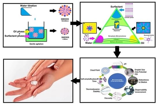
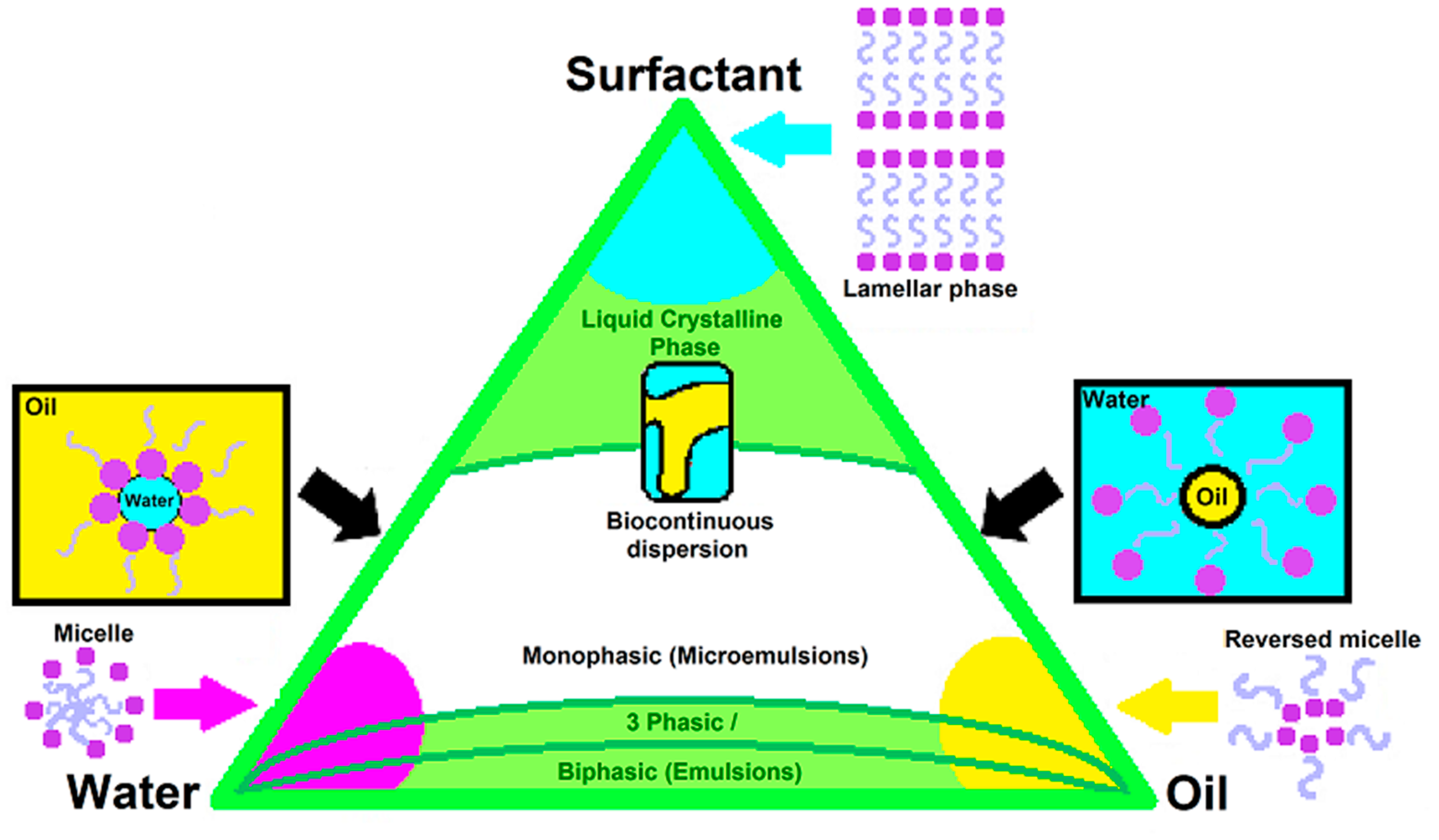
7. Pseudo-Ternary Phase Diagrams, Formerly Utilised Diagrams with Novel Potential

Pseudo-ternary phase diagrams are mostly constructed during the development of orally delivered SEDDSs, as is the case with most lipid-based drug delivery systems, since these diagrams provide a schematic representation of the region of self-emulsification when specific excipients are employed in combination [33,77]. This simplified process consists of preparing an optimised surfactant phase (fixed ratio of surfactant to co-surfactant) dissolved within a chosen oil phase in ratios of 9:1, 8:2, 7:3, 6:4, 5:5, 4:6, 3:7, 2:8, and 1:9 [83,98]. Hereafter, the water phase is added in a dropwise fashion while the mixtures are slightly stirred between water additions [99]. The point at which a mixture becomes turbid after moderate, continuous stirring marks the endpoint and, also, the concentration where spontaneous emulsification occurred. Next, the endpoint is plotted on the pseudo-ternary-phase diagram to illustrate the concentration range of excipient utilised in combination where self-emulsification is most likely to occur. Once the coordinates of the endpoint concentrations are plotted within the triangle, the area of spontaneous emulsification is termed heterogeneous due to the biphasic nature of this enclosed region, whereas the unenclosed area indicates the monophasic system known as the homogenous area of the tri-plot [33]. Additionally, pseudo-ternary-phase diagrams are functional instruments utilised to predict the phase behaviour of a potential SEDDS, as different segments of the diagram are indicative of certain behavioural characteristics of emulsions [100]. For instance, considering oral drug delivery, pseudo-ternary-phase diagrams can illustrate the robustness of a SEDDSs to dilution within the gastrointestinal environment [33,100]. However, during topical/transdermal drug delivery, different factors must be considered in order to optimise excipient concentrations and, subsequently, the phase behaviour of topically applied vehicles. The different phase behavioural regions can be seen in Figure 3.
As demonstrated in Figure 3, areas of a pseudo-ternary-phase diagram comprising either extensively high concentrations of the water-, oil-, or surfactant phase (combination of surfactant and co-surfactant) must be avoided, as these regions tend to generate unique structures such as lamellar phase formation in the presence of increased surfactant phase concentrations [100]. In addition, regions comprising increased surfactant concentrations must also be avoided to prevent skin irritation upon application of the drug delivery vehicle [73,74]. Moreover, reversed micelles are observed in oil rich regions of pseudo-ternary-phase diagrams [100]. Reversed micelles provide vast possibilities, since polar and nonpolar substances can be solubilised by these structures [101,102,103]. Additionally, promising applications for reversed micelles are known in the chemical industry in terms of enzymatic reactions for the purpose of certain drug delivery systems, as well as during the synthesis of nanomaterial [101,102,104]. However, reversed micelles do not necessarily contribute toward enhanced dermal drug delivery and are thus unwanted structures during the development of topical/transdermal SEDDSs. Micelles formed within water-rich areas of the tri-plot are considered highly unfavourable structures for dermal drug delivery due to their rigidity together with a decreased potential of deformability [105,106]. Therefore, all corners of pseudo-ternary-phase diagrams must be avoided during the development of topical/transdermal SEDDSs. Furthermore, the centre of the pseudo-ternary-phase diagram predicts the formation of unpredictable bi-continuous emulsions, as these emulsions refer to particle-solubilised systems comprising two continuous phases followed by a sustained inter-phase penetration [107].
The lipophilic or hydrophilic nature of incorporated drug(s) can also influence the choice of where the optimised formulation area is located, since the nature of the drug can contribute towards the time required to establish drug release from the formulation [108,109]. Hence, theoretically, a lipophilic drug will portray swift release if incorporated into a formulation containing an increased water concentration, as the lipophilic drug tends to desire escape from the hydrophilic vehicle into the lipophilic SC [108,110]. Therefore, pseudo-ternary-phase diagrams might be seen as an old authentication technique, but in terms of SEDDS development, it remains an irreplaceable prediction that enables the selection of desirable formulation properties [33]. After deciding which area of the obtained self-emulsification region is most suitable to enable topical/transdermal drug delivery, checkpoint formulations can be formulated and, subsequently, subjected to recognised characterisation experiments specifically created for oral SEDDSs, where a considerable twist is added to determine suitability for the topical/transdermal route.

8. Metamorphic Characterisation of Topical/Transdermal SEDDSs Versus Orally Delivered SEDDSs

8.1. Evaluation of Droplet Sizes, Zeta Potential, and Polydispersity Index

The characterisation of orally administered SEDDSs involves the evaluation of droplet size, zeta potential, studies relating to the surface morphology by means of electron microscopy, and an analysis of phase separation. The evaluation of droplet sizes, as well as zeta potential, provides insight into the chemical and physical profiles of the investigated SEDDSs. What is more, droplet sizes, together with the polydispersity index (PDI), influence the melting properties of oral lipid-based formulations, together with the rate of droplet penetration and dissolution. Therefore, these evaluations indicate a potential release of the encapsulated drugs from oral SEDDSs [33]. For the purpose of topical/transdermal drug delivery, droplet size determines an important aspect of diffusion, as smaller particles tend to portray faster, together with increasingly significant permeation across the SC [111,112,113]. Decreased droplet sizes can be supported by including surfactants that reduce interfacial tension in order to establish the formation of fine droplets [114]. Thus, the lipid-altering potential, along with droplet refinement potential, of surfactants can assist in the successful delivery of topical/transdermal SEDDSs [58,111,112,114]. Likewise, finer droplets comprising lipophilic drugs have demonstrated an increased affinity for subcutaneous lymphatic uptake and could assist in avoiding metabolic processes within the dermis [6,12]. The ideal range for subcutaneously injected substances considered suitable for lymphatic absorption is between 80–100 nm [6].
Dynamic light scattering is generally employed to analyse droplet size, zeta potential, and the PDI of SEDDSs, as these three characterisation assessments play an interchangeable part in predicting the SEDDSs performance in terms of dermal diffusion as well as formulation stability [33]. Zeta-potential measurements indicate the charge present on individual droplet surfaces within the chosen medium of dispersion [112]. This measurement identifies the degree of electrostatic repulsion present between droplets within the same dispersion, so as to contemplate the tendency of droplets to be repulsed by each other or to coagulate in order to establish larger droplets [115]. Hence, increased zeta-potential values predict increased droplet-droplet repulsion followed by increased emulsion stability [116]. Generally, the ideal zeta-potential value is defined as either >30 mV or <−30 mV for stable emulsions [117]. Interestingly, it has been reported in literature that the pH of a formulation can have a definite impact on zeta potential [118]. Therefore, if desired, pH adjustment can potentially provide more stable SEDDS formulations. Furthermore, the charge of droplets is not only indicative of formulation stability but also predicts the potential extent of dermal drug permeation [118,119]. Increased diffusivity can be driven by the affinity of the applied SEDDS for skin [118]. Theoretically, a positively charged formulation should portray increased skin surface affinity, since the net charge of the skin surface is of a negative nature [119]. Contrarily, the inclusion of natural oils in topical/transdermal SEDDSs renders negatively charged repulsion between droplets due to the presence of free fatty acids [120]. Ironically, free fatty acids are wanted components of topical/transdermal SEDDSs due to their inert skin penetration enhancement properties [58,63,70]. Besides, natural oils tend to carry a decreased risk of skin irritation reactions, as the fatty acids present in these oils are more compatible with the lipid matrix itself [63,70]. Therefore, negatively charged formulations will be able to cross the negatively charged skin surface through the inclusion of components such as free fatty acids [56,57,58]. Though, the permeation of negatively charged formulations can occur at a decreased rate compared to positively charged formulations [119].
A uniform droplet size distribution is likewise a predictive factor in terms of formulation stability [112]. The PDI indicates a droplet size distribution where a value of 0.0 specifies perfect sample homogeneity and a 1.0 grading is deliberated as a sample of high polydispersity in addition to portraying an unpredictable drug release. The pharmaceutical industry generally considers PDI values ranging from 0.05–0.7 acceptable depending on the type of formulation, as well as the application of said formulation. For example, in the field of topical/transdermal drug delivery, lipid-based carrier systems (PDI ≤ 0.3) and polymer-based nanoparticles (PDI≤0.2) should comply with predetermined PDI values in order to be considered suitable for topical/transdermal drug delivery [121].

8.2. Robustness to Dilution

Robustness to dilution refers to a simple evaluation test that consists of diluting SEDDSs 100-fold where, after, dilutions are left at an ambient temperature of approximately 25 °C for a period of 24 h prior to visual inspection to determine if phase separation has occurred [122]. The ability of a SEDDS to withstand drug precipitation as well as phase separation upon exposure to dilution indicates the stability of these simplified systems once introduced to gastrointestinal fluids [6]. Contrarily, the characterisation of SEDDSs destined for topical/transdermal drug delivery should rather be investigated for robustness towards dilution in fluids comprising different pH values. The purpose of this test in terms of dermal drug delivery would be to demonstrate the stability of a SEDDS as it diffuses through different skin layers while potentially exposed to sweat on the skin surface [77,123]. Remarkably, pH of the skin surface varies from 4.5–5.0, whereas the pH changes to approximately neutral within the final segments of the SC prior to reaching the underlying epidermis of the skin [123]. Therefore, it is recommended by the authors that SEDDSs destined for topical/transdermal drug delivery must be subjected to characterisation experiments, including dilution with fluids that vary in pH between a range of 5–7.4, to ensure the stability of these formulations upon dilution within different pH environments. Accordingly, this investigation will mimic the versatile conditions that SEDDSs can encounter while subjected to dermal diffusion [123].

8.3. Dispersibility Assessment

Evaluating the self-emulsification capacity of a spontaneous emulsion can contribute in predicting its in vitro performance after oral administration [33,124]. In order to achieve enhanced oral drug delivery, a rapid emulsification of the SEDDSs is desired, since spontaneous emulsification marks the rate-limiting step before drug absorption can occur [124]. Contrarily, the rate-limiting step for the majority of drugs intended for topical/transdermal delivery is identified as diffusion through the lipophilic shield established by the SC [53,56]. The differences between the diffusion of SEDDSs destined for topical/transdermal drug delivery and the absorption of orally administered SEDDSs are displayed in Figure 4 and Figure 5, respectively.
It is a priority during topical/transdermal drug delivery to increase the contact time between the skin surface and applied formulation in order to improve dermal drug diffusion [21,125]. Hence, orally administered SEDDSs should exhibit rapid self-emulsification, whereas topical/transdermal SEDDSs should not display prompt emulsification, as this portrays a superior capacity to withstand the exposure to exogenous water exposure [21,33,125]. If a SEDDS is able to restrict rapid emulsification once exposed to exogenous water sources or sweat secretion, it implies an enhanced occlusivity of the formulation, followed by an increased opportunity for dermal drug diffusion [21,125]. A grading system is utilised to categorise SEDDSs according to their exhibited dispersability potential, as depicted in Table 1 [126].
Rapid emulsification (Table 1) is either categorised as a Grade A or B emulsion, whereas decreased spontaneous emulsification properties are exhibited by Grade C, D, and E emulsions. Therefore, Grade A and B emulsions are deliberated as suitable for the development of oral SEDDSs [126], whereas the decreased tendency to self-emulsify of Grade C and D emulsions signifies an increased suitability for topical/transdermal drug delivery [21,125]. However, Grade E emulsions are not considered suitable for oral or topical/transdermal drug delivery, since their complete inability to form spontaneous emulsions predict unfavourable drug-release profiles.

8.4. Self-Emulsification Time

The time required by a system to achieve complete self-emulsification is related to the grading received by SEDDSs during the dispersability test [126]. Interestingly, spontaneous emulsification can ensue despite the presence of kinetic barriers between components, as the inclusion of surface-active agents allow miscibility of the lipophilic and hydrophilic phases of emulsified systems [32]. Therefore, instantaneous self-emulsification portrays the absence of kinetic barriers within the formulation, whereas increased self-emulsification times confirm the presence of kinetic barriers within the drug delivery system [32,33]. The grading thus received by emulsions destined for both oral and topical/transdermal drug delivery includes not only emulsion appearance but also the time required to render the complete dispersability of emulsions [33,126].

8.5. Viscosity

Internal friction that exists within a fluid itself—namely, viscosity—can facilitate resistance against flow, as well spontaneous emulsification [127,128]. For these reasons, determining viscosity profiles for SEDDSs, destined for topical/transdermal drug delivery, is considered highly important, as these results can contribute in identifying the influence of divergent oil phases as well as different surfactant concentrations on viscosity and self-emulsification potential [128,129]. The attraction forces within fluids that establish the degree of viscosity are sensitive toward changes in temperature [130]. Additionally, some systems can portray reversible or irreversible structural changes induced by the flow of fluids [131]. Flow behaviour is classified as either Newtonian or non-Newtonian [132]. Unchanged viscosity values are exhibited by Newtonian fluids upon exposure to different shear rates during viscosity experiments. On the other hand, the flow behaviours of non-Newtonian fluids are influenced by the applied shear rate [133]. Fluids that portray time-dependant flow behaviour changes are thixotropic- and rheopexy fluids, whereas time-independent flow-induced changes are observed in fluids of pseudo-plastic, dilatant-, and visco-elastic natures [133,134]. Pseudo-plastic flow behaviour is also termed shear-thinning and is considered the most favourable flow behaviour of non-Newtonian fluids destined for topical application since these fluids are known to show decreased viscosity once subjected to increased shear rates, as is the case with rubbing during topical application. Rubbing may enhance topical drug delivery due to the lipid disruption that is achieved by the rubbing action on the skin surface. Additionally, spreadability will most probably improve during rubbing, as attraction forces within the fluid are reduced due to the increased shear rate, which allows the formulation to cover and treat a larger affected area [135].
Viscosity profiles of SEDDSs assist in predicting stability, since certain flow behaviour causes irreversible structural changes to these formulations [136]. For instance, behaviour such as dilatancy is considered similar to the flocculation of SEDDSs during storage periods [137,138]. Dilatancy can be described as a shear-thickening behaviour, as these fluids tend to thicken to the point where a clay-like appearance is observed upon subjection to increased shear rates [139]. Therefore, dilatancy signifies risks such as injury to the skin, when applied with a rubbing action, as well as a tendency towards emulsion instability [135,137,138]. This can be attributed to an increased droplet size or the tendency of small droplets to form larger droplets [140]. Remarkably, the attraction forces present within SEDDSs that are evaluated during viscosity characterisation experiments can indicate the ease with which a SEDDS will spontaneously emulsify, since SEDDSs of increased viscosity are inclined to resist spontaneous emulsification due to powerful attraction forces within the emulsified system [127,128]. Therefore, SEDDSs of decreased viscosity can be expected to exhibit rapid, spontaneous emulsification, whereas SEDDSs of enhanced viscosity will more slowly self-emulsify and will thus be more suitable for oral drug administration [33,127,128].

8.6. Cloud Point Assessment

SEDDSs are sensitive towards changes in temperature [33,141]. However, excipients can remain functional while subjected to different temperatures that do not exceed the temperature at which the dehydration of components is observed [141]. Dehydration of excipients incorporated in emulsified systems may be visually identified as a sudden change of formulation appearance, from clear to turbid, once exposed to heightened temperatures [142]. This specific temperature is denoted the cloud point of SEDDSs [141,142]. Dehydration of excipients causes irreversible phase separation, which in turn risks erratic drug release, since spontaneous emulsification is destroyed by irreversible phase separation [141]. For the purpose of oral SEDDS development, formulations should own a cloud point temperature higher than 37 °C, similar to the systemic circulation temperature. This is to avoid the dehydration of excipients within the gastrointestinal tract [141]. Contrary, SEDDSs projected for topical drug delivery should portray cloud points exceeding 32 °C as to avoid formulation instability at the skin surface, which is known for an approximate temperature of 32 °C. However, SEDDSs intended for transdermal drug delivery should follow the same criteria as oral SEDDSs by exhibiting the dehydration of excipients at a temperature exceeding 37 °C, as these droplets should eventually reach the blood circulation in order to establish therapeutic effects [143]. Cloud points can be influenced by drugs, as well as the oil phase [144]. Hence, it is of significant importance to assess individual SEDDSs prior to dermal diffusion experiments. First, SEDDSs are diluted (1:100) through the addition of distilled water. Next, samples are placed in a water bath with a baseline temperature of 25 °C where the temperature will be slightly raised at 2 °C/min in order to determine cloud point temperatures of individual formulations [141].

8.7. Thermodynamic Stability Studies

Thermodynamic stability studies are performed to evaluate the capacity of spontaneous emulsions to remain stable under stressed conditions [97]. The results of these experiments predict the stability of a drug within the matrix created by excipients that can either enforce formulation stability together with reliable drug release or render instabilities such as aggregation, creaming, flocculation, Ostwald-ripening, and/or cracking during storage, as well as risk erratic drug release [97,144]. Experiments include exposure cycles of heating and cooling, subjection to centrifugation, and freeze-thaw stress cycles. Heating-cooling cycles refer to six cycles where SEDDSs are placed in environments of approximately 4 °C followed by heated conditions of approximately 45 °C. Exposure at each temperature should not exceed a period of 48 h. Next, SEDDSs that did not exhibit any form of instability during the heating-cooling cycles are subjected to centrifugation experiments comprising 3500 rpms for 30 min. Thereafter, formulations that did not depict phase separation post-centrifugation are further exposed to three freeze-thaw cycles. These experiments include alternate exposures to temperatures of approximately −20 °C, followed by temperatures approaching 25 °C [97]. If no phase separation, cracking, and/or creaming is observed after thermodynamic stability trials, the SEDDSs are considered suitable for further investigation for topical/transdermal drug delivery.

8.8. pH Measurement

Orally administered SEDDSs are drastically influenced by the pH of introduced fluids, since the pH of a fluid can either enhance or decrease ionisation of the included drug(s) and, thus, determine the solubility of incorporated components in the dissolution media [33]. Fortunately, the skin is able to accommodate formulations nearing neutrality up until formulations of high alkalinity, with a set range of 5.0–9.0 [51]. However, the ideal pH should only range between 4.5–5.0 in order to resemble the natural pH of the skin so as to ensure the optimum compatibility between the skin and formulation [123]. SEDDSs destined for dermal application must meet the pH requirements for the purpose of avoiding skin irritation [51].

9. Conclusions

In terms of oral drug delivery, lipid-based drug delivery systems, including SEDDSs, have received thorough investigation, as well as the refinement of specialised techniques such as muco-adhesive properties, zeta-potential changing capacities, and various solidification techniques [26,33,145]. However, SEDDSs destined for topical/transdermal drug delivery remain an undiscovered field of potential. The initial oral SEDDSs can transform the lipid-based drug delivery of hydrophobic drugs via the topical/transdermal route. This statement is supported by successful drug deliveries achieved by SEDDSs developed to improve drug delivery via alternative topical administration routes such as the ocular-, rectal-, vaginal-, and nasal routes of administration [19,20,21,22,23,24,25]. Moreover, SEDDSs remain noteworthy drug delivery vehicles, since approximately 30% of current drugs on the commercial market, together with up to 50% of newly discovered drugs, are of a noteworthy lipophilic nature [17,48,49,50,51]. Hence, the simplified formulation techniques of SEDDSs allow easy upscaling as well as more economically favourable manufacturing procedures [33,34]. Limitations of oral SEDDSs include decreased dissolution rates of lipophilic drugs within gut and mucosal fluids despite their ability to easily cross biological membranes. This leads to drug precipitation within mucosal- and gastrointestinal fluids that risk erratic drug releases [33]. However, topical/transdermal SEDDSs are not exposed to similar amounts of exogenous liquids and, therefore, might not present challenges similar to that of the oral route of administration [77,123]. Therefore, during the development of topical/transdermal SEDDSs, a detailed focus should be placed on an optimised formulation by including compatible excipients with multifunctional purposes, such as choosing natural oils for their natural skin penetration enhancement capabilities, while facilitating enhanced lipophilic drug solubilisation, together with a decreased tendency towards skin irritation [57,58,63,68].
Despite the fact that the literature cannot provide a clear explanation for the mechanism of spontaneous emulsification, the evident success of the self-emulsification drug delivery approach cannot be denied [19,20,21,22,23,24,25,32,33]. Additionally, pseudo-ternary-phase diagrams assist in predicting the behaviours of emulsions established by mysterious spontaneous emulsification [33,100]. Even though the limited understanding of spontaneous emulsification complicates formulation, it cannot necessarily be considered a factor for terminating the development of topical/transdermal SEDDSs and turning towards other topical/transdermal drug delivery systems such as nano-emulsions. SEDDSs have beneficial capacities compared to conventional topical/transdermal drug delivery systems; for example, nano-emulsions and liposomes, as SEDDSs, are prone toward an enhanced drug-loading capacity, decreased drug concentrations with similar therapeutic effects due to improved drug delivery, as well as an evident lymphatic uptake that renders hepatic metabolism evasion [146,147,148,149,150]. The topical/transdermal route is readily considered, as it naturally avoids hepatic metabolism, but the lymphatic uptake of lipophilic drugs from the dermis skin layer is another advantageous targeted treatment angle that can successfully render SEDDS the leading agents in topical/transdermal drug delivery [6,12]. The dermal lymphatic drug delivery principle can furthermore aid in diseases worsened by lymphatic dissemination, for example, human immunodeficiency virus (HIV), metastatic cancers, and endogenous extra-pulmonary TB [151].
The transfiguration of oral SEDDSs to topical/transdermal drug delivery vehicles is in early developmental stages with limited publications. Moreover, to the authors’ knowledge, this is the first clear review aimed at tailoring established characterisation techniques, utilised to obtain characterisation profiles of SEDDSs destined for oral drug delivery, to evaluate the characterisation profile of an ideal topical/transdermal SEDDS vehicle. The development of dermal drug delivery systems presents many obstacles due to the multi-layered nature of the largest organ of the human body [28,56,65]. Moreover, skin metabolism, as well as metabolomic evaluation techniques for dermal drug delivery vehicles, are relatively new and unestablished technologies [36]. Therefore, the challenges accompanied by the development of topical/transdermal SEDDSs might not be as simple as expected.

Conflicts of Interest

The authors declare no conflict of interest.

References

  1. Pouton, C.W. Formulation of self-emulsifying drug delivery systems. Adv. Drug Deliv. Rev. 1997, 25, 47–58. [Google Scholar] [CrossRef]
  2. Mandić, J.; Pobirk, A.Z.; Vrečer, F.; Gašperlin, M. Overview of solidification techniques for self-emulsifying drug delivery systems from industrial perspective. Int. J. Pharm. 2017, 533, 335–345. [Google Scholar] [CrossRef] [PubMed]
  3. Kazi, M.; Al-Swairi, M.; Ahmad, A.; Raish, M.; Alanazi, F.K.; Badran, M.M.; Ali Khan, A.; Alanazi, A.M.; Delwar Hussain, M. Evaluation of Self-Nanoemulsifying Drug Delivery Systems (SNEDDS) for Poorly Water-Soluble Talinolol: Preparation, in vitro and in vivo Assessment. Front. Pharmacol. 2019, 10, 459. [Google Scholar] [CrossRef] [PubMed] [Green Version]
  4. Kamal, M.M.; Nazzal, S. Development of a new class of sulforaphane-enabled self-emulsifying drug delivery systems (SFN-SEDDS) by high throughput screening: A case study with curcumin. Int. J. Pharm. 2018, 539, 147–156. [Google Scholar] [CrossRef]
  5. Ujhely, Z.; Vecsernyés, M.; Fehér, P.; Kósa, D.; Arany, P.; Nemes, D.; Sinka, D.; Vasvári, G.; Fenyvesi, F.; Váradi, J.; et al. Physico-chemical characterization of self-emulsifying drug delivery systems. Drug Discov. Today Technol. 2018, 27, 81–86. [Google Scholar] [CrossRef] [PubMed]
  6. Dokania, S.; Joshi, A.K. Self-microemulsifying drug delivery system (SMEDDS)—Challenges and road ahead. Drug Deliv. 2015, 22, 675–690. [Google Scholar] [CrossRef] [Green Version]
  7. Kale, A.A.; Patravale, V.B. Design and evaluation of self-emulsifying drug delivery systems (SEDDS) of nimodipine. AAPS PharmSciTech 2008, 9, 191–196. [Google Scholar] [CrossRef]
  8. Fatouros, D.G.; Karpf, D.M.; Nielsen, F.S.; Mullertz, A. Clinical studies with oral lipid based formulations of poorly soluble compounds. Ther. Clin. Risk Manag. 2007, 3, 591–604. [Google Scholar]
  9. Pouton, C.W. Lipid formulations for oral administration of drugs: Non-emulsifying, self-emulsifying and ‘self-microemulsifying’ drug delivery systems. Eur. J. Pharm. Sci. 2000, 11 (Suppl. 2), S93–S98. [Google Scholar] [CrossRef]
  10. Pouton, W.C.; Porter, J.H.C. Formulation of lipid-based delivery systems for oral administration: Materials, methods and strategies. Adv. Drug Deliv. Rev. 2008, 60, 625–637. [Google Scholar] [CrossRef]
  11. Chatterjee, B.; Almurisi, S.H.; Dukhan, A.A.M.; Mandal, U.K.; Sengupta, P. Controversies with self-emulsifying drug delivery system from pharmacokinetic point of view. Drug Deliv. 2016, 23, 3639–3652. [Google Scholar] [CrossRef] [PubMed] [Green Version]
  12. Syukri, Y.; Martien, R.; Lukitaningsih, E.; Nugroho, A.E. Novel self-nano emulsifying drug delivery system (SNEDDS) of andrographolide isolated from Andrographis paniculata Nees: Characterization, in-vitro and in-vivo assessment. J. Drug Deliv. Sci. Technol. 2018, 47, 514–520. [Google Scholar] [CrossRef]
  13. Xue, X.; Cao, M.; Qian, Y.; Chen, G. Preparation and optimization of rivaroxaban by self-nanoemulsifying drug delivery system (SNEDDS) for enhanced oral bioavailability and no food effect. AAPS PharmSciTech 2018, 19, 1847–1859. [Google Scholar] [CrossRef] [PubMed]
  14. Pouton, C.W. Formulation of poorly water-soluble drugs for oral administration: Physicochemical and physiological issues and the lipid formulation classification system. Eur. J. Pharm. Sci. 2006, 29, 278–287. [Google Scholar] [CrossRef]
  15. Čerpnjak, K.; Zvonar, A.; Gašperlin, M.; Vrečer, F. Lipid-based systems as a promising approach for enhancing the bioavailability of poorly water-soluble drugs. Acta Pharm. 2013, 63, 427–445. [Google Scholar] [CrossRef] [Green Version]
  16. Porter, C.J.H.; Pouton, C.W.; Cuine, J.F.; Charman, W.N. Enhancing intestinal drug solubilisation using lipid-based delivery systems. Adv. Drug Deliv. Rev. 2008, 60, 673–691. [Google Scholar] [CrossRef]
  17. Rahman, M.A.; Hussain, A.; Hussain, M.S.; Mirza, M.A.; Iqbal, Z. Role of excipients in successful development of self-emulsifying/microemulsifying drug delivery system (SEDDS/SMEDDS). Drug Dev. Ind. Pharm. 2013, 39, 1–19. [Google Scholar] [CrossRef]
  18. Prajapat, M.D.; Patel, N.J.; Bariya, A.; Patel, S.S.; Butani, S.B. Formulation and evaluation of self-emulsifying drug delivery system for nimodipine, a BCS class II drug. J. Drug Deliv. Sci. Technol. 2017, 39, 59–68. [Google Scholar] [CrossRef]
  19. Leichner, C.; Baus, R.A.; Jelkmann, M.; Plautz, M.; Barthelmes, J.; Dünnhaupt, S.; Bernkop-Schnürch, A. In vitro evaluation of a self-emulsifying drug delivery system (SEDDS) for nasal administration of dimenhydrinate. Drug Deliv. Transl. Res. 2019, 14, 945–955. [Google Scholar] [CrossRef] [Green Version]
  20. Kauss, T.; Gaubert, A.; Tabaran, L.; Tonelli, G.; Phoeung, T.; Langlois, M.H.; White, N.; Cartwright, A.; Gomes, M.; Gaudin, K. Development of rectal self-emulsifying suspension of a moisture-labile water-soluble drug. Int. J. Pharm. 2018, 30, 283–291. [Google Scholar] [CrossRef]
  21. Rohrer, J.; Zupančič, O.; Hetényi, G.; Kurpiers, M.; Bernkop-Schnürch, A. Design and evaluation of SEDDS exhibiting high emulsifying properties. J. Drug Deliv. Sci. Technol. 2018, 44, 366–372. [Google Scholar] [CrossRef]
  22. Köllner, S.; Nardin, I.; Markt, R.; Griesser, J.; Prüfert, F.; Bernkop-Schnürch, A. Self-emulsifying drug delivery systems: Design of a novel vaginal delivery system for curcumin. Eur. J. Pharm. Biopharm. 2017, 115, 268–275. [Google Scholar] [CrossRef] [PubMed]
  23. Pattewar, S.; Patil, D.; Sharma, S. Fabrication and characterization of self-microemulsifying mouth dissolving film for effective delivery of piroxicam. Indian J. Pharm. Sci. 2019, 81, 503–513. [Google Scholar] [CrossRef] [Green Version]
  24. ElKasabgy, N.A. Ocular supersaturated self-nanoemulsifying drug delivery systems (S-SNEDDS) to enhance econazole nitrate bioavailability. Int. J. Pharm. 2014, 460, 33–44. [Google Scholar] [CrossRef] [PubMed]
  25. Xiao, L.; Yi, T.; Liu, Y. A new self-microemulsifying mouth dissolving film to improve the oral bioavailability of poorly water soluble drugs. Drug Dev. Ind. Pharm. 2013, 39, 1284–1290. [Google Scholar] [CrossRef] [PubMed]
  26. Lupo, N.; Tkadlečková, V.N.; Jelkmann, M.; Laffleur, F.; Hetényi, G.; Kubová, K.; Bernkop-Schnürch, A. Self-emulsifying drug delivery systems: In vivo evaluation of their potential for oral vaccination. Acta Biomater. 2019, 94, 425–434. [Google Scholar] [CrossRef]
  27. Shaker, D.S.; Ishak, R.A.; Ghoneim, A.; Elhuoni, M.A. Nanoemulsion: A review on mechanisms for the transdermal delivery of hydrophobic and hydrophilic drugs. Sci. Pharm. 2019, 87, 17. [Google Scholar] [CrossRef] [Green Version]
  28. Bellefroid, C.; Lechanteur, A.; Evrard, B.; Piel, G. Lipid gene nanocarriers for the treatment of skin diseases: Current state-of-the-art. Eur. J. Pharm. Biopharm. 2019, 137, 95–111. [Google Scholar] [CrossRef]
  29. Nardin, I.; Köllner, S. Successful development of oral SEDDS: Screening of excipients from the industrial point of view. Adv. Drug Deliv. Rev. 2019, 142, 128–140. [Google Scholar] [CrossRef]
  30. Costagliola, C.; Semeraro, F.; Zeppa, L.; Bufalo, G.; Cardone, M.; Romano, M.; Ambrosone, L. Some physicochemical remarks on spontaneous emulsification of vitreal tamponades. BioMed Res. Int. 2014, 2014, 243056. [Google Scholar] [CrossRef]
  31. Silver, B.R.; Holub, K.; Mareček, V. Spontaneous emulsification at surfactantless liquid/liquid interfaces. J. Electroanal. Chem. 2017, 805, 91–97. [Google Scholar] [CrossRef]
  32. Solans, C.; Morales, D.; Homs, M. Spontaneous emulsification. Curr. Opin. Colloid Interface Sci. 2016, 22, 88–93. [Google Scholar] [CrossRef]
  33. Rani, S.; Rana, R.; Saraogi, G.K.; Kumar, V.; Gupta, U. Self-emulsifying oral lipid drug delivery systems: Advances and challenges. AAPS PharmSciTech 2019, 20, 129. [Google Scholar] [CrossRef] [PubMed]
  34. Gonҫalves, A.; Nikmaram, N.; Roohinejad, S.; Estevinho, B.N.; Rocha, F.; Greiner, R.; McClements, D.J. Production, properties and applications of solid self-emulsifying delivery systems (S-EDDS) in the food and pharmaceutical industries. Colloids Surf. 2018, 538, 108–126. [Google Scholar] [CrossRef]
  35. Feingold, K.R.; Schmuth, M.; Elias, P.M. The regulation of permeability barrier homeostasis. J. Investig. Dermatol. 2007, 127, 1574–1576. [Google Scholar] [CrossRef] [Green Version]
  36. Li, J.; Xu, W.; Liang, Y.; Wang, H. The application of skin metabolomics in the context of transdermal drug delivery. Pharmacol. Rep. 2017, 69, 252–259. [Google Scholar] [CrossRef]
  37. Oesch, F.; Fabian, E.; Oesch-Bartlomowicz, B.; Werner, C.; Landsiedel, R. Drug-metabolizing enzymes in the skin of man, rat, and pig. Drug Metab. Rev. 2017, 39, 659–698. [Google Scholar] [CrossRef]
  38. Manevski, N.; Swart, P.; Balavenkatraman, K.K.; Bertschi, B.; Camenisch, G.; Kretz, O.; Schiller, H.; Walles, M.; Ling, B.; Wettstein, R.; et al. Phase II metabolism in human skin: Skin explants show full coverage for glucuronidation, sulfation, N-acetylation, catechol methylation, and glutathione conjugation. Drug Metab. Dispos. 2015, 43, 126–139. [Google Scholar] [CrossRef] [Green Version]
  39. Svensson, C.K. Biotransformation of drugs in human skin. Drug Metab. Dispos. 2007, 37, 247–253. [Google Scholar] [CrossRef]
  40. Mojumdar, E.H.; Pham, Q.D.; Topgaard, D.; Sparr, E. Skin hydration: Interplay between molecular dynamics, structure and water uptake in the stratum corneum. Sci. Rep. 2017, 7, 1–3. [Google Scholar] [CrossRef] [Green Version]
  41. Das, A.; Ahmed, A.B. Natural permeation enhancer for transdermal drug delivery system and permeation evaluation: A review. Asian J. Pharm. Clin. Res. 2017, 10, 5–9. [Google Scholar] [CrossRef] [Green Version]
  42. N’Da, D.D. Prodrug strategies for enhancing the percutaneous absorption of drugs. Molecules 2014, 19, 20780–20807. [Google Scholar] [CrossRef] [PubMed] [Green Version]
  43. Burger, C.; Gerber, M.; Du Preez, J.L.; Du Plessis, J. Optimised transdermal delivery of pravastatin. Int. J. Pharm. 2015, 496, 518–525. [Google Scholar] [CrossRef] [PubMed]
  44. Jepps, O.G.; Dancik, Y.; Anissimov, Y.G.; Roberts, M.S. Modelling the human skin barrier—Towards a better understanding of dermal absorption. Adv. Drug Deliv. Rev. 2013, 65, 152–168. [Google Scholar] [CrossRef]
  45. King, M.J.; Michel, D.; Foldvari, M. Evidence for lymphatic transport of insulin by topically applied biphasic vesicles. J. Pharm. Pharmacol. 2003, 55, 1339–1344. [Google Scholar] [CrossRef]
  46. Gurram, A.K.; Deshpande, P.B.; Kar, S.S.; Nayak, U.Y.; Udupa, N.; Reddy, M.S. Role of components in the formation of self-microemulsifying drug delivery systems. Indian J. Pharm. Sci. 2015, 77, 249–257. [Google Scholar] [CrossRef] [Green Version]
  47. Tang, J.; Sun, J.; Cui, F.; Zhang, T.; Liu, X.; He, Z. Self-emulsifying drug delivery systems for improving oral absorption of ginkgo biloba extracts. Drug Deliv. 2008, 15, 477–484. [Google Scholar] [CrossRef] [Green Version]
  48. Schmid-Wendtner, M.H.; Korting, H.C. The pH of the skin surface and its impact on the barrier function. Skin Pharmacol. Physiol. 2006, 19, 296. [Google Scholar] [CrossRef] [Green Version]
  49. Nair, A.; Jacob, S.; Al-Dhubiab, B.; Attimarad, M.; Harsha, S. Basic considerations in the dermatokinetics of topical formulations. Braz. J. Pharm. Sci. 2013, 49, 423–434. [Google Scholar] [CrossRef] [Green Version]
  50. Holm, R. Bridging the gaps between academic research and industrial product developments of lipid-based formulations. Adv. Drug Deliv. Rev. 2019, 142, 118–127. [Google Scholar] [CrossRef]
  51. Naik, A.; Kalia, Y.N.; Guy, R.H. Transdermal drug delivery: Overcoming the skin’s barrier function. PSTT 2000, 3, 318–326. [Google Scholar] [CrossRef]
  52. Wen, H.; Jung, H.; Li, X. Drug delivery approaches in addressing clinical pharmacology-related issues: Opportunities and challenges. AAPS J. 2015, 17, 1327–1340. [Google Scholar] [CrossRef] [PubMed]
  53. Parhi, R.; Swain, S. Transdermal evaporation drug delivery system: Concept to commercial products. Adv. Pharm. Bull. 2018, 8, 535. [Google Scholar] [CrossRef]
  54. Hauss, D.J.; Fogal, S.E.; Ficorilli, J.V.; Price, C.A.; Roy, T.; Jayaraj, A.A.; Keirns, J.J. Lipid-based delivery systems for improving the bioavailability and lymphatic transport of a poorly water-soluble LTB4 inhibitor. J. Pharm. Sci. 1998, 87, 164–169. [Google Scholar] [CrossRef] [PubMed]
  55. Kimura, M.; Shizuki, M.; Miyoshi, K.; Sakai, T.; Hidaka, H.; Takamura, H.; Matoba, T. Relationship between the molecular structures and emulsification properties of edible oils. Biosci. Biotechnol. Biochem. 1994, 58, 1258–1261. [Google Scholar] [CrossRef]
  56. Lane, M.E. Skin penetration enhancers. Int. J. Pharm. 2013, 447, 12–21. [Google Scholar] [CrossRef]
  57. Van Zyl, L.; Du Preez, J.; Gerber, M.; Du Plessis, J.; Viljoen, J. Essential fatty acids as transdermal penetration enhancers. J. Pharm. Sci. 2016, 105, 188–193. [Google Scholar] [CrossRef]
  58. Viljoen, J.M.; Cowley, A.; Du Preez, J.; Gerber, M.; Du Plessis, J. Penetration enhancing effects of selected natural oils utilized in topical dosage forms. Drug Dev. Ind. Pharm. 2015, 41, 2045–2054. [Google Scholar] [CrossRef]
  59. Lundborg, M.; Wennberg, C.L.; Narangifard, A.; Lindahl, E.; Norlén, L. Predicting drug permeability through skin using molecular dynamics simulation. JCR 2018, 283, 269–279. [Google Scholar] [CrossRef]
  60. Knothe, G.; Dunn, R.O. A comprehensive evaluation of the melting points of fatty acids and esters determined by differential scanning calorimetry. J. Am. Oil Chem. Soc. 2009, 86, 843–856. [Google Scholar] [CrossRef]
  61. Chi, S.-C.; Park, E.S.; Kim, H. Effect of penetration enhancers on flurbiprofen permeation through rat skin. Int. J. Pharm. 1995, 126, 267–274. [Google Scholar] [CrossRef]
  62. Aungst, B.J. Structure/effect studies of fatty acids isomers as skin penetration enhancers and skin irritants. Pharm. Res. 1989, 3, 244–247. [Google Scholar] [CrossRef] [PubMed]
  63. Lin, T.K.; Zhong, L.; Santiago, J. Anti-inflammatory and skin barrier repair effects of topical application of some plant oils. Int. J. Mol. 2017, 19, 70. [Google Scholar] [CrossRef] [PubMed] [Green Version]
  64. ElMasry, S.R.; Hathout, R.M.; Abdel-Halim, M.; Mansour, S. In Vitro transdermal delivery of sesamol using oleic acid chemically-modified gelatin nanoparticles as a potential breast cancer medication. J. Drug Deliv. Sci. Technol. 2018, 48, 30–39. [Google Scholar] [CrossRef]
  65. Hadgraft, J.; Lane, M.E. Advanced topical formulations (ATF). Int. J. Pharm. 2016, 514, 52–57. [Google Scholar] [CrossRef] [Green Version]
  66. Longhi, R. Omega Fatty Acids in Brain and Neurological Health, 2nd ed.; Academic Press: London, UK, 2019; pp. 457–477. [Google Scholar]
  67. Cicero, N.; Albergamo, A.; Salvo, A.; Bua, G.D.; Bartolomeo, G.; Mangano, V.; Rotondo, A.; Di Stefano, V.; Di Bella, G.; Dugo, G. Chemical characterization of a variety of cold-pressed gourmet oils available on the Brazilian market. Food Res. Int. 2018, 109, 517–525. [Google Scholar] [CrossRef]
  68. Rueda, A.; Seiquer, I.; Olalla, M.; Giménez, R.; Lara, L.; Cabrera-Vique, C. Characterization of fatty acid profile of argan oil and other edible vegetable oils by gas chromatography and discriminant analysis. J. Chem. 2014, 843908. [Google Scholar] [CrossRef]
  69. Hu, W.; Fitzgerald, M.; Topp, B.; Alam, M.; O’Hare, T.J. A review of biological functions, health benefits, and possible de novo biosynthetic pathway of palmitoleic acid in macadamia nuts. J. Funct. Foods 2019, 62, 103520. [Google Scholar] [CrossRef]
  70. Vaughn, A.R.; Clark, A.K.; Sivamani, R.K.; Shi, V.Y. Natural oils for skin-barrier repair: Ancient compounds now backed by modern science. Am. J. Clin. Dermatol. 2018, 19, 103–117. [Google Scholar] [CrossRef]
  71. Gore, E.; Picard, C.; Savary, G. Spreading behavior of cosmetic emulsions: Impact of the oil phase. Biotribology 2018, 16, 17–24. [Google Scholar] [CrossRef]
  72. Vaz, S.; Silva, R.; Amaral, M.H.; Martins, E.; Lobo, J.S.; Silva, A.C. Evaluation of the biocompatibility and skin hydration potential of vitamin E-loaded lipid nanosystems formulations: In vitro and human in vivo studies. Colloids Surf. B 2019, 179, 242–249. [Google Scholar] [CrossRef] [PubMed]
  73. Mojeiko, G.; de Brito, M.; Salata, G.C.; Lopes, L.B. Combination of microneedles and microemulsions to increase celecoxib topical delivery for potential application in chemoprevention of breast cancer. Int. J. Pharm. 2019, 560, 365–376. [Google Scholar] [CrossRef] [PubMed]
  74. Kiselmann, C.; Dobler, D.; Schmidts, T.; Eicher, A.C.; Möbs, C.; Pfützner, W.; Runkel, F. Development of a skin-friendly microemulsion for dermal allergen-specific immunotherapy. Int. J. Pharm. 2018, 550, 463–469. [Google Scholar] [CrossRef] [PubMed]
  75. Effendy, I.; Maibach, H.I. Surfactants and experimental irritant contact dermatitis. Contact Dermat. 1995, 33, 217–225. [Google Scholar] [CrossRef]
  76. Ou, B.; Huang, D.; Hampsch-Woodill, M.; Flanagan, J.A.; Deemer, E.K. Analysis of antioxidant activities of common vegetables employing oxygen radical absorbance capacity (ORAC) and ferric reducing antioxidant power (FRAP) assays: A comparative study. J. Agric. Food Chem. 2002, 5, 223–228. [Google Scholar] [CrossRef]
  77. Margetts, L.; Sawyer, R. Transdermal drug delivery: Principles and opioid therapy. BJA CEPD 2007, 7, 171–176. [Google Scholar] [CrossRef]
  78. Ibrahim, T.M.; Abdallah, M.H.; El-Megrab, N.A.; El-Nahas, H.M. Upgrading of dissolution and anti-hypertensive effect of Carvedilol via two combined approaches: Self-emulsification and liquisolid techniques. Drug Dev. Ind. Pharm. 2018, 44, 873–885. [Google Scholar] [CrossRef]
  79. Haque, T.; Talukder, M.M.U. Chemical enhancer: A simplistic way to modulate barrier function of the stratum corneum. Adv. Pharm. Bull. 2018, 8, 169–179. [Google Scholar] [CrossRef] [Green Version]
  80. Chintalapudi, R.; Murthy, T.E.G.K.; Lakshmi, K.R.; Manohar, G.G. Formulation, optimization, and evaluation of self-emulsifying drug delivery systems of nevirapine. Int. J. Pharm. Investig. 2015, 5, 205–213. [Google Scholar] [CrossRef]
  81. Viljoen, J.M.; Botes, D.; Steenekamp, J.H. Formulation and evaluation of selected transmucosal dosage forms containing a double fixed-dose of acyclovir and ketoconazole. Eur. J. Pharm. Sci. 2018, 111, 503–513. [Google Scholar] [CrossRef]
  82. Braissant, O.; Wirz, D.; Göpfert, B.; Daniels, A.U. Biomedical use of isothermal microcalorimeters. Sensors 2010, 10, 9369–9383. [Google Scholar] [CrossRef] [PubMed]
  83. Simončič, Z.; Zupančič, P.; Roškar, R.; Gartner, A.; Kogej, K.; Kmetec, V. Use of microcalorimetry in determination of stability of enalapril maleate and enalapril maleate tablet formulations. Int. J. Pharm. 2007, 342, 145–151. [Google Scholar] [CrossRef] [PubMed]
  84. Ferreira-Ermita, D.A.C.; Valente, F.L.; Carlo-Reis, E.C.; Araújo, F.R.; Ribeiro, I.M.; Cintra, C.C.V.; Borges, A.P.B. Characterization and in vivo biocompatibility analysis of synthetic hydroxyapatite compounds associated with magnetite nanoparticles for a drug delivery system in osteomyelitis treatment. Res. Mater. 2020, 5, 100063. [Google Scholar] [CrossRef]
  85. Naahidi, S.; Jafari, M.; Logan, M.; Wang, Y.; Yuan, Y.; Bae, H.; Dixon, B.; Chen, P. Biocompatibility of hydrogel-based scaffolds for tissue engineering applications. Biotechnol. Adv. 2017, 35, 530–544. [Google Scholar] [CrossRef]
  86. Naahidi, S.; Jafari, M.; Edalat, F.; Raymond, K.; Khademhosseini, A.; Chen, P. Biocompatibility of engineered nanoparticles for drug delivery. J. Control. Release 2013, 166, 182–194. [Google Scholar] [CrossRef]
  87. Kohane, D.S.; Langer, R. Biocompatibility and drug delivery systems. Chem. Sci. 2010, 1, 441–446. [Google Scholar] [CrossRef]
  88. Atarés, L.; Chiralt, A. Essential oils as additives in biodegradable films and coatings for active food packaging. Trends Food Sci. Technol. 2016, 48, 51–62. [Google Scholar] [CrossRef]
  89. McWilliam, V.; Peters, R.; Tang, M.L.; Dharmage, S.; Ponsonby, A.L.; Gurrin, L.; Perrett, K.; Koplin, J.; Allen, K.J.; Dwyer, T.; et al. Patterns of tree nut sensitization and allergy in the first 6 years of life in a population-based cohort. J. Allergy Clin. Immunol. 2019, 143, 644–650. [Google Scholar] [CrossRef]
  90. Ménard, N.; Tsapis, N.; Poirier, C.; Arnauld, T.; Moine, L.; Lefoulon, F.; Péan, J.; Fattal, E. Drug solubilization and in vitro toxicity evaluation of lipoamino acid surfactants. Int. J. Pharm. 2012, 423, 312–320. [Google Scholar] [CrossRef]
  91. Dimitrijevic, D.; Shaw, A.J.; Florence, A.T. Effects of some non-ionic surfactants on transepithelial permeability in Caco-2 cells. J. Pharm. Pharmacol. 2000, 52, 157–162. [Google Scholar] [CrossRef]
  92. Sigward, E.; Mignet, N.; Rat, P.; Dutot, M.; Muhamed, S.; Guigner, J.; Scherman, D.; Brossard, D.; Crauste-Manciet, S. Formulation and cytotoxicity evaluation of new self-emulsifying multiple W/O/W nanoemulsions. Int. J. Nanomed. 2013, 8, 611–625. [Google Scholar] [CrossRef] [Green Version]
  93. Nemes, D.; Ujhelyi, Z.; Arany, P.; Pető, A.; Feher, P.; Varadi, J.; Fenyvesi, F.; Vecsernyes, M.; Bacskay, I. Biocompatibility investigation of different pharmaceutical excipients used in liquid dosage forms. Pharmazie 2018, 73, 16–18. [Google Scholar] [CrossRef] [PubMed]
  94. Festing, S.; Wilkinson, R. The ethics of animal research. Talking Point on the use of animals in scientific research. EMBO Rep. 2007, 8, 526–530. [Google Scholar] [CrossRef] [PubMed] [Green Version]
  95. Ashmawy, A.M.; Ayoub, I.M.; Eldahshan, O.A. Chemical composition, cytotoxicity and molecular profiling of Cordia africana Lam. on human breast cancer cell line. Nat. Prod. Res. 2020. [Google Scholar] [CrossRef]
  96. Moniruzzaman, M.; Tamura, M.; Tahara, Y.; Kamiya, N.; Goto, M. Ionic liquid-in-oil microemulsion as a potential carrier of sparingly soluble drug: Characterization and cytotoxicity evaluation. Int. J. Pharm. 2010, 400, 243–250. [Google Scholar] [CrossRef]
  97. Jalil, A.; Asim, M.H.; Akkus, Z.B.; Schoenthaler, M.; Matuszczak, B.; Bernkop-Schnürch, A. Self-emulsifying drug delivery systems comprising chlorhexidine and alkyl-EDTA: A novel approach for augmented antimicrobial activity. J. Mol. Liq. 2019, 295, 111649. [Google Scholar] [CrossRef]
  98. Ke, Z.; Hou, X.; Jia, X.-B. Design and optimisation of self-nanoemulsifying drug delivery systems for improved bioavailability of cyclovirobuxine D. Drug Des. Dev. Ther. 2016, 10, 2049–2060. [Google Scholar] [CrossRef] [Green Version]
  99. Dash, P.N.; Habibuddin, M.; Humaira, T.; Ramesh, D. Design, optimisation and elevation of glipizide solid self-nanoemulsifying drug delivery for enhanced solubility and dissolution. SPJ 2015, 23, 528–540. [Google Scholar] [CrossRef] [Green Version]
  100. Hegde, R.R.; Verma, A.; Ghosh, A. Microemulsion: New insights into the ocular drug delivery. ISRN Pharm. 2013. [Google Scholar] [CrossRef]
  101. Lépori, C.M.O.; Correa, N.M.; Silber, J.J.; Chávez, F.V.; Falcone, R.D. Interfacial properties modulated by the water confinement in reverse micelles created by the ionic liquid-like surfactant bmim-AOT. Soft Matter 2019, 15, 947–955. [Google Scholar] [CrossRef]
  102. Silva, O.F.; Fernández, M.A.; Silber, J.J.; De Rossi, R.H.; Correa, N.M. Inhibited phenol ionisation in reverse micelles: Confinement effect at the nanometre scale. ChemPhysChem 2012, 3, 124–130. [Google Scholar] [CrossRef] [PubMed]
  103. Barauch, B.; Roden, J.M.; Sedgnick, M.; Correa, N.M.; Levinger, N.E.; Crans, D.S. When is water not water? Exploring water confined in large reverse micelles using a highly charged inorganic molecular probe. J. Am. Chem. Soc. 2006, 128, 12758–12765. [Google Scholar] [CrossRef]
  104. Kogan, A.; Garti, N. Microemulsions as transdermal drug delivery vehicles. Adv. Colloid Interface Sci. 2006, 16, 123–126. [Google Scholar] [CrossRef] [PubMed]
  105. Abdel-Messih, H.A.; Ishak, R.A.; Geneidi, A.S.; Mansour, S. Tailoring novel soft nano-vesicles ‘Flexosomes’ for enhanced transdermal drug delivery: Optimization, characterization and comprehensive ex vivo–in vivo evaluation. Int. J. Pharm. 2019, 560, 101–115. [Google Scholar] [CrossRef] [PubMed]
  106. El Zaafarany, G.M.; Awad, G.A.; Holayel, S.M.; Mortada, N.D. Role of edge activators and surface charge in developing ultradeformable vesicles with enhanced skin delivery. Int. J. Pharm. 2010, 397, 164–172. [Google Scholar] [CrossRef] [PubMed]
  107. Cai, D.; Clegg, P.S.; Li, T.; Rumble, K.A.; Tavacoli, J.W. Bijels formed by direct mixing. Soft Matter 2017, 13, 4824–4829. [Google Scholar] [CrossRef] [PubMed] [Green Version]
  108. Jones, J.G.; White, K.A.J.; Delgado-Charro, M.B. A mechanistic approach to modelling the formation of a drug reservoir in the skin. Math. Biosci. 2016, 281, 36–45. [Google Scholar] [CrossRef] [Green Version]
  109. Ita, K.B. Transdermal drug delivery: Progress and challenges. J. Drug Deliv. Sci. Technol. 2014, 24, 245–250. [Google Scholar] [CrossRef]
  110. Czajkowska-Kośnik, A.; Szekalska, M.; Winnicka, K. Nanostructured lipid carriers: A potential use for skin drug delivery systems. Pharmacol. Rep. 2019, 71, 156–166. [Google Scholar] [CrossRef]
  111. Hardiningtyas, S.D.; Nagao, S.; Yamamoto, E.; Shirakigawa, N.; Wakabayashi, R.; Goto, M.; Ijima, H.; Kamiya, N. A non-sized gel in-in-oil suspension for transcutaneous protein delivery. Int. J. Pharm. 2019, 567, 118495. [Google Scholar] [CrossRef]
  112. Kaur, R.; Ajitha, M. Formulation of transdermal nanoemulsion gel drug delivery system of lovastatin and its in vivo characterization in glucocorticoid induced osteoporosis rat model. J. Drug Deliv. Sci. Technol. 2019, 52, 968–978. [Google Scholar] [CrossRef]
  113. Zhou, X.; Hao, Y.; Yuan, L.; Pradhan, S.; Shrestha, K.; Pradhan, O.; Liu, H.; Li, W. Nano-formulations for transdermal drug delivery: A review. CCL 2018, 29, 1713–1724. [Google Scholar] [CrossRef]
  114. Ali, H.H.; Hussein, A.A. Oral solid self-nanoemulsifying drug delivery systems of candesartan citexetil: Formulation, characterization and in vitro drug release studies. AAPS Open 2017, 3, 6. [Google Scholar] [CrossRef] [Green Version]
  115. Gupta, V.; Trivedi, P. Lipid Nanocarriers for Drug Targeting, 1st ed.; William Andrew Applied Science Publishers: Kidlington, UK, 2018; pp. 563–627. [Google Scholar]
  116. Ding, Z.; Jiang, Y.; Liu, X. Nanotechnology-Based Targeted Drug Delivery System for Brain Tumors, 1st ed.; Academic Press: London, UK, 2018; pp. 327–358. [Google Scholar]
  117. Sharma, S.; Shukla, P.; Misra, A.; Mishra, P.R. Colloid and Interface Science in Pharmaceutical Research and Development, 1st ed.; Elsevier: Kidlington, UK, 2014; pp. 149–172. [Google Scholar]
  118. Gumustas, M.; Sengel-Turk, C.T.; Gumustas, A.; Ozkan, S.A.; Uslu, B. Multifunctional Systems for Combined Delivery, Biosensing and Diagnostics, 1st ed.; Elsevier: Amsterdam, The Netherlands, 2017; pp. 67–108. [Google Scholar]
  119. Carter, P.; Narasimhan, B.; Wang, Q. Biocompatible nanoparticles and vesicular systems in transdermal drug delivery for various skin diseases. Int. J. Pharm. 2019, 555, 49–62. [Google Scholar] [CrossRef] [PubMed]
  120. Umerska, A.; Cassisa, V.; Matougui, N.; Joly-Guillou, M.L.; Eveillard, M.; Saulnier, P. Antibacterial action of lipid nanocapsules containing fatty acids or monoglycerides as co-surfactants. Eur. J. Pharm. Biopharm. 2016, 108, 100–110. [Google Scholar] [CrossRef]
  121. Danaei, M.; Dehghankhold, M.; Ataei, S.; Hasanzadeh Davarani, F.; Javanmard, R.; Dokhani, A.; Khorasani, S.; Mozafari, M. Impact of particle size and polydispersity index on the clinical applications of lipidic nanocarrier systems. Pharmaceutics 2018, 10, 57. [Google Scholar] [CrossRef] [PubMed] [Green Version]
  122. Czajkowska-Kośnik, A.; Szekalska, M.; Amelian, A.; Szymańska, E.; Winnicka, K. Development and evaluation of liquid and solid self-emulsifying drug delivery systems for atorvastatin. Molecules 2015, 20, 21010–21022. [Google Scholar] [CrossRef] [Green Version]
  123. Sheshala, R.; Anuar, N.K.; Samah, N.H.A.; Wong, T.W. In vitro drug dissolution/permeation testing of nanocarriers for skin application: A comprehensive review. AAPS PharmSciTech 2019, 20, 164. [Google Scholar] [CrossRef]
  124. Nasr, A.; Gardouh, A.; Ghorab, M. Novel solid self-nanoemulsifying drug delivery system (S-SNEDDS) for oral delivery of olmesartan medoxomil: Design, formulation, pharmacokinetic and bioavailability evaluation. Pharmaceutics 2016, 8, 20. [Google Scholar] [CrossRef]
  125. Kathe, K.; Kathpalia, H. Film forming systems for topical and transdermal drug delivery. Asian J. Pharm. Sci. 2017, 12, 487–497. [Google Scholar] [CrossRef]
  126. Chaudhari, K.S.; Akamanchi, K.G. Novel bicephalous heterolipid based self-microemulsifying drug delivery system for solubility and bioavailability enhancement of efavirenz. Int. J. Pharm. 2019, 560, 205–218. [Google Scholar] [CrossRef] [PubMed]
  127. Perazzo, A.; Preziosi, V.; Guido, S. Phase inversion emulsification: Current understanding and applications. Adv. Colloid Interface Sci. 2015, 222, 581–599. [Google Scholar] [CrossRef] [PubMed]
  128. Eid, A.M.; El-Enshasy, H.A.; Aziz, R.; Elmarzugi, N.A. The preparation and evaluation of self-nanoemulsifying systems containing Swietenia oil and an examination of its anti-inflammatory effects. Int. J. Nanomed. 2014, 9, 4685. [Google Scholar] [CrossRef] [Green Version]
  129. Nowak, E.; Kovalchuk, N.M.; Che, Z.; Simmons, M.J. Effect of surfactant concentration and viscosity of outer phase during the coalescence of a surfactant-laden drop with a surfactant-free drop. Colloids Surf. A Physicochem. Eng. Asp. 2016, 505, 124–131. [Google Scholar] [CrossRef] [Green Version]
  130. Akilu, S.; Baheta, A.T.; Kadirgama, K.; Padmanabhan, E.; Sharma, K.V. Viscosity, electrical and thermal conductivities of ethylene and propylene glycol-based β-SiC nanofluids. J. Mol. Liq. 2019, 284, 780–792. [Google Scholar] [CrossRef]
  131. Mendonsa, N.S.; Pradhan, A.; Sharma, P.; Prado, R.M.; Murthy, S.N.; Kundu, S.; Repka, M.A. A quality by design approach to develop topical creams via hot-melt extrusion technology. Eur. J. Pharm. Sci. 2019, 136, 104948. [Google Scholar] [CrossRef]
  132. Picchi, D.; Poesio, P.; Ullmann, A.; Brauner, N. Characteristics of stratified flows of Newtonian/non-Newtonian shear-thinning fluids. Int. J. Multiph. Flow 2017, 97, 109–133. [Google Scholar] [CrossRef] [Green Version]
  133. Kondratiev, A.; Ilyushechkin, A. Flow behaviour of crystallising coal ash slags: Shear viscosity, non-newtonian flow and temperature of critical viscosity. Fuel 2018, 224, 783–800. [Google Scholar] [CrossRef]
  134. Shanthilal, J.; Bhattacharya, S. Characterisation of time-independent and time-dependent rheological behaviour simultaneously by multiple loop experimentation. J. Food Sci. Technol. 2016, 53, 4106–4109. [Google Scholar] [CrossRef] [Green Version]
  135. Nguyen, H.X.; Puri, A.; Banga, A.K. Methods to simulate rubbing of topical formulation for in vitro skin permeation studies. Int. J. Pharm. 2017, 519, 22–33. [Google Scholar] [CrossRef]
  136. Mewis, J.; Wagner, N.J. Thixotropy. Colloid Interface Sci. 2009, 147, 214–227. [Google Scholar] [CrossRef] [PubMed]
  137. Dickinson, E. Strategies to control and inhibit the flocculation of protein-stabilized oil-in-water emulsions. Food Hydrocoll. 2019, 96, 209–223. [Google Scholar] [CrossRef]
  138. Rognon, P.G.; Einav, I.; Gay, C. Flowing resistance and dilatancy of dense suspensions: Lubrication and repulsion. J. Fluid Mech. 2011, 689, 75–96. [Google Scholar] [CrossRef] [Green Version]
  139. Chen, Q.; Liu, M.; Xuan, S.; Jiang, W.; Cao, S.; Gong, X. Shear dependent electrical property of conductive shear thickening fluid. Mater. Des. 2017, 121, 92–100. [Google Scholar] [CrossRef]
  140. Ma, P.; Zeng, Q.; Tai, K.; He, X.; Yao, Y.; Hong, X.; Yuan, F. Development of stable curcumin nanoemulsions: Effects of emulsifier type and surfactant-to-oil ratios. J. Food Sci. Technol. 2018, 55, 3485–3497. [Google Scholar] [CrossRef] [PubMed]
  141. Zupančič, O.; Rohrer, J.; Thanh Lam, H.; Grießinger, J.A.; Bernkop-Schnürch, A. Development and in vitro characterization of self-emulsifying drug delivery system (SEDDS) for oral opioid peptide delivery. Drug Dev. Ind. Pharm. 2017, 43, 1694–1702. [Google Scholar] [CrossRef] [PubMed] [Green Version]
  142. AboulFotouh, K.; Allam, A.A.; El-Badry, M.; El-Sayed, A.M. Role of self-emulsifying drug delivery systems in optimizing the oral delivery of hydrophilic macromolecules and reducing interindividual variability. Colloids Surf. B 2018, 167, 82–92. [Google Scholar] [CrossRef]
  143. Heylings, J.R.; Davies, D.J.; Burton, R. Dermal absorption of testosterone in human and pig skin in vitro. Toxicol. Vitro 2018, 48, 71–77. [Google Scholar] [CrossRef]
  144. Liu, H.; Han, G.; Zhang, H.; Liu, Q.; Kong, B. Improving the physical and oxidative stability of emulsions based on the interfacial electrostatic effects between porcine bone protein hydrolysates and porcine bone protein hydrolysate-rutin conjugates. Food Hydrocoll. 2019, 94, 418–427. [Google Scholar] [CrossRef]
  145. Salimi, E.; Le-Vinh, B.; Zahir-Jouzdani, F.; Matuszczak, B.; Ghaee, A.; Bernkop-Schnürch, A. Self-emulsifying drug delivery systems changing their zeta-potential via a flip-flop mechanism. Int. J. Pharm. 2018, 550, 200–206. [Google Scholar] [CrossRef]
  146. Leonaviciute, G.; Adamovic, N.T.; Lam, H.T.; Rohrer, J.; Partenhauser, A. Bernkop-Schnürch, A. Self-emulsifying drug delivery systems (SEDDS): Proof-of-concept how to make them mucoadhesive. Eur. J. Pharm. Biopharm. 2017, 112, 51–57. [Google Scholar] [CrossRef] [PubMed]
  147. Valicherla, G.R.; Dave, K.M.; Syed, A.A.; Riyazuddin, M.; Gupta, A.P.; Singh, A.; Mitra, K.; Datta, D.; Gayen, J.R. Formulation optimization of Docetaxel loaded self-emulsifying drug delivery system to enhance bioavailability and anti-tumor activity. Sci. Rep. 2016, 6, 26895. [Google Scholar] [CrossRef] [PubMed]
  148. Niu, Z.; Conejos-Sanchez, I.; Griffin, B.T.; O’Driscoll, C.M.; Alonso, M.J. Lipid-based nanocarriers for oral peptide delivery. Adv. Drug. Deliv. Rev. 2016, 106, 337–354. [Google Scholar] [CrossRef] [PubMed]
  149. Jaiswal, M.; Dudhe, R.; Sharma, P.K. Nanoemulsion: An advanced mode of drug delivery system. 3 Biotech 2015, 5, 123–127. [Google Scholar] [CrossRef] [PubMed] [Green Version]
  150. Bhosale, R.R.; Osmani, R.A.; Ghodake, P.P.; Shaikh, S.M.; Chavan, S.R. Nanoemulsion: A review on novel profusion in advanced drug delivery. IJPBR 2014, 2, 122–127. [Google Scholar] [CrossRef]
  151. Khadke, S.; Roces, C.B.; Cameron, A.; Devitt, A.; Perrie, Y. Formulation and manufacturing of lymphatic targeting liposomes using microfluidics. JCR 2019, 307, 211–220. [Google Scholar] [CrossRef]
Figure 1. Drug penetration pathways (1, 2 and 3); sites of drug metabolism (a, b and c); and skin anatomy.
Scipharm 88 00017 g001
Figure 2. Processes and excipients utilised during the formulation of self-emulsifying drug delivery systems (SEDDSs). SMEDDS = self-micro-emulsifying drug delivery systems and SNEDDS = self-nanoemulsifying drug delivery systems.
Scipharm 88 00017 g002
Figure 3. Hypothetical pseudo-ternary-phase diagram of phase behaviour exhibited by emulsions.
Scipharm 88 00017 g003
Figure 4. Dermal/transdermal uptake of SEDDSs. SC = stratum corneum.
Scipharm 88 00017 g004
Figure 5. Schematic representation of different uptake pathways exhibited by oral SEDDSs.
Scipharm 88 00017 g005
Table 1. Grading system of spontaneous emulsification.
GradingDescription of Visual Observation
Grade ASwift emulsification, presents a clear/bluish appearance (60 s).
Grade BRapid emulsion, with bluish appearance (60 s).
Grade CEmulsion exhibits fine, milky appearance (120 s).
Grade DDull, greyish-white appearance with an additional oily layer at emulsion surface together with slow emulsification (>120 s).
Grade EPoor or minimal emulsification noted with large oil droplets noticed on the surface.

