

Deep-COVID: Detection and Analysis of COVID-19 Outcomes Using Deep Learning
 , , ,
, , ,
Abstract
:1. Introduction
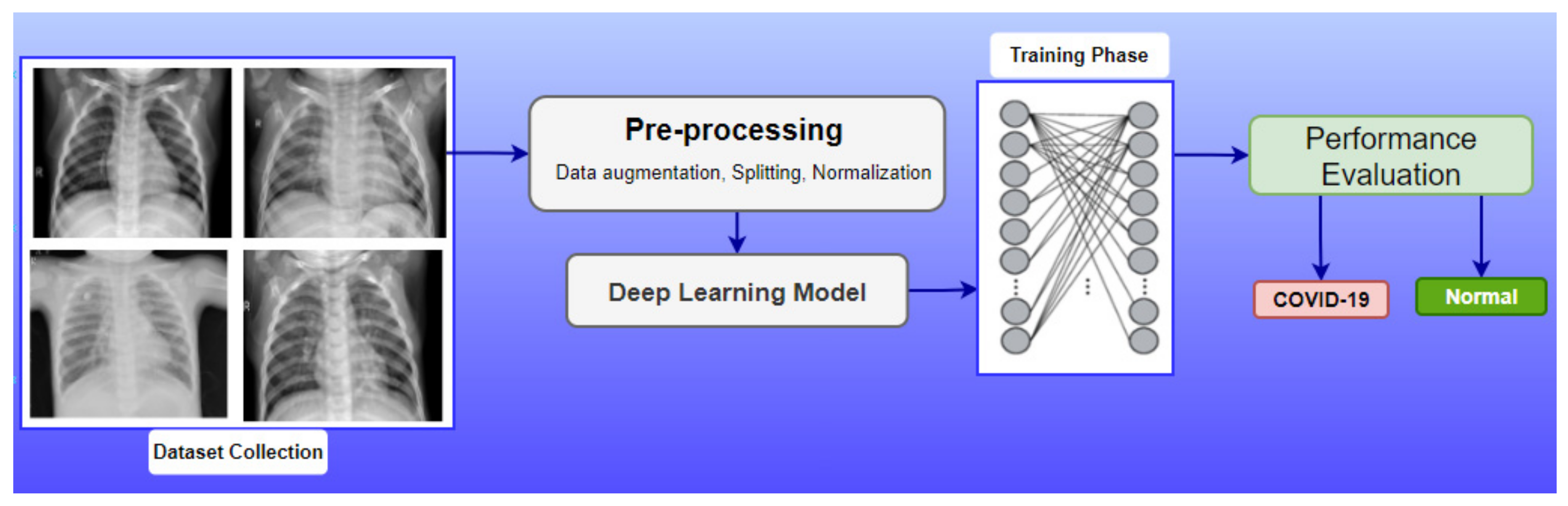
- By analyzing CXR images, the DL mode using TL is employed to categorize COVID-19-infected individuals.
- The recommended model is used to automatically extract features from the ImageNet dataset employing its weights and a model structure.
- Thorough experiments are carried out to assess the efficiency of the proposed solution using the COVID-19 CXR dataset.
- The aim of this study is to propose a robust DL architecture that can also be used for other clinical datasets.
- Comparative analysis is also presented by taking into account various renowned supervised learning approaches for COVID-19 detection.
2. Literature Review
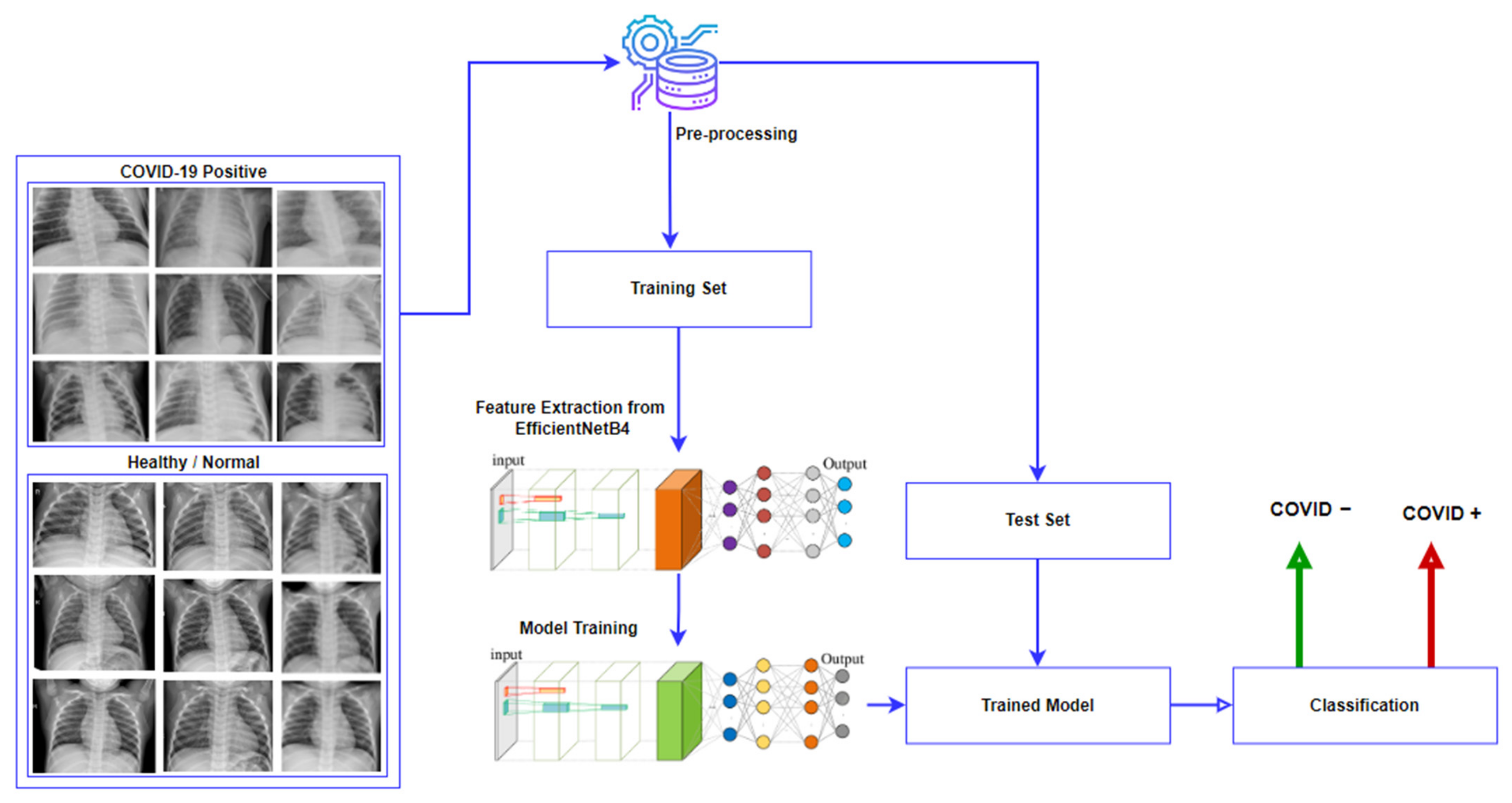
3. Proposed Methodology
| Algorithm 1: Proposed methodology steps |
| Let d = dataset, α = augmentation, i = image, pp = pre-Processing, r = rotate s = scale, sm = shifting methods, ia = image augmentation |
| Begin |
| 1: Get(d) |
| 2: α(i) w.r.t. r, s, st |
| 3: Perform (pp (i)) |
| 3.1. Perform (ia) |
| 3.2. Resize |
| 3.3. Normalize (i)/interval [0, 1] |
| 3.3.1. Conversion |
| 3.3.2. Computation (mean) |
| 3.3.3. Scaling(i) |
| 3.3.4. Conversion back |
| 3.4. Dataset splitting for training, testing, and validation |
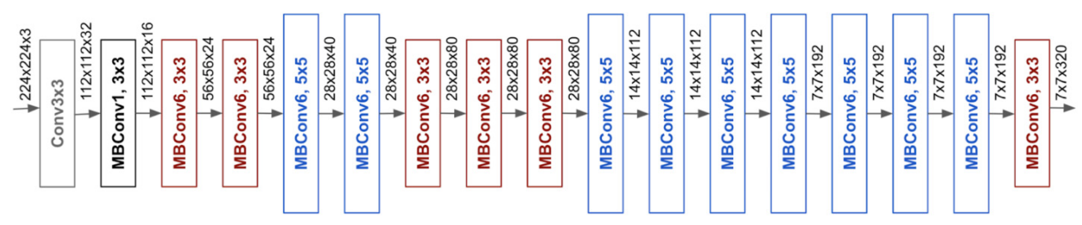
| 3.5. Feature extraction from EfficientNetB4 |
| 3.6. Optimize (epochs, batch size, model layers, learning weights) |
| Step 4: Evaluation metrics (accuracy, precision, F1 score, and recall) |
| End |
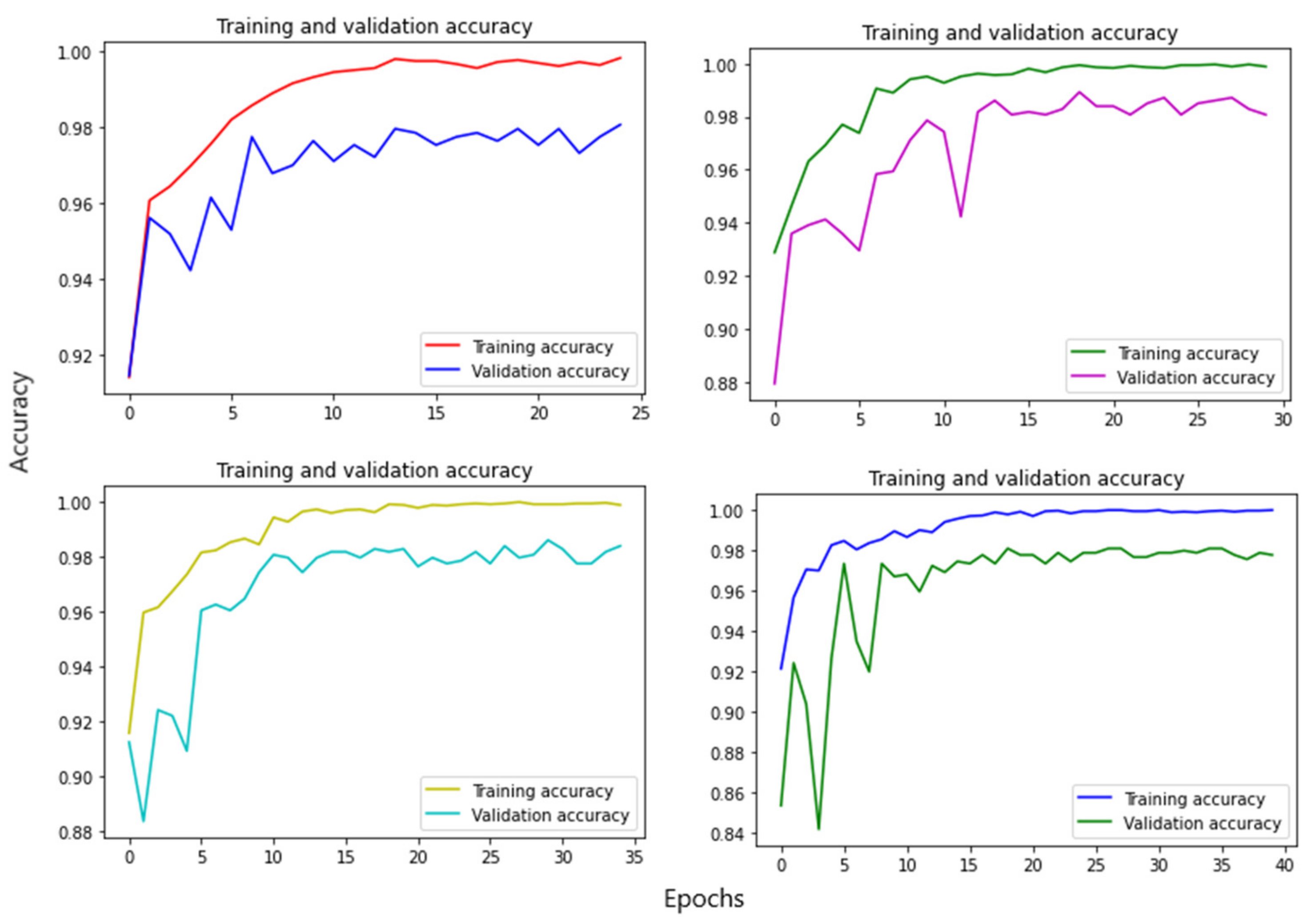
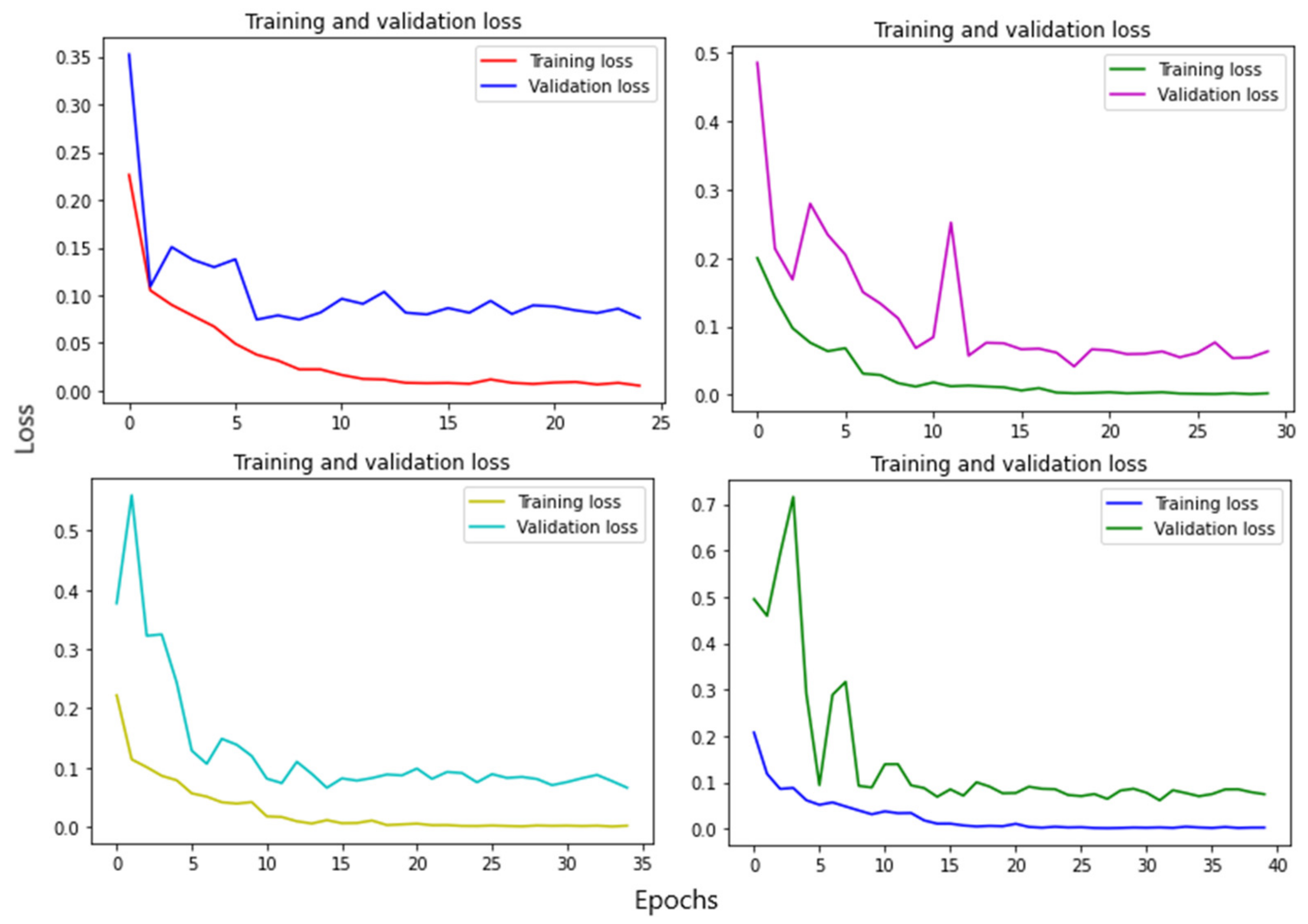
4. Experiment and Results
5. Conclusions
Author Contributions
Funding
Data Availability Statement
Conflicts of Interest
References
- Wu, F.; Zhao, S.; Yu, B.; Chen, Y.-M.; Wang, W.; Song, Z.-G.; Hu, Y.; Tao, Z.-W.; Tian, J.-H.; Pei, Y.-Y.; et al. A new coronavirus associated with human respiratory disease in China. Nature 2020, 579, 265–269. [Google Scholar] [CrossRef] [Green Version]
- Kong, W.; Agarwal, P.P. Chest Imaging Appearance of COVID-19 Infection. Radiol. Cardiothorac. Imaging 2020, 2, e200028. [Google Scholar] [CrossRef] [Green Version]
- Bernheim, A.; Mei, X.; Huang, M.; Yang, Y.; Fayad, Z.A.; Zhang, N.; Diao, K.; Lin, B.; Zhu, X.; Li, K.; et al. Chest CT Findings in Coronavirus Disease-19 (COVID-19): Relationship to Duration of Infection. Radiology 2020, 295, 200463. [Google Scholar] [CrossRef] [Green Version]
- Long, C.; Xu, H.; Shen, Q.; Zhang, X.; Fan, B.; Wang, C.; Zeng, B.; Li, Z.; Li, X.; Li, H. Diagnosis of the Coronavirus disease (COVID-19): rRT-PCR or CT? Eur. J. Radiol. 2020, 126, 108961. [Google Scholar] [CrossRef]
- Shi, H.; Han, X.; Jiang, N.; Cao, Y.; Alwalid, O.; Gu, J.; Fan, Y.; Zheng, C. Radiological findings from 81 patients with COVID-19 pneumonia in Wuhan, China: A descriptive study. Lancet Infect. Dis. 2020, 20, 425–434. [Google Scholar] [CrossRef]
- Zhao, W.; Zhong, Z.; Xie, X.; Yu, Q.; Liu, J. Relation Between Chest CT Findings and Clinical Conditions of Coronavirus Disease (COVID-19) Pneumonia: A Multicenter Study. Am. J. Roentgenol. 2020, 214, 1072–1077. [Google Scholar] [CrossRef]
- Humayun, M.; Alsayat, A. Prediction Model for Coronavirus Pandemic Using Deep Learning. Comput. Syst. Sci. Eng. 2022, 40, 947–961. [Google Scholar] [CrossRef]
- Gorbalenya, A.E.; Baker, S.C.; Baric, R.; Groot, R.J.D.; Drosten, C.; Gulyaeva, A.A.; Haagmans, B.L.; Lauber, C.; Leontovich, A.M.; Neuman, B.W.; et al. Severe acute respiratory syndrome-related coronavirus: The species and its viruses—A statement of the Coronavirus Study Group. bioRxiv 2020, 1–15. [Google Scholar] [CrossRef] [Green Version]
- Serte, S.; Demirel, H. Deep learning for diagnosis of COVID-19 using 3D CT scans. Comput. Biol. Med. 2021, 132, 104306. [Google Scholar] [CrossRef]
- Panwar, H.; Gupta, P.K.; Siddiqui, M.K.; Morales-Menendez, R.; Bhardwaj, P.; Singh, V. A deep learning and grad-CAM based color visualization approach for fast detection of COVID-19 cases using chest X-ray and CT-Scan images. Chaos Solitons Fractals 2020, 140, 110190. [Google Scholar] [CrossRef]
- Chavez, S.; Long, B.; Koyfman, A.; Liang, S.Y. Coronavirus Disease (COVID-19): A primer for emergency physicians. Am. J. Emerg. Med. 2021, 44, 220–229. [Google Scholar] [CrossRef]
- Minaee, S.; Kafieh, R.; Sonka, M.; Yazdani, S.; Soufi, G.J. Deep-COVID: Predicting COVID-19 from chest X-ray images using deep transfer learning. Med. Image Anal. 2020, 65, 101794. [Google Scholar] [CrossRef]
- Nayak, S.R.; Nayak, D.R.; Sinha, U.; Arora, V.; Pachori, R.B. Application of deep learning techniques for detection of COVID-19 cases using chest X-ray images: A comprehensive study. Biomed. Signal Process. Control 2021, 64, 102365. [Google Scholar] [CrossRef]
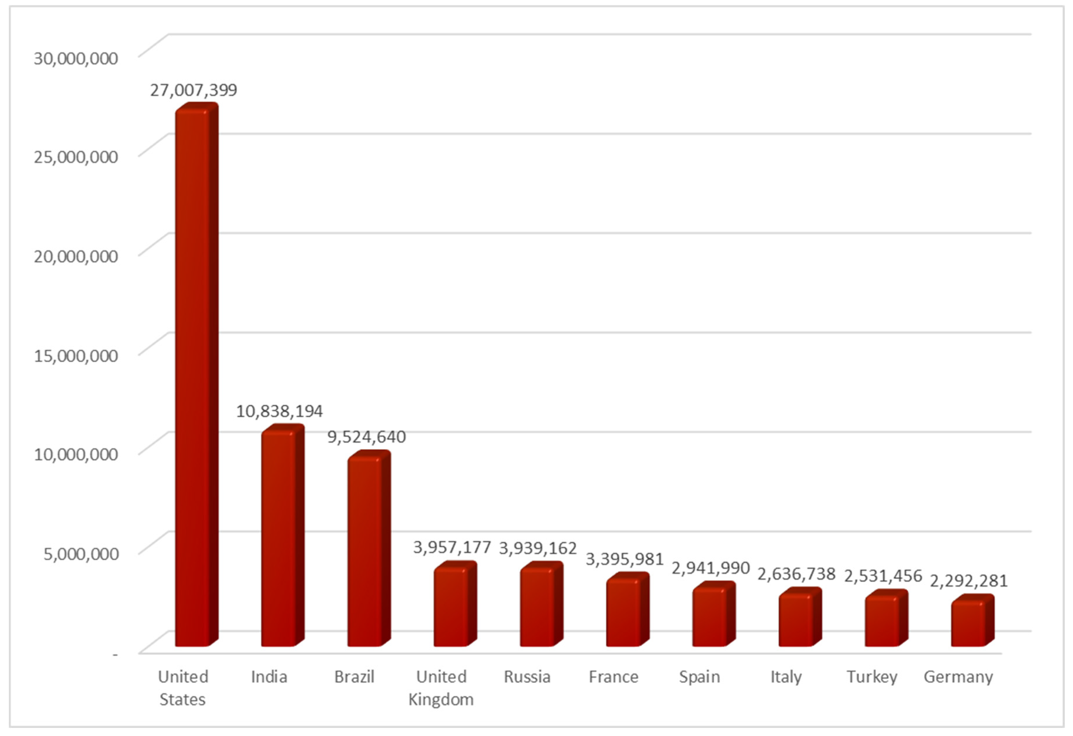
- Armstrong, M. The Countries with the Most COVID-19 Cases. 2021. Available online: https://www.statista.com/chart/21467/coutries-most-covid-19-cases/ (accessed on 13 July 2022).
- Al-Waisy, A.S.; Mohammed, M.A.; Al-Fahdawi, S.; Maashi, M.S.; Garcia-Zapirain, B.; Abdulkareem, K.H.; Mostafa, S.A.; Kumar, N.M.; Le, D.N. COVID-DeepNet: Hybrid multimodal deep learning system for improving COVID-19 pneumonia detection in chest X-ray images. Comput. Mater. Contin. 2021, 67, 2409–2429. [Google Scholar] [CrossRef]
- Arora, P.; Kumar, H.; Panigrahi, B.K. Prediction and analysis of COVID-19 positive cases using deep learning models: A descriptive case study of India. Chaos Solitons Fractals 2020, 139, 110017. [Google Scholar] [CrossRef] [PubMed]
- Alakus, T.B.; Turkoglu, I. Comparison of deep learning approaches to predict COVID-19 infection. Chaos Solitons Fractals 2020, 140, 110120. [Google Scholar] [CrossRef] [PubMed]
- Alazab, M.; Awajan, A.; Mesleh, A.; Abraham, A.; Jatana, V.; Alhyari, S. COVID-19 prediction and detection using deep learning. Int. J. Comput. Inf. Syst. Ind. Manag. Appl. 2020, 12, 168–181. [Google Scholar]
- Khalil, M.I.; Tehsin, S.; Humayun, M.; Jhanjhi, N.; AlZain, M.A. Multi-Scale Network for Thoracic Organs Segmentation. Comput. Mater. Contin. 2022, 70, 3251–3265. [Google Scholar] [CrossRef]
- Khalil, M.I.; Humayun, M.; Jhanjhi, N.Z.; Talib, M.N.; Tabbakh, T.A. Multi-class segmentation of organ at risk from abdominal CT images: A deep learning approach. In Intelligent Computing and Innovation on Data Science; Springer: Singapore, 2021; pp. 425–434. [Google Scholar]
- Zech, J.R.; Badgeley, M.A.; Liu, M.; Costa, A.B.; Titano, J.J.; Oermann, E.K. Variable generalization performance of a deep learning model to detect pneumonia in chest radiographs: A cross-sectional study. PLoS Med. 2018, 15, e1002683. [Google Scholar] [CrossRef] [Green Version]
- Shi, F.; Wang, J.; Shi, J.; Wu, Z.; Wang, Q.; Tang, Z.; He, K.; Shi, Y.; Shen, D. Review of Artificial Intelligence Techniques in Imaging Data Acquisition, Segmentation, and Diagnosis for COVID-19. IEEE Rev. Biomed. Eng. 2020, 14, 4–15. [Google Scholar] [CrossRef] [Green Version]
- Farhat, H.; Sakr, G.E.; Kilany, R. Deep learning applications in pulmonary medical imaging: Recent updates and insights on COVID-19. Mach. Vis. Appl. 2020, 31, 1–42. [Google Scholar] [CrossRef] [PubMed]
- Pathak, Y.; Shukla, P.; Tiwari, A.; Stalin, S.; Singh, S. Deep Transfer Learning Based Classification Model for COVID-19 Disease. Irbm 2020, 43, 87–92. [Google Scholar] [CrossRef]
- Afshar, P.; Heidarian, S.; Naderkhani, F.; Oikonomou, A.; Plataniotis, K.N.; Mohammadi, A. COVID-CAPS: A capsule network-based framework for identification of COVID-19 cases from X-ray images. Pattern Recognit. Lett. 2020, 138, 638–643. [Google Scholar] [CrossRef] [PubMed]
- Brunese, L.; Mercaldo, F.; Reginelli, A.; Santone, A. Explainable deep learning for pulmonary disease and coronavirus COVID-19 detection from X-rays. Comput. Methods Programs Biomed. 2020, 196, 105608. [Google Scholar] [CrossRef] [PubMed]
- Khan, A.I.; Shah, J.L.; Bhat, M.M. CoroNet: A deep neural network for detection and diagnosis of COVID-19 from chest x-ray images. Comput. Methods Programs Biomed. 2020, 196, 105581. [Google Scholar] [CrossRef]
- Houssein, E.H.; Abohashima, Z.; Elhoseny, M.; Mohamed, W.M. Hybrid quantum convolutional neural networks model for COVID-19 prediction using chest X-Ray images. arXiv 2021, arXiv:2102.06535. [Google Scholar] [CrossRef]
- Apostolopoulos, I.D.; Aznaouridis, S.I.; Tzani, M.A. Extracting Possibly Representative COVID-19 Biomarkers from X-ray Images with Deep Learning Approach and Image Data Related to Pulmonary Diseases. J. Med. Biol. Eng. 2020, 40, 462–469. [Google Scholar] [CrossRef]
- Oh, Y.; Park, S.; Ye, J.C. Deep Learning COVID-19 Features on CXR Using Limited Training Data Sets. IEEE Trans. Med. Imaging 2020, 39, 2688–2700. [Google Scholar] [CrossRef]
- Medhi, K.; Jamil, M.; Hussain, M.I. Automatic detection of COVID-19 infection from chest x-ray using deep learning. medrxiv 2020. [Google Scholar]
- Toraman, S.; Alakus, T.B.; Turkoglu, I. Convolutional capsnet: A novel artificial neural network approach to detect COVID-19 disease from X-ray images using capsule networks. Chaos Solitons Fractals 2020, 140, 110122. [Google Scholar] [CrossRef]
- Sahinbas, K.; Catak, F.O. Transfer learning-based convolutional neural network for COVID-19 detection with X-ray images. In Data Science for COVID-19; Academic Press: Cambridge, MA, USA, 2021; pp. 451–466. [Google Scholar]


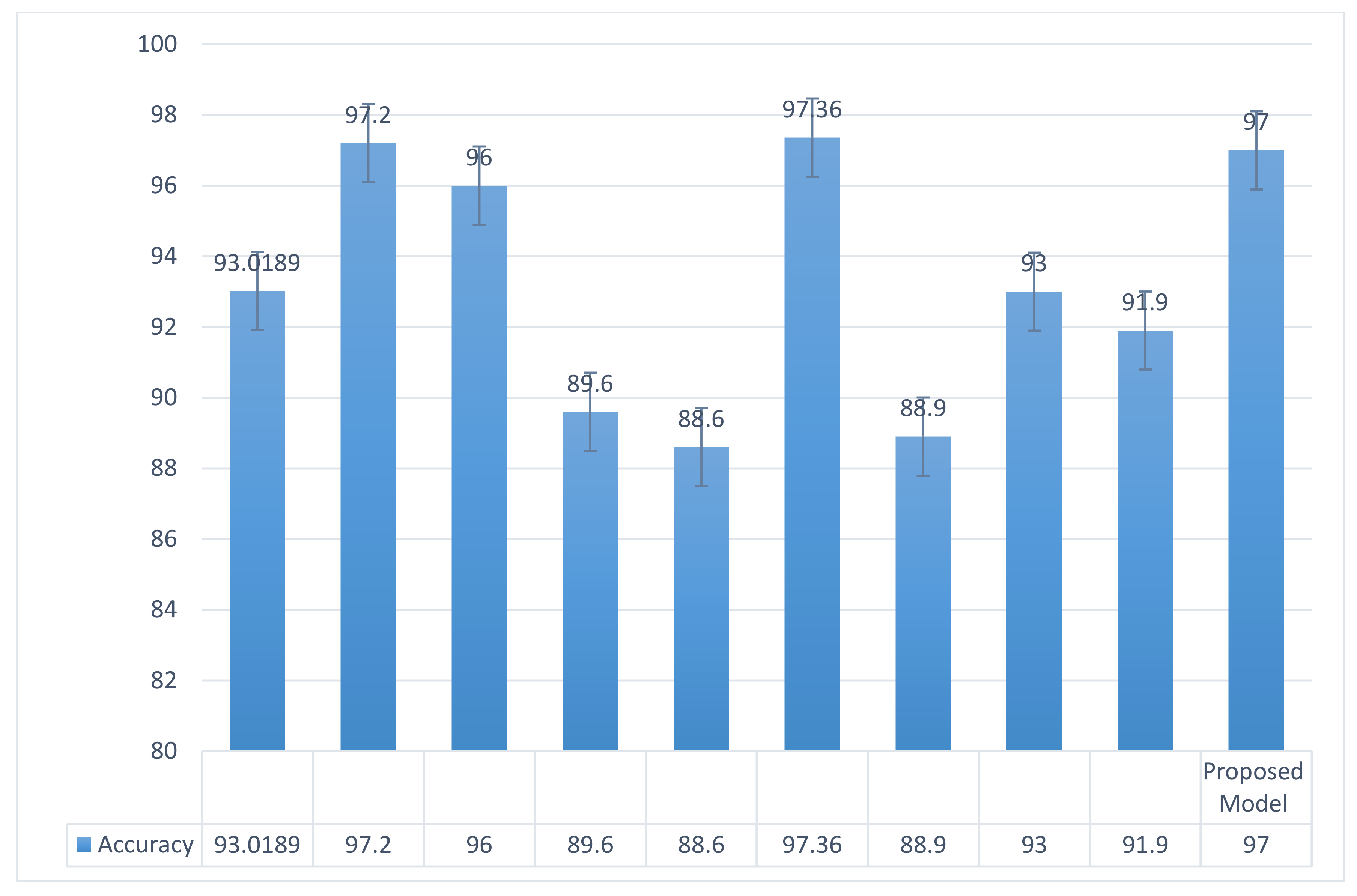
| Article | Methodology | Total Classes | Research Findings | Accuracy |
|---|---|---|---|---|
| Pathak et al. [24] | ResNet-50 | 2 | Lightweight DL model. | 93.0189% |
| Afshar, P. et al. [25] | Capsule-based network | 2 | Modified loss function to handle class imbalance. | 97.2% |
| Brunese et al. [26] | VGG-16 | 3 | Decreases the time window around 2.5 s. | 96% |
| Khan et al. [27] | CoroNet | 4 | Improves on existing radiology-based methods for smaller datasets. | 89.6% |
| Essam H. et al. [28] | CNN based on hybrid quantum | 3 | Integrates the random quantum circuits with CNNs. | 88.6% |
| Ioannis D et al. [29] | MobileNet V2 | 3 | Trained the CNN from scratch for detection of COVID-19. | 97.36% |
| Y Oh et al. [30] | ResNet-18 | 3 | Patch-based CNN with comparatively few trainable parameters. | 88.9% |
| Kishore Medhi et al. [31] | Deep CNN | 2 | COVID-19 classification with the DNN approach as quick and reliable. | 93% |
| Toraman et al. [32] | Capsule networks | 2 | Convolutional CapsNet method employed to facilitate fast screening for COVID-19. | 97.24% |
| Kevser Sahinbas et al. [33] | VGG-16 | 2 | DTL approach with multiple DL models scores considerable results on limited data. | 80% |
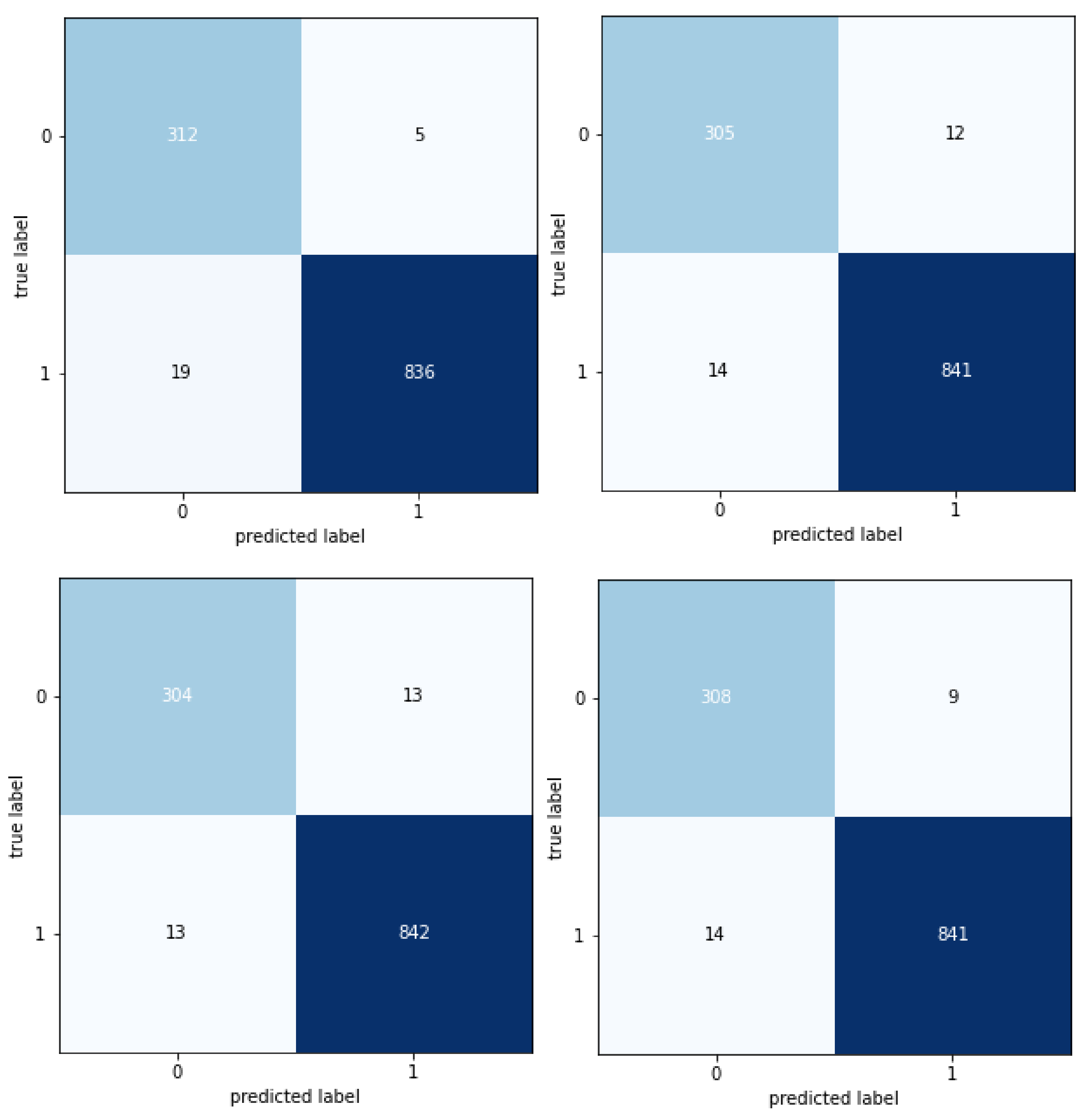
| Evaluation | Results | |||
|---|---|---|---|---|
| Accuracy | 0.97 | |||
| precision | recall | f1-score | support | |
| COVID | 0.96 | 0.97 | 0.97 | 217 |
| NORMAL | 0.96 | 0.99 | 0.99 | 1580 |
| PNEUMONIA | 0.96 | 0.97 | 0.98 | 423 |
Publisher’s Note: MDPI stays neutral with regard to jurisdictional claims in published maps and institutional affiliations. |
© 2022 by the authors. Licensee MDPI, Basel, Switzerland. This article is an open access article distributed under the terms and conditions of the Creative Commons Attribution (CC BY) license (https://creativecommons.org/licenses/by/4.0/).
Share and Cite
Khalil, M.I.; Rehman, S.U.; Alhajlah, M.; Mahmood, A.; Karamat, T.; Haneef, M.; Alhajlah, A. Deep-COVID: Detection and Analysis of COVID-19 Outcomes Using Deep Learning. Electronics 2022, 11, 3836. https://doi.org/10.3390/electronics11223836
Khalil MI, Rehman SU, Alhajlah M, Mahmood A, Karamat T, Haneef M, Alhajlah A. Deep-COVID: Detection and Analysis of COVID-19 Outcomes Using Deep Learning. Electronics. 2022; 11(22):3836. https://doi.org/10.3390/electronics11223836
Chicago/Turabian StyleKhalil, Muhammad Ibrahim, Saif Ur Rehman, Mousa Alhajlah, Awais Mahmood, Tehmina Karamat, Muhammad Haneef, and Ashwaq Alhajlah. 2022. "Deep-COVID: Detection and Analysis of COVID-19 Outcomes Using Deep Learning" Electronics 11, no. 22: 3836. https://doi.org/10.3390/electronics11223836