
Intracranial Monitoring to Verify Novel Transcranial Electric Stimulation in an Epileptic Swine Model

 and
and 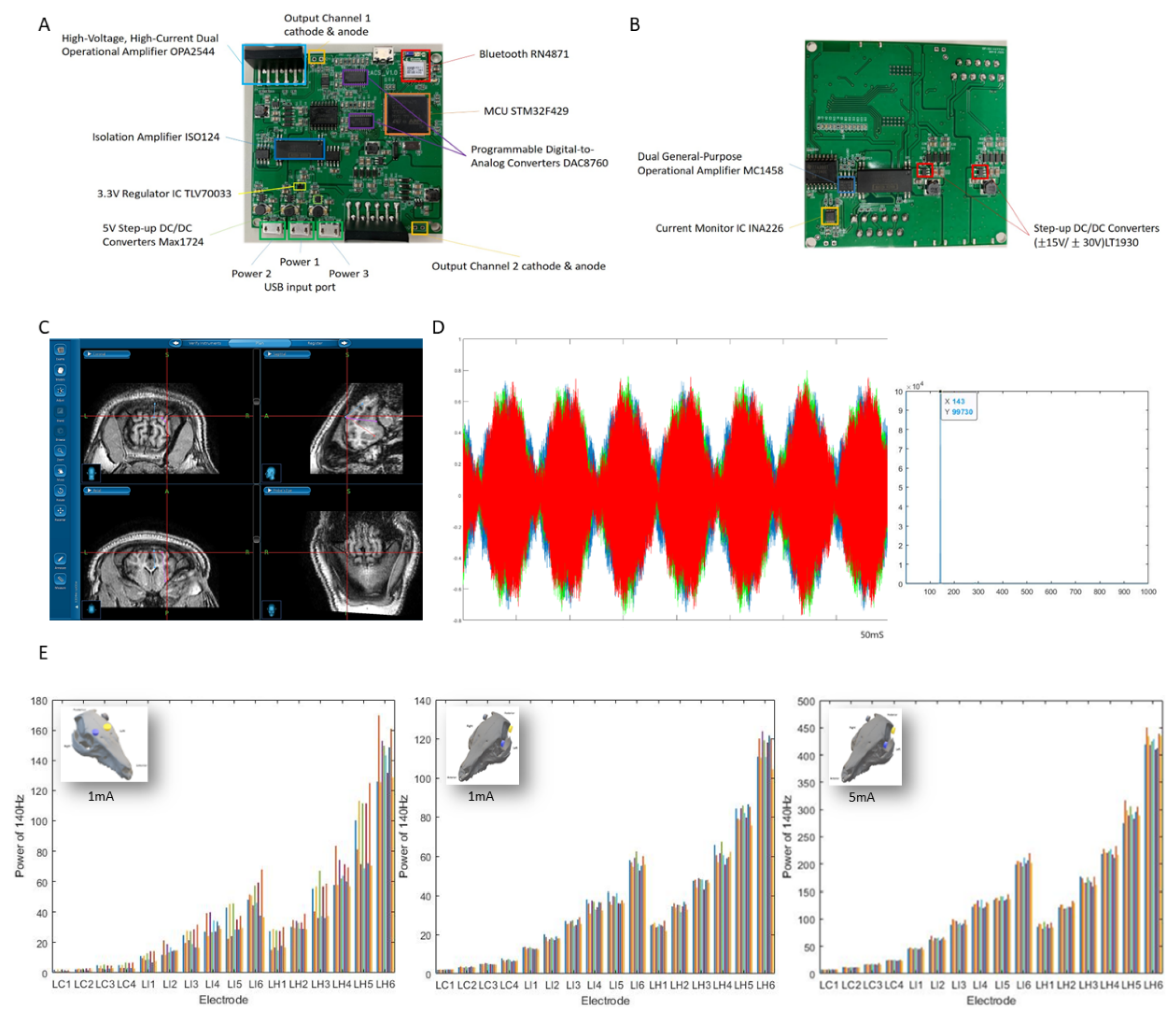
Abstract
:1. Introduction
2. Materials and Methods
2.1. Protocol of the Swine Model
2.2. Transcranial Electric Stimulation
2.3. Depth Electrode Implantation with Neuro-Navigation
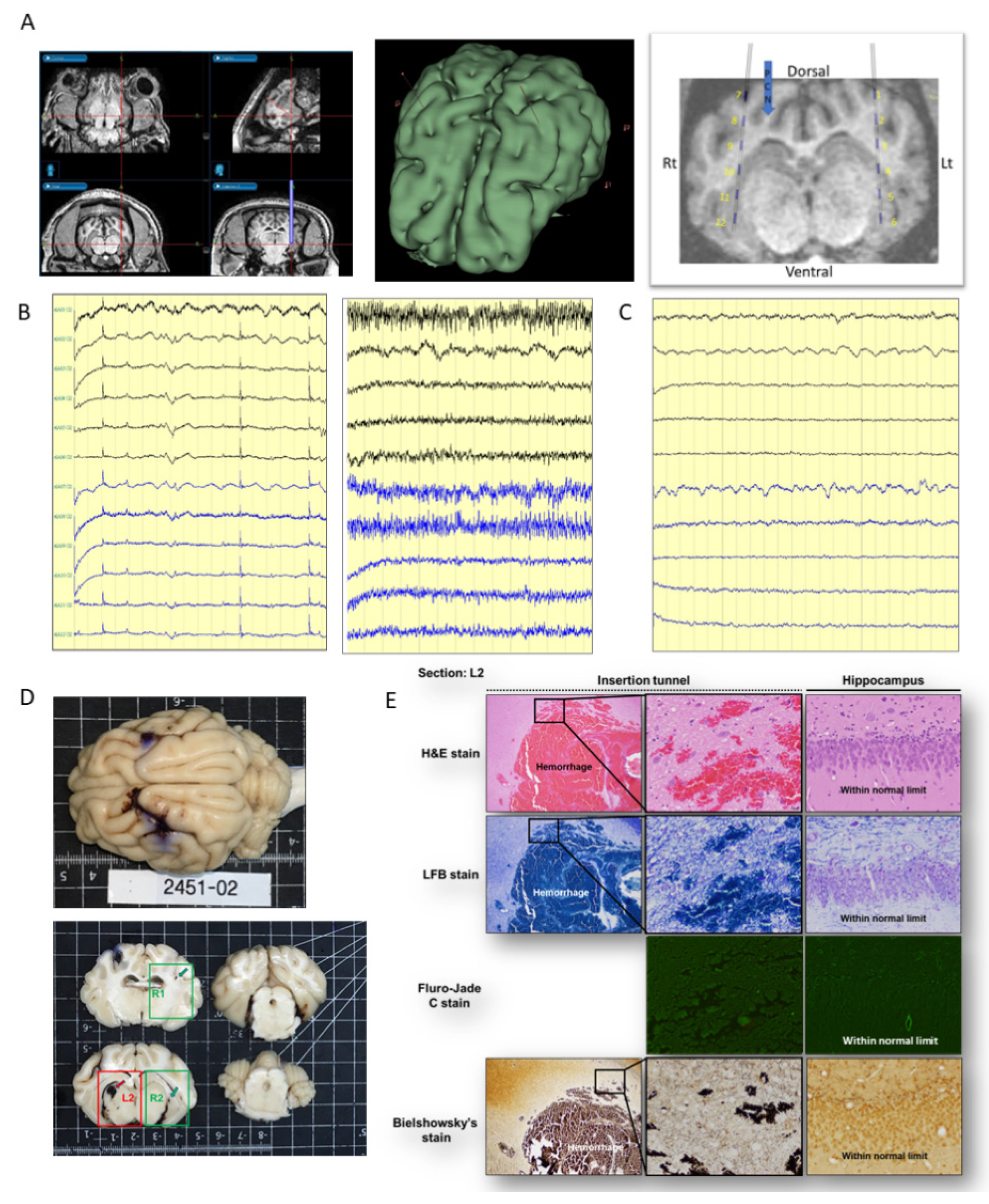
2.4. Histopathological Examination
3. Results
Histopathology Changes after Electrical Stimulation
4. Discussion
Limitation
5. Conclusions
Author Contributions
Funding
Institutional Review Board Statement
Informed Consent Statement
Data Availability Statement
Acknowledgments
Conflicts of Interest
References
- Vöröslakos, M.; Takeuchi, Y.; Brinyiczki, K.; Zombori, T.; Oliva, A.; Fernández-Ruiz, A.; Kozák, G.; Kincses, Z.T.; Iványi, B.; Buzsáki, G.; et al. Direct effects of transcranial electric stimulation on brain circuits in rats and humans. Nat. Commun. 2018, 9, 483. [Google Scholar] [CrossRef] [PubMed] [Green Version]
- Bianco, M.G.; Pullano, S.A.; Citraro, R.; Russo, E.; de Sarro, G.; de Villers Sidani, E.; Fiorillo, A.S. Neural Modulation of the Primary Auditory Cortex by Intracortical Microstimulation with a Bio-Inspired Electronic System. Bioengineering 2020, 7, 23. [Google Scholar] [CrossRef] [PubMed] [Green Version]
- Li, M.C.H.; Cook, M.J. Deep brain stimulation for drug-resistant epilepsy. Epilepsia 2018, 59, 273–290. [Google Scholar] [CrossRef] [PubMed]
- Fisher, R.; Salanova, V.; Witt, T.; Worth, R.; Henry, T.; Gross, R.; Oommen, K.; Osorio, I.; Nazzaro, J.; LaBar, D.; et al. Electrical stimulation of the anterior nucleus of thalamus for treatment of refractory epilepsy. Epilepsia 2010, 51, 899–908. [Google Scholar] [CrossRef]
- Feurra, M.; Bianco, G.; Santarnecchi, E.; Del Testa, M.; Rossi, A.; Rossi, S. Frequency-Dependent Tuning of the Human Motor System Induced by Transcranial Oscillatory Potentials. J. Neurosci. 2011, 31, 12165–12170. [Google Scholar] [CrossRef] [Green Version]
- Sivaramakrishnan, A.; Datta, A.; Bikson, M.; Madhavan, S. Remotely supervised transcranial direct current stimulation: A feasibility study for amyotrophic lateral sclerosis. NeuroRehabilitation 2019, 45, 369–378. [Google Scholar] [CrossRef]
- Nitsche, M.A.; Paulus, W. Excitability changes induced in the human motor cortex by weak transcranial direct current stimulation. J. Physiol. 2000, 527 Pt 3, 633–639. [Google Scholar] [CrossRef]
- Nitsche, M.A.; Paulus, W. Sustained excitability elevations induced by transcranial DC motor cortex stimulation in humans. Neurology 2001, 57, 1899–1901. [Google Scholar] [CrossRef]
- Ozen, S.; Sirota, A.; Belluscio, M.A.; Anastassiou, C.A.; Stark, E.; Koch, C.; Buzsáki, G. Transcranial Electric Stimulation Entrains Cortical Neuronal Populations in Rats. J. Neurosci. 2010, 30, 11476–11485. [Google Scholar] [CrossRef]
- Helfrich, R.F.; Schneider, T.R.; Rach, S.; Trautmann-Lengsfeld, S.A.; Engel, A.K.; Herrmann, C.S. Entrainment of Brain Oscillations by Transcranial Alternating Current Stimulation. Curr. Biol. 2014, 24, 333–339. [Google Scholar] [CrossRef] [Green Version]
- Antal, A.; Boros, K.; Poreisz, C.; Chaieb, L.; Terney, D.; Paulus, W. Comparatively weak after-effects of transcranial alternating current stimulation (tACS) on cortical excitability in humans. Brain Stimul. 2008, 1, 97–105. [Google Scholar] [CrossRef] [PubMed]
- Zaehle, T.; Rach, S.; Herrmann, C.S. Transcranial Alternating Current Stimulation Enhances Individual Alpha Activity in Human EEG. PLoS ONE 2010, 5, e13766. [Google Scholar] [CrossRef] [Green Version]
- Fröhlich, F.; McCormick, D.A. Endogenous Electric Fields May Guide Neocortical Network Activity. Neuron 2010, 67, 129–143. [Google Scholar] [CrossRef] [PubMed] [Green Version]
- Grossman, N.; Bono, D.; Dedic, N.; Kodandaramaiah, S.B.; Rudenko, A.; Suk, H.-J.; Cassara, A.M.; Neufeld, E.; Kuster, N.; Tsai, L.-H.; et al. Noninvasive Deep Brain Stimulation via Temporally Interfering Electric Fields. Cell 2017, 169, 1029–1041.e16. [Google Scholar] [CrossRef] [Green Version]
- Vadera, S.; Mullin, J.; Bulacio, J.; Najm, I.; Bingaman, W.; Gonzalez-Martinez, J. Stereoelectroencephalography Following Subdural Grid Placement for Difficult to Localize Epilepsy. Neurosurgery 2013, 72, 723–729. [Google Scholar] [CrossRef] [PubMed]
- Cossu, M.; Cardinale, F.; Castana, L.; Citterio, A.; Francione, S.; Tassi, L.; Benabid, A.L.; Lo Russo, G. Stereoelectroencephalography in the presurgical evaluation of focal epilepsy: A retrospective analysis of 215 procedures. Neurosurgery 2005, 57, 706–718. [Google Scholar] [CrossRef]
- Guenot, M.; Isnard, J.; Ryvlin, P.; Fischer, C.; Ostrowsky, K.; Mauguiere, F.; Sindou, M. Neurophysiological monitoring for epilepsy surgery: The Talairach SEEG method. StereoElectroEncephaloGraphy. Indications, results, complications and therapeutic applications in a series of 100 consecutive cases. Stereotact. Funct. Neurosurg. 2001, 77, 29–32. [Google Scholar] [CrossRef]
- Garcia-Lorenzo, B.; del Pino-Sedeno, T.; Rocamora, R.; Lopez, J.E.; Serrano-Aguilar, P.; Trujillo-Martin, M.M. Stereoelectroencephalography for Refractory Epileptic Patients Considered for Surgery: Systematic Review, Meta-Analysis, and Economic Evaluation. Neurosurgery 2019, 84, 326–338. [Google Scholar] [CrossRef] [Green Version]
- Opitz, A.; Falchier, A.; Yan, C.G.; Yeagle, E.M.; Linn, G.S.; Megevand, P.; Thielscher, A.; Deborah, A.R.; Milham, M.P.; Mehta, A.D.; et al. Spatiotemporal structure of intracranial electric fields induced by transcranial electric stimulation in humans and nonhuman primates. Sci. Rep. 2016, 6, 31236. [Google Scholar] [CrossRef] [Green Version]
- You, C.; Yao, L.; Yao, P.; Li, L.; Ding, P.; Liang, S.; Liu, C.; Xue, N. An iEEG Recording and Adjustable Shunt-Current Conduction Platform for Epilepsy Treatment. Biosensors 2022, 12, 247. [Google Scholar] [CrossRef]
- Van Gompel, J.J.; Bower, M.R.; Worrell, G.A.; Stead, M.; Meier, T.R.; Goerss, S.J.; Chang, S.-Y.; Kim, I.; Meyer, F.B.; Marsh, W.R.; et al. Swine model for translational research of invasive intracranial monitoring. Epilepsia 2011, 52, e49–e53. [Google Scholar] [CrossRef] [PubMed]
- Reato, D.; Rahman, A.; Bikson, M.; Parra, L.C. Low-Intensity Electrical Stimulation Affects Network Dynamics by Modulating Population Rate and Spike Timing. J. Neurosci. 2010, 30, 15067–15079. [Google Scholar] [CrossRef] [PubMed] [Green Version]
- Anastassiou, C.A.; Perin, R.; Markram, H.; Koch, C. Ephaptic coupling of cortical neurons. Nat. Neurosci. 2011, 14, 217–223. [Google Scholar] [CrossRef] [PubMed]
- Jackson, M.P.; Rahman, A.; Lafon, B.; Kronberg, G.; Ling, D.; Parra, L.C.; Bikson, M. Animal models of transcranial direct current stimulation: Methods and mechanisms. Clin. Neurophysiol. 2016, 127, 3425–3454. [Google Scholar] [CrossRef] [Green Version]
- Esmaeilpour, Z.; Schestatsky, P.; Bikson, M.; Brunoni, A.R.; Pellegrinelli, A.; Piovesan, F.X.; Santos, M.M.; Menezes, R.B.; Fregni, F. Notes on Human Trials of Transcranial Direct Current Stimulation between 1960 and 1998. Front. Hum. Neurosci. 2017, 11, 71. [Google Scholar] [CrossRef] [Green Version]
- Radman, T.; Ramos, R.L.; Brumberg, J.C.; Bikson, M. Role of cortical cell type and morphology in subthreshold and suprathreshold uniform electric field stimulation in vitro. Brain Stimul. 2009, 2, 215–228. [Google Scholar] [CrossRef] [Green Version]
- Velasco, A.L.; Velasco, F.; Velasco, M.; Trejo, D.; Castro, G.; Carrillo-Ruiz, J.D. Electrical Stimulation of the Hippocampal Epileptic Foci for Seizure Control: A Double-Blind, Long-Term Follow-Up Study. Epilepsia 2007, 48, 1895–1903. [Google Scholar] [CrossRef]
- Wang, Y.-C.; Kremen, V.; Brinkmann, B.H.; Middlebrooks, E.H.; Lundstrom, B.N.; Grewal, S.S.; Guragain, H.; Wu, M.-H.; Van Gompel, J.J.; Klassen, B.T.; et al. Probing circuit of Papez with stimulation of anterior nucleus of the thalamus and hippocampal evoked potentials. Epilepsy Res. 2020, 159, 106248. [Google Scholar] [CrossRef]
- Rashid, S.; Pho, G.; Czigler, M.; Werz, M.A.; Durand, D.M. Low frequency stimulation of ventral hippocampal commissures reduces seizures in a rat model of chronic temporal lobe epilepsy. Epilepsia 2012, 53, 147–156. [Google Scholar] [CrossRef] [Green Version]
- Berényi, A.; Belluscio, M.; Mao, D.; Buzsáki, G. Closed-Loop Control of Epilepsy by Transcranial Electrical Stimulation. Science 2012, 337, 735–737. [Google Scholar] [CrossRef] [Green Version]
- Moliadze, V.; Atalay, D.; Antal, A.; Paulus, W. Close to threshold transcranial electrical stimulation preferentially activates inhibitory networks before switching to excitation with higher intensities. Brain Stimul. 2012, 5, 505–511. [Google Scholar] [CrossRef] [PubMed]
- Chaieb, L.; Antal, A.; Paulus, W. Transcranial alternating current stimulation in the low kHz range increases motor cortex excitability. Restor. Neurol. Neurosci. 2011, 29, 167–175. [Google Scholar] [CrossRef] [PubMed]
- Ladas, T.P.; Chiang, C.-C.; Gonzalez-Reyes, L.E.; Nowak, T.; Durand, D.M. Seizure reduction through interneuron-mediated entrainment using low frequency optical stimulation. Exp. Neurol. 2015, 269, 120–132. [Google Scholar] [CrossRef] [PubMed] [Green Version]
- Chen, T.; Si, Y.; Chen, D.; Zhu, L.; Xu, D.; Chen, S.; Zhou, D.; Liu, L. The value of 24-hour video-EEG in evaluating recurrence risk following a first unprovoked seizure: A prospective study. Seizure 2016, 40, 46–51. [Google Scholar] [CrossRef] [PubMed] [Green Version]
- Cambiaghi, M.; Magri, L.; Cursi, M. Importance of EEG in validating the chronic effects of drugs: Suggestions from animal models of epilepsy treated with rapamycin. Seizure 2015, 27, 30–39. [Google Scholar] [CrossRef] [Green Version]


Publisher’s Note: MDPI stays neutral with regard to jurisdictional claims in published maps and institutional affiliations. |
© 2022 by the authors. Licensee MDPI, Basel, Switzerland. This article is an open access article distributed under the terms and conditions of the Creative Commons Attribution (CC BY) license (https://creativecommons.org/licenses/by/4.0/).
Share and Cite
Wang, Y.-C.; Wang, P.-F.; Pan, H.-C.; Lin, C.-Y.; Hsu, H.-T.; Liu, Z.-H.; Lee, P.-L. Intracranial Monitoring to Verify Novel Transcranial Electric Stimulation in an Epileptic Swine Model. Electronics 2022, 11, 2195. https://doi.org/10.3390/electronics11142195
Wang Y-C, Wang P-F, Pan H-C, Lin C-Y, Hsu H-T, Liu Z-H, Lee P-L. Intracranial Monitoring to Verify Novel Transcranial Electric Stimulation in an Epileptic Swine Model. Electronics. 2022; 11(14):2195. https://doi.org/10.3390/electronics11142195
Chicago/Turabian StyleWang, Yu-Chi, Po-Fang Wang, Han-Chi Pan, Chuan-Yi Lin, Hao-Teng Hsu, Zhuo-Hao Liu, and Po-Lei Lee. 2022. "Intracranial Monitoring to Verify Novel Transcranial Electric Stimulation in an Epileptic Swine Model" Electronics 11, no. 14: 2195. https://doi.org/10.3390/electronics11142195