Communication

The Natural Centella asiatica Extract Acts as a Stretch Mark Eraser: A Biological Evaluation

1
Givaudan Active Beauty, Science and Technology, 51110 Pomacle, France
2
Givaudan Active Beauty, Science and Technology, 31000 Toulouse, France
*
Author to whom correspondence should be addressed.
Cosmetics 2024, 11(1), 15; https://doi.org/10.3390/cosmetics11010015
Submission received: 4 December 2023 / Revised: 19 January 2024 / Accepted: 21 January 2024 / Published: 23 January 2024
(This article belongs to the Special Issue Application of Plant-Based Molecules and Materials in Cosmetics)

Abstract

:
Stretch marks are far from exclusively appearing on pregnant women and appear whenever the body experiences rapid growth. Collagen fibres are altered in the dermis, which is associated with a loss of orientation, and the elastic network is disrupted, leading to a fibrotic organisation. This results in epidermal tearing that produces skin lesions. Centella asiatica (CAST) is a well-known medicinal plant rich in active triterpenic molecules and traditionally used to treat wounds and help skin repair. The aim of this study was to evaluate CAST extract as a natural way to solve stretch mark concerns and understand its mechanism of action. Fibroblast proliferation based on scratch assay model and their gene expression by RT-qPCR was first evaluated. At the ex vivo level, elastin fibres were quantified by immunofluorescence. The orientation of the collagen fibres and their occupation of the dermis were analysed after Sirius red staining and specific software analysis. We showed that CAST stimulated fibroblast proliferation and reduced extracellular matrix degradation and fibrosis. On a stretch-marked skin explant, CAST increased the occupation of collagen fibres and elastin production. Based on the mechanisms behind the formation of stretch marks, CAST restored the dermis network by optimising fibre organisation for a visible skin remodelling effect.

1. Introduction

Stretch marks, also called striae, are a form of tissue damage on the skin due to an excessive stretching of the dermis [1,2]. Stretch marks are commonly associated with pregnancy and affect between 60% and 90% of women during their pregnancy [3]. In reality, they occur as the result of tearing of the dermis during periods of rapid growth of the body or some body parts. Therefore, around 27% of adolescents are concerned by stretch marks [3]. As they are caused by the sudden expansion of the skin, especially in areas where fat is most likely to be stored in our body, obesity may lead to the apparition of stretch marks. Stretch marks are thus usually found on the breasts, thighs, and belly, especially near the navel, upper arms, underarms, and lower back, both in women and men [4].
Stretch marks affect the dermis by preventing the fibroblasts from organising collagen fibres to keep up with the skin’s stretching. The collagen bundles are altered, lose their orientation, and are organised into a fibrotic structure, while the elastic network is also disrupted [5]. As a result, the skin appears less firm, less deformable, and less plastic. This normally leads to epidermal tearing, which can produce a lesion in the form of a visible stretch mark. Stretch marks could be classified into six different types depending on their appearance and epidemiology; striae atrophicans (thinned skin), striae gravidarum (following pregnancy), striae distensae (stretched skin), striae rubrae (red), striae albae (white), striae nigra (black), and striae caerulea (dark blue) [4]. Over time, stretch marks tend to atrophy and lose pigmentation, depending on how recent they are on the skin. Thus, hyper-pigmented stretch marks related to an acute stage are characterised by the initial erythematous, and a chronic stage is characterised by a hypo-pigmented and atrophic lesion [6].
Current treatment involves invasive methods such as laser therapy, light therapy, collagen injection, laser lipolysis, radiofrequency techniques, and microdermabrasion [1,6,7]. A topical solution may be used, and among these, a formulation with CAST has been described to improve stretch marks, but its mechanism of action is poorly described [8,9,10,11]. CAST is one of the most known traditional medicinal plants. It has long been used to promote skin repair and wound healing, and it is a medicinal remedy present in the Ayurvedic system and traditional Chinese medicine [12]. It grows in cultivated fields in Madagascar and other Asian and African countries. It expresses, in its tiny and round leaves, as secondary metabolites, some biologically active triterpenic molecules (asiaticoside, asiatic acid and madecassic acid) [13]. CAST is a promising candidate to solve stretch mark concerns as it enhances cell production and matrix components and improves tensile strength [12]. Clinical studies have shown that the application of a formulation with CAST significantly improved stretch mark appearance [9,14]. We have also previously conducted a clinical study on 54 women divided into two groups (the control group applying a placebo formula without CAST and a group using a formulation with CAST [15]). The volunteers used the product three times per day for one month. After four weeks of using the CAST formulation, skin thickness at the edge and in the centre of the stretch marks was significantly increased in comparison to the placebo, such as skin vascularisation and elasticity for a visual reduction in stretch marks appearance [15]. Following this preliminary work, we wanted to explore the mechanism of action of CAST further, and the aim of this study was to unveil potential unknown pathways that can be impacted by CAST at the in vitro and ex vivo levels. Transcriptomic analysis was performed in order to identify potential new actors in stretch mark-related fibrosis, such as CGTF. Modulation of its expression by CAST could be the result of improvement of the dermal matrix, improving the quality of the dermis and thus reducing stretch mark damages.

2. Materials and Methods

The aim of this study was to evaluate the active ingredient obtained from Centella asiatica (supplied by Givaudan Active Beauty, Argenteuil, France) as a new way to solve stretch mark concerns.
Centella CAST (INCI: asiaticoside, asiatic acid, madecassic acid) was obtained by Indena Spa, Viale Ortles 12, Milan, Italy. The product is obtained through the extraction, fractionation, and recombination of terpenes typical of Centella asiatica, which are the major bioactive constituents. These include asiaticoside, in which a trisaccharide moiety is linked to the aglycone asiatic acid, madecassoside, and madecassic acid. Centella asiatica is sourced in Madagascar, and leaves are hand collected from spontaneous growth during their growing season, harvested, and naturally dried. After a preliminary extraction of Centella soft extract, the product is concentrated and purified to the first fraction of asiaticoside. Discarded solutions are concentrated, the pH is adjusted, and the fraction is purified to obtain the madecassic and asiatic acid fractions. The asiaticoside fraction and the acids fraction are then mixed to match the specifications. This natural extract has a very fixed composition and is composed of asiaticoside (about 10–16%), asiatic acid (about 6–9%) and madecassossic acid (about 10–14%).

2.1. Wound Healing on Fibroblast

Fibroblasts (NIH-3T3, ATCC CRL-1658) were maintained in supplemented DMEM medium (Gibco®, Life Technologies, Carlsbad, CA, USA) containing 10% fetal bovine serum (FBS), (Hyclone, Logan, UT, USA) and 1% antibiotics (Sigma-Aldrich, St. Louis, MO, USA) (DMEM complete medium) at 37 °C 5% CO2. NIH-3T3 (105 cells/well) were seeded in 96-well Essen ImageLock plates (Essen BioScience, Ann Arbor, MI, USA) and grown to 80% confluence in a CO2-humidified incubator. After 24 h, the scratch was made using the 96-pin WoundMaker (Essen BioScience, Ann Arbor, MI, USA). Fibroblasts were treated with CAST at 1, 5, 10, 25, or 50 µg/mL. FBS 10% was used as a positive control. Wound images were taken every hour for 36 h, and the data were analysed using the integrated metric “relative wound density” from the live-content cell imaging system IncuCyte HD (Essen BioScience, Ann Arbor, MI, USA). The experiments were performed in triplicate wells.

2.2. Transcriptomic Analysis on Fibroblast

Normal human dermal fibroblasts (NHDFs) freshly isolated from non-stretch-marked or from stretch-marked areas from the same donor were seeded at 300,000 cells per well in 6-well plates and cultivated in DMEM medium (Gibco®, Life Technologies, Carlsbad, CA, USA) supplemented by FBS with 10% and 1% antibiotics (Sigma-Aldrich, St. Louis, MO, USA). After 48 h of culture, NHDFs were rinsed two times with PBS (Gibco®, Life Technologies, Carlsbad, CA, USA) and allowed to rest in FCS-free medium overnight before stimulation. Cells were stimulated with CAST extract at 10 µg/mL versus untreated condition. After 24 h of stimulation, total RNA was extracted using the extract-all method (Fisher Scientific, Hampton, NH, USA). RNA quality was controlled, and reverse transcription was performed to obtain cDNA using the Verso cDNA kit (Thermo Fisher Scientific, Waltham, MA, USA). RT-qPCR was performed on specific pre-coated plates (Applied Biosystems, Foster City, CA, USA) designed to study the transcriptomic expression of different genes involved in fibrosis and matrix remodelling with 10 ng of cDNA per well using CFX96 Touch (Biorad, Hercules, CA, USA) and Universal Taqman mix (Quantabio, Beverly, MA, USA). CTGF: Unique Assay ID qHsaCED0002044, FAK: Unique Assay ID qHsaCED0001879, MMP1: Unique Assay ID qHsaCID0017039, MMP7: Unique Assay ID qHsaCID0011537. The relative quantification (RQ) of gene expression was calculated according to POP4 (Ribonuclease/MRP subunit) (POP4: qHsaCID0015127) and B2M (Beta-2-Microglobulin) (B2M: qHsaCID0015347) housekeeping genes. The data are expressed in fold change relative to normal fibroblasts or to untreated stretch-marked fibroblasts.

2.3. Collagen Network Analysis on Skin Explant

Skin explants were obtained according to ethical and regulatory rules and under the agreement of the participants. Skin explants with stretch marks from a 60-year-old female volunteer donor (Biopredic International, Saint Grégoire, France) were treated topically with CAST at 0.5% for 5 days. Each day, treatment and medium (Genoskin, Toulouse, France) were renewed, and explants were incubated at 37 °C 5% CO2.
INCI placebo: AQUA/WATER, CETYL ALCOHOL, GLYCERYL STEARATE, PEG-75 STEARATE, CETETH-20, STEARETH-20, ISODECYL NEOPENTANOATE, PHENOXYETHANOL.
After 5 days, skin biopsies were fixed in formalin (Sigma-Aldrich, St. Louis, MO, USA) for 48 h and then dehydrated overnight by automated dehydration using Histocore Pearl (Leica, Wetzlar, Germany). After dehydration, skin explants were included in paraffin (Leica, Wetzlar, Germany), and skin sections of 4 µm were obtained with a microtome (Leica, Wetzlar, Germany). Paraffin was removed thanks to xylene baths (Sigma-Aldrich, St. Louis, MO, USA) followed by ethanol (VWR International, Radnor, PA, USA) dehydration. Skin slices were stained with a ready-to-use Sirius red colouration (Labo Modern, Gennevilliers, France) before mounting on a thin glass slide. Dermis structure was observed using a bright field, and collagen fibre orientation was analysed in polarised light. Under polarised light, collagen I fibres were detected using the red channel of a slide scanner (Olympus, Tokyo, Japan). The quantity of collagen I was quantified using ImageJ 1.53k software on the whole dermis. Furthermore, the images were oriented with the epidermal part up at 90° of the x-axis. The images were then processed to remove a 200 μm thickness from the outer-most stratum corneum to eliminate the papillary dermis for the orientation analysis of collagen fibres. The conditioned images were processed by a segmentation algorithm, allowing the analysis to focus only on the collagen fibres. Based on a plane parallel to the epidermis, collagen I fibres were quantified at 10° intervals in order to evaluate collagen I occupation in the dermis.

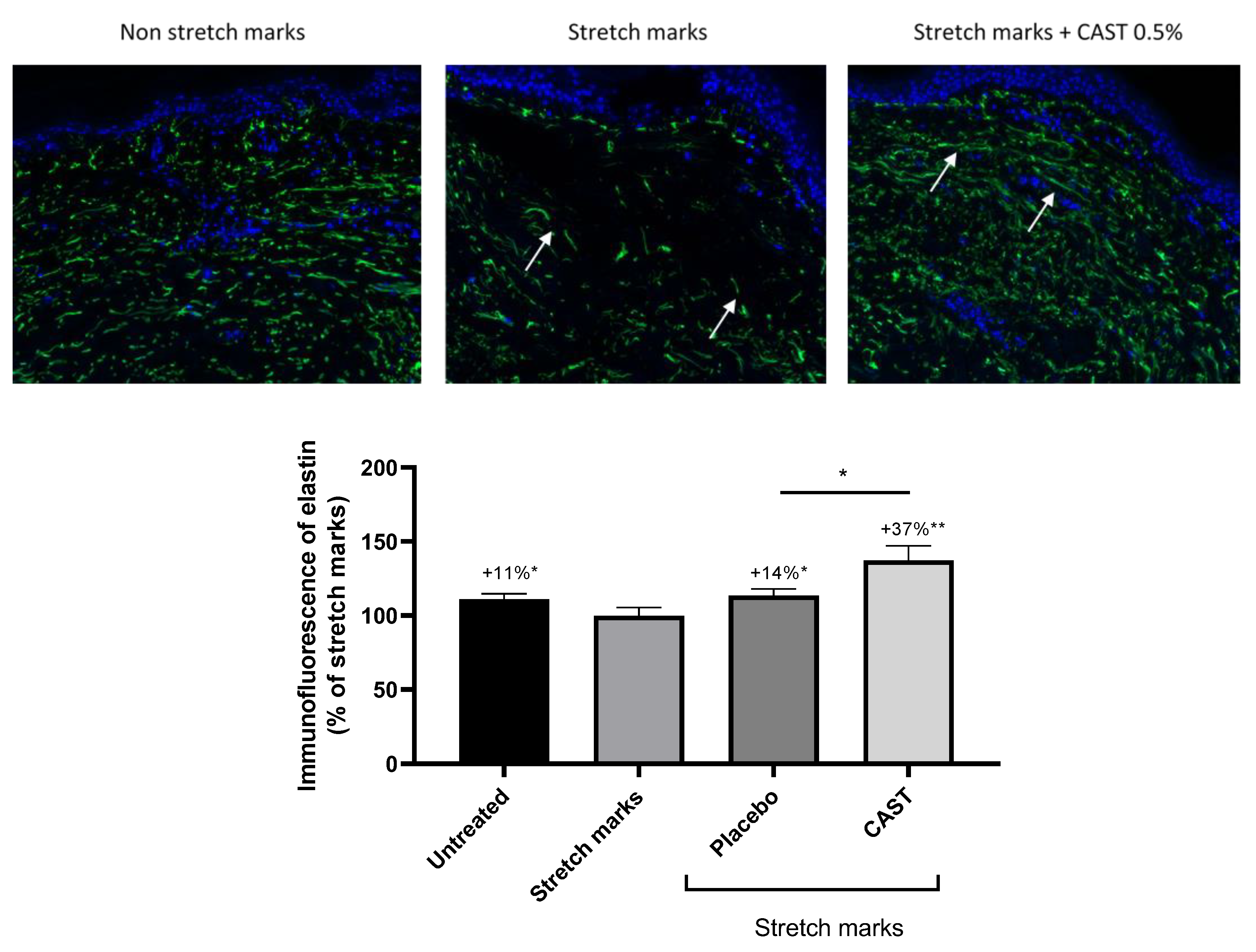
2.4. Elastin Immunofluorescence on Skin Explant

The skin explants were cultivated and treated as previously described. Paraffin was removed thanks to xylene baths followed by ethanol dehydration. Antigenic sites were revealed with a citrate buffer (Sigma-Aldrich, St. Louis, MO, USA). Then, skin sections were BSA-saturated (Sigma-Aldrich, St. Louis, MO, USA) for 30 min, followed by immunostaining to specifically detect elastin (1/75e, Santa-Cruz, Dallas, TX, USA). After overnight incubation of the primary antibody at 4 °C, skin sections were washed and incubated for 1 h with secondary antibody (Anti-mouse, 1/500e, Abcam, Cambridge, UK). The staining was observed using fluorescent microscopy (Zeiss, Axio observer, Iena, Germany). Relative fluorescence was quantified for all images using ImageJ 1.53k software.

2.5. Statistical Analysis

For all studies, a Shapiro-Wilk test was used to verify whether the raw data followed the Gaussian Law. In the case of normally distributed data, the mean values were compared with untreated conditions or normal skin conditions (no stretch marks) using an unpaired t-student test. In the case of non-normally distributed data, a Mann–Whitney U test was used. For all statistical tests, we considered significant results as follows: # p < 0.1, * p < 0.05, ** p < 0.01 and *** p < 0.001.

3. Results

3.1. Centella asiatica Effect on Fibroblast Migration

NIH-3T3 monolayer cell cultures were wounded and then treated with non-cytotoxic concentrations (1, 5, 10, 25, and 50 µg/mL) of CAST. NIH-3T3 fibroblast migration was observed following CAST treatment, even at the lower concentration tested. After 36 h, a wound closure of up to 85% was induced by CAST at a tested concentration between 1 and 25 µg/mL. However, significant inhibition of basal wound healing in NIH-3T3 cells was demonstrated at the higher concentration tested (50 µg/mL) of CAST (Figure 1). Interestingly, after 24 h of incubation, better wound closure was observed on cells treated with CAST at 10 µg/mL comparatively with FBS at 10% (positive reference), as observed in illustrative pictures and graphs. Our results demonstrated that CAST initiated a more rapid cell migration process for better wound-healing activity.

3.2. Anti-Fibrotic Effect of Centella asiatica

Stretch-marked fibroblasts were treated with CAST at 10 µg/mL for 24 h. An RT-qPCR analysis was performed with a focus on matrix preservation and fibrosis markers. In comparison to normal fibroblasts, stretch-marked fibroblasts significantly increased fibrosis markers by 63% and 66% regarding the expression of CTGF and PTK2, respectively. Regarding matrix remodelling, MMP1 and MMP7 were significantly increased by 873% and 1230%, respectively, in comparison to normal fibroblasts. After CAST treatment, fibrosis and matrix degradation markers were significantly decreased by 128%, 58%, 149%, and 50% for CTGF, PTK2, MMP1, and MMP7, respectively, in comparison to the stretch-marked fibroblasts (Table 1).
On stretch-marked skin explants, the fibrosis is visible in the bright field after Sirius red staining, and the accumulation of collagen I deposits was revealed by an intense red staining and an anarchic orientation of collagen bundles. The treatment with CAST at 0.5% reduced the fibrosis, and collagen bundles were well organised, as observed by the less impacted and better-defined papillary and reticular dermis organisations in Figure 2C. Moreover, the special Sirius red staining highlights the natural birefringence of collagen fibres when exposed to polarised light. Collagen type I would show a yellow–red colour, and the quantification showed a significant increase in collagen for the stretch-marked area in comparison to the non-stretch-marked region by 10%. After treatment with CAST at 0.5%, the quantity of collagen I was significantly reduced by 10% in comparison to untreated stretch marks. The placebo showed a strong impact on collagen and reduced it by 18% in comparison to stretch marks. CAST was able to increase the amount of collagen I in comparison to placebo, reaching a normal collagen quantity (Figure 2).
This special Sirius red staining highlights the configuration of the collagen, as well as the heterogeneity of the direction of the fibres in the tissues. Image analysis after polarised light acquisition allows the quantification of the number of collagen bundles in each direction, starting from a plane parallel to the epidermis. Stretch marks significantly decreased the occupation of the collagen fibre in the dermis by 33% in comparison to the normal skin. CAST at 0.5% significantly increased their occupation by 49% in comparison to the stretch mark explants (Figure 3). In addition, stretch marks impacted the preferential direction of the fibres, especially between 20° and 60° relative to the epidermis. This preferential direction of the collagen bundles was restored by CAST at 0.5% (Figure 4).

3.3. Centella asiatica Improves Skin Elasticity

The elastin network was stained with immunofluorescence on human skin sections. The elastic fibres significantly increased by 11% in normal skin in comparison to stretch marks. After treatment with CAST 0.5%, elastin significantly increased by 37% in comparison to the untreated stretch-marked skin (Figure 5).

4. Discussion

Stretch mark concerns are mainly characterized by fibrotic skin. We showed that fibroblasts isolated from stretch-marked tissue expressed fibrosis markers (CTGF and PTK2), leading to an intense Sirius red staining on stretch-marked skin sections. CAST has been described to promote in vitro fibroblast migration, matrix components, and collagen synthesis [13,16]. In 2023, Leite Diniz et al. described tissue regeneration, cell migration, and wound repair processes that were mediated by CAST extract, where, among the active compounds, asiatic acid is especially involved in this activity [17]. Interestingly, during our study, we confirmed that CAST enhanced fibroblast migration, but on stretch-marked skin explants, the quantity of collagen I was not impacted by the treatment. We hypothesized that the effect of CAST at the in vitro level may be different than the effect on ex vivo skin explants depending on the physio-pathological environment and expression of complex markers. For example, a previous study using animal models showed that CAST fastens skin healing by the formation of a thick epidermal layer but with a moderate formation of granulation tissues and collagen, which corroborates our observation in humans [11,18]. In the context of pulmonary fibrosis, CAST was able to decrease fibrosis by decreasing collagen I content as well [19]. However, in addition to the quantity of collagen, the orientation and maturation of the collagen beams are essential. Using three-dimensional video, a study has demonstrated that collagen bundles are disrupted in stretch marks with marked separation of bundles and disorganisation of collagen fibrils, leading to this fibrotic organisation [20]. By using specific software that quantifies collagen fibres in all directions, we showed that the occupation of the dermis by collagen bundles was significantly improved following treatment with CAST. Finally, regarding collagen, CAST was able to promote maturation and restore the orientation of the bundles without enhancing the quantity of collagen. The elastic fibre network is also disrupted in stretch marks associated with micro fragmentation, and newly formed immature elastic fibres are found in those gaps [20,21,22,23]. By increasing elastin synthesis, CAST is able to promote elastic fibre networks, and staining analysis has shown the restoration of long, thick, and mature elastic fibres. Indeed, CAST was able to reduce fibrotic gene expression and also matrix degradation via MMPs to prevent this anarchic dermis organisation. Among them, connective-tissue growth factor (CTGF) is upregulated in various types of physio-pathological fibrosis [24]. Interestingly, an upregulation of CTGF in a stretch mark context has never been described. During our work, we compared a gene expression profile from normal skin and stretch-mark skin, which demonstrated that stretch-mark-related fibrosis involved a strong overexpression of gene coding for CTGF. This CTGF upregulation, which is reversed by topical application of CAST, showed evidence of an improvement in fibrosis through the control of the CTGF pathway. Overexpression of metalloproteinase within a fibrosis context has already been described in the literature by Giannandrea et al. in 2014. They are key proteases for matrix remodelling, but upregulation is the result of fibrosis [25]. Likewise, our study has revealed that by promoting dermis organisation among the components, CAST works on the physio-pathological pathways of stretch marks.

5. Conclusions

Centella asiatica acts as a natural remodelling partner that is useful for solving stretch mark concerns. Our study has unveiled the mechanism of action behind the efficacy of CAST. This traditional key plant significantly reduced fibrotic markers and extracellular matrix degradation. By decreasing collagen I degradation, the occupation of the beams and their preferential direction were restored for a plumping effect in the centre of the stretch mark. CAST has also stimulated micro-circulation and tropoelastin synthesis to fully replenish the dermis network, which led to a previously published clinical improvement in skin elasticity and a visible erasure of stretch marks.

Author Contributions

Conceptualisation, C.B., M.M., A.S. and R.R.; methodology, C.B., M.M., A.S. and R.R.; investigation, C.B., M.M. and M.B.; writing—original draft preparation, C.B. All authors have read and agreed to the published version of the manuscript.

Funding

This research received no external funding.

Institutional Review Board Statement

Not applicable.

Informed Consent Statement

Not applicable.

Data Availability Statement

Previous clinical study available at https://doi.org/10.23736/S1128-9155.18.00460-0, accessed on 20 June 2018.

Acknowledgments

The authors would like to acknowledge Biomeca for their support of this study.

Conflicts of Interest

Authors Cloe Boira, Marie Meunier, Marine Bracq, Amandine Scandolera and Romain Reynaud were employed by the company Givaudan Active Beauty. The remaining authors declare that the research was conducted in the absence of any commercial or financial relationships that could be construed as a potential conflict of interest.

References

  1. Ud-Din, S.; McGeorge, D.; Bayat, A. Topical management of striae distensae (stretch marks): Prevention and therapy of striae rubrae and albae. J. Eur. Acad. Dermatol. Venereol. 2016, 30, 211–222. [Google Scholar] [CrossRef]
  2. Borrelli, M.R.; Griffin, M.; Ngaage, L.M.; Longaker, M.T.; Lorenz, H.P. Striae Distensae: Scars without Wounds. Plast. Reconstr. Surg. 2021, 148, 77–87. [Google Scholar] [CrossRef]
  3. García Hernández, J.Á.; Madera González, D.; Padilla Castillo, M.; Figueras Falcón, T. Use of a specific anti-stretch mark cream for preventing or reducing the severity of striae gravidarum. Randomized, double-blind, controlled trial. Int. J. Cosmet. Sci. 2013, 35, 233–237. [Google Scholar] [CrossRef]
  4. Oakley, A.M.; Patel, B.C. Stretch Marks; StatPearls Publishing: Treasure Island, FL, USA, 2023. [Google Scholar]
  5. Veronese, S.; Picelli, A.; Zoccatelli, A.; Zadra, A.; Faccioli, N.; Smania, N.; Sbarbati, A. The pathology under stretch marks? An elastosonography study. J. Cosmet. Dermatol. 2022, 21, 859–864. [Google Scholar] [CrossRef]
  6. Lokhande, A.; Mysore, V. Striae distensae treatment review and update. Indian Dermatol. Online J. 2019, 10, 380–395. [Google Scholar]
  7. Liu, L.; Ma, H.; Li, Y. Interventions for the treatment of stretch marks: A systematic review. Cutis 2014, 94, 66–72. [Google Scholar]
  8. Draelos, Z.D.; Gold, M.H.; Kaur, M.; Olayinka, B.; Grundy, S.L.; Pappert, E.J.; Hardas, B. Evaluation of an onion extract, Centella asiatica, and hyaluronic acid cream in the appearance of striae rubra. Skinmed 2010, 8, 80–86. [Google Scholar]
  9. Rawlings, A.V.; Bielfeldt, S.; Lombard, K.J. A review of the effects of moisturizers on the appearance of scars and striae. Int. J. Cosmet. Sci. 2012, 34, 519–524. [Google Scholar] [CrossRef]
  10. Farahnik, B.; Park, K.; Kroumpouzos, G.; Murase, J. Striae gravidarum: Risk factors, prevention, and management. Int. J. Women’s Dermatol. 2017, 3, 77–85. [Google Scholar] [CrossRef]
  11. Young, G.; Jewell, D. Creams for preventing stretch marks in pregnancy. Cochrane Database Syst. Rev. 1996, 2, CD000066. [Google Scholar] [CrossRef]
  12. Sun, B.; Wu, L.; Wu, Y.; Zhang, C.; Qin, L.; Hayashi, M.; Kudo, M.; Gao, M.; Liu, T. Therapeutic Potential of Centella asiatica and Its Triterpenes: A Review. Front. Pharmacol. 2020, 11, 568032. [Google Scholar] [CrossRef]
  13. Bylka, W.; Znajdek-Awiżeń, P.; Studzińska-Sroka, E.; Brzezińska, M. Centella asiatica in cosmetology. Adv. Dermatol. Allergol. 2013, 1, 46–49. [Google Scholar] [CrossRef]
  14. Korgavkar, K.; Wang, F. Stretch marks during pregnancy: A review of topical preventi-on. Br. J. Dermatol. 2015, 172, 606–615. [Google Scholar] [CrossRef]
  15. Togni, S.; Maramaldi, G.; Pagin, I.; Riva, A.; Eggenhoffner, R.; Giacomelli, L.; Cesarone, M.R.; Belcaro, G. Postpartum stretch marks treated with Centella asiatica cream: Report of efficacy from a pilot registry study. Esperienze Dermatol. 2018, 20, 23–26. [Google Scholar] [CrossRef]
  16. Maquart, F.X.; Chastang, F.; Simeon, A.; Birembaut, P.; Gillery, P.; Wegrowski, Y. Triterpenes from Centella asiatica stimulate extracellular matrix accumulation in rat experimental wounds. Eur. J. Dermatol. 1999, 9, 289–296. [Google Scholar]
  17. Leite Diniz, L.R.; Calado, L.L.; Duarte, A.B.S.; de Sousa, D.P. Centella asiatica and its metabolite Asiatic Acid: Wound healing effects and therapeutic potential. Metabolites 2023, 13, 276. [Google Scholar] [CrossRef]
  18. Sh. Ahmed, A.; Taher, M.; Mandal, U.K.; Jaffri, J.M.; Susanti, D.; Mahmood, S.; Zakaria, Z.A. Pharmacological properties of Centella asiatica hydrogel in accele-rating wound healing in rabbits. BMC Complement. Altern. Med. 2019, 19, 213. [Google Scholar]
  19. Dong, S.-H.; Liu, Y.-W.; Wei, F.; Tan, H.-Z.; Han, Z.-D. Asiatic acid ameliorates pulmo-nary fibrosis induced by bleomycin (BLM) via suppressing pro-fibrotic and inflammatory signaling pathways. Biomed. Pharmacother. 2017, 89, 1297–1309. [Google Scholar] [CrossRef]
  20. Wang, F.; Calderone, K.; Do, T.; Smith, N.; Helfrich, Y.; Johnson, T.; Kang, S.; Voorhees, J.; Fisher, G. Severe disruption and disorganization of dermal collagen fibrils in early striae gravidarum. Br. J. Dermatol. 2018, 178, 749–760. [Google Scholar] [CrossRef]
  21. Johnson, T.M.; Lowe, L.; Brown, M.D.; Sullivan, M.J.; Nelson, B.R. Histology and Physiology of Tissue Expansion. J. Dermatol. Surg. Oncol. 1993, 19, 1074–1078. [Google Scholar] [CrossRef]
  22. Wang, F.; Calderone, K.; Smith, N.; Do, T.; Helfrich, Y.; Johnson, T.; Kang, S.; Voorhees, J.; Fisher, G. Marked disruption and aberrant regulation of elastic fibres in early striae gravidarum. Br. J. Dermatol. 2015, 173, 1420–1430. [Google Scholar] [CrossRef] [PubMed]
  23. Basile, F.V.; Basile, A.V.; Basile, A.R. Striae Distensae After Breast Augmentation. Aesthetic Plast. Surg. 2012, 36, 894–900. [Google Scholar] [CrossRef] [PubMed]
  24. Yanagihara, T.; Tsubouchi, K.; Gholiof, M.; Chong, S.G.; Lipson, K.E.; Zhou, Q.; Scallan, C.; Upagupta, C.; Tikkanen, J.; Keshavjee, S.; et al. Connective-Tissue Growth Factor contributes to TGF-β1-induced lung fibrosis. Am. J. Respir. Cell Mol. Biol. 2022, 66, 260–270. [Google Scholar] [CrossRef]
  25. Giannandrea, M.; Parks, W.C. Diverse functions of matrix metalloproteinases during fibrosis. Dis. Model. Mech. 2014, 7, 193–203. [Google Scholar] [CrossRef]
Figure 1. Effect of CAST on NIH-3T3 in wound healing. Wound closure kinetics after 36 h of treatment with FBS 10% (positive control) and CAST at 1, 5, 10 and 25 µg/mL starting just after the injury (top). Representative pictures of healing after 24 h following the treatment with FCS 10% (positive control) and CAST at 10 µg/mL (bottom).
Cosmetics 11 00015 g001
Figure 2. Effect of CAST on skin fibrosis. Dermis network visualisation in bright field (×20) after Sirius red staining (top). (A) Control; (B) untreated stretch mark skin explant (the square highlights a fibrotic organisation); (C) CAST 0.5% treatment (the arrows illustrate normal collagen bundles). Quantification of collagen I quantity in the whole dermis after Sirius red staining observed under polarised light (bottom). Mann-Whitney test was performed to analyse the comparison with untreated stretch mark conditions with * p < 0.05.
Cosmetics 11 00015 g002aCosmetics 11 00015 g002b
Figure 3. Collagen I fibres occupation in the dermis after Sirius red staining and analysis in polarized light. Mann–Whitney test with * p < 0.05.
Cosmetics 11 00015 g003
Figure 4. Collagen I orientation in the dermis. The fibres were quantified every 10° based on a parallel plane to the epidermis. (A) Non-stretch-marked skin, (B) untreated stretch-marked skin, and (C) stretch-marked skin explant treated with CAST at 0.5%.
Cosmetics 11 00015 g004
Figure 5. Elastin immunofluorescence visualisation and quantification. The arrows indicate, in the case of stretch marks, a damaged and short elastic fibre, whereas the treatment with CAST increased elastin content and fibril maturation. Mann–Whitney test with * p < 0.05, ** p < 0.01.
Cosmetics 11 00015 g005
Table 1. Gene expression (fold change) on NHDFs from a non-stretch-marked donor or stretch-marked donor. Fibroblasts with stretch marks were treated with CAST at 10 µg/mL. Unpaired t-test was performed to analyse the comparison with non-stretch marks or untreated conditions with # p < 0.1, * p < 0.05, ** p < 0.01 and *** p < 0.001.
GenesFold Change
Stretch Marks versus Non-Stretch-Marked
Fold Change
CAST versus Stretch Marks
FibrosisCTGF (CCN2)1.6 *0.44 **
PTK2 (FAK)1.7 #0.63 **
Extra Cellular MatrixMMP19.7 *0.40 **
MMP713.3 ***0.67 *
Disclaimer/Publisher’s Note: The statements, opinions and data contained in all publications are solely those of the individual author(s) and contributor(s) and not of MDPI and/or the editor(s). MDPI and/or the editor(s) disclaim responsibility for any injury to people or property resulting from any ideas, methods, instructions or products referred to in the content.

