Article

Analysis of Cosmetic Products Containing Serratula coronata Herb Extract

1
Department and Division of Practical Cosmetology and Skin Diseases Prophylaxis, Poznan University of Medicinal Sciences, 3 Rokietnicka St., 60-806 Poznań, Poland
2
Department of Chemistry, Adam Mickiewicz University, 8 Uniwersytetu Poznańskiego St., 61-614 Poznań, Poland
3
Chair and Department of Pharmaceutical Technology, Faculty of Pharmacy, Poznan University of Medical Sciences, 6 Grunwaldzka Street, 60-780 Poznań, Poland
*
Author to whom correspondence should be addressed.
Cosmetics 2023, 10(1), 18; https://doi.org/10.3390/cosmetics10010018
Submission received: 12 December 2022 / Revised: 8 January 2023 / Accepted: 10 January 2023 / Published: 16 January 2023

Abstract

:
Phytoecdysteroids exert significant anti-inflammatory effects, which makes them valuable ingredients in pharmaceutical and cosmetic products. However, data on their use in cosmetics are limited. Here, a new formulation with the extract of the Serratula coronata herb containing phytoecdysteroids was developed. The aim of this study was to perform physicochemical characteristics and evaluate the safety of the creams with S. coronata. Chromatography was used to detect the dominant phytoecdysteroids in the extract. The chemical and physical description of the creams was performed using the following parameters: viscosity, pH, and stability. The microbiological purity (pharmacopoeial methods) and transdermal permeability (Raman spectroscopy) were assessed to ensure the safety of the plant extracts used in the creams. The study confirmed the presence of phytoecdysteroid fractions of the S. coronata herb in the creams (20-hydroxyecdysone, polypodine B, and ajugasterone C). The results indicated that the cosmetics containing the S. coronata extract were chemically and microbiologically stable, thereby contributing to their safety. Their effectiveness is the result of transdermal permeability of 20-hydroxyecdysone. In this study, we demonstrated the importance of the S. coronata extract as a source of bioactive phytoecdysteroids and proved that the extract’s characteristics may make it the key ingredient of safe and stable skincare products that support the treatment of various inflammatory skin diseases. These results were a continuation of those presented in our earlier publication.

Graphical Abstract

1. Introduction

Recent years have witnessed a noticeable increase in the search for plant ingredients that may be applicable in numerous fields such as the cosmetic industry. However, plant extracts still lack homogeneity and present a considerable challenge in all attempts at standardization and description [1,2]. In addition, plant extract content affects the stability of cosmetic formulations and their transdermal permeability.
Serratula coronata L. (S. coronata), genus Serratula, is a perennial herb from the Asteraceae family [3]. Its overground parts are rich in ascorbic acid, alkaloids, and flavonoids. The leaves contain numerous amino acids, chief among them aspartic and glutamic acids as well as leucine, whereas the buds are rich in L-arginine. S. coronata is also a source of phytoecdysteroids (PEs) such as 20-hydroxyecdysone (20-HE), polypodine B, ajugasterone C, integristerone A, ecdysterone 2,3,20,22-diacetonide, and ecdysterone 20,22-monoacetonide, which belong to the group of polyhydroxylated steroids characterized by low toxicity and broad-spectrum biological activity [4,5]. Total PE content in the overground part of S. coronata depends on the vegetative phase of the plant and has been estimated at 0.5–1.5% [4]. So far, various sources have confirmed beneficial effects of preparations containing PEs such as ecdysterone and its esters as well as plant extracts rich in these compounds. Their beneficial effect on the skin is associated with their ability to regulate the process of keratinocyte differentiation, fortify the natural epidermal barrier, reduce transepidermal water loss (TEWL), increase epithelial hydration and strength, and improve exfoliation of the corneal epithelium, thus restoring skin smoothness. Owing to these properties, cosmetics with PEs are suitable for individuals with dry and very dry skin, psoriasis, and seborrheic dermatitis [5,6].
Various analytical techniques may be used for PE characterization [7,8,9] because their polarity and weak crystallization make separation difficult [10]. The use of combined techniques (HPLC-UV, HPLC-MS, and HPLC-NMR) increases the sensitivity and selectivity of PE analysis [7]; HPLC-NMR is seen as the most reliable source of information on PE structure and content in a sample [5].
Microbiological purity of cosmetics offers safety to their users during application and prevents physicochemical changes in the preparation as well as infections and skin diseases. Testing for stability ensures that a cosmetic product maintains its intended quality. Unfortunately, studies on the microbial quality and stability of cosmetics containing an S. coronata herb extract are limited at best. Thus, physicochemical and microbiological assessments are needed to control the quality of the existing cosmetics and to develop new formulations. This study aimed to evaluate the physicochemical and microbiological characteristics of new creams containing the extract of the S. coronata herb. A new formulation of the S. coronata herb extract containing phytoecdysteroids was developed.

2. Results and Discussion

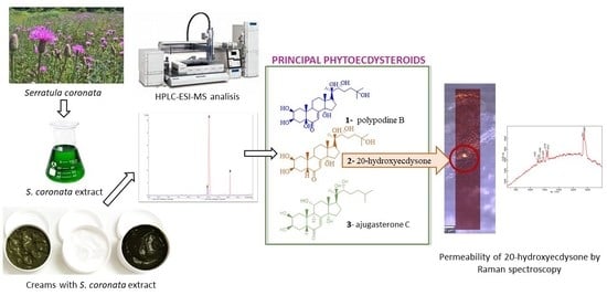
2.1. Chromatographic Analysis

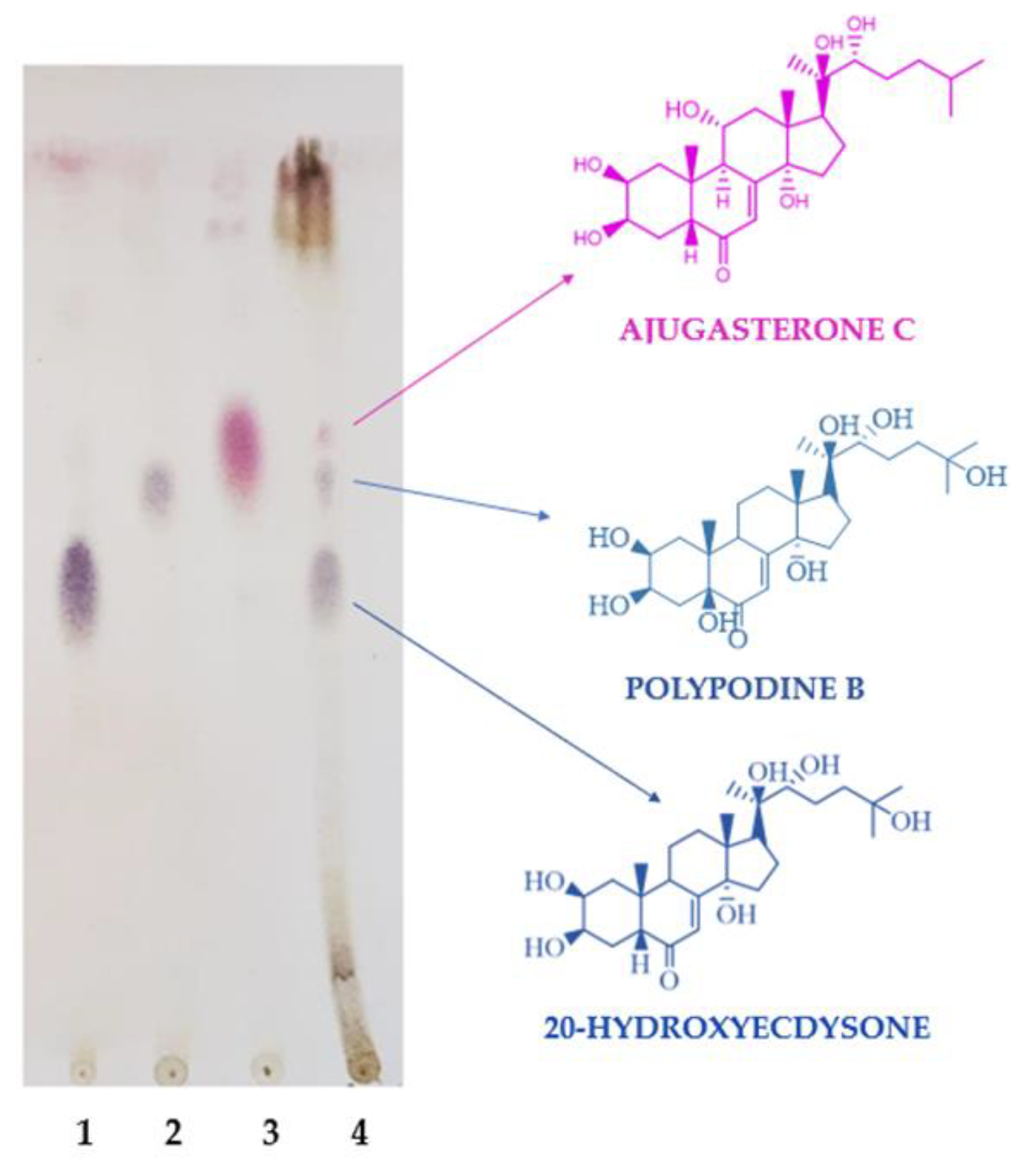
2.1.1. Analysis of the Chemical Content of the Extract Using Thin-Layer Chromatography

TLC confirmed the presence of the dominant PEs in the S. coronata extract. The compounds were identified by means of a comparison between the retention parameters of the extract compounds and those of the model compounds (Figure 1).

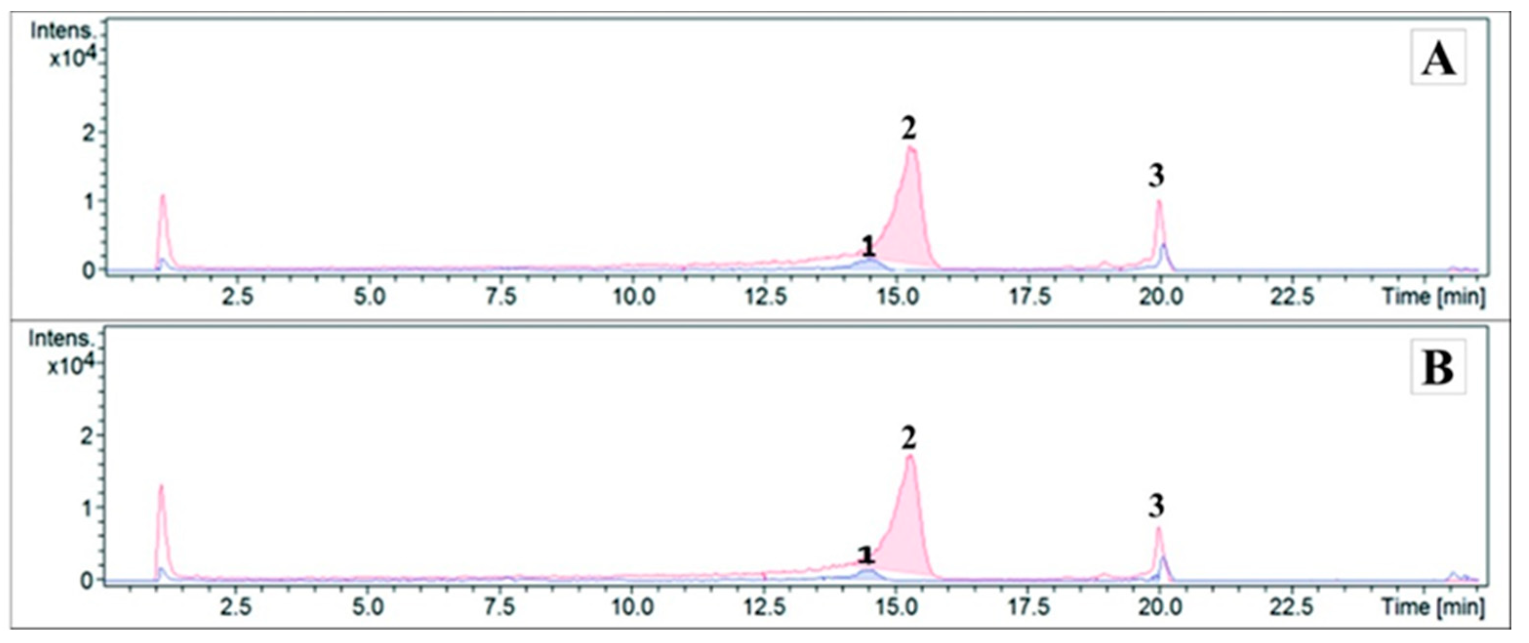
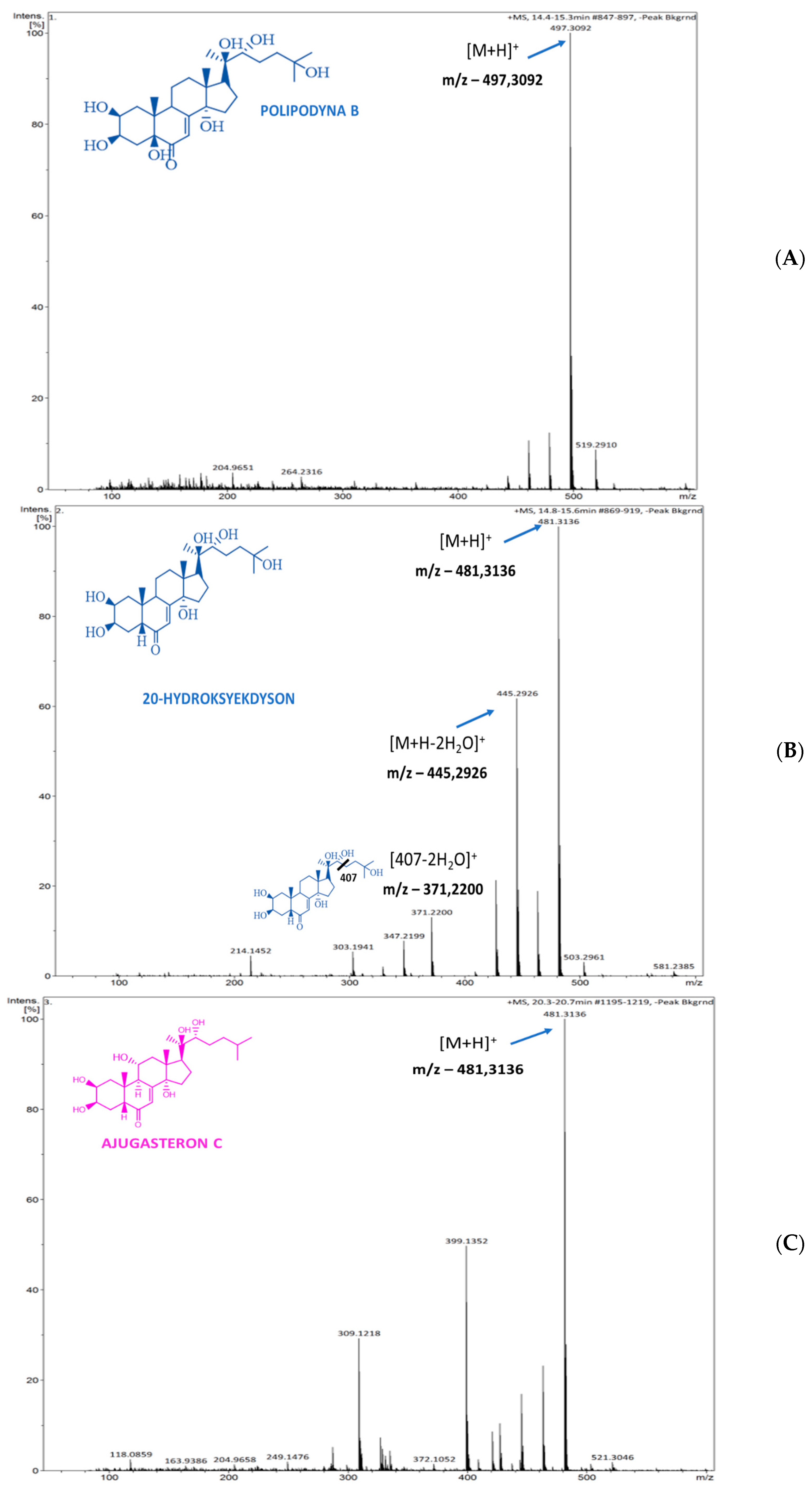
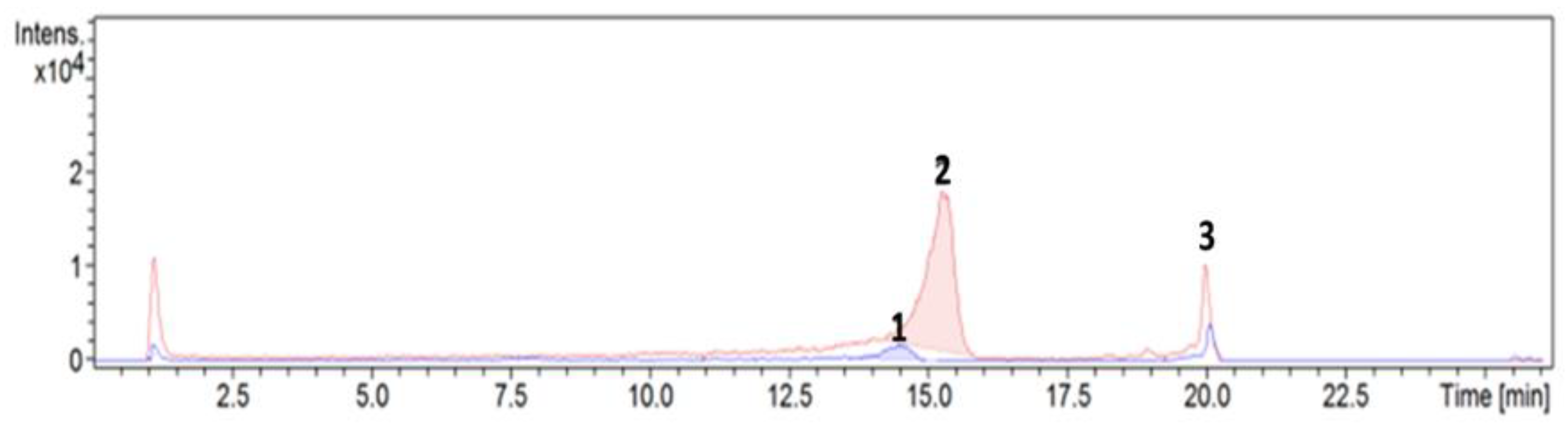
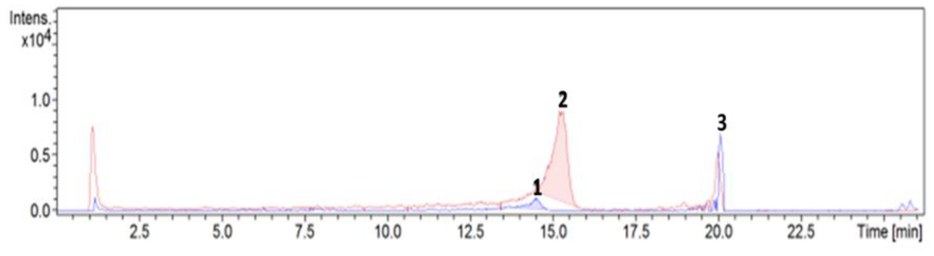
2.1.2. Analysis of the Dominant Phytoecdysteroids in the Creams Using HPLC-ESI-MS

The compositions of the prepared cosmetics were as follows: Cream 0 (Lekobaza® and placebo), Cream 1 (Lekobaza® and S. coronata extract), Cream 2 (Lekobaza® and salicylic acid), and Cream 3 (Lekobaza®, S. coronata extract, and salicylic acid) (Figure 2. Altogether, 80 samples were prepared (20 samples of 30 g for each examined cream).
Chromatograms (Figure 3) and mass spectra (Figure 4) were used to present the results of the qualitative analysis of Creams 1 and 3 using HPLC-ESI-MS. Three phytoecdysteroids (20-HE, ajugasterone C, and polypodine B) were found.
The following were observed in the ESI-MS (+) spectra in the positive mode: the m/z 497.3092 ion (m/z represents the mass-to-charge ratio) was identified as the polypodine B molecule (Figure 4A); the m/z 481.3136 ion was attributable to 20-HE [M-H]+ (Figure 4B); and the m/z 481,3136 [M-H]+ ion was identified as the ajugasterone C molecule (Figure 4C). The presence of the subsequently observed ions; e.g., the m/z 445.2926 [M-H-2H2O]+ ion, was attributed to the loss of one hydroxyl group, although it may have been the result of a split between C-23 and C-24 at the side chain (481 > 407). The split was not visible in the spectra because it occurred immediately after the loss of two water molecules, thereby resulting in m/z 371.2200.

2.2. Physical and Chemical Analysis of the Creams

Introduction of a cosmetic preparation to the market requires numerous tests such as physical and chemical analyses. The pH of a cosmetic product significantly affects the barrier function of the skin and its overall condition. The pH values vary depending on the chemical composition of a product and its purpose [11].

2.2.1. Chemical and Microbiological Stability of the Creams

Cosmetic products must remain safe up until their expiry date, which is determined by the manufacturer. Although the composition of the cosmetics is regulated by law, there are no legal requirements regarding stability tests. Most cosmetic preparations are tested for microbiological stability, and chemical stability is only tested only in some cases. Without testing for chemical stability, a manufacturer cannot be certain of a product’s safety and stability throughout its entire shelf life. In accordance with the EU directive on cosmetic products, a safety protocol is required for each cosmetic formulation (annex I to the regulation) [12]. Cosmetic preparations are at risk for microbiological contamination during production and usage [13]. Microbes that have been isolated from the contaminated cosmetics include Staphylococcus (S. aureus, S. epidermidis, and S. warneri), Escherichia coli, and Pseudomonas aeruginosa [14]. Both the Food and Drug Administration (FDA) and the EU directive on cosmetic products require the microbial population to be low, stable, and free from harmful microorganisms [15]. The production process should be compliant with the ISO Good Manufacturing Practice (GMP) on the production, monitoring, and transport of cosmetic products to minimize the risk for microbial contamination. The effects of microbial contamination of a cosmetic product may range from moderate (change in color and/or texture) to severe (altered activity/toxicity) [16].
Chemical stability is another key element that ensures product safety. Several testing methods for assessing the chemical stability of cosmetics under predictable transport, storage, and usage conditions have been designed [17]. According to the guidelines of the International Council on Harmonization of Technical Requirements for Registration of Pharmaceuticals for Human Use (ICH) (International Cooperation on Pharmaceuticals), these factors include humidity, temperature, pH, the presence of oxidants, and exposure to light [18]. The so-called “stress tests,” during which the product is exposed to the abovementioned factors, are the most important stability tests for active components. Degradation of the active substances in cosmetic formulations is associated with numerous chemical reactions that might occur under the influence of various environmental factors (i.e., hydrolysis, oxidation, isomerization, hydration, dimerization, or decarboxylation). In addition, photostability testing constitutes an integral part of the stability analysis [18].

Chemical Stability

The chemical stability of the creams was determined by exposing the samples to certain temperatures and humidity levels. Chemical stability was confirmed by using the qualitative analysis of the creams (Figure 5 and Figure 6). PEs’ presence in the samples after stability testing indicated that the compounds did not undergo degradation processes under the influence of the abovementioned environmental factors.

Microbiological Purity

The microbiological acceptance criteria of non-sterile topicals depend on the microbial count in a sample. Their limits are based on the Total Aerobic Count (TAMC) and Total Yeast/Mold Count (TYMC). The acceptance criteria for microbiological quality are established by considering TAMC ≤ 1 × 10² CFU/g and TYMC ≤ 1 × 101 CFU/g (CFU = colony-forming unit) [19]. The results of microbiological testing are presented in Table 1.
According to the Polish Pharmacopoeia XII, the acceptable germ counts for non-sterile topicals are 102 CFU for aerobic bacteria and 101 CFU for yeast and mold. Therefore, our results were indicative of microbiological stability [20].

Viscosity and pH

The analysis of the pH revealed an acidic reaction of the plant extract (pH 4.13 ± 0.05). The values of the cream viscosity and pH are presented in Table 2.
The active component lowered the pH of the cosmetic medium (Lekobaza®), and salicylic acid increased the acidic pH of the cosmetic (Table 2). The extract, the pH of which was similar to that of the medium, caused a slight decrease in the pH of the cosmetic formulation. A physiologically slightly acidic pH of the stratum corneum (4.1–5.8) impacts the barrier function of the skin, synthesis and aggregation of the lipids, keratinocyte differentiation, and desquamation, as well as microbiome and antibody activation in the skin [21]. A low skin pH, especially an acidic pH of the stratum corneum, inhibits skin colonization with pathogens such as Staphylococcus aureus or Streptococcus pyogenes and supports normal microbial growth [22]. An increased skin pH has been observed in various skin dermatoses. In psoriasis, an increase in pH by 0.3–0.4 is associated with a drop in the hydrogen ion (H+) concentration by half, which affects the biochemical processes of the skin. An elevated pH and increased skin dryness stimulate proteinase-activated receptor-2, which leads to pruritus in psoriatic patients because extra- and intra-cellular concentration of the protons modulates the afferent (pruritus and pain) and the efferent (growth, differentiation, and survival of the cells) functions [21]. Beneficial effects of the creams containing the S. coronata extract have been confirmed by applied research [1]. The method of decreasing the skin pH by using acidification has long been applied to wound healing because it increases anti-bacterial activity, changes protease activity and oxygen release, decreases the toxicity of the bacterial end products, and stimulates epithelization and angiogenesis [23].
Rheology plays a vital role in the determination of the stability of cosmetic products. Rheological properties such as viscosity significantly impact the quality of a cosmetic because they determine its consistency and spreadability on the skin [24,25]. The results of the viscosity tests are presented in Table 2. A high relative viscosity of a cosmetic formulation is perceived by the users as the equivalent of high concentration of the active substances and their effectiveness. Rheological properties are often selected to match the type of a cosmetic product and how it affects the skin. Cosmetics that regenerate the skin and soothe skin irritation (e.g., those designed for dry and flaky skin) should remain on the skin’s surface for a prolonged time [25,26], and these are most often supplied as creams [27].

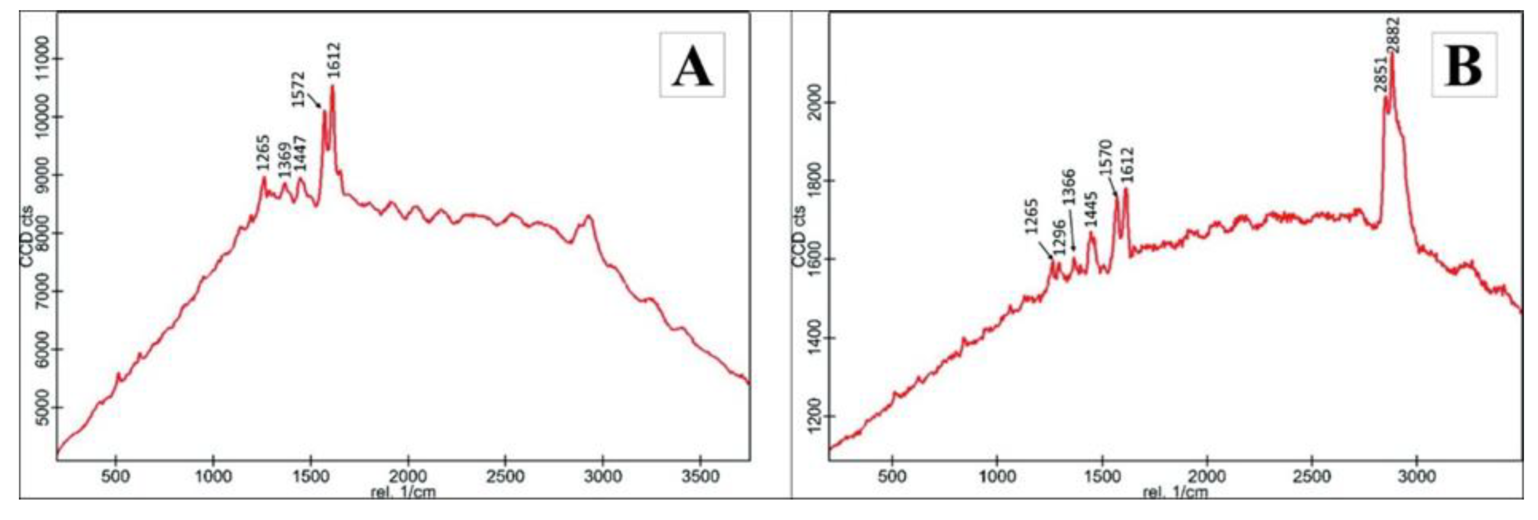
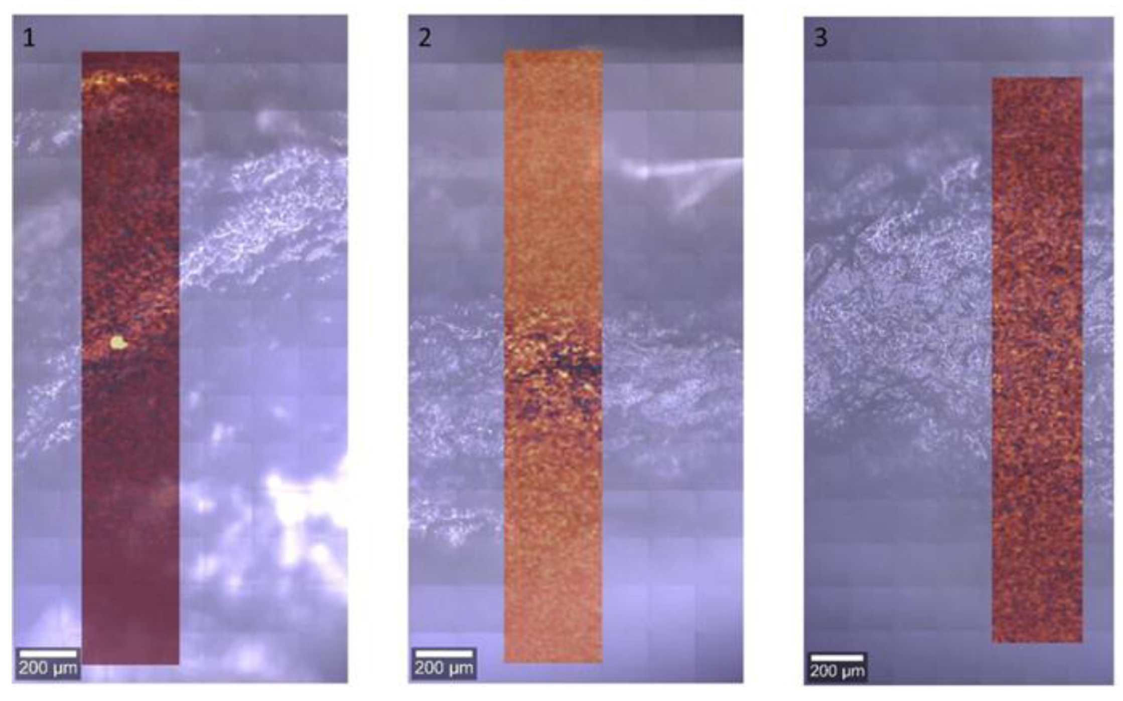
2.3. Permeability of 20-Hydroxyecdysone by Raman Spectroscopy in an In Vitro Model

The permeability factor of the active components significantly affects the effectiveness of a cosmetic product. Biologically active substances may be transported through the skin at three levels: topical (absorption), intracutaneous (penetration), and deep (resorption). Active substances need to pass the epithelial barrier and reach the deep layers of the skin to achieve the desired effect [28]. Active components may penetrate the skin via the intercellular and transcellular route as well as through skin appendages. The permeability of the active substances depends on the physical and chemical properties of the diffusion substance, the properties of the medium, and biological (overall condition and thickness of the skin, age and metabolism, and blood flow) and physical (temperature, time of day, and climate) factors [29,30]. The methods that allow the determination of the degree of skin penetration by the active components may be subdivided into two groups: quantitative methods and qualitative or semi-quantitative methods. The former includes diffusion chambers and the Parallel Artificial Membrane Permeability Assay (PAMPA) test; the latter includes microscopic and spectroscopic methods as well as combinations of the two. Quantitative in vitro tests are regularly performed to measure how much of an active component passed through the membrane over time versus the diffusion area associated with the amount of the active component obtained in the acceptor chamber. As far as qualitative tests are concerned, they are used to identify the active substance and will allow the determination of the presence or the relative amount of the active component in various skin layers [31]. A combination of the chemometric methods with spectroscopic imaging on transversal skin sections allows for the precise localization of the molecules in the layers of the skin [32].
In our study, that method was applied to determine the permeability of 20-HE, the dominant PE in the S. coronata creams. The test was conducted in an in vitro human skin model using Raman spectroscopy, an efficient spectroscopic technique that detects the vibrational modes of molecules. The obtained Raman spectra constituted highly specific spectroscopic molecule models, thereby allowing to identify them and monitor their passage through the skin. The method offers a non-invasive and dynamic technique to analyze the permeability of active substances through skin layers with a spatial resolution of a few micrometers [32].
According to the available literature, 20-HE remains the dominant ecdysteroid in both the plant and animal kingdoms [33,34]. Thus, a 3% 20-HE cream was composed for our study and investigated via confocal Raman spectroscopy (CRS) to measure the penetration rate of the 20-HE. Based on the CRS spectra, 1570 and 1612 cm−1 were identified as the most characteristic bands. Less specific bands were observed at positions 1265, 1369, and 1447 cm−1. The CRS spectra for 20-HE and for the 3 wt.% 20-HE cream are presented in Figure 7A,B, respectively.
Raman maps plotting the distribution of the cream with 3 wt.% 20-HE in the cross-section of the skin for a photomicrograph taken in visible light are presented in Figure 8.
The obtained results of the CRS indicted that the transdermal penetration rate was 1200 µm (subcutaneous tissue).

3. Materials and Methods

3.1. Standard and Chemicals

Standard 20-HE, ajugasterone C, and polypodine B were previously purified and characterized for S. coronata [35]. All solvents were of HPLC grade and all other chemicals were of analytical reagent grade, and they were used as received.

3.2. Material Extraction [35]

The fragmented and dried sample of S. coronata (2000 g) was soaked three times with 10,250 mL of ethanol (96%, POCH). The extract was filtered, and its solvent was evaporated using a rotary evaporator (Rotavapor® R-100, BUCHI, Flawil, Switzerland) at 40 °C. Next, the extract was concentrated to dryness at room temperature. A total of 136.42 g of the extract (extractum siccum) with a dark green color was obtained.

3.2.1. Analysis of the Chemical Composition of the Extract Using Thin-Layer Chromatography

The dominant PEs in the S. coronata extract were 20-HE, ajugasterone C, and polypodine B [9,35]. Thin-layer chromatography (TLC) on aluminum slates coated with silica gel (Merck, Germany) was used to confirm the presence of these compounds in the extract. A mixture (5:1) of dichloromethane (p.a., POCH) and methanol (p.a., POCH) was used as the mobile phase. The dishes were sprayed with p-anisaldehyde and heated at 105 °C for visualization [36]. The model compounds used in the TLC analysis (20-HE, ajugasterone C, and polypodine B) were established in our previous study [35].

3.2.2. Analysis of the Dominant Phytoecdysteroids in the S. Coronata Extract Using Liquid Chromatography–Electrospray Ionization–Mass Spectrometry (HPLC-ESI-MS)

HPLC-ESI-MS was used to detect the dominant PEs in the extract. Earlier analyses of the model compounds helped to establish the following retention times (RT): 20-HE—15.2 min, ajugasterone C—20.4 min, and polypodine B—14.8 min. The extract was diluted 1000-fold before testing. The measurements were conducted using a quadrupole time-of-flight mass spectrometer (QTOF, Impact HD, Bruker Daltonics, Bremen, Germany) in positive mode and a liquid chromatograph (LC) Ultimate 3000 (Thermo Scientific/Dionex). The LC conditions are presented in Table 3. We developed and validated a simple, sensitive, and precise method for the isolation and quantification of the dominant ecdysteroids—ajugasterone C, polypodine B, and 20-HE—from the S. coronata herb in an earlier study [35].

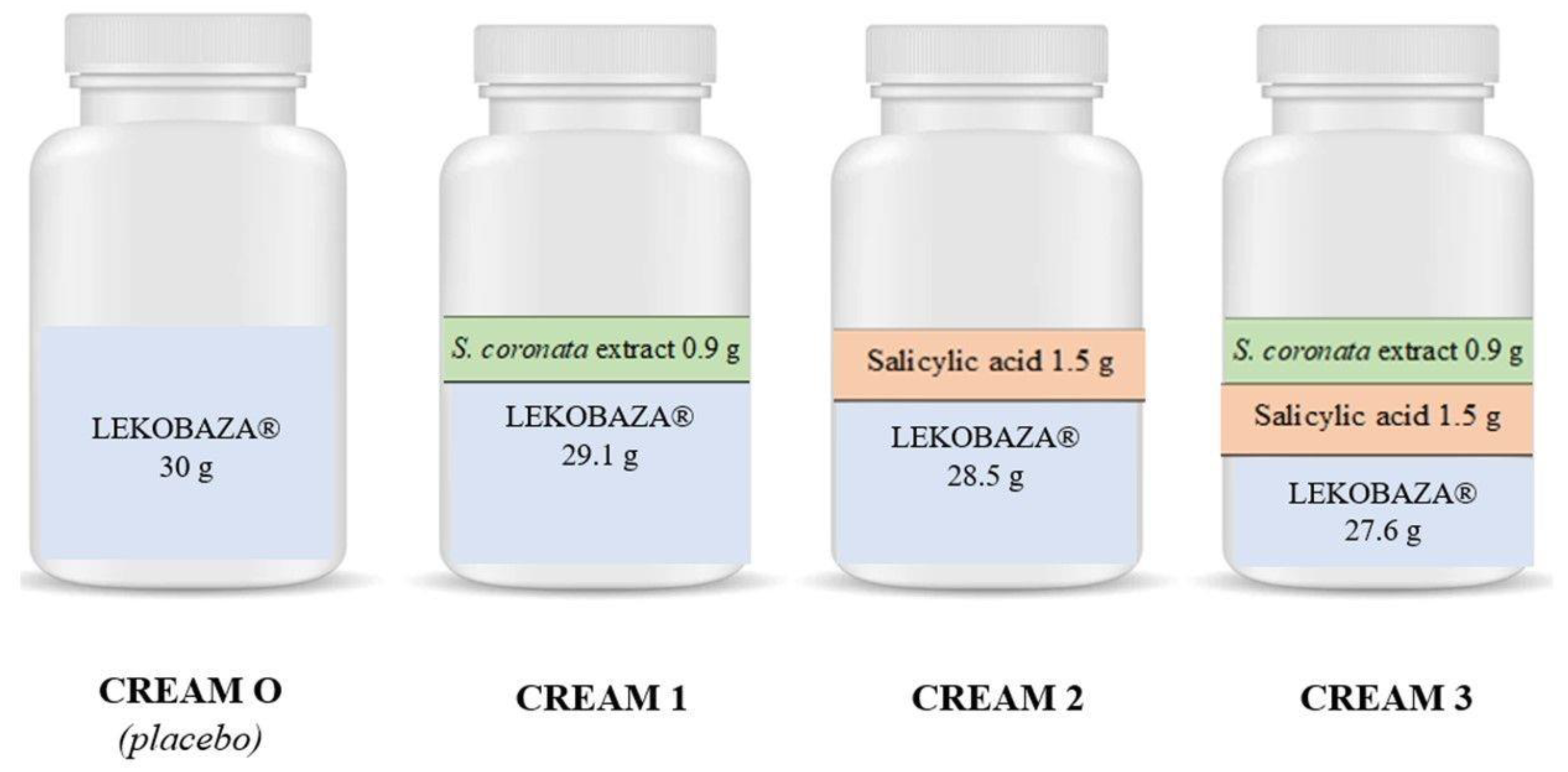
3.3. Cream Preparation

Four creams (0—placebo, 2—cream with salicylic acid, 1 and 3—creams with the extract; 30 g each) were prepared. The composition of the creams is presented in Figure 4. Lekobaza® Pharma Cosmetic (Fagron, Krakow, Poland) was used as the cream base and as the placebo. It is a multi-component medium with a pH of 5.5 containing white Vaseline, glycerol monostearate, and cetyl alcohol, Miglyol® 812, macrogol-20-glycerol-monostearate, propylene glycol, and purified water. It is a universal base that penetrates the skin easily, is less greasy, and creates a protective film on the skin’s surface.
Salicylic acid was used as an ingredient in the creams due to its keratolytic properties and to enhance phytoecdysteroid penetration [37].

3.4. Viscosity and pH

The pH of the creams was determined using a VWR ORP15 PEN/PH 10 PEN pH-meter (VWR International, Gdansk, Poland) with a predefined setting range of 4–7 pH. All pH measurements were conducted at 21 °C. The cream viscosity was determined using a RheoTec RCO2 rotational viscometer with an adjustable coagulation threshold (Messtechnik GmbH, Ottendorf-Okrilla, Germany). The creams were placed in narrow glass beakers. Next, the appropriate rotational spindle was mounted and inserted. The measurements were conducted at 25 ± 2 °C using the R5 spindle for the creams with the extract (Creams 1 and 3) and the R7 spindle for Cream 0 at a rotational speed of 3 rpm. The viscosity and pH were measured three times and used to calculate the mean and standard deviation values.

3.5. Chemical Stability

Creams 1 and 3 were placed in the stability chamber (TH-ICH-300, Jeiotech Co., Ltd., Daejeon, Republic of Korea) for 21 days at a temperature of 45 °C and 75% relative humidity (RH) to determine the effects of humidity and temperature on the stability of the dominant PEs in the S. coronata extract [38]. Next, all samples were analyzed using the qualitative HPLC-ESI-MS method.

3.6. Microbiological Stability

The microbiological analysis of the creams was conducted in accordance with the method described in the European Pharmacopoeia (FP) [39]: 10 g of the preparation was mixed with sterile phosphate buffer (PB, Sigma-Aldrich, Darmstadt, Germany) + 2.5% Tween80 (Sigma-Aldrich, Darmstadt, Germany) to 100 mL (1:10 dilution). The sample was vigorously stirred for approximately 15 min and then further diluted using a 0.2% Tween80 phosphate buffer. Each of the four samples (1:10, 1:100, 1:1000, and 1:10,000 dilutions) was then transferred to a separate Petri dish using 1 ml of the homogenate each (stirred for 5 s before placement). Next, 15–20 mL of liquid medium—Tryptone-Soy-Agar (TSA, 2 dishes) and Sabouraud Dextrose Agar (SDA, 2 dishes)—at 40 °C were added. After mixing and medium solidification, the TSA cultures were incubated at 30–35 °C for 3–5 days and the SDA cultures at 20–25 °C for 5–7 days. Next, the aerobic bacteria, yeast, and mold counts per 1 g of the sample were calculated while taking into the account the dilution factor.

3.7. Analysis of the Dominant Phytoecdysteroids Using HPLC-ESI-MS

A qualitative HPLC-ESI-MS analysis was used to confirm the presence of the dominant PEs in the S. coronata creams. We also analyzed the preparations that had been tested in the climate chamber to determine the effects of temperature and humidity on the stability of these components.

3.7.1. Sample Preparation

Before the chromatographic assay, 5.0 g of Creams 1 and 3 were placed in 25 mL glass vials and soaked in 5 mL of 96% ethanol (POCH). The samples were stirred vigorously, placed in an ultrasonic washer (Bandelin Sonorex RK 255 H), and filtered through syringe filters (0.45 µm pore size). Next, 1 mL of the filtrate was measured and placed in a glass vial for the HPLC-ESI-MS analysis.

3.7.2. Conditions of the Analysis

The designed method was subjected to validation in accordance with the ICH guidelines [40].
A qualitative analysis of Creams 1 and 3 was conducted using the same equipment and the same measurement parameters as in the case of the extract analysis (Section 3.2.2).

3.8. Permeability of 20-Hydroxyecdysone as Measured by Raman Spectroscopy in An In Vitro Model

Skin penetration by 20-HE, the dominant compound of the S. coronata extract, was examined using confocal Raman spectroscopy: 1 g of 20-HE was dissolved in 5.6 mL of water with 26.6 g of Lekobaza® to prepare the emulsified cream and to analyze its penetration through the skin layers. The study using Raman spectroscopy was performed using a WITec Alpha 300 spectrometer equipped with a confocal microscope (WITec alpha300 R, Ulm, Germany), a TrueSurface attachment, and an electron-multiplying CCD (EMCCD) camera for ultra-fast and sensitive imaging. The measurements were taken with the use of excitation with frequency-doubled Nd:YAG laser line with a wavelength of 532 nm. The radiation power at the focal point was approximately 10 mW. The spectral resolution of the collected spectra was approximately 3 cm−1. The measurements were made using an air objective with a magnification of 20× and a numerical aperture of 0.4. The measurements of the spectra of the samples were recorded for the following parameters: laser power—10 mW, number of accumulations—10, and accumulation time of a single spectrum—0.5 s. In case of mapping, the measurement procedure consisted of the sequential collection of individual spectra from a specific area each time the spectrum was measured over the entire spectral range; i.e., 0–3600 cm−1, using the excitation line with a wavelength of 532 nm. The image measurements were recorded for the following parameters: laser power—10 mW, accumulation time of a single spectrum—0.15 s, and sampling density—10 µm. The analysis of the mapping results consisted of the integration of the observed bands in the spectra. The Raman spectra were collected in the range of 0–3600 cm−1 for 532 nm. The permeability analysis was performed on cross-sections through the layers of the skin after incubation with the samples. Skin samples were obtained from excess skin during abdominoplasty from healthy females. Samples were prepared in a 6-well plate with phosphate-buffered saline (PBS) to prevent tissue dehydration. Afterward, the skin was treated with the 3% 20-HE cream and incubated for 6 h at 37 °C (5% CO2), and then the samples were mounted on slides for the Raman spectroscopy. All experiments were performed in three biological repetitions with each of them in three technical repeats.

4. Conclusions

Based on the results obtained, it was shown that the herb Serratula coronata is a source of ecdysone compounds that can be used for the development of formulations for skin care. All of the developed cosmetic formulations showed chemical and microbiological stability. In addition, the biological activity of 20-hydroxyecdysone can be related to its proven transdermal permeation, which was confirmed using Raman spectroscopy in an in vitro model. The regenerative properties of phytoecdysteroids, as already confirmed in our previous work, make cosmetic preparations containing Serratula coronata extracts useful for skin problems such as psoriasis.

Author Contributions

Conceptualization, J.G.-P. and M.P.; methodology, J.G.-P., M.P., A.K. and A.F.-G.; investigation, A.K., M.U., A.F.-G. and T.O.; data curation, A.K. and A.F.-G.; writing—original draft preparation, J.G.-P. and M.P.; writing—review and editing, A.K., A.F.-G., M.U., T.O., I.M. and J.N.; supervision, J.G.-P. and M.P.; project administration, A.K. and A.F.-G. All authors have read and agreed to the published version of the manuscript.

Funding

This research received no external funding.

Institutional Review Board Statement

Not applicable.

Informed Consent Statement

Not applicable.

Data Availability Statement

The data presented in this study are available upon request.

Conflicts of Interest

The authors declare no conflict of interest.

References

  1. Kroma, A.; Pawlaczyk, M.; Feliczak-Guzik, A.; Urbańska, M.; Jenerowicz, D.; Seraszek-Jaros, A.; Kikowska, M.; Gornowicz-Porowska, J. Phytoecdysteroids from Serratula coronata L. for Psoriatic Skincare. Molecules 2022, 27, 3471. [Google Scholar] [CrossRef] [PubMed]
  2. Srivastava, P.; Singh, M.; Devi, G.; Chaturvedi, R. Herbal Medicine and Biotechnology for the Benefit of Human Health. In Animal Biotechnology: Models in Discovery and Translation; Verma, A.S., Sing, A., Eds.; Elsevier Science Publishing Co. Inc.: San Diego, CA, USA, 2014; pp. 563–575. [Google Scholar]
  3. Olennikov, D.N. Metabolites of Serratula L. and Klasea Cass. (Asteraceae): Diversity, Separation Methods, and Bioactivity. Separations 2022, 9, 448. [Google Scholar] [CrossRef]
  4. Ivashchenko, I.; Rakhmetov, D.B. Biomorphological Features of Serratula coronata L. (Asteraceae) Introduced in ZHNAEU’s Botanical Garden. Mod. Phytomorphol. 2016, 10, 69–80. [Google Scholar]
  5. Das, N.; Mishra, S.K.; Bishayee, A.; Ali, E.S.; Bishayee, A. The Phytochemical, Biological, and Medicinal Attributes of Phytoecdysteroids: An Updated Review. Acta Pharm. Sin. B 2021, 11, 1740–1766. [Google Scholar] [CrossRef] [PubMed]
  6. Laekeman, G.; Vlietinck, A. Phytoecdysteroids: Phytochemistry and Pharmacological Activity. In Natural Products: Phyto-Chemistry, Botany and Metabolism of Alkaloids, Phenolics and Terpenes; Ramawat, K.G., Mérillon, J.M., Eds.; Springer: Berlin/Heidelberg, Germany, 2013; pp. 3827–3849. [Google Scholar]
  7. Ványolós, A.; Báthori, M. New Perspectives in the Analysis of Ecdysteroids: A Promising Group of Biologically Active Compounds. Curr. Pharm. Anal. 2008, 4, 162–175. [Google Scholar] [CrossRef]
  8. Garg, A.; Sharma, R.; Dey, P. Analysis of Triterpenes and Triterpenoids. In Recent Advances in Natural Products Analysis; Silva, A.S., Nabavi, S.F., Saeedi, M., Nabavi, S.M., Eds.; Elsevier: Amsterdam, The Netherlands, 2020; pp. 393–426. [Google Scholar]
  9. Odinokov, V.N.; Galyautdinov, I.V.; Nedopekin, D.V.; Khalilov, L.M.; Shashkov, A.S.; Kachalab, V.V.; Dinanc, L.; Lafont, R. Phytoecdysteroids from the juice of Serratula coronata L. (Asteraceae). Insect Mol. Biol. 2002, 32, 161–165. [Google Scholar] [CrossRef]
  10. Ghosh, D.; Laddha, K.S. Extraction and Monitoring of Phytoecdysteroids Through HPLC. J. Chromatogr. Sci. 2006, 44, 22–26. [Google Scholar] [CrossRef] [Green Version]
  11. Lukić, M.; Pantelić, I.; Savić, S.D. Towards Optimal pH of the Skin and Topical Formulations: From the Current State of the Art to Tailored Products. Cosmetics 2021, 8, 69. [Google Scholar] [CrossRef]
  12. European Union. Regulation (EC) No 1223/2009 of the European Parliament and of the Council of 30 November 2009 on Cosmetic Products; European Union: Brussels, Belgium, 2009. [Google Scholar]
  13. Michalek, I.M.; John, S.M.; Caetano Dos Santos, F.L. Microbiological Contamination of Cosmetic Products—Observations from Europe, 2005–2018. J. Eur. Acad. Dermatol. Venereol. 2019, 33, 2151–2157. [Google Scholar] [CrossRef]
  14. Kim, H.W.; Seok, Y.S.; Cho, T.J.; Rhee, M.S. Risk Factors Influencing Contamination of Customized Cosmetics Made On-the-Spot: Evidence from the National Pilot Project for Public Health. Sci. Rep. 2020, 10, 1561. [Google Scholar] [CrossRef] [Green Version]
  15. Choubey, S.; Godbole, S. Methods for Evaluation of Microbiological Safety, Guidelines Governing the Quality and Survey on Microbial Contamination of Commercial Cosmetic Products—A Review. WJPMR 2017, 3, 85–94. [Google Scholar]
  16. Noor, R.; Zerin, N.; Das, K.K.; Nitu, L.N. Safe usage of cosmetics in Bangladesh: A quality perspective based on microbiological attributes. J. Biol. Res. Thessalon. 2015, 22, 10. [Google Scholar] [CrossRef] [PubMed]
  17. Drechsel, D.; Towle, K.; Fung, E.; Novick, R.; Paustenbach, D.; Monnot, A. Chemical Stability Analysis of Hair Cleansing Conditioners under High-Heat Conditions Experienced during Hair Styling Processes. Cosmetics 2018, 5, 23. [Google Scholar] [CrossRef] [Green Version]
  18. Jamrógiewicz, M.; Merchel, M. A History of the Physical and Chemical Stability of Pharmaceuticals—A Review. Acta Pol. Pharm. 2018, 75, 297–304. [Google Scholar]
  19. Wirotesangthong, M. Microbiological Limits in Cosmetics. Isan J. Pharm. Sci. 2021, 17, 1–22. [Google Scholar] [CrossRef]
  20. Registration of Medicinal Products, Medical Devices and Biocidal Products. Polish Pharmacopoeia, 12th ed.; Registration of Medicinal Products, Medical Devices and Biocidal Products: Warsaw, Poland, 2020; Volume I.
  21. Proksch, E. pH in Nature, Humans and Skin. J. Dermatol. 2018, 45, 1044–1052. [Google Scholar] [CrossRef]
  22. Chan, A.; Mauro, T. Acidification in the Epidermis and the Role of Secretory Phospholipases. Dermato-Endocrinol. 2011, 3, 84–90. [Google Scholar] [CrossRef] [Green Version]
  23. Nagoba, B.S.; Suryawanshi, N.M.; Wadher, B.; Selkar, S. Acidic Environment and Wound Healing: A Review. Wounds 2015, 27, 5–11. [Google Scholar]
  24. Davies, A.; Amin, S. Rheology of Cosmetic Products: Surfactant Mesophases, Foams and Emulsions. J. Cosmet. Sci. 2020, 71, 481–496. [Google Scholar]
  25. Gräbner, D.; Hoffmann, H. Rheology of Cosmetic Formulations. In Cosmetic Science and Technology: Theoretical Principles and Applications; Sakamoto, K., Lochhead, R.Y., Maibach, H.I., Eds.; Elsevier Inc.: Amsterdam, The Netherlands, 2017; pp. 471–488. [Google Scholar]
  26. Moravkova, T.; Stern, P. Rheological and Textural Properties of Cosmetic Emulsions. Appl. Rheol. 2011, 21, 1–6. [Google Scholar] [CrossRef]
  27. Sarkar, D.K. Creams and Ointments. In Pharmaceutical Emulsions: A Drug Developer’s Toolbag; Sarkar, D.K., Ed.; John Wiley & Sons: Hoboken, NJ, USA, 2013; pp. 69–76. [Google Scholar]
  28. Kim, B.; Cho, H.-E.; Moon, S.H.; Ahn, H.-J.; Bae, S.; Cho, H.-D.; An, S. Transdermal Delivery Systems in Cosmetics. Biomed. Dermatol. 2020, 4, 10. [Google Scholar] [CrossRef]
  29. Shabbir, M.; Ali, S.; Shahid, N.; Rehman, K.; Umair, A.; Moosa, R. Formulation Considerations And Factors Affecting Transdermal Drug Delivery System. Int. J. Pharm. Integr. Sci. 2014, 2, 20–35. [Google Scholar]
  30. Marwah, H.; Garg, T.; Goyal, A.K.; Rath, G. Permeation enhancer strategies in transdermal drug delivery. Drug Deliv. 2016, 23, 564–578. [Google Scholar] [CrossRef] [PubMed]
  31. Zsikó, S.; Csányi, E.; Kovács, A.; Budai-Szűcs, M.; Gácsi, A.; Berkó, S. Methods to Evaluate Skin Penetration In Vitro. Sci. Pharm. 2019, 87, 19. [Google Scholar] [CrossRef] [Green Version]
  32. Essendoubi, M.; Gobinet, C.; Reynaud, R.; Angiboust, J.F.; Manfait, M.; Piot, O. Human Skin Penetration of Hyaluronic Acid of Different Molecular Weights as Probed by Raman Spectroscopy. Skin. Res. Technol. 2015, 22, 55–62. [Google Scholar] [CrossRef] [PubMed]
  33. Buniam, J.; Chukijrungroat, N.; Rattanavichit, Y.; Surapongchai, J.; Weerachayaphorn, J.; Bupha-Intr, T.; Saengsirisuwan, V. 20-Hydroxyecdysone Ameliorates Metabolic and Cardiovascular Dysfunction in High-Fat-High-Fructose-Fed Ovariectomized Rats. BMC Complement. Med. Ther. 2022, 20, 140. [Google Scholar] [CrossRef]
  34. Festucci-Buselli, R.A.; Contim, L.A.S.; Barbosa, L.C.A.; Stuart, J.; Otoni, W.C. Biosynthesis and Potential Functions of the Ecdysteroid 20-Hydroxyecdysone—A Review. Canad. J. Bot. 2008, 86, 978–987. [Google Scholar] [CrossRef]
  35. Napierała, M.; Nawrot, J.; Gornowicz-Porowska, J.; Florek, E.; Moroch, A.; Adamski, Z.; Kroma, A.; Miechowicz, I.; Nowak, G. Separation and HPLC Characterization of Active Natural Steroids in a Standardized Extract from the S. Coronata Herb with Antiseborrheic Dermatitis Activity. Int. J. Environ. Res. Public Health 2020, 17, 6453. [Google Scholar] [CrossRef]
  36. Nowak, G.; Nawrot, J.; Latowski, K. Arbutin in Serratula quinquefolia M.B. (Asteraceae). Acta Soc. Bot. Pol. 2009, 78, 137–140. [Google Scholar] [CrossRef]
  37. Arif, T. Salicylic acid as a peeling agent: A comprehensive review. Clin. Cosmet. Investig. Dermatol. 2015, 8, 455–461. [Google Scholar] [CrossRef] [Green Version]
  38. González-González, O.; Ramirez, I.O.; Ramirez, B.I.; O’Connell, P.; Ballesteros, M.P.; Torrado, J.J.; Serrano, D.R. Drug Stability: ICH versus Accelerated Predictive Stability Studies. Pharmaceutics 2022, 14, 2324. [Google Scholar] [CrossRef] [PubMed]
  39. European Directorate for the Quality of Medicines EDQM. European Pharmacopoeia, 7th ed.; The European Directorate for the Quality of Medicines & HealthCare: Strasbourg, France, 2010; pp. 163–167, 519–520. [Google Scholar]
  40. ICH. Q2 (R2): Validation of Analytical Procedures–Text and Methodology. In Proceedings of the International Conference on Harmonization (ICH), Geneva, Switzerland, 31 March 2022. [Google Scholar]
Figure 1. Thin layer-chromatography of the S. coronata extract: 1—20-hydroxyecdysone; 2—polypodine B; 3—ajugasterone C; 4—S. coronata extract.
Cosmetics 10 00018 g001
Figure 2. Compositions of the prepared creams.
Cosmetics 10 00018 g002
Figure 3. Chromatogram of Cream 1 (A) and Cream 3 (B) containing 3 wt.% S. coronata: (1) polypodine B; (2) 20-hydroxyecdysone; (3) ajugasterone C (red—first analysis of cream 1, blue—second analysis of cream 1).
Cosmetics 10 00018 g003
Figure 4. Polypodine B (A), 20-hydroxyecdysone (B), and ajugasterone C (C) in 3 wt.% S. coronata creams identified via ESI-MS.
Cosmetics 10 00018 g004
Figure 5. Chromatogram of Cream 1 with the Serratula coronata extract after 3 weeks in a climatic chamber: 1—polypodine B; 2—20-hydroxyecdysone; 3—ajugasterone C (red first analysis of Cream 1, blue second analysis of Cream 1).
Cosmetics 10 00018 g005
Figure 6. Chromatogram of Cream 3 with the S. coronata extract after 3 weeks in a climatic chamber: 1—polypodine B, 2—20-hydroxyecdysone, 3—ajugasterone C (red—first analysis of Cream 3, blue—second analysis of Cream 3).
Cosmetics 10 00018 g006
Figure 7. Raman spectra of 20-hydroxyecdysone (A) and 3 wt.% 20-hydroxyecdysone cream (B).
Cosmetics 10 00018 g007
Figure 8. Representative Raman images of the distribution of 3 wt.% 20-hydroxyecdysone cream prepared on the basis of the integration one of the characteristic bands (1612 cm−1) for each of the three biological replicates.
Cosmetics 10 00018 g008
Table 1. Results of microbiological testing of the creams.
CreamsTotal Aerobic Count Test ResultTotal Yeast
and Mold Count Test Result
Method
0—Lekobaza®<10 CFU/g<10 CFU/gAccording to the Polish
Pharmacopoeia XII
1—Lekobaza®
+ S. coronata extract
<10 CFU/g<10 CFU/g
2—Lekobaza®
+ salicylic acid
<10 CFU/g<10 CFU/g
3—Lekobaza®
+ salicylic acid
+ S. coronata extract
<10 CFU/g<10 CFU/g
Table 2. Viscosities and pHs of the creams.
CreamspH
M ± SD
Viscosity (mPA × S)
M ± SD
0—Lekobaza5.03 ± 0.05152,900 ± 33,517
1—Lekobaza® + S. coronata extract4.30 ± 0.06121,737 ± 1744
2—Lekobaza® + salicylic acid3.07 ± 0.12 194,200 ± 17665
3—Lekobaza® + salicylic acid2.47 ± 0.06134,607 ± 2119
+S. coronata extract
Abbreviations: SD, standard deviation; M, mean; mPA, minipascal; s, second.
Table 3. LC conditions using a Kinetex 2.6µ C18 column (100 × 210 mm): temp. = 30 °C, injection volume = 10 µL, and mobile phase flow rate = 0.3 mL/min.
Time (min)Mobile Phase
H2O + 0.1% FA (%)ACN + 0.1% FA (%)
090.010.0
190.010.0
1287.512.5
1487.512.5
2055.045.0
2255.045.0
2410.090.0
2610.090.0
2890.010.0
3590.010.0
Abbreviations: ACN, acetonitrile; FA, formic acid.
Disclaimer/Publisher’s Note: The statements, opinions and data contained in all publications are solely those of the individual author(s) and contributor(s) and not of MDPI and/or the editor(s). MDPI and/or the editor(s) disclaim responsibility for any injury to people or property resulting from any ideas, methods, instructions or products referred to in the content.

