Article

Surface Modification of Hemoglobin-Based Oxygen Carriers Reduces Recognition by Haptoglobin, Immunoglobulin, and Hemoglobin Antibodies

1
Institute of Transfusion Medicine, Charité-Universitätsmedizin Berlin, 10117 Berlin, Germany
2
Department of Radiological Technology, Faculty of Allied Health Sciences, Naresuan University, Phitsanulok 65000, Thailand
3
Division of Biochemistry and Nutrition, School of Medical Sciences, University of Phayao, Phayao 56000, Thailand
4
Division of Clinical Immunology and Transfusion Sciences, School of Allied Health Sciences, University of Phayao, Phayao 56000, Thailand
5
Department of Pharmaceutical Technology, Faculty of Pharmacy, Payap University, Chiang Mai 50000, Thailand
6
Department of Medical Physics, Biophysics and Radiology, Medical Faculty, Trakia University, Stara Zagora 6000, Bulgaria
*
Author to whom correspondence should be addressed.
Coatings 2019, 9(7), 454; https://doi.org/10.3390/coatings9070454
Submission received: 3 July 2019 / Revised: 12 July 2019 / Accepted: 17 July 2019 / Published: 21 July 2019

Abstract

:
Hemoglobin-based oxygen carriers (HBOCs) represent a propitious type of blood substitute to transport oxygen throughout the body while acting as a carrier in biomedical applications. However, HBOCs in blood are recognized and rapidly scavenged by the body’s innate immune systems. To overcome this problem, HBOCs require a surface modification that provides protection against detection and elimination in order to prolong their circulation time after administration. In this study, we investigated different surface modifications of hemoglobin submicron particles (HbMPs) by double/triple precipitation, as well as by adsorption of human serum albumin (HSA), hyaluronic acid (HA), and pluronic (Plu) to discover how diverse surface modifications influence the oxygen binding capacity and the binding of anti-hemoglobin (Hb) antibodies, immunoglobulin G (IgG), and haptoglobin (HP) to HbMPs. The particle size and zeta potential of the six types of HbMP modifications were analyzed by zeta sizer, confocal laser scanning microscopy, and transmission electron microscopy (TEM), and were compared to the unmodified HbMPs. The results revealed that all surface-modified HbMPs had a submicron size with a negative charge. A slight decrease in the oxygen binding capacity was noticed. The specific binding of anti-Hb antibodies, IgG, and HP to all surface-modified HbMPs was reduced. This indicates a coating design able to protect the particles from detection and elimination processes by the immune system, and should lead to a delayed clearance and the required and essential increase in half-life in circulation of these particles in order to fulfill their purpose. Our surface modification method reflects a promising strategy for submicron particle design, and can lead the way toward novel biomedical applications.

Graphical Abstract

1. Introduction

Blood transfusion is a potentially life-saving procedure and is used in many therapeutic applications. Several risks, such as transmission of infectious disease, immunomodulation etc., have accelerated the development of blood substitutes as alternatives to human blood [1,2]. The development of blood substitutes has mainly focused on the requirements that they carry oxygen, have no toxicity or side effects, and have a sufficient circulation time [3,4,5]. During recent decades, the use of blood substitutes has been extensively investigated especially with the attention to hemoglobin-based oxygen carriers (HBOCs). Interestingly, the development and modification of HBOCs for clinical trials is still in progress [5,6,7]. Currently, the different approaches to fabricating HBOCs that have been investigated have had the primary consideration of stabilizing the hemoglobin (Hb) molecule and improving the oxygen-carrying capabilities by cross-linked hemoglobin, polymerized hemoglobin, hemoglobin conjugated to macromolecules, and encapsulated hemoglobin [8,9]. HBOCs are expected to increase the total oxygen carrying capacity and oxygen delivery of blood because of an increase in total Hb concentration. Therefore, the oxygen carrying behavior of HBOCs should be considered and investigated.
However, one main problem seems to be the short half-life of HBOCs after their transfusion [10]. Surprisingly, there are no literature data available, which could explain the reason for the elimination of the HBOCs. It is known that Hb is detected by haptoglobin (HP), and the Hb-HP complex is recognized by Kupffer cells very fast [11,12]. Therefore, HBOCs should preclude Hb–HP complex formation to avoid liver uptake and depletion of HP in the plasma. Additionally, we have to consider the opsonization of HBOCs, which would result in a rapid clearance by the reticuloendothelial system (RES). To overcome this obstacle, modifications of the surface properties of carriers, also known as “stealth coatings,” have been developed and applied to prevent particles from protein absorption and immune recognition [13,14]. The properties of these polymers should include high flexibility and high hydrophilicity. Many materials and coating approaches have been utilized for modifying the surface of particles. For instance, the natural polysaccharides (e.g., hyaluronic acid) or synthetic polymers (e.g., polyethylene glycol (PEG)-based copolymers such as pluronic) have been widely used as coating materials. Hyaluronic acid (HA) or hyaluronan is a hydrophilic linear glycosaminoglycan composed of alternating (1→4)-β-d-glucuronic acid and (1→3)-β-N-acetyl-d-glucosamine [15], which has been used for the fabrication of hyaluronic-acid-based layer-by-layer capsules for the intracellular uptake of the capsules by macrophages, indicating their potential as a carrier for intracellular drug delivery [16]. Pluronics (poloxamers) are a class of water-soluble and non-ionic A-B-A triblock copolymers, where A is poly (ethylene oxide) (PEO) and B is poly (propylene oxide) (PPO). Pluronic block copolymers have been used and applied as a micellar nano-carrier for drug delivery system, resulting in increased solubility and metabolic stability, and improving circulation time. Gyulai et al. reported that the presence of pluronics on the surface of poly lactic-co-glycolic acid (PLGA) nanoparticles preserved the aggregation stability and improved the nonspecific membrane affinity of the system [17].
Amongst the available coating materials, human serum albumin (HSA) is also a promising molecule for surface modification. HSA is the product of hepatic protein synthesis and is the most abundant plasma protein (molecular weight (MW) = 66.5 kDa). These properties, including high stability, preferential uptake in tumor and inflamed tissue, biodegradability, and lack of toxicity and immunogenicity indicate a good material for drug delivery carriers [18,19]. Esfandyari-Manes et al. also reported that the surface modification of particles with albumin improves long-circulating and stealth particles for drugs delivery [20].
To date, there has been a great interest in the development of HBOCs in their versatile properties, including biocompatibility, biodegradability, and low immunogenicity. Previously published data of hemoglobin particles synthesized by a co-precipitation-crosslinking-dissolution technique (CCD-technique) demonstrated HBOCs with high oxygen affinity and low immunogenicity [21,22].
The aim of this study was to clarify in which way several modifications of the surface of HbMP influence the oxygen binding and the adsorption of antibodies, especially the anti-Hb antibodies, immunoglobulin, and the glycoprotein HP. The surface modifications were performed by double/triple precipitation, as well as by adsorption of human serum albumin (HSA), hyaluronic acid (HA), and pluronic (Plu). As mentioned above, the utilization of HSA, HA, and Plu as surface coating materials is a promising approach for drug delivery carriers. We hypothesized that these polymers could be applied to the particle and HbMP would become coated with a layer that would allow it to avoid anti-Hb antibody, IgG, and HP binding.

2. Materials and Methods

2.1. Materials

HSA was purchased from the Baxalta Deutschland GmbH, Unterschleissheim, Germany. Manganese chloride tetrahydrate (MnCl2·4H2O), sodium carbonate (Na2CO3), ethylene-diaminetetraacetic acid (EDTA), sodium borohydride (NaBH4), glutaraldehyde (GA), hyaluronic acid sodium salt, kolliphor®P407, and potassium ferricyanide (K3(Fe(CN)6)) were purchased from Sigma-Aldrich, Munich, Germany. Sodium chloride (NaCl) was purchased from Merck KGaA, Darmstadt, Germany. Ringer’s acetate solution (consisting of sodium chloride, potassium chloride, calcium chloride dehydrate, magnesium chloride hexahydrate, and sodium acetate trihydrate in water) was purchased from Serumwerk Bernburg AG, Bernburg, Germany. Ampuwa® (aqua ad injectable) was purchased from Fresenius Kabi Deutschland GmbH, Bad Homburg, Germany. Pronase was purchased from Roche Diagnostics GmbH, Mannheim, Germany. Rabbit anti-bovine hemoglobin antibody (LS-C294442) and allophycocyanin (APC)-conjugated rabbit polyclonal antibody to human haptoglobin (LS-C236382) were purchased from LS Bio, Washington, WA, USA. Goat anti-rabbit IgG conjugated to SureLight® APC (D3-114-1mg) were purchased from Columbia Biosciences, Frederick, MD, USA. Haptoglobin from pooled human plasma (H3536) was purchased from Sigma, Saint Louis, MO, USA. Human IgG isotype control (biotin), (NBP1-96855) was purchased from Novus Biologicals a Bio-Techne Brand, Centennial, CO, USA. APC-streptavidin (Cat#405243, Lot B223923) was purchased from BioLegend Inc., San Diego, CA, USA.
Hemoglobin (Hb) was extracted from bovine red blood cells by hypotonic hemolysis [23]. Fresh bovine whole blood (received from Biophyll GmbH, Dietersburg, Germany), anticoagulated with EDTA, was centrifuged (2500 g, 10 min, 4 °C), was washed three times with phosphate-buffered saline (PBS), and then five volumes of ice-cold Ampuwa were added to one volume of washed red blood cells. The solution was stirred at 4 °C overnight and then centrifuged (10,000 g, 4 °C) for 1 h. The supernatant was filtered through a 0.1 μm polyethersulfone filter (Sartorius AG, Göttingen, Germany) and stored at −80 °C as a stock solution for further use.

2.2. Particle Preparation

The synthesis and surface modification of HbMPs are schematically displayed in Figure 1. We fabricated six different types of surface-modified HbMP. A short description of each particle type and the corresponding abbreviations are listed in Table 1.

2.2.1. Preparation of Hemoglobin Submicron Particles (HbMPs)

HbMPs were fabricated using a protocol based on the CCD technique, as previously described [24,25]. Briefly, 5 mg/mL Hb in 0.125 M MnCl2 solution was mixed rapidly with 0.125 M Na2CO3 and stirred for 30 s to obtain Hb-MnCO3 particles (first precipitation). These particles were washed twice with distilled water by centrifugation (3000 g, 3 min) and re-dispersed in 0.9% NaCl solution. The Hb-MnCO3 particles were incubated with 0.02% GA solution for 1 h at room temperature. After that, the obtained particles were washed and re-suspended in distilled water. These particles were used for a second as well as a third precipitation.

2.2.2. Preparation of HSA-d-HbMPs and HSA-t-HbMPs

Double precipitated HbMPs (HSA-d-HbMP): For the second co-precipitation step, the washed Hb-MnCO3 particles were suspended in 0.04 M MnCl2 and 0.4% HSA was added to adsorb onto the surface of the particles for 5 min. After adding 0.04 M Na2CO3, the obtained particles were washed with distilled water by centrifugation (3000 g, 3 min) and were re-suspended in 0.9% NaCl solution. The double co-precipitated HSA-d-Hb-MnCO3 particles were cross-linked again with 0.02% GA at room temperature under moderate stirring. A measure of 0.25 M EDTA was used to dissolve the MnCO3 templates. A measure of 0.2 mg/mL NaBH4 in 0.1 M NaOH solution was added and incubated for 30 min. The resulting HSA-d-HbMPs were washed three times with 0.9% NaCl solution supplemented with 0.2% HSA (centrifugation 10,000 g, 10 min), and were re-suspended in Ringer’s acetate solution and stored at 4 °C for further use.
Triple precipitated HbMPs (HSA-t-HbMP): The additional co-precipitation step of MnCO3 with HSA for fabrication of HSA-t-HbMPs was performed after the second crosslinking step, followed by cross-linking with GA in the same manner and concentrations before performing the dissolution step.

2.2.3. Preparation of HA-d-HbMPs and HA-t-HbMPs

The HA-d-HbMPs were prepared in the same manner as HSA-d-HbMPs, using 0.04% HA in addition to the 0.4% HSA. The fabrication of HA-t-HbMPs was performed after the second crosslinking step by co-precipitation of MnCO3 with 0.04% HA and 0.4% has, followed by cross-linking with GA in the same manner and concentrations before performing the dissolution step.

2.2.4. Preparation of Plu-d-HbMPs and Plu-t-HbMPs

The Plu-d-HbMPs were also prepared in the same manner as HSA-d-HbMPs, using 0.4% pluronic in addition to the 0.4% HSA. The fabrication of Plu-t-HbMPs was performed after the second crosslinking step by co-precipitation of MnCO3 with 0.4% pluronic and 0.4% has, followed by cross-linking with GA in the same manner and concentrations before performing the dissolution step.

2.3. Particle Characterization

2.3.1. Size and Zeta-Potential

The hydrodynamic size and zeta potential of each surface-modified HbMPs was determined using a Zetasizer Nano ZS instrument (Malvern Instruments Ltd., Worcestershire, UK). Each sample was diluted 1:200 using PBS solutions. All the measurements were carried out in triplicate.

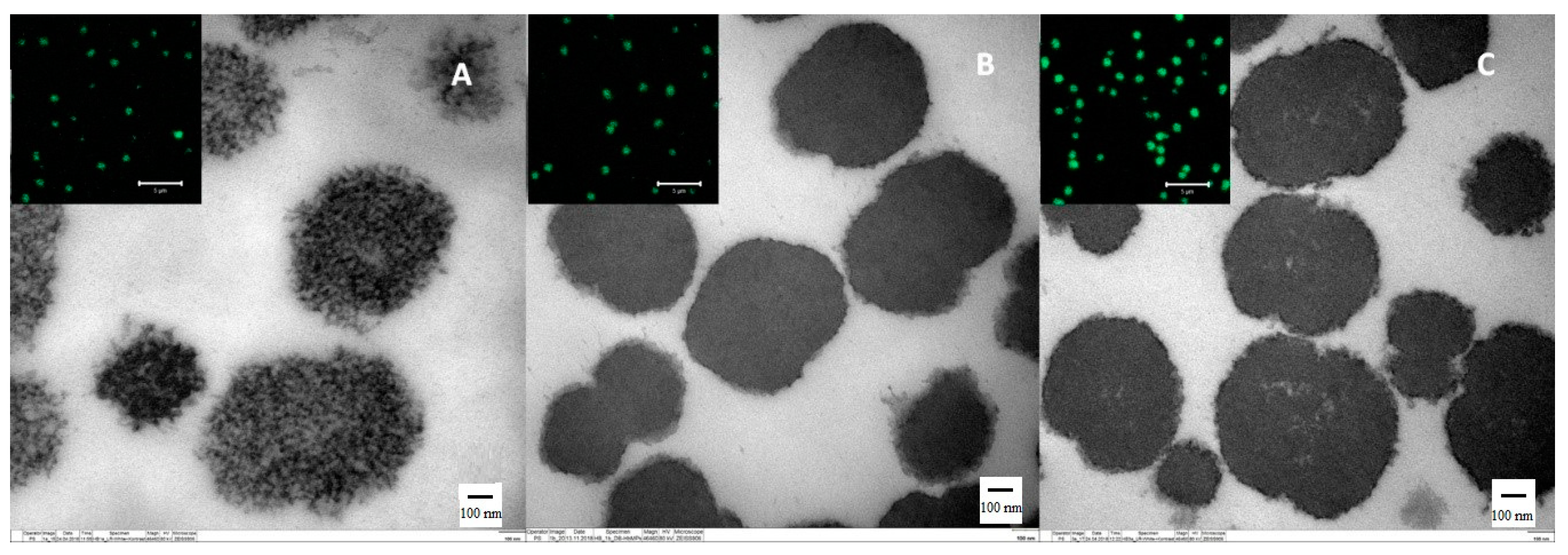
2.3.2. Transmission Electron Microscopy (TEM)

TEM imaging was performed using a Zeiss EM 906 Omega microscope (Carl Zeiss AG, Oberkochen, Germany) at an acceleration voltage of 80 kV. Sample preparation was as follows: The water of the particle suspensions was exchanged with ethanol by washing steps with gradually increasing ethanol concentrations (10%, 20%, 40%, 60%, 80%, 99.9%). After the last centrifugation (1800 g, 6 min), the sample was infiltrated with LR-White Resin (ratio of Ethanol: LR-WR = 1:1) and incubated overnight at room temperature. After that, the sample was exchanged with fresh LR-WR and was transferred into gelatin capsules. Polymerization of the resin was performed with UV light for 48 h at room temperature. At the end of the process, ultrathin sections (60–70 nm on mesh-grids) were prepared. Sections were stained with 1% phosphotungstic acid for 15 min and 4% aqueous uranyl acetate for 15 min.

2.3.3. Confocal Laser Scanning Microscopy (CLSM)

CLSM images of each surface-modified HbMPs preparation were taken with LSM 510 Meta (Carl Zeiss MicroImaging GmbH, Jena, Germany) microscope with a 100× oil-immersion objective (numerical aperture 1.3), applying excitation wavelength of 488 nm and a long pass emission filter of 505 nm.

2.3.4. Determination of Hb Content

The total hemoglobin concentration of the surface-modified HbMPs was determined by the modified alkaline hematin-D method (AHD method) [26,27]. Briefly, 2% (v/v) particle suspensions were mixed with pronase to obtain a final concentration of 0.5 mg/mL pronase, and were incubated at 45 °C for 30 min. After that, AHD reagent (25 mg/mL Triton X-100 in 0.1 M NaOH) was added at the volume ratio 1:1, then thoroughly mixed and incubated for 15 min at room temperature. All samples were measured at the absorption spectra at wavelength 574 nm using a UV–vis spectrophotometer (Hitachi U2800, Hitachi High-Technologies Corporation, Tokyo, Japan), and an average of three individual measurements were recorded for calculation of the Hb concentration.

2.3.5. Determination of Oxygenated HbMPs (Oxy-Hbs)

The ferricyanide method was applied for measurement of the oxygen content on the surface-modified HbMPs [28,29]. Briefly, 2% (v/v) particle suspensions was placed into a glass vial containing a magnetic stirrer. A miniaturized optical needle type oxygen sensor (oxygen micro-sensor NTH-PSt7, PreSens-Precision Sensing GmbH, Regensburg, Germany) connected to a portable oxygen meter with data logging (Microx 4, PreSens-Precision Sensing GmbH, Regensburg, Germany) was inserted into the stirred sample, and the concentration of dissolved oxygen was recorded. After 3 s, 10% potassium ferricyanide was added and the change of the concentration of dissolved oxygen was measured until a stable value was reached. The difference between the final pO2 value and the initial pO2 corresponds to the oxygen bound to hemoglobin in the particles. The change in pO2 was converted into the concentration of released oxygen (cO2) with the following formula, known as Henry’s Law:
c O 2   =   a O 2 × p O 2
where aO2 is the solubility coefficient of oxygen in blood: 0.0031 mL O2/mmHg O2/dL blood. Having derived the mass of released oxygen, the concentration of the Oxy-Hb was calculated as follows:
c ( Oxy - Hb ) = m O 2 ρ ( O 2 )   × H b s
where ρO2=1.43 g/L is the density of O2 and Hbs is the saturated Oxy-Hb content which equals 1.34 mL of O2 per gram of Hb.

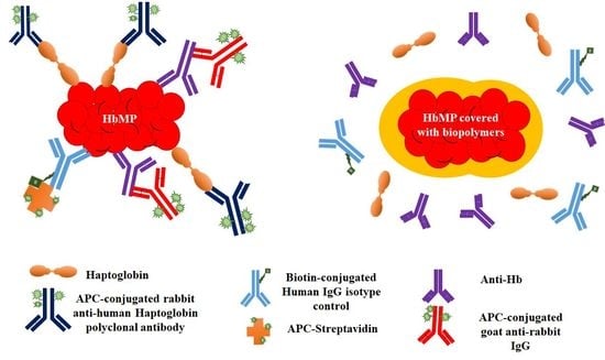
2.4. In Vitro Antibody Binding Assays

To confirm a successful surface modification of HbMP and detect the Hb on the surface of HbMP, the adsorption of anti-bovine Hb antibodies, immunoglobulins (IgG), and haptoglobin (HP) was performed by using flow cytometry (FACS Canto II, Becton & Dickinson, Franklin Lakes, NJ, USA). In this experiment, we used the six different types of surface modified HbMPs and the unmodified HbMPs as a control (description is shown in Table 1). Staining the particles with only secondary antibodies provided the non-specific binding, staining with both primary and secondary antibodies provided the total binding. Specific binding was calculated by subtracting the non-specific binding from the total binding.

2.4.1. Binding Assay of Anti-Hb Antibodies

Samples of 1 mL of 0.2% (v/v) particle suspensions were incubated for 2 h with the primary rabbit anti-bovine hemoglobin antibody at a final concentration of 2.5 µg/mL. The particle suspensions were then incubated with the secondary APC-conjugated goat anti-rabbit IgG (final concentration 1 µg/mL). Incubations were performed at room temperature. Between each incubation step, the particle suspensions were washed 2–3 times with PBS solution. Subsequently, the particle suspensions were incubated again in darkness for 2 h, washed, and re-suspended in 300 µL PBS solutions for investigation by flow cytometry.

2.4.2. Binding Assay of Immunoglobulin (IgG)

The binding of IgG to particles was also performed, and comprised the same step procedure described above used for the binding assay of anti-Hb antibodies. The primary biotin conjugated human IgG isotype control was added into 1 mL of particle suspension at a final concentration of 2.5 µg/mL, and was incubated for 2 h at room temperature followed by PBS washings. After that, the particle suspensions were incubated for 2 h in the dark with APC-streptavidin at a final concentration of 1 µg/mL.

2.4.3. Binding Assay of Haptoglobin (HP)

The accessibility of Hb of each surface-modified HbMPs for the HP was investigated in the same manner as described above. Firstly, HP from pooled human plasma was added to 1 mL of particle suspension at a final concentration of 2.5 µg/mL HP. Next, APC-conjugated rabbit polyclonal antibody to human haptoglobin (HP antibody) was added at a final concentration of 1 µg/mL (secondary antibody).

2.5. Statistical Analysis

The results are presented as means ± standard deviation (SD). The data were compared using ANOVA-like test. GraphPad Prism 6 software (GraphPad, La Jolla, CA, USA) was employed for graphs and statistical analyses. p-value < 0.05 were considered statistically significant.

3. Results and Discussion

3.1. Particle Characterization

3.1.1. Particle Morphology and Size

The morphology of HbMPs and surface-modified HSA-d-HbMPs and HSA-t-HbMPs was investigated by TEM. The HbMPs, HSA-d-HbMPs, and HSA-t-HbMPs showed a peanut-like shape (Figure 2A–C, respectively). The unmodified particles (Figure 2A) clearly showed a more porous structure compared to the surface-modified HbMPs with HSA. The HSA-d-HbMPs and HSA-t-HbMPs appeared denser and were surrounded by a thin dark rim, which confirms the successful coating with HSA. Due to the autofluorescence caused by the protein crosslinking with GA, the HbMPs, HSA-d-HbMPs and HSA-t-HbMPs could be additionally visualized by CLSM. The corresponding CLSM images (Figure 2 inserts) confirm the suspension stability of the samples, e.g., absence of aggregation. This was also found for HbMPs with HA and pluronic surface modification, respectively (Supplementary Figure S1).
The hydrodynamic diameter of all seven samples was determined by dynamic light scattering, and was in the range of 0.7 to 1 µm with a low polydispersity index (PDI < 0.3), indicating a narrow size distribution and an absence of aggregation. The hydrodynamic diameters of the surface-modified particles did not significantly differ from that of HbMPs (Figure 3A).

3.1.2. Zeta Potential

The mean values of the zeta-potential of all surface-modified HbMPs suspended in PBS solutions (conductivity 18–20 mS/cm) were negative (in the range −11 to −13 mV for HSA-d-HbMPs, HSA-t-HbMPs, Plu-d-HbMPs, and Plu-t-HbMPs). No significant differences in the zeta potential of the surface-modified HbMPs in presence of HSA and Plu could be found compared to HbMPs (p < 0.05). However, the zeta potential of HA-d-HbMPs and HA-t-HbMPs became significantly more negative (increased by 36%–38%) compared to HbMP (Figure 3B). Similar results are reported in our previous studies about the influence of HA on MnCO3-templated Hb/BSA-HA particles [30]. The enhanced zeta potential values of the HA-d-HbMPs and HA-t-HbMPs confirm the attachment of HA, a strongly negatively charged polysaccharide, on the surface of the particles.

3.1.3. Hemoglobin Content and Oxygen Binding Capacity

The total Hb concentration of all surface-modified HbMPs revealed no statistically significant difference compared to HbMPs (35–40 mg/mL as calculated for suspensions with a hematocrit (Hct) of 20%, p value < 0.05) (Figure 4A).
The oxygen binding capacity of HBOC is commonly determined by oxygen dissociation curves (ODC), which demonstrate the relationship between oxygen partial pressure and the oxygen fractional saturation [8]. However, this method requires specific and expensive equipment, gas flow apparatus, and gas tanks, and therefore, some research groups have attempted to overcome the high-cost instrument problem and have proposed a more simple method for analysis of oxygen binding capacity in the laboratory [28,31,32].
In this study, we applied the ferricyanide method to determine the concentration of Oxy-Hb as a simple and inexpensive approach. The ferricyanide causes a conversion from oxyhemoglobin into met-hemoglobin (Met-Hb), and therefore a release of oxygen (as determined by the change in pO2) into the surrounding media, corresponding to the oxygen amount bound to hemoglobin in the particles. The oxygen binding capacity of the particles was determined from the content of Oxy-Hb (Figure 4B). For all samples, the Oxy-Hb calculated for a suspension with a Hct of 20% was in a range between 9 to 12 mg/mL. For the double precipitated HbMPs (HSA-d-HbMPs, HA-d-HbMPs and Plu-d-HbMP) the Oxy-Hb content showed a slight decreasing tendency, which was not statistically significant compared to the Oxy-Hb content of HbMPs. In contrast, the Oxy-Hb in the triple precipitated HbMPs showed a significant decrease by 33%, 32% and 35% for HSA-t-HbMPs, HA-t-HbMPs and Plu-t-HbMPs, respectively, when compared to HbMPs. This was most probably caused by the increased preparation time due to the additional coprecipitation and GA crosslinking steps during the surface modification, which can lead to partial oxidation of Hb to methemoglobin (MetHb), where the iron in the heme group is in the ferric state [25,33].

3.2. Binding of Anti-Hb Antibodies to Surface-Modified HbMPs

The quality of the surface modification of the HbMPs was tested using indirect immunofluorescence staining with anti-Hb antibodies (Figure 5A). Unmodified HbMPs present Hb molecules on their surface that will be recognized by the rabbit anti-bovine Hb antibodies (primary antibodies). The fluorescently labeled secondary goat anti-rabbit antibodies subsequently recognize the primary antibodies on the surface of the HbMPs, and the fluorescence signal can be detected and analyzed by flow cytometry.
Figure 5B shows the percentage of non-specifically (only with secondary antibodies) and specifically stained particles with anti-Hb antibodies in each group (% events). The % events positive for non-specific binding were low (5%–10%), with no statistically significant difference between the different samples. The % events positive for specific anti-Hb antibody binding were very high for the non-modified HbMPs (~80%) and for HSA-d-HbMPs (~70%), with no statistically significant difference. In the samples (HA-d-HbMP and Plu-d-HbMP) where double precipitation was performed, the % positive events of specific anti-Hb antibody binding was statistically significantly decreased when compared to HbMPs (by 36% and 33%, respectively). Most importantly, the specific anti-Hb antibody binding in the samples modified by triple precipitation, HSA-t-HbMP, HA-t-HbMP and Plu-t-HbMP, was decreased by 54%, 56% and 67%, respectively, when compared to the unmodified HbMPs. Additionally, the values of the main fluorescence intensity measured in all surface modified samples were lower than that measured for the HbMP sample (Supplementary Figure S2A). This is illustrated by representative histograms (Figure 5C–E) showing the distribution of the fluorescence intensity among the events of the HbMP, HSA-d-HbMP and HSA-t-HbMP samples. Therefore, the surface modifications by additional precipitations, especially by the triple precipitations, shielded the Hb molecules and inhibited their recognition by the anti-Hb antibodies.

3.3. Binding of IgG to Surface-Modified HbMPs

The binding of IgG on the surface of particles leads to opsonization and recognition of the particles by the immunocompetent cells in the blood and tissues. Therefore, a high-quality surface modification should protect the particles from IgG adsorption. Here, we incubated our unmodified and modified HbMPs with biotinylated human IgG, which was subsequently detected by APC-labelled streptavidin (Figure 6A) and analyzed by flow cytometry.
The results presented in Figure 6B,C show that IgG bound to the unmodified HbMPs (roughly 50% positive events of specific binding). In contrast, all surface modified samples showed statistically significant decreased events of specific binding by 80%–95%. The values of the main fluorescence intensity measured in the surface modified samples were lower than that measured for the HbMP sample (Supplementary Figure S2B). This is illustrated by representative histograms (Figure 6C–E) showing the distribution of the fluorescence intensity among the events of the HbMP, HSA-d-HbMP, and HSA-t-HbMP samples. Therefore, the surface modification by additional precipitations successfully inhibited IgG binding to the particle surface.

3.4. Binding of Haptoglobin to Surface-Modified HbMPs

HP is a glycoprotein that is mostly synthesized and secreted by liver cells, and plays a vital role with a high affinity to Hb in the circulation [34]. It is responsible for the rapid clearance of acellular Hb from the blood circulation, forming a complex with the β-chains of Hb which is then recognized by the Kupffer cells. Presumably, it can bind also to the Hb molecules on the surface of the HbMPs, which has to be avoided in order to achieve long circulation times of the HBOC.
We incubated our unmodified and modified HbMPs with human HP, which was subsequently detected by APC-labelled rabbit anti-human-HP antibodies (Figure 7A) and analyzed by flow cytometry. The results presented in Figure 7B,C show that HP bound to the unmodified HbMP (roughly 25%–30% positive events of specific binding). In contrast, all surface modified samples showed statistically significant decreased events of specific binding by 80%–95%. The values of the main fluorescence intensity measured in the surface modified samples were lower than that measured for the HbMP sample (Supplementary Figure S2C). The representative histograms (Figure 7C–E) illustrate the distribution of the fluorescence intensity among the events of the HbMP, HSA-d-HbMP and HSA-t-HbMP samples. Therefore, the surface modification by additional precipitations successfully inhibited HP binding to the particle surface.
Our data demonstrated that a double/triple precipitation of HSA, HA and Plu on the surface of HbMPs can be successfully employed to shield the Hb, resulting in a significant decrease of the specific binding of antibodies. In particular, the triple precipitated HbMPs showed the most promising results, reducing the binding of anti-Hb antibodies, IgG and HP, which can protect the HbMPs from endogeneous Hb removal mechanisms and rapid clearance from circulation.
The current development of blood substitutes, especially HBOCs, has amplified applications through investigation of the ability to transport oxygen, non-immunogenicity, extended half-life in circulation, and lack of toxicity. In this study, we modified the surface of HbMP by additional co-precipitation steps with three biopolymers (HSA, HA and Plu) and characterized their properties. Albumin and HA form complexes [35] and the amino groups of albumin are crosslinked by GA at the surface of HbMPs. That means HA will be immobilized at the surface, which was demonstrated by the zeta potential. In presence of HA, the zeta potential became more negative compared with that of only albumin. Pluronic also interacts with albumin [36], and the same mechanism should work as for HA, although only an indirect proof of the immobilization of Plu could be performed.
HbMPs were fabricated using the highly promising CCD technique based on MnCO3, which has been shown to be very simple and effective for entrapment of proteins into submicron particles [21,25,30,37].
HBOCs may induce severe side effects and toxicity after administration, due to scavenging of vascular endothelial nitric oxide (NO) and heme-mediated oxidative reactions [38,39]. Based on our previous reports of preclinical studies, HbMPs-700 fabricated by the CCD technique are a very promising approach, demonstrating a higher affinity to oxygen, avoidance of vasoconstriction, and non-mutagenicity [22,25].
Although HBOCs have shown a good biocompability and safety in pre-clinical studies, the circulatory clearance after administration is still a limitation, and it should be investigated for further clinical trials. In this study, we showed that surface modification of HbMPs could be a promising approach toward production of HBOCs with prolonged circulation.

4. Conclusions

Surface-modified HbMPs can be successfully prepared by modified CCD technique, including biopolymers, as shown here for HSA, HA and Plu, by means of double and triple co-precipitation. The obtained particles remained in the same size range as the unmodified HbMPs, with the typical peanut-like shape and more dense morphology.
The content of functional Hb of the surface-modified HbMPs was slightly decreased due to increased number of precipitations and additional GA crosslinking steps, leading to partial oxidation of Hb to MetHb. This obstacle could be avoided by applying antioxidants during the preparation or by performing the fabrication under an oxygen free atmosphere.
The immunofluorescence assays revealed that our surface modification technique was efficient to reduce the specific binding of anti-Hb antibodies, IgG and HP. These results can be useful for predicting a longer half-life of HbMPs in circulation.

Supplementary Materials

The following are available online at https://www.mdpi.com/2079-6412/9/7/454/s1, Figure S1: Confocal laser scanning micrograph of HA-d-HbMPs (A,B), Plu-d-HbMPs (C,D), HA-t-HbMPs (E,F), and Plu-t-HbMPs (G,H) in fluorescence mode (left) and transmission mode (right) (Scale bar: 5 µm), Figure S2: The mean fluorescence intensity (MFI) of non-specific and specific binding of anti-Hb antibodies (A), IgG (B), and haptoglobin (C) in each surface-modified HbMP is shown. Specific antibody binding values were calculated by subtracting the non-specific binding from total binding. Data represent the mean ± SD, and asterisks indicate the significance of differences when compared to HbMPs (* p < 0.05).

Author Contributions

Experiments, Data Analysis and Writing, A.P. (Aussanai Prapan); Writing and Data Analysis, I.K.; Data Analysis, N.S., C.K., S.C., A.S. and Y.X.; Conceptualization, Design of Experiments, Performing Specific Experiments, Interpretation and Discussion of Results, Revision of Manuscript, R.G.; Evaluation and Discussion of Results, A.P. (Axel Pruß); Conceptualization, Advised all Research, Design of Experiments, Evaluation and Discussion of Results, Revision of Manuscript, H.B.

Funding

A.P. (Aussanai Prapan) hold an academic development scholarship from Naresuan University, N.S. and C.K. from University of Phayao, S.C. from Payap University. This research received no external funding.

Acknowledgments

We are grateful to Petra Schrade, Core Facility of Electron Microscopy, Institute of Anatomy for the TEM images. We thank Chutamas Thepmalee and Patcharin Thammasit for suggestion on the result of immunofluorescence staining. We acknowledge support from the German Research Foundation (DFG) and the Open Access Publication Funds of Charité–Universitätsmedizin Berlin

Conflicts of Interest

The authors declare no conflict of interest.

References

  1. Sharma, S.; Sharma, P.; Tyler, L.N. Transfusion of blood and blood products: Indications and complications. Am. Fam. Physician 2011, 83, 719–724. [Google Scholar] [CrossRef] [PubMed]
  2. Rawn, J. The silent risks of blood transfusion. Curr. Opin. Anaesthesiol. 2008, 21, 664–668. [Google Scholar] [CrossRef] [PubMed]
  3. Moradi, S.; Jahanian-Najafabadi, A.; Roudkenar, M.H. Artificial Blood Substitutes: First steps on the long route to clinical utility. Clin. Med. Insights Blood Disord. 2016, 9, 33–41. [Google Scholar] [CrossRef] [PubMed]
  4. Seifried, E.; Mueller, M.M. The present and future of transfusion medicine. Blood Transfus. 2011, 9, 371–376. [Google Scholar] [CrossRef] [PubMed]
  5. Winslow, R.M. Current status of oxygen carriers. Vox Sang. 2006, 91, 102–110. [Google Scholar] [CrossRef]
  6. Keipert, P.E. Hemoglobin-Based Oxygen Carrier (HBOC) Development in Trauma: Previous Regulatory Challenges, Lessons Learned, and a Path Forward; Advances in Experimental Medicine and Biology Series 977; Springer: Cham, Switzerland, 2017; pp. 343–350. [Google Scholar] [CrossRef]
  7. Chen, J.-Y.; Scerbo, M.; Kramer, G. A review of blood substitutes: Examining the history, clinical trial results, and ethics of hemoglobin-based oxygen carriers. Clinics 2009, 64, 803–814. [Google Scholar] [CrossRef]
  8. Alayash, A.I. Blood substitutes: Why haven’t we been more successful? Trends Biotechnol. 2014, 32, 177–185. [Google Scholar] [CrossRef]
  9. Jia, Y.; Duan, L.; Li, J. Hemoglobin-based nanoarchitectonic assemblies as oxygen carriers. Adv. Mater. 2016, 28, 1312–1318. [Google Scholar] [CrossRef]
  10. Estep, T.N. Pharmacokinetics and mechanisms of plasma removal of hemoglobin-based oxygen carriers. Artif. Cells Nanomed. Biotechnol. 2015, 43, 203–215. [Google Scholar] [CrossRef]
  11. Shih, A.W.Y.; Mcfarlane, A.; Verhovsek, M. Haptoglobin testing in hemolysis: Measurement and interpretation. Am. J. Hematol. 2014, 89, 443–447. [Google Scholar] [CrossRef]
  12. Schaer, D.J.; Vinchi, F.; Ingoglia, G.; Tolosano, E.; Buehler, P.W. Haptoglobin, hemopexin, and related defense pathways—basic science, clinical perspectives, and drug development. Front. Physiol. 2014, 5, 415. [Google Scholar] [CrossRef] [PubMed]
  13. Salmaso, S.; Caliceti, P. Stealth properties to improve therapeutic efficacy of drug nanocarriers. J. Drug Deliv. 2013, 2013, 374252. [Google Scholar] [CrossRef] [PubMed]
  14. Amoozgar, Z.; Yeo, Y. Recent advances in stealth coating of nanoparticle drug delivery systems. Wiley Interdiscip. Rev. Nanomed. Nanobiotechnol. 2012, 4, 219–233. [Google Scholar] [CrossRef] [PubMed] [Green Version]
  15. Chong, B.F.; Blank, L.M.; Mclaughlin, R.; Nielsen, L.K. Microbial hyaluronic acid production. Appl. Microbiol. Biotechnol. 2005, 66, 341–351. [Google Scholar] [CrossRef] [PubMed]
  16. Szarpak, A.; Cui, D.; Dubreuil, F.; De Geest, B.G.; De Cock, L.J.; Picart, C.; Auzély-Velty, R. Designing hyaluronic acid-based layer-by-layer capsules as a carrier for intracellular drug delivery. Biomacromolecules 2010, 11, 713–720. [Google Scholar] [CrossRef] [PubMed]
  17. Gyulai, G.; Magyar, A.; Rohonczy, J.; Orosz, J.; Yamasaki, M.; Bősze, S.; Kiss, E. Preparation and characterization of cationic pluronic for surface modification and functionalization of polymeric drug delivery nanoparticles. Express Polym. Lett. 2016, 10, 216–226. [Google Scholar] [CrossRef]
  18. Nitta, S.K.; Numata, K. Biopolymer-based nanoparticles for drug/gene delivery and tissue engineering. Int. J. Mol. Sci. 2013, 14, 1629–1654. [Google Scholar] [CrossRef] [PubMed]
  19. Liu, F.; Mu, J.; Xing, B. Recent advances on the development of pharmacotherapeutic agents on the basis of human serum albumin. Curr. Pharm. Des. 2015, 21, 1866–1888. [Google Scholar] [CrossRef]
  20. Esfandyari-manesh, M.; Mostafavi, S.H.; Majidi, R.F.; Koopaei, M.N.; Ravari, N.S.; Amini, M.; Darvishi, B.; Ostad, S.N.; Atyabi, F.; Dinarvand, R. Improved anticancer delivery of paclitaxel by albumin surface modification of PLGA nanoparticles. DARU J. Pharm. Sci. 2015, 23, 28. [Google Scholar] [CrossRef]
  21. Bäumler, H.; Xiong, Y.; Liu, Z.Z.; Patzak, A.; Georgieva, R. Novel hemoglobin particles-promising new-generation hemoglobin-based oxygen carriers. Artif. Organs 2014, 38, 708–714. [Google Scholar] [CrossRef]
  22. Kao, I.; Xiong, Y.; Steffen, A.; Smuda, K.; Zhao, L.; Georgieva, R.; Pruss, A.; Bäumler, H. Preclinical in vitro safety investigations of submicron sized hemoglobin based oxygen carrier HbMP-700. Artif. Organs 2018, 42, 549–559. [Google Scholar] [CrossRef] [PubMed]
  23. Haney, C.R.; Buehler, P.W.; Gulati, A. Purification and chemical modifications of hemoglobin in developing hemoglobin based oxygen carriers. Adv. Drug Deliv. Rev. 2000, 40, 153–169. [Google Scholar] [CrossRef]
  24. Bäumler, H.; Georgieva, R. Coupled enzyme reactions in multicompartment microparticles. Biomacromolecules 2010, 11, 1480–1487. [Google Scholar] [CrossRef] [PubMed]
  25. Xiong, Y.; Steffen, A.; Andreas, K.; Müller, S.; Sternberg, N.; Georgieva, R.; Bäumler, H. Hemoglobin-based oxygen carrier microparticles: Synthesis, properties, and in vitro and in vivo investigations. Biomacromolecules 2012, 13, 3292–3300. [Google Scholar] [CrossRef] [PubMed]
  26. Kloypan, C.; Prapan, A.; Suwannasom, N.; Chaiwaree, S.; Kaewprayoon, W.; Steffen, A.; Xiong, Y.; Baisaeng, N.; Georgieva, R.; Bäumler, H. Improved oxygen storage capacity of haemoglobin submicron particles by one-pot formulation. Artif. Cells Nanomed. Biotechnol. 2018, 46, S964–S972. [Google Scholar] [CrossRef] [PubMed] [Green Version]
  27. Anchinmane, V.T.; Sankhe, S.V. Evaluation of hemoglobin estimation with non-cyanide alkaline haematin D-575 method. Int. J. Res. Med. Sci. 2016, 44, 4297–4299. [Google Scholar] [CrossRef]
  28. Haldane, J. The ferricyanide method of determining the oxygen capacity of blood. J Physiol. 1900, 25, 295–302. [Google Scholar] [CrossRef]
  29. Baidukova, O.; Wang, Q.; Chaiwaree, S.; Freyer, D.; Prapan, A.; Georgieva, R.; Zhao, L.; Baumler, H. Antioxidative protection of haemoglobin microparticles (HbMPs) by polydopamine. Artif. Cells Nanomed. Biotechnol. 2018, 46, S693–S701. [Google Scholar] [CrossRef]
  30. Xiong, Y.; Georgieva, R.; Steffen, A.; Smuda, K.; Bäumler, H. Structure and properties of hybrid biopolymer particles fabricated by co-precipitation cross-linking dissolution procedure. J. Colloid Interface Sci. 2018, 514, 156–164. [Google Scholar] [CrossRef]
  31. Cook, S.F. The action of potassium cyanide on certain respiratory pigments. J. Gen. Physiol. 1928, 11, 339–348. [Google Scholar] [CrossRef]
  32. Zhang, X.; Liu, C.; Yuan, Y.; Shan, X.; Sheng, Y.; Xu, F. A noninvasive method for measuring the oxygen binding-releasing capacity of hemoglobin-loaded polymeric nanoparticles as oxygen carrier. J. Mater. Sci. Mater. Med. 2009, 20, 1025–1030. [Google Scholar] [CrossRef] [PubMed]
  33. Wu, M.; Feng, K.; Li, Q.; Ma, H.; Zhu, H.; Xie, Y.; Yan, G.; Chen, C.; Yan, K. Glutaraldehyde-polymerized hemoglobin and tempol (PolyHb-Tempol) has superoxide dismutase activity that can attenuate oxidative stress on endothelial cells induced by superoxide anion. Artif. Cells Nanomed. Biotechnol. 2018, 46, 47–55. [Google Scholar] [CrossRef] [PubMed]
  34. Jia, Y.; Wood, F.; Buehler, P.W.; Alayash, A.I. Haptoglobin preferentially binds β but not α subunits cross-linked hemoglobin tetramers with minimal effects on ligand and redox reactions. PLoS ONE 2013, 8, e59841. [Google Scholar] [CrossRef] [PubMed]
  35. Pigman, W.; Gramling, E.; Holley, H.L. Interactions of hyaluronic acid with serum albumin. Biochim. Biophys. Acta 1961, 46, 100–107. [Google Scholar] [CrossRef]
  36. Neacsu, M.V.; Matei, I.; Micutz, M.; Staicu, T.; Precupas, A.; Popa, V.T.; Salifoglou, A.; Ionita, G. Interaction between albumin and pluronic F127 block copolymer revealed by global and local physicochemical profiling. J. Phys. Chem. B 2016, 120, 4258–4267. [Google Scholar] [CrossRef] [PubMed]
  37. Xiong, Y.; Liu, Z.Z.; Georgieva, R.; Smuda, K.; Steffen, A.; Sendeski, M.; Voigt, A.; Patzak, A.; Bäumler, H. Nonvasoconstrictive hemoglobin particles as oxygen carriers. ACS Nano 2013, 7, 7454–7461. [Google Scholar] [CrossRef] [PubMed]
  38. Alayash, A.I. Mechanisms of toxicity and modulation of hemoglobin-based oxygen carriers (HBOCs). Shock 2017, 3, 1–16. [Google Scholar] [CrossRef]
  39. Toma, V.A.; Farca, A.D.; Roman, I.; Sevastre, B.; Hathazi, D.; Scurtu, F.; Damian, G.; Silaghi-Dumitrescu, R. Comparative in vivo effects of hemoglobin- based oxygen carriers ( HBOC ) with varying prooxidant and physiological reactivity. PLoS ONE 2016, 11, e0153909. [Google Scholar] [CrossRef]
Figure 1. Fabrication scheme of surface-modified hemoglobin submicron particles (HbMPs) by triple precipitation in presence of polymers (human serum albumin (HSA), hyaluronic acid (HA), or pluronic (Plu)). For fabrication of the double precipitated surface-modified HbMP, the particles were dissolved with ethylene-diaminetetraacetic acid (EDTA) after the second crosslinking step.
Coatings 09 00454 g001
Figure 2. Transmission electron microscope (TEM) images (scale bar: 100 nm) together with confocal laser scanning microscope (CLSM) imaging (inserts, scale bar: 5 µm) of unmodified hemoglobin submicron particles (HbMPs) (A), double precipitated HbMPs (HSA-d-HbMPs) (B), and triple precipitated HbMPs (HSA-t-HbMPs) (C).
Coatings 09 00454 g002
Figure 3. The hydrodynamic diameter (A) and zeta-potential (B) of the surface-modified HbMPs. Data are presented as mean ± SD (n = 3). ’’NS’’ indicates not significant, whereas the asterisk indicates a significant difference when compared to HbMPs (* p < 0.05).
Coatings 09 00454 g003
Figure 4. The Hb (A) and oxygenated Hb (Oxy-Hb) (B) content of surface-modified HbMPs as calculated for a suspension with an Hct of 20%. Data are represented as mean ± SD (n = 3). ’’NS’’ indicates not significant, whereas the asterisk indicates a significant difference when compared to HbMPs (* p < 0.05).
Coatings 09 00454 g004
Figure 5. Immunofluorescence assay with rabbit anti-bovine-Hb antibodies. (A) Schematic drawing representing a model of indirect immunofluorescence staining with anti-Hb antibodies. (B) The % positive events of non-specific and specific binding of anti-Hb antibodies on each surface-modified HbMP. Specific binding was calculated by subtracting the non-specific binding from the total binding. Data represent the mean ± SD, and asterisks indicate the significance of differences when compared to HbMPs (* p < 0.05). Representative histograms show examples of flowcytometric analysis of anti-Hb antibodies binding on HbMPs (C), HSA-d-HbMPs (D) and HSA-t-HbMPs (E). Gray areas represent the histograms of particles that were not exposed to antibodies. Dash lines are histograms of particles with only secondary antibodies staining, as a non-specific binding. The solid lines are the histograms of particles with both primary and secondary antibody staining, as a total binding.
Coatings 09 00454 g005
Figure 6. Immunofluorescence assay of the binding of IgG (biotinylated) to surface-modified HbMPs. (A) Schematic drawing representing the staining procedure with APC-Streptavidin. (B) The % positive events of non-specific and specific binding of APC-Streptavidin on each surface-modified HbMP. Specific binding was calculated by subtracting the non-specific binding from the total binding. Data represent the mean ± SD, and asterisks indicate the significance of differences when compared to HbMPs (* p < 0.05). Representative histograms show examples of flowcytometric analysis of the binding of APC-Streptavidin on HbMPs (C), HSA-d-HbMPs (D) and HSA-t-HbMPs (E). Gray areas represent the histograms of particles that were not exposed to antibodies. Dash lines are histograms of particles with only APC-Streptavidin staining, as a non-specific binding. The solid lines are the histograms of particles with both biotinylated IgG and APC-Streptavidin staining, as a total binding.
Coatings 09 00454 g006
Figure 7. Immunofluorescence assay of haptoglobin (HP) binding to surface-modified HbMPs. (A) Schematic drawing representing the staining procedure. (B) The % positive events of non-specific and specific binding of anti-HP antibodies on each surface-modified HbMP. Specific binding was calculated by subtracting the non-specific binding from the total binding. Data represent the mean ± SD, and asterisks indicate the significance of differences when compared to HbMPs (* p < 0.05). Representative histograms show example of flowcytometric analysis of anti-HP antibodies binding on HbMP (C), HSA-d-HbMP (D) and HSA-t-HbMP (E) samples. Gray areas represent the histogram of particles that were not exposed to antibodies. Dash lines are histograms of particles with only anti-HP antibody staining, as a non-specific binding. The solid lines are the histograms of particles with both HP and anti-HP antibodies staining, as a total binding.
Coatings 09 00454 g007
Table 1. Specification of the different surface modifications of HbMPs.
Particle TypeAbbreviation
Hemoglobin submicron particlesHbMPs
Double precipitated HbMP in presence of HSA:
Hb precipitation first, HSA absorption before second precipitation step.
HSA-d-HbMP
Double precipitated HbMP in presence of HA:
Hb precipitation first, HA and HSA absorption before second precipitation step.
HA-d-HbMP
Double precipitated HbMP in presence of pluronic:
Hb precipitation first, Plu and HSA absorption before second precipitation step.
Plu-d-HbMP
Triple precipitated HbMP in presence of HSA:
Hb precipitation first, HSA absorption before second and third precipitation step.
HSA-t-HbMP
Triple precipitated HbMP in presence of HA:
Hb precipitation first, HA and HSA absorption before second and third precipitation step.
HA-t-HbMP
Triple precipitated HbMP in presence of pluronic:
Hb precipitation first, Plu and HSA absorption before second and third precipitation step.
Plu-t-HbMP

