The CRATI Project: New Insights on the Consolidation of Salt Weathered Stone and the Case Study of San Domenico Church in Cosenza (South Calabria, Italy)

 , ,
, ,  , ,
, ,  and
and
Abstract
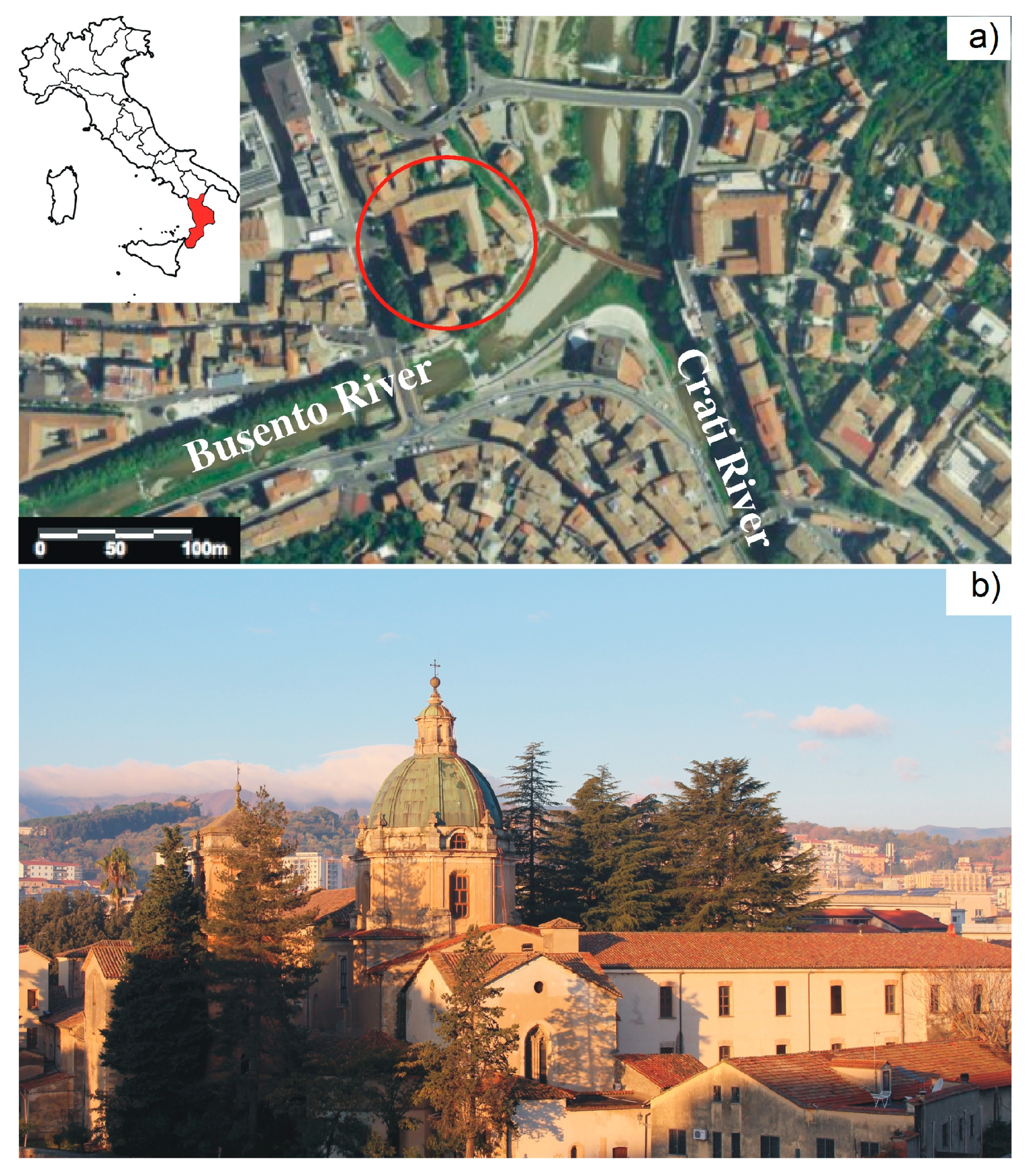
:1. Introduction and Historical Setting
2. Materials and Analytical Methods
2.1. Sampling and Macroscopic Characterization
2.2. Diagnostic Analysis
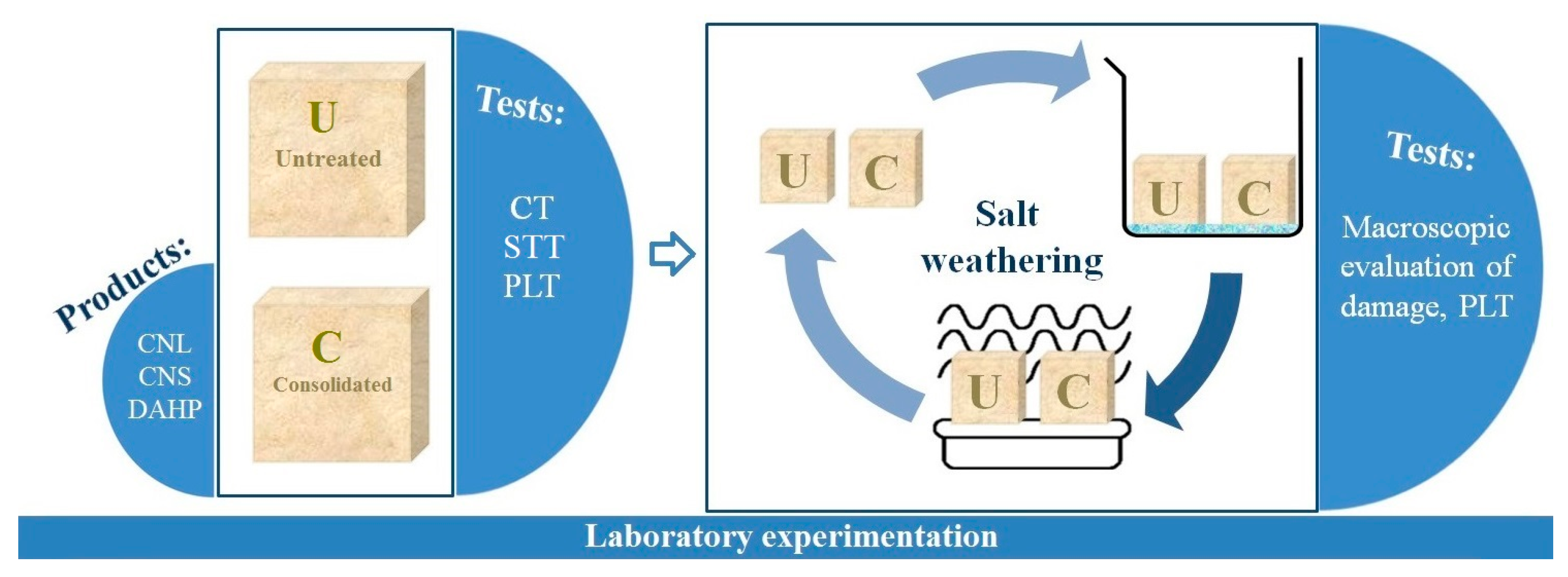
2.3. Laboratory Tests
3. Results and Discussion
3.1. Characterization of Stone Materials & Alteration Forms
3.1.1. Polarising Optical Microscopy and X-ray Diffraction Analysis
3.1.2. Fourier Transform Infrared Spectroscopy
3.1.3. EMPA-EDS Analysis
3.2. Laboratory Tests
3.2.1 Colorimetric Tests (CT)
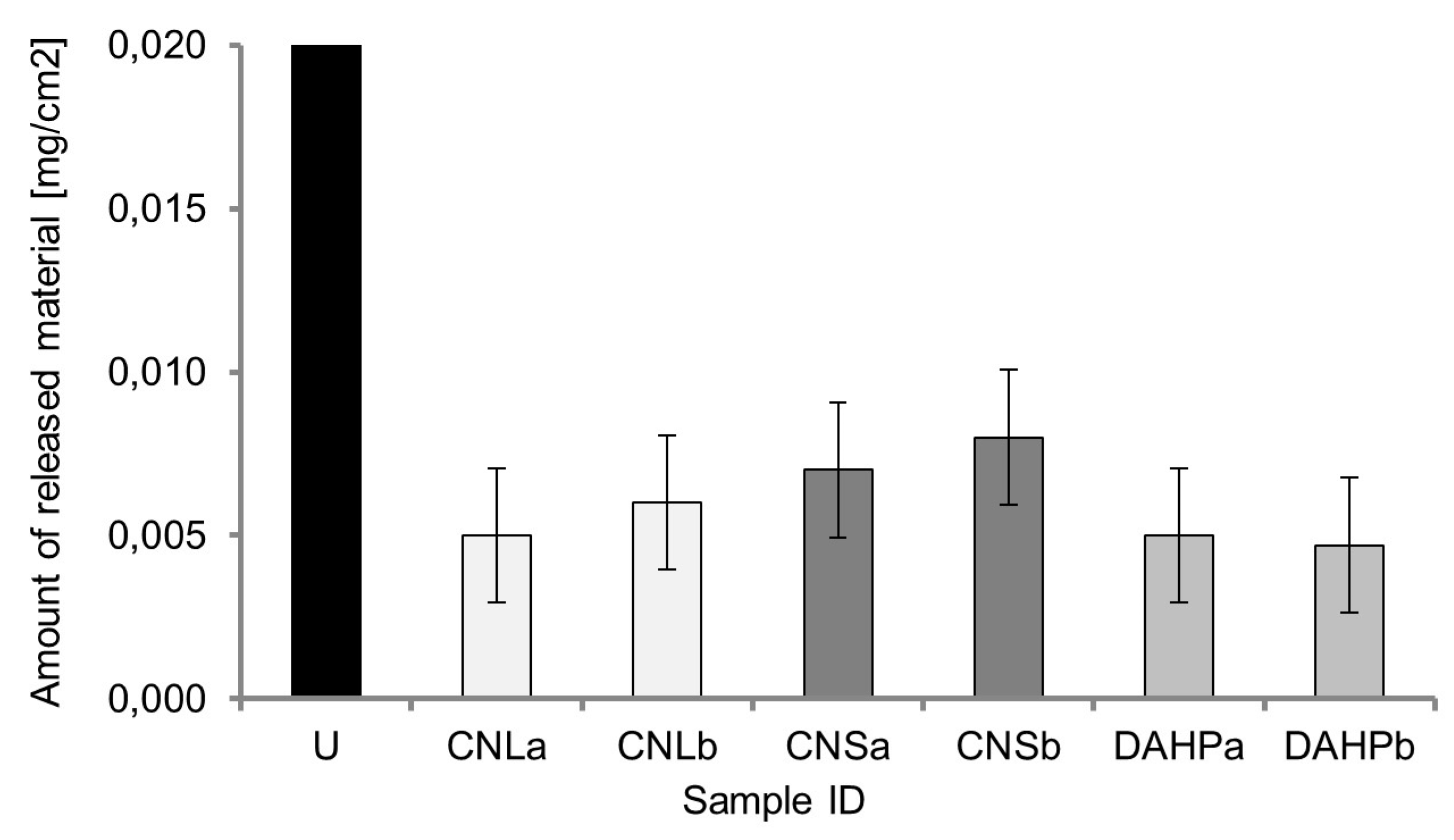
3.2.2. Scotch Tape Test (SST)
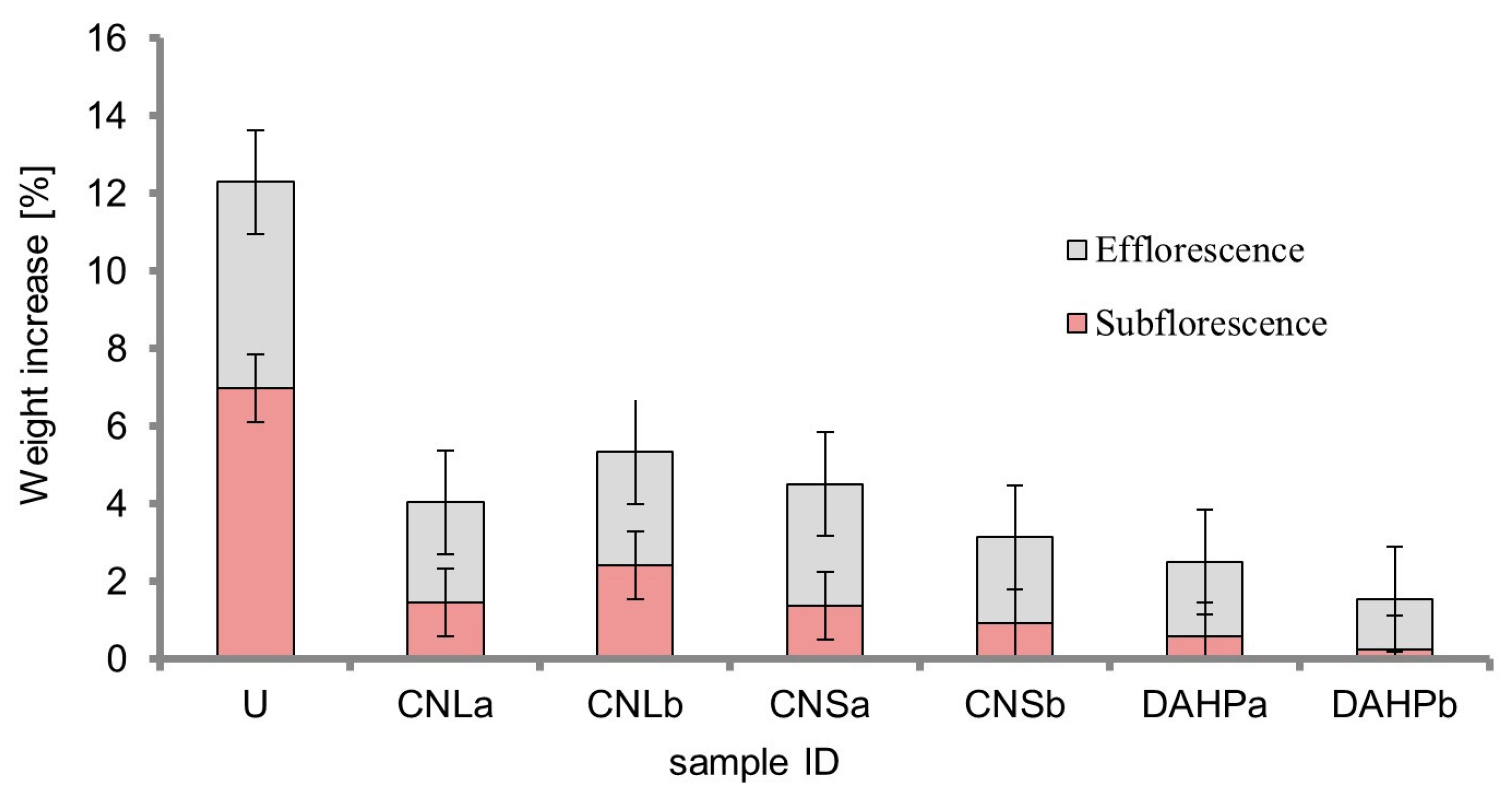
3.2.3. Salt Weathering and Consolidation Performance Tests by Point Load Test (PLT)
4. Final Remarks
Author Contributions
Funding
Conflicts of Interest
References
- La Russa, M.F.; Rovella, N.; Ruffolo, S.A.; Scarciglia, F.; Macchia, A.; Licchelli, M.; Malagodi, M.; Khalilli, F.; Randazzo, L. Consolidation of earthen building materials: A comparative study. Archaeol. Anthropol. Sci. 2019. [Google Scholar] [CrossRef]
- Ruffolo, S.A.; La Russa, M.F.; Ricca, M.; Belfiore, C.M.; Macchia, A.; Comite, V.; Pezzino, A.; Crisci, G.M. New insights on the consolidation of salt weathered limestone: The case study of Modica stone. Bull. Eng. Geol. Environ. 2017, 76, 11–20. [Google Scholar] [CrossRef]
- La Russa, M.F.; Ruffolo, S.A.; Rovella, N.; Belfiore, C.M.; Pogliani, P.; Pelosi, C.; Andaloro, M.; Crisci, G.M. Cappadocian ignimbrite cave churches: Stone degradation and conservation strategies. Period. Mineral. 2014, 83, 187–206. [Google Scholar]
- Lazzarini, L.; Laurenzi Tabasso, M. II Restauro della Pietra; Casa Editrice Dott Antonio Milan: Padova, Italy, 1986. [Google Scholar]
- Cultrone, G.; Sebastian, E. Laboratory simulation showing the influence of salt efflorescence on the weathering of composite building materials. Environ. Geol. 2008, 56, 729–740. [Google Scholar] [CrossRef]
- Rodriguez-Navarro, C.; Doehne, E.; Sebastian, E. How does sodium sulfate crystallize? Implications for the decay and testing of building materials. Cem. Concr. Res. 2000, 30, 1527–1534. [Google Scholar] [CrossRef] [Green Version]
- Benavente, D.; García del Cura, M.A.; Bernabéu, A.; Ordóńez, S. Quantification of salt weathering in porous stones using an experimental continuos partial immersion method. Eng. Geol. 2001, 59, 313–325. [Google Scholar] [CrossRef]
- Flatt, R.J. Salt damage in porous materials: How high supersaturations are generated. J. Cryst. Growth 2002, 242, 435–454. [Google Scholar] [CrossRef]
- Angeli, M.; Bigas, J.P.; Benavente, D.; Menendez, B.; Herbert, R.; David, C. Salt crystallization in pores: Quantification and estimation of damage. Environ. Geol. 2007, 52, 187–195. [Google Scholar] [CrossRef]
- Angeli, M.; Hébert, R.; Menéndez, B.; David, C.; Bigas, J.P. Influence of temperature and salt concentration on the salt weathering of a sedimentary stone with sodium sulphate. Geol. Soc. 2010, 333, 35–42. [Google Scholar]
- Rodolico, F. Le pietre delle città d’Italia, 2nd ed.; Felice Le Monnier: Firenze, Italy, 1995; pp. 427–429. [Google Scholar]
- Bruno, E. Scalpellini di Calabria—I Cantieri e le Scuole; La petite Académie: Fuscaldo Marina, Italy, 1995. [Google Scholar]
- Lico, A. Materiali Lapidei e Cave di Approvvigionamento Degli Scalpellini Roglianesi: Risorse in Calabria e Nella Provincia di Cosenza. In La Pietra, Il Mestiere e L’arte del Decorare. Storia Della Lavorazione Della Pietra Nella Provincia di Cosenza; Pellegrini Editore: Cosenza, Italy, 2015; pp. 74–91. [Google Scholar]
- Milella, O. L’architettura dei Domenicani in Storia della Calabria nel Rinascimento. In Le Arti Nella Storia; Valtieri, S., Ed.; Gangemi: Roma, Italy, 2002; pp. 549–580. [Google Scholar]
- Esposito, G.L. San Domenico di Cosenza (MD, n. s. V); Iliesi Ed.: Pistoia, Italy, 1974; p. 338. [Google Scholar]
- Crisci, G.M.; De Francesco, A.M.; Gattuso, C.; Miriello, D. Un metodo geochimico per la determinazione della provenienza di lapidei macroscopicamente omogenei. Un esempio di applicazione sui monumenti del centro storico di Cosenza. Arkos—Scienze e Restauro Dell’architettura 2003, 2, 52–59. [Google Scholar]
- Mastandrea, A.; Muto, F.; Neri, C.; Papazzoni, C.A.; Perri, E.; Russo, F. Deep-Water Coral Banks: An Example from the “Calcare di Mendicino” (Upper Miocene, Northern Calabria, Italy). Facies 2002, 47, 27–42. [Google Scholar] [CrossRef]
- Critelli, S.; Le Pera, E. Geological Map of Calabria, scale 1:330,000. In Valutazione delle Piene in Calabria. Caratteristiche Morfometriche dei Bacini della Calabria; Gabriele, S., Ed.; Rubbettino: Soveria Mannelli, Italy, 2000. [Google Scholar]
- Colella, A. Sedimentation, deformational events and eustacy in the perithyrrenian Amantea Basin: Preliminary synthesis. Giornale di Geologia 1995, 57, 179–193. [Google Scholar]
- Forestieri, G.; Tedesco, A.; Ponte, M.; Olivito, R.S. Local building stones used in Calabrian architecture: Calcarenite and sandstone of the Thyrrenian Coastal Range of Cosenza Province (Italy). In Proceedings of the XIV International Forum Le Vie dei Mercanti: World Heritage and Degradation, Smart Design, Planning and Technologies, Aversa/Naples/Capri, Italy, 16–18 June 2016. [Google Scholar]
- Weththimuni, M.L.; Licchelli, M.; Malagodi, M.; Rovella, N.; La Russa, M.F. Consolidation of bio-calcarenite stone by treatment based on diammonium hydrogenphosphate and calcium hydroxide nanoparticles. Measurement 2018, 127, 396–405. [Google Scholar] [CrossRef]
- Normal 43/93L: Misure Colorimetriche di Superfici Opache. In Raccomandazioni Normal: Alterazioni dei Materiali Lapidei e Trattamenti Conservativi: Proposte per L’unificazione dei Metodi Sperimentali di Studio e di Controllo; CNR: Roma, Italy; ICR: Roma, Italy, 1993.
- Drdàcky, M.; Lesàk, J.; Rescic, S.; Slízkovà, Z.; Tiano, P.; Valach, J. Standardization of peeling test for assessing the cohesion and consolidation characteristics of historic stone surfaces. Mater. Struct. 2013, 45, 505–520. [Google Scholar] [CrossRef]
- Standard Test Method for Determination of the Point Load Strength Index of Rock; ASTM D5731; ASTM International: West Conshohocken, PA, USA, 2002.
- Natural Stone Test Methods-Determination of Resistance to Salt Crystallization; EN 12370; European Committee for Standardization (CEN): Brussels, Belgium, 2001; pp. 108–121.
- Folk, R.L. Spectral Subdivision of Limestone Types. In M 1: Classification of Carbonate Rocks—A Symposium; Ham, W.E., Ed.; American Association of Petroleum Geologists: Tulsa, OK, USA, 1962; pp. 62–84. [Google Scholar]
- Dunham, R.J. Classification of Carbonate Rocks According to Depositional Texture. In M 1: Classification of Carbonate Rocks; Ham, W.E., Ed.; American Association of Petroleum Geologists: Tulsa, OK, USA, 1962; pp. 108–121. [Google Scholar]
- Wilson, M.J. Clay Mineralogy: Spectroscopic and Chemical Determinative Methods; Chapman & Hall: London, UK, 1994; pp. 11–67. [Google Scholar]
- Comite, V.; Fermo, P. The effects of air pollution on cultural heritage: The case study of Santa Maria delle Grazie al Naviglio Grande (Milan). EPJ Plus 2018, 133, 556. [Google Scholar] [CrossRef]
- La Russa, M.F.; Ricca, M.; Cerioni, A.; Chilos, M.G.; Comite, V.; De Santis, M.; Rovella, N.; Ruffolo, S.A. The colors of the Fontana di Trevi: An analytical approach. Int. J. Archit. Herit. 2018, 12, 114–124. [Google Scholar] [CrossRef]
- La Russa, M.F.; Comite, V.; Aly, N.; Barca, D.; Fermo, P.; Rovella, N.; Antonelli, F.; Tesser, E.; Aquino, M.; Ruffolo, S.A. Black crusts on Venetian built heritage, investigation on the impact of pollution sources on their composition. EPJ Plus 2018, 133, 370. [Google Scholar] [CrossRef]
- Barca, D.; Comite, V.; Belfiore, C.M.; Bonazza, A.; La Russa, M.F.; Ruffolo, S.A.; Crisci, G.M.; Pezzino, A.; Sabbioni, C. Impact of air pollution in deterioration of carbonate building materials in Italian urban environments. Appl. Geochem. 2014, 48, 122–131. [Google Scholar] [CrossRef]
- La Russa, M.F.; Ruffolo, S.A.; Belfiore, C.M.; Aloise, P.; Randazzo, L.; Rovella, N.; Pezzino, A.; Montana, G. Study of the effects of salt crystallization on degradation of limestone rocks. Period Miner. 2013, 82, 113–127. [Google Scholar]
- Brimblecombe, P.; Grossi, C.M. Aesthetic thresholds and blackening of stone buildings. Sci. Total Environ. 2005, 349, 175–189. [Google Scholar] [CrossRef]
- Rampazzi, L.; Andreotti, A.; Bonaduce, I.; Colombini, M.P.; Colombo, C.; Toniolo, L. Analytical investigation of calcium oxalate films on marble monuments. Talanta 2005, 63, 966–977. [Google Scholar] [CrossRef]
- Sabbioni, C.; Zappia, G. Oxalate patinas on ancient monument: the biological hypothesis. Aerobiologia 1991, 7, 31–37. [Google Scholar] [CrossRef]
- UNI 10921:2001 Beni Culturali—Materiali Lapidei Naturali ed Artificiali—Prodotti Idrorepellenti—Applicazione su Provini e Determinazione in Laboratorio delle Loro Caratteristiche; UNI: Milano, Italy, 2001.
- Witzel, R.F.; Burnham, R.W.; Onley, J.W. Threshold and suprathreshold perceptual color differences. JOSA 1973, 63, 615–625. [Google Scholar] [CrossRef]
- La Russa, M.F.; Ruffolo, S.A.; Álvarez de Buergo, M.; Ricca, M.; Belfiore, C.M.; Pezzino, A.; Crisci, G.M. The behaviour of consolidated Neapolitan yellow Tuff against salt weathering. Bull. Eng. Geol. Environ. 2017, 76, 115–124. [Google Scholar] [CrossRef]
- Benavente, D.; Martinez Martinez, J.; Cueto, N.; Garcia del Cura, M.A. Salt weathering in dual-porosity building dolostones. Eng. Geol. 2007, 94, 215–226. [Google Scholar] [CrossRef]
- Evans, I.S. Salt crystallization and rock weathering: A review. Revue de Geomorphologie Dynamique 1970, 19, 153–177. [Google Scholar]
- Benavente, D.; Garcia del Cura, M.A.; Fort, R.; Ordonez, S. Durability estimation of porous building stones from pore structure and strength. Eng. Geol. 2004, 74, 113–127. [Google Scholar] [CrossRef]
- Benavente, D.; Garcia del Cura, M.A.; Ordonez, S. Salt influence on evaporation from porous building rocks. Construct. Build. Mater. 2003, 17, 113–122. [Google Scholar] [CrossRef]
- Yang, F.; Liu, Y.; Zhu, Y.; Long, S.; Zuo, G.; Wang, C.; Guo, F.; Zhang, B.; Jiang, S. Conservation of weathered historic sandstone with biomimetic apatite. Chin. Sci. Bull. 2012, 57, 2171–2176. [Google Scholar] [CrossRef] [Green Version]
- Yang, F.; Zhang, B.; Liu, Y.; Wei, G.; Zhang, H.; Chen, W.; Xu, Z. Biomimic conservation of weathered calcareous stones by apatite. New J. Chem. 2011, 35, 887–892. [Google Scholar] [CrossRef]


| Historical Stone Samples | |||
|---|---|---|---|
| ID Sample | Description | Sampling Point | Employed Techniques |
| SD1 | Fragment of calcarenite, no macroscopic evidence of superficial alteration forms. | Prothyrum. Base of the cluster pillar, right side. | POM, XRD |
| SD2 | Fragment of calcarenite, no macroscopic evidence of superficial alteration forms. | Prothyrum. Base of the cluster pillar, right side. | POM, XRD |
| SD3 | Fragment of calcarenite, no macroscopic evidence of superficial alteration forms. | Prothyrum. First ashlar on the lower part of the cluster pillar, right side. | POM, XRD |
| SD4 | Fragment of calcarenite, macroscopic evidence of a greyish superficial layer. | Prothyrum. Base of the cluster pillar, left side. | POM, XRD, FT-IR, EMPA-EDS |
| SD6 | Fragment of calcarenite, no macroscopic evidence of superficial alteration forms. | Prothyrum. Along the intersection between the left cluster pillar and the lateral front. | POM, XRD |
| SD8 | Fragment of calcarenite, macroscopic evidence of a blackish superficial layer. | Main portal. Base of the cluster pillar, left side. | POM, XRD, FT-IR |
| SD9 | Fragment of calcarenite, macroscopic evidence of a blackish superficial layer. | Main portal. Base of the cluster pillar, left side. | POM, XRD, FT-IR, EMPA-EDS |
| SD10 | Fragment of calcarenite, macroscopic evidence of a greyish superficial layer. | Main portal. Cluster pillar, left side. | POM, XRD, FT-IR |
| SD11 | Fragment of calcarenite, macroscopic evidence of a greyish superficial layer. | Main portal. Pier, left side. | POM, XRD, FT-IR |
| SD12 | Fragment of calcarenite, macroscopic evidence of a blackish superficial layer. | Main portal. Cluster pillar, right side. | POM, XRD, FT-IR |
| SD13 | Fragment of calcarenite, macroscopic evidence of a blackish superficial layer. | Main portal. Base of the cluster pillar, right side. | POM, XRD, FT-IR |
| Sample Group ID. | Products/Formulations | Experimentation on Specimens | ||||
|---|---|---|---|---|---|---|
| I Step | II Step | III Step | ||||
| Preliminary Characterization Tests | Consolidant Treatment (g/cm2) | Consolidation Performance Tests | Salt Weathering (cycles) | Consolidation Performance Tests | ||
| CNLa | CaLoSiL® | Colorimetric Test, Scotch Tape Test, Point Load Test | 0.36 | Colorimetric Test, Scotch Tape Test, Point Load Test | 15 | Macroscopic evaluation of the damage and Point Load Test |
| CNLb | CaLoSiL® | 0.18 | 15 | |||
| CNSa | Nano Estel® | 0.32 | 15 | |||
| CNSb | Nano Estel® | 0.16 | 15 | |||
| DAHPa | Mixture of Ca(OH)2 and diammonium hydrogen phosphate | 0.32 | 15 | |||
| DAHPb | Mixture of Ca(OH)2 and diammonium hydrogen phosphate | 0.16 | 15 | |||
| U | Untreated | – | 15 | |||
| Mineralogical Phases by XRD | |||||||||||
|---|---|---|---|---|---|---|---|---|---|---|---|
| Sample | Cal | Dol | Fds | Mic | Chl | Pl | Qtz | Clay min. | Fe-ox | Gy | Wed |
| SD1 | ++++ | (-) | (-) | ++ | + | (-) | ++ | (-) | + | (-) | (-) |
| SD2 | ++++ | (-) | (-) | (-) | (-) | + | ++ | (-) | (-) | (-) | (-) |
| SD3 | ++++ | (-) | (-) | ++ | (-) | + | ++ | (-) | + | (-) | (-) |
| SD4 | ++++ | (-) | (-) | (-) | (-) | (-) | ++ | + | + | + | (-) |
| SD6 | ++++ | (-) | + | + | (-) | + | ++ | (-) | + | (-) | (-) |
| SD8 | ++++ | (-) | + | ++ | (-) | + | ++ | (-) | + | + | + |
| SD9 | ++++ | (-) | (-) | ++ | (-) | (-) | ++ | (-) | (-) | + | (-) |
| SD10 | ++++ | (-) | ++ | + | (-) | (-) | ++ | (-) | (-) | + | (-) |
| SD11 | ++++ | (-) | (-) | (-) | (-) | (-) | + | (-) | (-) | + | (-) |
| SD12 | ++++ | (-) | (-) | (-) | (-) | (-) | + | (-) | (-) | + | + |
| SD13 | ++++ | +++ | + | + | (-) | + | ++ | (-) | + | + | + |
© 2019 by the authors. Licensee MDPI, Basel, Switzerland. This article is an open access article distributed under the terms and conditions of the Creative Commons Attribution (CC BY) license (http://creativecommons.org/licenses/by/4.0/).
Share and Cite
Ricca, M.; Le Pera, E.; Licchelli, M.; Macchia, A.; Malagodi, M.; Randazzo, L.; Rovella, N.; Ruffolo, S.A.; Weththimuni, M.L.; La Russa, M.F. The CRATI Project: New Insights on the Consolidation of Salt Weathered Stone and the Case Study of San Domenico Church in Cosenza (South Calabria, Italy). Coatings 2019, 9, 330. https://doi.org/10.3390/coatings9050330
Ricca M, Le Pera E, Licchelli M, Macchia A, Malagodi M, Randazzo L, Rovella N, Ruffolo SA, Weththimuni ML, La Russa MF. The CRATI Project: New Insights on the Consolidation of Salt Weathered Stone and the Case Study of San Domenico Church in Cosenza (South Calabria, Italy). Coatings. 2019; 9(5):330. https://doi.org/10.3390/coatings9050330
Chicago/Turabian StyleRicca, Michela, Emilia Le Pera, Maurizio Licchelli, Andrea Macchia, Marco Malagodi, Luciana Randazzo, Natalia Rovella, Silvestro A. Ruffolo, Maduka L. Weththimuni, and Mauro F. La Russa. 2019. "The CRATI Project: New Insights on the Consolidation of Salt Weathered Stone and the Case Study of San Domenico Church in Cosenza (South Calabria, Italy)" Coatings 9, no. 5: 330. https://doi.org/10.3390/coatings9050330