Review

Mechanisms of the Antibacterial Effects of TiO2–FeOx under Solar or Visible Light: Schottky Barriers versus Surface Plasmon Resonance

Ecole Polytechnique Fédérale de Lausanne, EPFL-SB-GPAO Station 6, CH-1015 Lausanne, Switzerland
*
Author to whom correspondence should be addressed.
Coatings 2018, 8(11), 391; https://doi.org/10.3390/coatings8110391
Submission received: 4 October 2018 / Revised: 13 October 2018 / Accepted: 1 November 2018 / Published: 4 November 2018
(This article belongs to the Special Issue Photocatalytic Thin Films)

Abstract

:
This study reports the significant mechanistic difference between binary-oxide antibacterial films with the same composition but different microstructures. Binary TiO2-FeOx films were found to present a faster bacterial inactivation kinetics under visible light irradiation than each single oxide acting independently. The interaction between the film active surface species and the bacteria within the disinfection period was followed by X-ray photoelectron spectroscopy (XPS) and provided the evidence for a redox catalysis taking place during the bacterial inactivation time. The optical and surface properties of the films were evaluated by appropriate surface analytical methods. A differential mechanism is suggested for each specific microstructure inducing bacterial inactivation. The surface FeOx plasmon resonance transferred electrons into the conduction band of TiO2 because of the Schottky barrier after Fermi level equilibration of the two components. An electric field at the interface between TiO2 and FeOx, favors the separation of the photo-generated charges leading to a faster bacterial inactivation by TiO2–FeOx compared to the bacterial inactivation kinetics by each of the single oxides.

1. Introduction

Photocatalytic nanomaterials such as TiO2 are receiving a great deal of attention owing to their potential applications in environmental remediation [1,2]. Nonetheless, the low efficiency of this class of materials under solar irradiation, absorbing <5% of the incident light, limits their performance in photo-induced processes. At the present time, there is a need to develop more efficient TiO2 composite photocatalysts, active under visible light [3,4,5]. Hybrid nanostructured composite photocatalysts like binary oxides seem a fruitful solution. The present mini-review addresses hybrid, stable, adhesive antibacterial TiO2–FeOx films leading to visible light-driven bacterial inactivation. We describe briefly the properties of TiO2 and Fe oxides (FeOx) and address the preparation, evaluation, and properties of TiO2–FeOx films having the same composition but different microstructures.
TiO2 is known to exhibit photocatalytic antimicrobial activity over a broad spectrum of microorganisms. The antimicrobial properties of TiO2 are attributed to the high redox potential of reactive oxygen species (ROS) generated on TiO2 under band-gap irradiation. Foster et al. [6], Yadav et al. [7], Kiwi et al. [8], Kubacka et al. [9], Verdier et al. [10], Al-Hazmi et al. [11], and others have recently reported a comprehensive review of the photocatalytic disinfection properties of TiO2. The first report on bacterial sterilization by TiO2 powders was published by Matsunaga et al. [12]. TiO2 under band-gap irradiation photo-induces charge carriers that subsequently in the presence of O2 lead to ROS with high oxidative potentials. These ROS are effective in pollutant abatement, bacterial inactivation, or both. When the oxidant used exceeds the natural antioxidant defenses in a bacterial strain, death cell follows through a complex set of redox reactions. Natural antioxidants like carotene, ascorbic acid, tocopherol inhibit lipid peroxidation or O-singlet effects [13] and the effects of ROS radicals such as HO2 and OH which are effective in biological inactivation.
Iron oxide thin film have extensive applications in semiconductor devices, magneto-optic memories, audio-video systems, computer chips, and memory storage devices. Iron oxide exists in three phases: α-Fe2O3, β-Fe2O3, γ-Fe2O3 [14]. α-Fe2O3 with a band gap of 2.2 eV absorbs in the visible up to 570 nm and its charges present a low hole diffusion length and short exciton lifetime (~10 ps). Iron oxide shows antibacterial behavior [15]. ROS generated by α-Fe2O3 induce physical damage by contact leading to bacterial reduction [16,17,18]. The genotoxicity of Fe oxides is currently investigated in medical research against cancer. The oxidative damage introduced by Fe, FeOx, Fe2O3, and Fe3O4 nanoparticles (NPs) reduces or destroys cancer cells but concomitantly induces cellular injury or death in normal cells. Fe oxides lead to genetically regulated cell death (apoptosis) and to an increase of the ROS levels by damage within cells, followed by autophagy. The potential damage to tissues located behind cellular barriers needs to be considered when using Fe NPs for targeting tumors [19,20,21]. Super-paramagnetic iron oxide particles combined with certain chemicals are labelled SPIONS and increase ROS stress up to a factor of 10 during cancer cell treatment. Coupling SPIONS with X-ray radiotherapy amplifies the cytotoxicity on tumors and cancer cells. This is a synergistic strategy [22]. Because of the complexity of this field, this mini-review addresses only the interaction of binary oxides containing FeOx NPs with simpler probes like bacteria and the details related to the bacterial damage and destruction.
Fe(III)-modified titania and Fe(III)–Ti(IV) binary oxides have received little attention as photocatalyst films compared to their parent single metal oxides, Fe2O3 and TiO2. This is due to the difficulty of obtaining a pure mono-phase TiO2. In addition, the effect of different preparative conditions on films prepared by sol–gel as well as their surface properties, catalytic and photocatalytic activity have been the object of very few studies [23,24,25,26,27]. In colloidal formulations up to 10%, FeOx can be added and will disperse well in the TiO2 lattice. The addition of higher percentages of FeOx leads to Fe-phase segregation. Very little work has been reported on the detailed microstructure of TiO2–FeOx stable, uniform, adhesive films. This moved us to investigate the films microstructure effect on the bacterial inactivation kinetics. Surface properties and reaction mechanism leading to bacterial killing is reported by the binary-oxide composites.

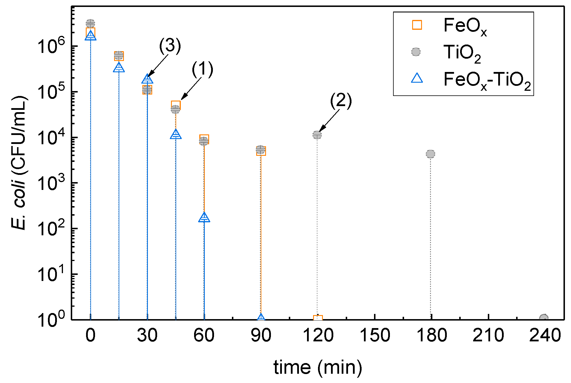
2. TiO2–FeOx Surfaces Leading to Bacterial Inactivation under Solar Light with a Faster Kinetics Compared to Either TiO2 or FeOx Films

Dispersions of FeCl3 and TiO2 Degussa P25 were prepared using FeCl3 (100 mg L−1) and TiO2 (5 g L−1) and were irradiated to photo-corrode polyethylene (PE) and introduce negatively charged oxidative sites able to bind both oxides by exchange–adsorption and electrostatic interaction. The photo-corrosion of the TiO2 powder surfaces was carried out under UV irradiation for 15 h. After UV irradiation, the films were sonicated in aqueous solution for 10 min to remove loosely bound oxide particles, washed, and dried for 10 min at 80 °C. This operation was repeated two times, and the films were dried at 60 °C. Stable TiO2–FeOx films were obtained by this procedure.
Figure 1 shows the kinetics of bacterial inactivation under low-intensity solar irradiation for TiO2, FeOx, and TiO2–FeOx. It is readily seen that the binary composite induced a faster bacterial inactivation kinetics compared to each of the single oxides evaluated separately [28]. The main reactions leading to bacterial inactivation on TiO2 are suggested below in Equations (1)–(5) [2,29]:
bacteria + [ Ti O 2 - PE ] light [ Ti O 2 * - PE ] bacteria [ bacteria * Ti O 2 - PE ] cbe  
TiO 2 ( cbe ) - PE + O 2 ads     O 2 ads E 0 0.16   NHE  
TiO 2 ( cbe ) - PE + O 2 + H + H O 2   E 0 0.05 NHE  
Ti O 2 ( vbh + ) - PE + OH ads OH   E 0 1.90 NHE  
Ti O 2 ( vbh + ) - PE + H 2 O ads OH ads + E +
Bacterial inactivation by FeOx under solar simulated light irradiation could be obtained as noted in Equations (6)–(9):
[ PE - Fe O x ] h υ [ bacteria * Fe O x ] - PE [ bacteria + + Fe O x ] - PE + cbe  
Fe O x + hv Fe O x ( vbh + )  
vbh + + H 2 O ads   OH ads + H +
cbe + H 2 O ads     OH + H +
The acceleration of bacterial inactivation in Figure 1 by the TiO2–FeOx photocatalyst can be rationalized in terms of the intervention of FeOx, injecting e into TiO2, as noted below:
FeO x   +   light     FeO x ( e )   +   FeO x ( h + )  
FeO x ( e )   +   TiO 2     FeO x   +   TiO 2 ( e trapping   sites )  
FeO x ( e )   +   O 2     FeO x   +   O 2  
O 2   +   H +     HO 2     ROS  
TiO 2 ( e trapped   sites )   +   O 2     TiO 2   +   O 2     ROS  
TiO 2 ( h + ) + bacteria CO 2 + H 2 O + inorganic residues
The mechanism of the reaction between TiO2–FeOx–PE and the bacteria under visible light proceeded noted in Equation (1). Under visible light, FeOx (mainly Fe2O3), as it will be described in the paragraph below, would transfer the photo-generated electrons in the conduction band (cbe) to the lower-lying TiO2 sites, since FeOx presents a conduction band (cb) positioned at potential energy values 0.4–0.6 eV, below the anatase trapping states [30]. Leytner et al. [31] identified the electron-trapping sites in anatase positioned at ~0.8 eV below the anatase (cb) by time-resolved photo-acoustic spectroscopy (TRPAS). Gray et al. [32] used electron paramagnetic resonance (EPR) spectroscopy and reported anatase trapping sites located ~0.5–0.8 eV below the anatase (cb). The mechanism for the interfacial charge transfer (IFCT) between Fe2O3 and TiO2 on sol–gel films is suggested in Figure 2.
It is interesting, at this point, to look into the oxidation states of the FeOx species before and after bacterial inactivation. Figure 3 presents the changes in the Fe oxidation states of PE–FeOx within the 120 min bacterial inactivation reported in Figure 1, trace (1). Figure 3 shows that the initial Fe(III)/Fe2O3 at 712.2 eV decreased from ~80.0% at time zero to ~53.0% after bacterial inactivation. Concomitantly, an increase in the Fe3O4 at 713.6 eV and Fe(II) at 709.7 eV was observed. The X-ray photoelectron spectroscopy (XPS) shifts were referenced by the values found in reference [33]. Therefore, during the bacterial inactivation period, redox reactions occurred on the catalyst surface during bacterial inactivation. After bacterial inactivation, the XPS peak positions for Fe(III), FeO(II/III), and Fe(II) peaks were: 711.4, 708.6, and 713.8 eV, respectively [34]. The three Fe-oxides in Figure 3 intervened with a different potential during bacterial reduction as shown in Figure 3. The surface concentration of the elements were observed to remain fairly stable within the period of bacterial inactivation (see Table 1).

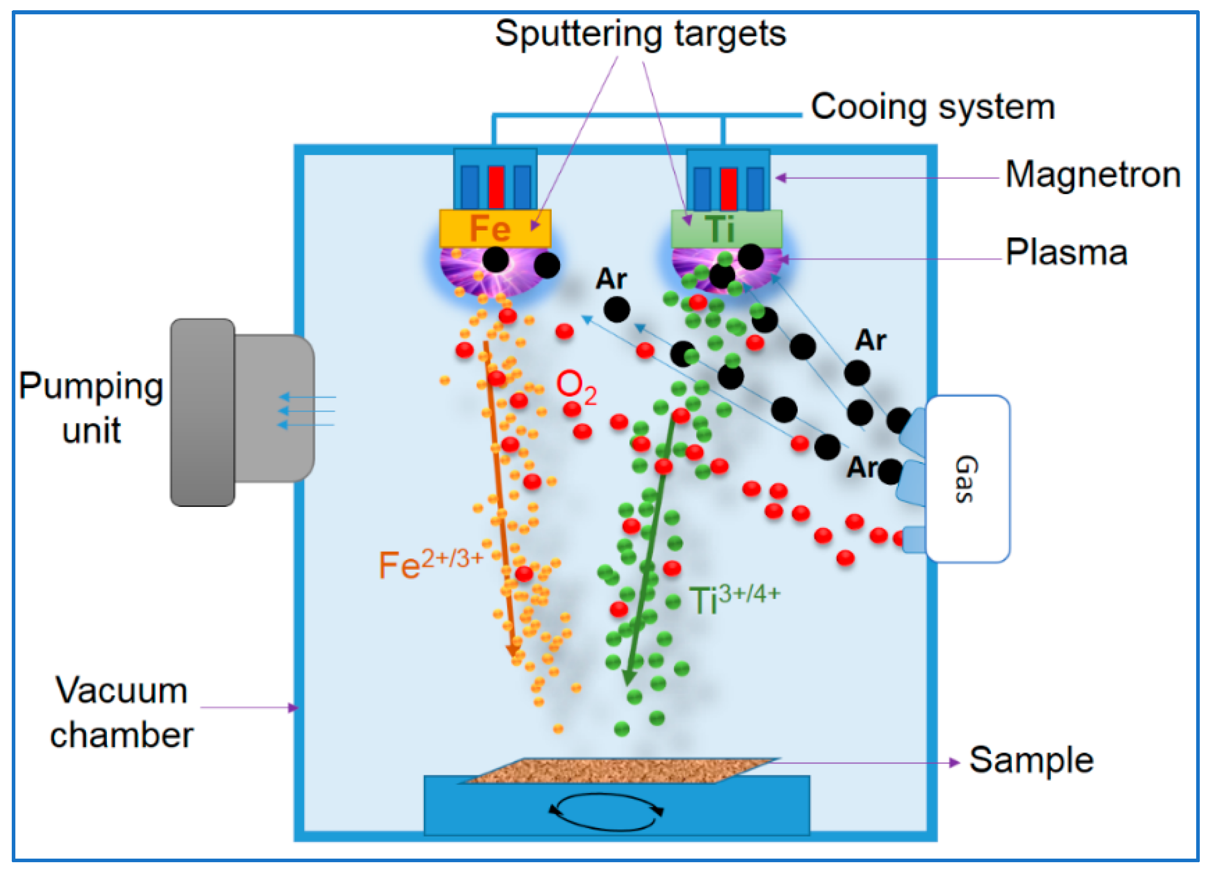
3. Sputtering of TiO2–Fe2O3 Microstructure to Accelerate the Bacterial Inactivation Kinetics: Process Optimization

Films prepared by sol–gel are not perfectly reproducible, are not robust, are weakly adhesive, and lack uniformity. This is a serious hindrance for antibacterial applications over long periods, for which film stability is a primary consideration. Work by Kelly et al. [35,36] addressed the preparation of stable antibacterial films by sputtering metals and oxides. Sputtering active metals and oxides leads to highly oxidative species, leading to bacterial death. A magnetron sputtering unit is shown in Figure 4. The amounts of the point defects in the aggregates/crystallites films on the substrate are considerably higher than in similar films prepared by sol–gel because of the higher energies during the sputtering process [37]. Defects in oxide crystals and nuclei are located at energies within the band gaps, giving raise to intermediate mid-gap energy states mediating the photo-excited electron transition from the cb band to the valence band (vb) band [38]. Alternatively, they can act as recombination centers depending on their concentration. The rapid inclusion during sputtering, of Fe and O atoms during the sputtering time into the TiO2 film lattice creates O vacancies and interstitial defects. [39]. The higher activity of the sputtered films is in part attributed to an increase in the defects in the crystallites. Oxides like TiO2 and FeOx have been extensively reported in the literature presenting four type defects: (a) O vacancies, (b) Ti or Fe vacancies, (c) O interstitials, and (d) Ti or Fe interstitials [40,41]. The work in the field of bacterial inactivation films need a more advanced catalysts design and preparation leading to a faster kinetics and a higher absorption in the visible range. Also, a higher reusability threshold is needed for antibacterial films for large scale applications. Section 4 below illustrates how the microstructure of TiO2–FeOx profoundly affects the bacterial inactivation kinetics.

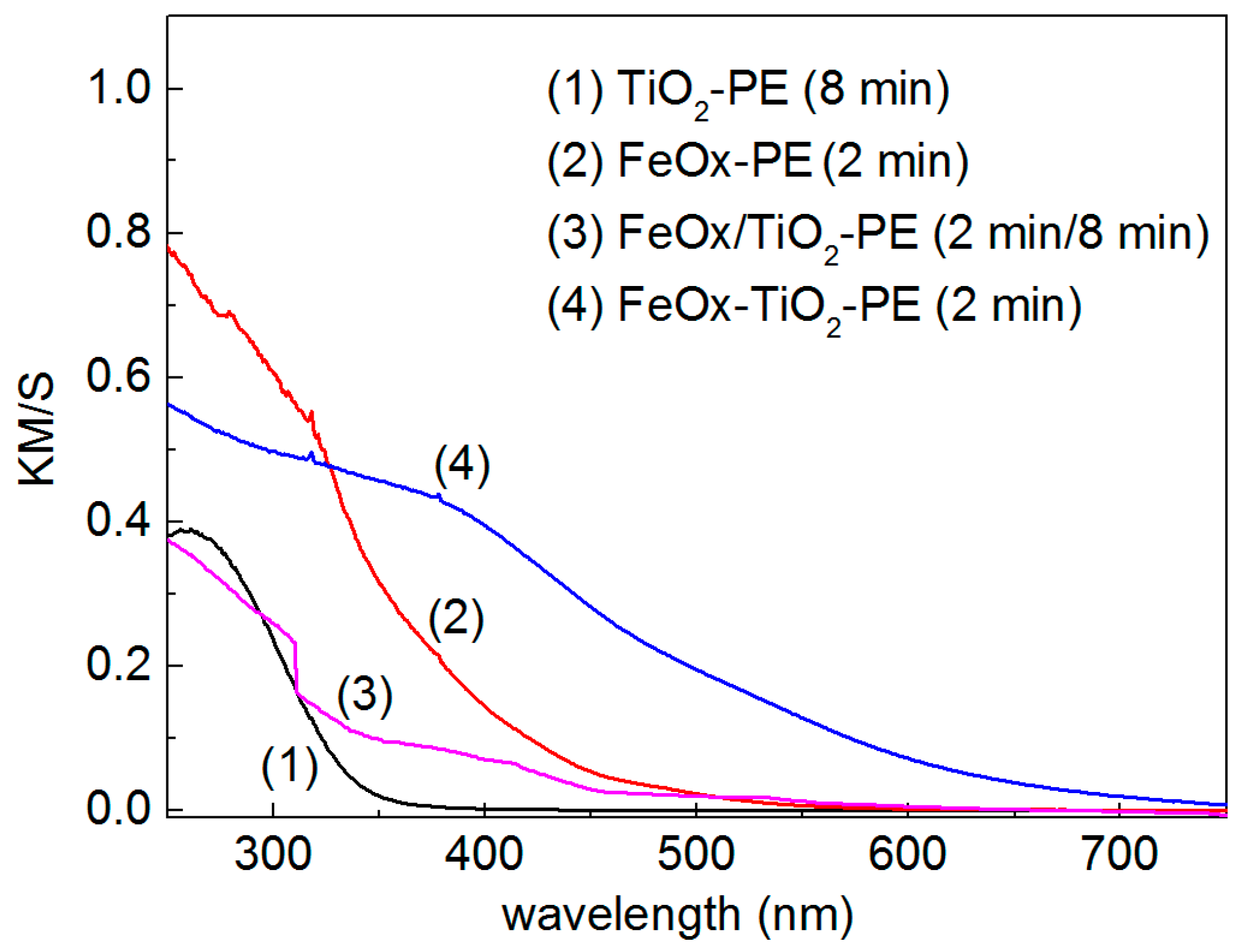
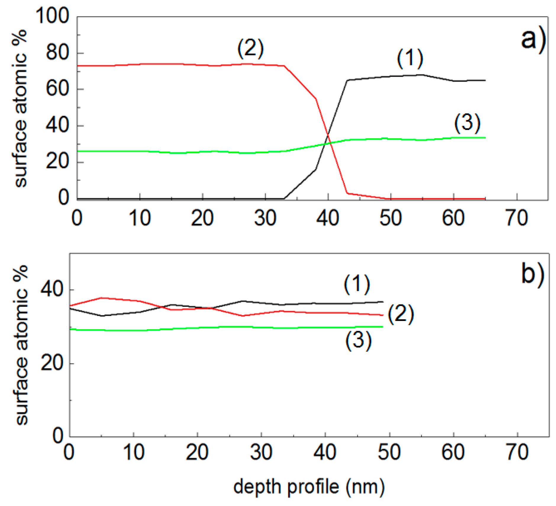
4. Optical and Surface Properties of Co-Sputtered and Sequentially Sputtered TiO2–FeOx Films Active in Bacterial Inactivation

Figure 5 shows the diffuse reflection spectroscopy (DRS) spectra in Kubelka–Munk units for TiO2–PE, FeOx–PE, (a) sequentially sputtered TiO2/FeOx–PE films, and (b) co-sputtered TiO2–FeOx–PE films. The sputtering times noted in the caption of Figure 5 were optimized to find the most suitable ratio TiO2/FeOx for a film leading to bacterial inactivation kinetics. The TiO2–FeOx composite absorbs in the visible region >400 nm, inducing TiO2–FeOx charge transfer bands [42]. The light absorption in the spectral region between 400 and 500 nm in Figure 5 is attributed to IFCT between TiO2 and FeOx. The weak absorption >500 nm is due to the short-lived Fe d–d inter-band transitions. The electron pair-deficient oxygen vacancy was suggested to be able to react with Ti4+-ions to form Ti3+ centers, by Serpone et al. [43]. The amount of vacancies was reported to be one-half of the Fe(III) found in the TiO2 (Ti4+) network [44,45,46,47,48,49].
The roughness (Rg) of the co-sputtered TiO2–FeOx–PE films was ~24 nm, as determined by atomic force microscopy (AFM). A value of ~11 nm was found for the sequentially sputtered TiO2/FeOx–PE films. The co-sputtered films showed FeOx nano-particle sizes of 15–30 nm and TiO2 nano-particle sizes of 10–15 nm. The particle size, diffusion, and mass transport determine particle growth and surface roughness [50]. The sequential sputtered films showed FeOx NPs sizes of 20–40 nm. The bigger size of the sequentially sputtered NPs compared to the co-sputtered films is attributed to an easier collective diffusion in the FeOx top-most layers of the latter samples [51]. The deposition of the co-sputtered TiO2–FeOx–PE films and the sequentially sputtered TiO2/FeOx–PE films was carried in a similar way. Bacterial inactivation mediated by the co-sputtered TiO2–FeOx–PE sample was completed within 60 min compared with the 120 min required by the sequentially sputtered samples. Both samples were sputtered for 2 min. Both samples were close in specific surface area (SSA) and that the difference in the bacterial inactivation times could not be ascribed to a difference in the surface area between the samples. The photo-sensitizing role of FeOx unexpectedly led to similar inactivation times under solar simulated light and under visible light in both samples (404 nm cut-off filter).

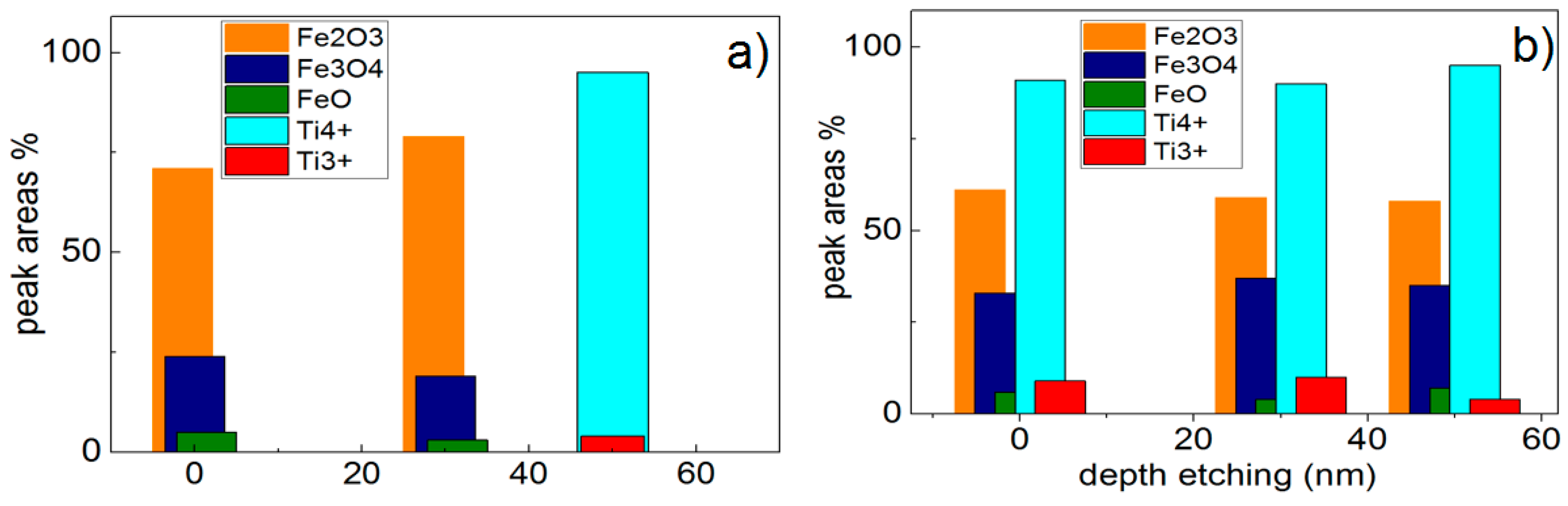
5. Evidence by XPS of Bacterial Inactivation Inducing Differentiated Redox Interactions with TiO2–FeOx Samples

Figure 6a presents the changes in the Fe oxidation states for sequentially sputtered TiO2/FeOx–PE films. The initial Fe2O3 was seen to increase from ~70% at time zero to ~80% after 30 min at the expense of Fe3O4 and FeO. The Fe oxide peaks were referenced by the values reported in reference [33]. Figure 6b shows the changes of FeOx and TiO2 oxidation states in the co-sputtered films within the disinfection time. The initial 60% Fe2O3 percentage remained constant during the disinfection time, and the Fe3O4 and FeO percentages were conserved up to 60 min, while TiO2 (Ti4+) slightly increased with a concomitant decrease of Ti3+-oxidation state. Electrostatic attraction occurs between the negatively charged Escherichia coli at pH 6–7 and the slightly positive TiO2–FeOx–PE surface. The interaction between reactants at distances below 4–8 Å is accompanied by a strong polarization at these short distances [52,53]. Fe2O3 presents a cb at +0.1 eV and a vb at +2.2 eV [1,2]. The valence band holes (vbh+) interact with the adsorbed –OH surface groups but do not have a potential high enough to lead to the formation of OH radicals, since the transformation OH–OH requires 1.90 eV. The HO2 radicals oxidize bacteria undergoing concomitantly HO2–HO2 reduction at 0.75 eV. This is a significant lower potential compared to that required by the transformation OH–OH. The HO2 decomposes at pH > 4.8, driving the pH to acidic values, as shown below in Equation (16):
HO 2 +   Fe 3 + Fe 2 + +   O 2 +   H +  
Figure 7a presents the XPS for the sequentially sputtered films. Figure 7a shows the atomic percentage composition as a function of the etching depth for Fe, Ti, and O. The etching of the film surface was carried out by sputtering Ar ions of 5 kV. These Ar ions are able to reach a depth of ~50 nm (~250 layers). The TiO2 under-layers in Figure 7 were only detected up to 30 nm and reached an atomic concentration of 60% after sputtering Ar ions for 45 nm. The O-enrichment level was stable at 25%–30% up to 65 nm. Figure 7b shows that the surface atomic percentage of the Ti and Fe layers was similar within 50 nm (250 atomic layers). The amount of O in the surface was close to those of TiO2 and FeOx at ~30%. Figure 7a,b show the drastic differences in the microstructure of the TiO2–FeOx films for the sequential and co-sputtered samples.

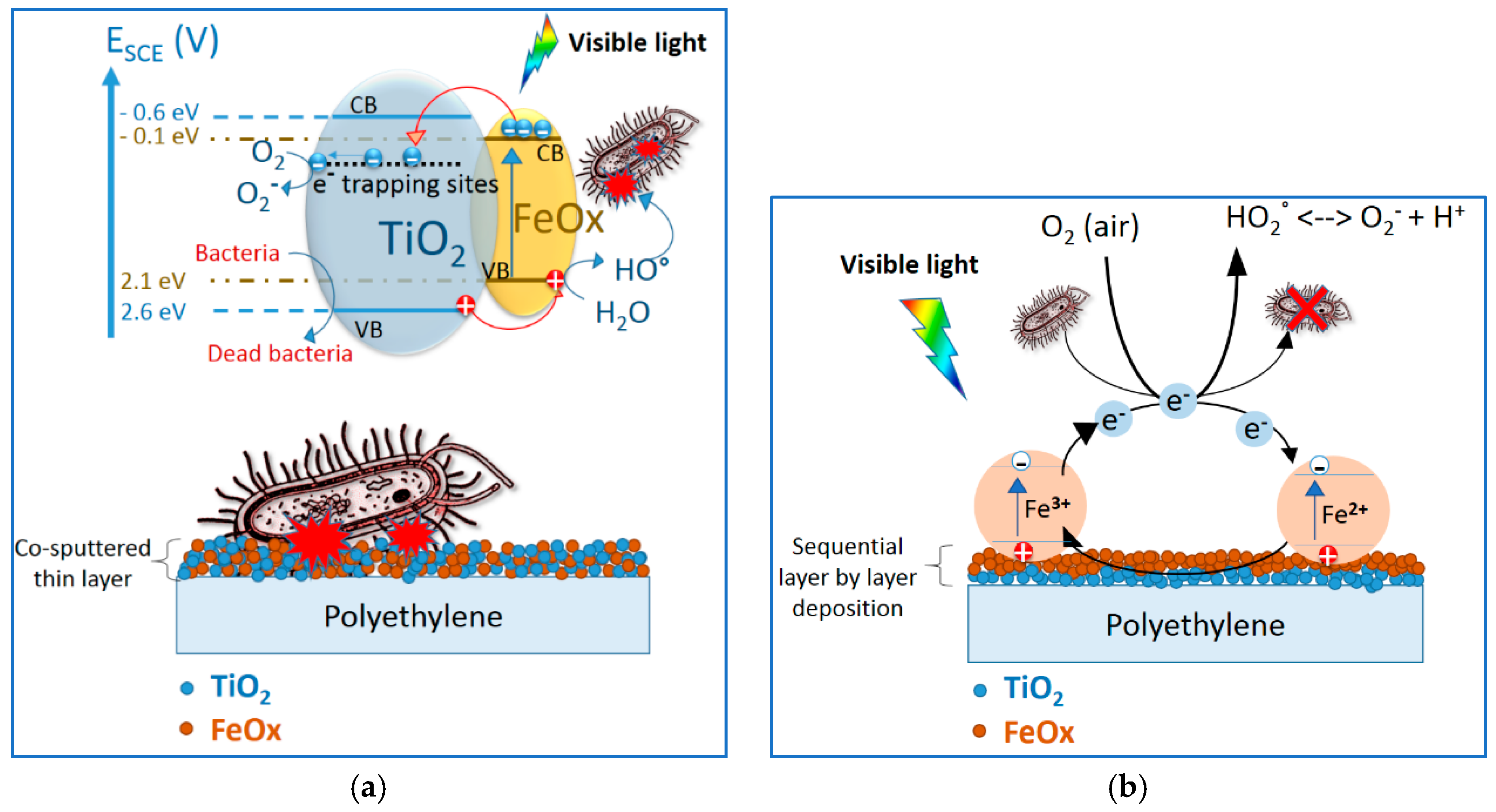
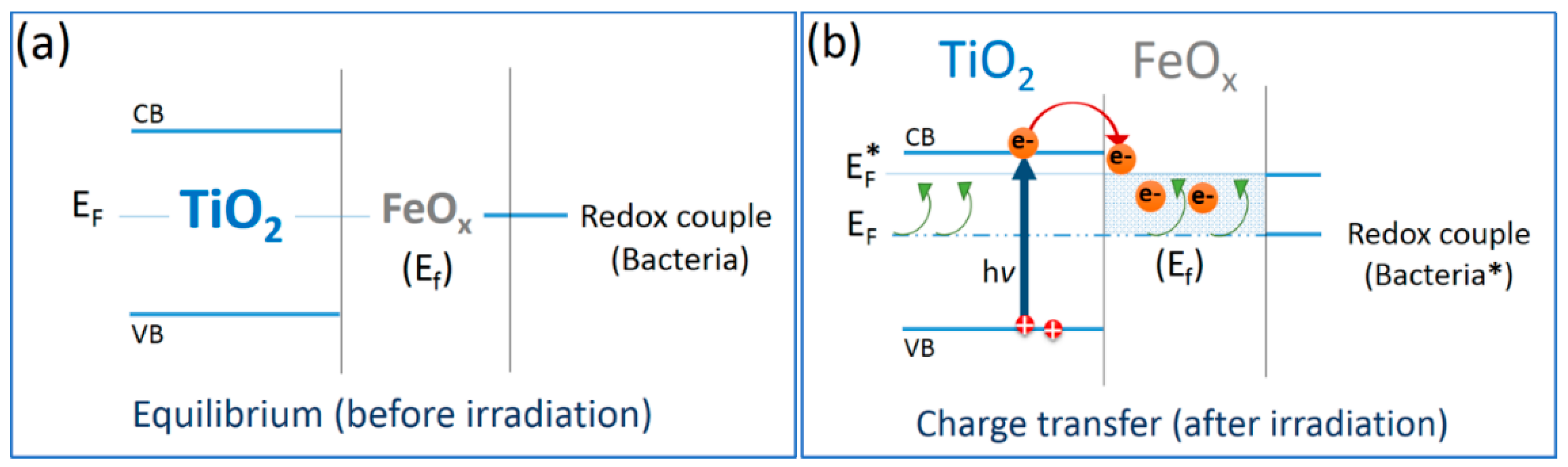
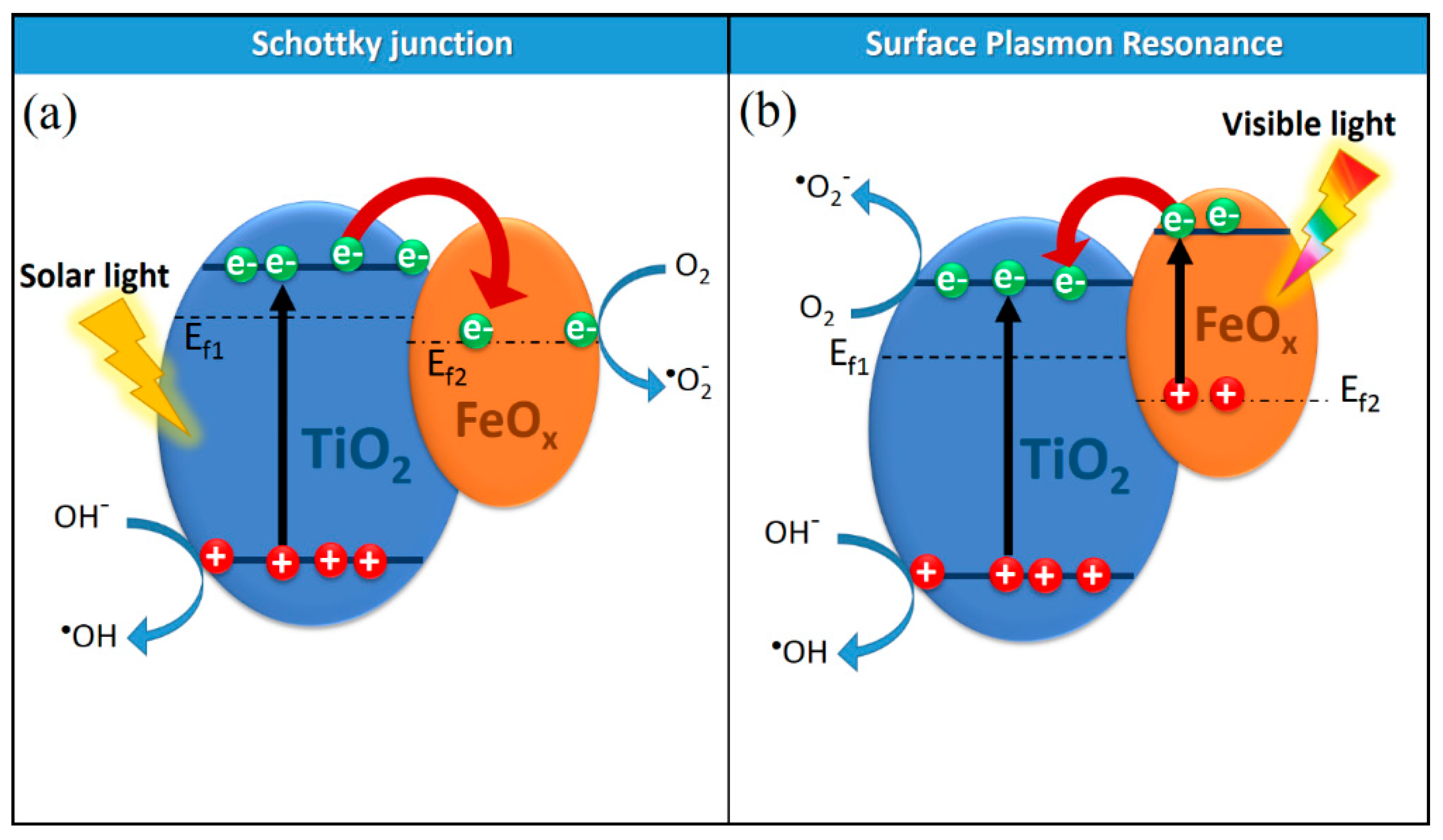
6. The Role of the Microstructure Controlling the Bacterial Inactivation Mechanism: Critical Issues

The schematic intervention of co-sputtered FeOx–TiO2 leading to bacterial inactivation is suggested in Figure 8a. The mixed TiO2–FeOx–PE film led to charge separation and involved quasi-Fermi equilibration between the two oxides. The FeOx electron transfer to low-lying TiO2 trapping states is shown in Figure 8a. The co-sputtered TiO2–FeOx–PE films induced a faster bacterial inactivation compared to the sequentially sputtered TiO2/FeOx–PE. A decrease in the film FeOx electron-hole recombination rate occurred in the co-sputtered TiO2-FeOx films. Some steps involving the transfer of Fe2O3 cb (e) into the TiO2 trapping sites are noted below:
Fe O x +   visible   light FeO x ( e )   +   FeO x ( h + )  
FeO x ( e )   +   TiO 2 FeO x +   TiO 2 ( e trapping-sites )  
TiO 2 ( h + )   +   bacteria inorganic / organic   residues  
In the sequentially sputtered TiO2/FeOx–PE film as shown in Figure 8b, the FeOx topmost layers absorbed the visible light reaching the sample surface [36,39,54]. The incorporation of Fe oxides into the crystal lattice of wide band-gap semiconductors (such as TiO2) improved the photocatalytic activity of TiO2 in the visible-light region because of the surface plasmon resonance (SPR) effect of FeOx [55,56]. The surface plasmon resonance arises from the collective oscillations of electrons on the surfaces of metals and oxides, and these surface transfer electrons into the conduction band of TiO2 because of the Schottky barrier. The Schottky barrier between FeOx and TiO2 arises as a consequence of the Fermi level equilibration between TiO2 and FeOx. This favors the separation of the photo-generated charges, as noted in Figure 9 [57,58].
Figure 10a shows that the TiO2 nanocomposites irradiated with solar energy photons, having energy higher than the TiO2 band gap, photo-excited electrons from the valance band to the conduction band, leaving holes in the valance band. Figure 10b shows the IFCT at the TiO2-FeOx heterojunction under visible light. The close contact between FeOx NPs and TiO2 in the sputtered films acts as an electron sink to promote the reduction of oxygen on their surfaces. Subsequently, the holes in the valence band of TiO2 migrated, inducing bacterial oxidation. A local electric field developed by the SPR of FeOx in contact with TiO2. The increased charge separation, due to the FeOx NPs sputtered on TiO2, increased the lifetime of the TiO2 charge-carriers. This was due to FeOx partly substituting the lattice Ti4+ sites in TiO2, which modifies the visible light absorbance of TiO2. Takeuchi [59] has recently reported metal implantation on TiO2 films, increasing the film photocatalytic activity. Figure 8b consisting only of FeOx NPs (in the TiO2/FeOx–PE film) is a film made up by a single component. In this case, a faster charge recombination of the photo-induced charges occurs, limiting the amount of charges available for the photocatalytic reactions leading to bacterial inactivation [60].

7. Conclusions

This work describes the modification strategies of TiO2 to prepare more performing binary oxides employable in photocatalysis. Basic concepts related to the surface modification of TiO2 by FeOx are discussed. Further, this review suggests basic mechanisms for photo-chemical processes as a function of the film microstructure. The heterojunction between FeOx and TiO2 promotes a directional electron flow in the co-sputtered films, leading to a faster bacterial inactivation. FeOx and TiO2 deposition in the films follow a random distribution. The photochemical intervention in bacterial inactivation processes were a function of the sputtering time and applied sputtering energy. The redox reactions taking place during bacterial inactivation were monitored by XPS within the disinfection time. The co-sputtered FeOx–TiO2–PE films were shown to lead to a faster bacterial inactivation kinetic. These films show the potential to prevent biofilm formation under sun or visible light. This mini-review may be useful to orient the work on low-cost, stable TiO2-films for pollutants degradation and bacterial inactivation with enhanced absorption in the visible region.

Author Contributions

S.R. carried out the sputtering, biological evaluation, and surface characterization of the films. J.K. oriented the content of the work and the layout of the final write-up. Both authors approved the submitted manuscript and are accountable for the material presented.

Funding

This review received no external funding.

Acknowledgments

The authors thank the financial support of the EPFL that made possible the studies described in this mini-review.

Conflicts of Interest

The authors declare no conflict of interest.

Abbreviations

cbconduction band
vbvalence band
cbephoto-generated electrons in the conduction band
vbh+photo-generated holes in the valence band
FeOxiron oxides
TiO2titanium dioxide
PEpolyethylene
TiO2/FeOx–PEsequentially sputtered TiO2 followed by FeOx deposition
TiO2–FeOx–PEco-deposition of TiO2 and FeOx (at the same time)
EPRelectron paramagnetic resonance
IFCTinterfacial charge transfer
ROSreactive oxygen species
XPSX-ray Photo-electron Spectroscopy
Rgroughness
DRSdiffuse reflectance spectroscopy
SSAspecific surface area
TRPAStime-resolved photo-acoustic spectroscopy

References

  1. Byrne, J.A.; Dunlop, P.S.M.; Hamilton, J.W.J.; Fernandez-Ibanes, P.; Polo-Lopez, I.; Sharma, K.P.; Vennard, M.S.A. A review of heterogeneous photocatalysis for water and surface disinfection. Molecules 2015, 20, 5574–5615. [Google Scholar] [CrossRef] [PubMed]
  2. Rtimi, S.; Sanjines, R.; Pulgarin, C.; Kulik, A.; Kiwi, J. Innovative transparent non-scattering TiO2 bactericide films inducing increased E. coli cell fluidity. Surf. Coat. Technol. 2014, 254, 333–343. [Google Scholar] [CrossRef]
  3. Pelaez, M.; Nolan, N.T.; Pillai, S.C.; Seery, M.K.; Falaras, P.; Kontos, A.G.; Dunlop, P.S.M.; Hamilton, J.W.J.; Byrne, A.J.; O’Shea, K.; et al. A review on the visible light active titanium dioxide photocatalysts for environmental applications. Appl. Catal. B 2012, 125, 331–349. [Google Scholar] [CrossRef] [Green Version]
  4. Banerjee, S.; Pillai, C.S.; Falaras, P.; O’Shea, K.; Byrne, A.-J.; Dionysiou, D. New insights into the mechanism of visible light photocatalysis. J. Phys. Chem. Lett. 2014, 5, 2543–2554. [Google Scholar] [CrossRef] [PubMed] [Green Version]
  5. Banerjee, S.; Dionysiou, D.; Pillai, C.S. Self-cleaning applications of TiO2 by photo-induced hydrophilicity and photocatalysis. Appl. Catal. B 2015, 176–177, 396–428. [Google Scholar] [CrossRef]
  6. Foster, H.; Ditta, I.; Varghese, S.; Steele, A. Photocatalytic disinfection using titanium dioxide: Spectrum and mechanism of antimicrobial activity. Appl. Microbiol. Biotechnol. 2011, 90, 1847–1868. [Google Scholar] [CrossRef] [PubMed]
  7. Yadav, H.; Kiwi, J.; Pawar, S.H. Development in photocatalytic antibacterial activity of nano TiO2: A review. Korean J. Chem. 2016, 33, 1989–1998. [Google Scholar] [CrossRef]
  8. Rtimi, S.; Pulgarin, C.; Kiwi, J. Recent developments in accelerated antibacterial inactivation on 2D Cu-titania surfaces under indoor visible light. Coatings 2017, 7, 20. [Google Scholar] [CrossRef]
  9. Kubacka, A.; Diez, M.; Rojo, D.; Ciordia, S.; Zapico, I.; Albar, J.; Barbas, C.; Martins dos Santos, V.; Fernandez-Garrcia, M.; Ferrer, M. Understanding the Antimicrobial mechanism of TiO2 based nano-composite films in a pathogenic bacterium. Sci. Rep. 2014, 4, 4134–4143. [Google Scholar] [CrossRef] [PubMed]
  10. Verdier, T.; Coutand, M.; Bertron, A. Antibacterial activity of TiO2 photocatalyst alone or in coatings on E. coli: The influence of methodological aspects. Coatings 2014, 4, 670–686. [Google Scholar] [CrossRef]
  11. Alhadrami, H.A.; Al-Hazmi, F. Antibacterial activities of titanium oxide nanoparticles. J. Bioelectron. Nanotechnol. 2017, 2, 5. [Google Scholar]
  12. Matsunaga, T.; Tomoda, R.; Nakajima, T.; Wake, H. Photoelectrochemcal sterilization of microbial cells by semiconductor powders. FEMS Microbiol. Lett. 1985, 29, 211–214. [Google Scholar] [CrossRef]
  13. Jones, D.P. Redefining oxidative stress. Antioxid. Redox Signal. 2006, 8, 1865–1879. [Google Scholar] [CrossRef] [PubMed]
  14. Yogi, A.; Varshney, D. Magnetic and structural properties of pure and Cr-doped hematite: α-Fe2−xCrxO3 (0 ≤ x ≤ 1). J. Adv. Ceram. 2013, 2, 360–369. [Google Scholar] [CrossRef]
  15. Basnet, P.; Larsen, G.K.; Jadeja, R.P.; Hung, Y.C.; Zhao, Y. α-Fe2O3 nano-columns and nano-rods fabricated by electron beam evaporation for visible light photocatalytic and antimicrobial applications. ACS Appl. Mater. Inter. 2013, 5, 2085–2095. [Google Scholar] [CrossRef] [PubMed]
  16. Zhang, W.; Rittmann, B.; Chen, Y. Size effects on adsorption of hematite nanoparticles on E. coli cells. Environ. Sci. Technol. 2011, 45, 2172–2178. [Google Scholar] [CrossRef] [PubMed]
  17. Ubale, A.U.; Belkehdkar, M.R. Size dependent physical properties of nanostructured α-Fe2O3 thin films grown by successive ionic-layer deposition and reaction method for antibacterial application. J. Mater. Sci. Technol. 2015, 31, 1–9. [Google Scholar]
  18. Jana, T.K.; Pal, A.; Mandal, A.K.; Sarwar, S.; Chakrabarti, P.; Chatterjee, K. Photocatalytic and antibacterial performance of α-Fe2O3 nanostructures. ChemistrySELECT 2017, 2, 3068–3077. [Google Scholar]
  19. Golbamaki, N.; Rasuley, B.; Cassano, A.; Robinson, N.; Benfebati, E.; Leszczynski, J.; Cronin, T. Genotoxicity of metal oxide nanomaterials: Review of recent data and discussion of possible mechanisms. Nanoscale 2015, 14, 2154–2198. [Google Scholar] [CrossRef] [PubMed]
  20. Bhabra, B. Nanoparticles can cause DNA damage across a cellular barrier. Nat. Nanotechnol. 2009, 4, 876–883. [Google Scholar] [CrossRef] [PubMed]
  21. Schulz, E.; Wenzel, P.H.; Munzel, T.H.; Daiber, A. Mitochondrial redox signaling: Interaction of mitochondrial reactive oxygen species with other sources of oxidative stress. Antioxid. Redox Signal. 2014, 20, 308–324. [Google Scholar] [CrossRef] [PubMed]
  22. Klein, S.; Sommer, A.; Distel, V.; Hazeman, L.; Kroner, W.; Neuhuber, W.; Muller, P.; Proux, O.; Kryshxhi, C. Superparamagnetic iron oxide nanoparticles as novel X-ray enhancer for low-dose radiation therapy. J. Phys. Chem. B 2014, 118, 6159–6166. [Google Scholar] [CrossRef] [PubMed]
  23. Wachs, I. Recent conceptual advances in the catalysis science of mixed metal oxide catalytic materials. Catal. Today 2005, 100, 79–94. [Google Scholar] [CrossRef]
  24. Pal, B.; Maheshwar, S.; Gyoichi, N. Preparation and characterization of TiO2/Fe2O3 binary mixed oxides and its photocatalytic properties. Mater. Chem. Phys. 1999, 59, 254–261. [Google Scholar] [CrossRef]
  25. Rong, L.; Jia, Y.; Bu, N.; Wu, J.; Zhen, Q. Photocatalytic degradation of methyl blue using Fe2O3/TiO2 composite ceramics. J. Alloy. Compd. 2015, 643, 88–93. [Google Scholar]
  26. Tung, W.; Daoud, W. New approach toward nanozised ferrous ferric oxide and Fe3O4-doped titanium dioxide photocatalysts. Appl. Mat. Interfaces 2009, 11, 2453–2461. [Google Scholar] [CrossRef] [PubMed]
  27. Lazar, M.; Daoud, W. Achieving selectivity in TiO2-based photocatalysis. RSC Adv. 2013, 3, 4130–4135. [Google Scholar] [CrossRef]
  28. Camarasa-Mena, A.; Rtimi, S.; Pulgarin, C.; Lavanchy, C.-J.; Kiwi, J. Grafted semiconductors on PE-films leading to bacterial inactivation: Synthesis characterization and mechanism. Colloids Surf. A 2017, 519, 231–237. [Google Scholar] [CrossRef]
  29. Wardman, P. Reduction potentials of one electron couples involving free radicals in aqueous solutions. J. Phys. Chem. Ref. Data 1989, 18, 1637–1755. [Google Scholar] [CrossRef]
  30. Fujishima, A.; Zhang, X.; Tryk, D. TiO2 photocatalysis and related surface phenomena. Surf. Sci. Reps. 2008, 63, 515–546. [Google Scholar] [CrossRef]
  31. Leytner, S.; Hupp, T. Evaluation of the energetics of electron trap states at the nano-crystalline titanium dioxide/aqueous solution interface via time-resolved photo-acoustic spectroscopy. Chem. Phys. Lett. 2000, 330, 231–236. [Google Scholar] [CrossRef]
  32. Hurum, C.; Agrios, A.; Gray, K.; Rajh, T.; Thurnauer, M. Explaining the enhanced photocatalytic activity of Degussa P25 mixed-phase TiO2 using EPR. J. Phys. Chem. B 2003, 107, 4545–4549. [Google Scholar] [CrossRef]
  33. Wagner, D.; Riggs, M.; Davis, E.; Mullenberg, G. Handbook of X-ray Photoelectron Spectroscopy; PerkinElmer: Eden Prairie, MN, USA, 1979. [Google Scholar]
  34. Kelly, P.; Li, H.; Benson, P.; Whitehead, K.; Verran, J.; Arnell, R.; Iordanova, I. Comparison of the tribological and antimicrobial properties of CrN/Ag, ZrN/Ag TiN/Ag and Tin/Cu nanocomposite coatings. Surf. Coat. Technol. 2010, 205, 1606–1610. [Google Scholar] [CrossRef]
  35. Rtimi, S.; Pulgarin, C.; Sanjines, R.; Kiwi, J. Novel FeOx polyethylene transparent films: Synthesis and mechanism of surface regeneration. RSC Adv. 2015, 5, 80203–80211. [Google Scholar] [CrossRef]
  36. Fisher, L.; Ostovapour, S.; Kelly, P.; Whitehead, K.; Cooke, K.; Storgards, E.; Verran, J. Molybdenum doped titanium oxide photocatalytic coating; for use as hygienic surfaces: The effect of soiling on antimicrobial activity. Biofouling 2014, 30, 911–919. [Google Scholar] [CrossRef] [PubMed]
  37. Mathews, W.J. Nucleation of Thin Films. In Epitaxial Growth, 1st ed.; Venables, J., Price, G., Eds.; Elsevier: New York, NY, USA, 1975; pp. 382–435. [Google Scholar]
  38. Koster, G.; Huijben, M.; Rijnders, G. Epitaxial Growth of Complex Metal Oxides; Elsevier: Burlington, VT, USA, 2015. [Google Scholar]
  39. Rtimi, S.; Sanjines, R.; Kiwi, J.; Pulgarin, C.; Bensimon, M.; Khmehl, I.; Nadtochenko, V. Innovative photocatalyst (FeOx-TiO2): Transients induced by femtosecond laser pulse leading to bacterial inactivation under visible light. RSC Adv. 2015, 5, 101751–101759. [Google Scholar] [CrossRef]
  40. Bak, T.; Nowotny, J.; Nowotny, M. Defect Disorder of Titanium Dioxide. J. Phys. Chem. B 2006, 110, 21560–21567. [Google Scholar] [CrossRef] [PubMed]
  41. Kremenović, A.; Antić, B.; Blanuša, J.; Čomor, M.; Colomban, P.H.; Mazerolles, L.; Bozin, E. Heterogeneity and disorder in Ti1-xFeyO2-d nanocrystal rutile-based flower like aggregates: Detection of anatase. J. Phys. Chem. C 2011, 115, 4395–4403. [Google Scholar] [CrossRef]
  42. Yu, H.; Irie, H.; Shimodaira, Y.; Hisogi, Y.; Kuroda, Y.; Miyauchi, M.; Hashimoto, K. An efficient visible-light-sensitive Fe(III)-grafted TiO2 photocatalyst. J. Phys. Chem. C 2010, 114, 16481–16486. [Google Scholar] [CrossRef]
  43. Kusnetsov, N.V.; Serpone, N. Visible light absorption by various titanium dioxide specimens. J. Phys. Chem. B 2006, 110, 25203–25209. [Google Scholar] [CrossRef] [PubMed]
  44. Etacheri, V.; Di Valentin, C.; Schneider, J.; Bahnemann, D.; Pillai, C.S. Visible-light activation of TiO2 photocatalysts: Advances in theory and experiments. J. Photochem. Photobiol. C Rev. 2015, 25, 1–29. [Google Scholar] [CrossRef]
  45. Novabpour, P.; Ostorvapour, S.; Tattershall, C.; Cooke, K.; Kelly, P.; Verran, J.; Whitehead, K.; Hill, C.; Raulio, M.; Priha, O. Photocatalytic TiO2 and doped TiO2 coatings to improve the hygiene of surfaces in food and beverage processing—A study of the physical and chemical resistance of the coatings. Coatings 2014, 4, 433–449. [Google Scholar] [CrossRef]
  46. Truppi, A.; Petronella, F.; Placido, T.; Striccoli, M.; Agostiano, A.; Curri, M.L.; Comparelli, R. Visible light active TiO2 based hybrid nanocatalyst for environmental applications. Catalysts 2017, 7, 100. [Google Scholar] [CrossRef]
  47. Shakeri, A.; Yip, D.; Badv, M.; Imani, S.; Sanjari, M.; Didar, T. Self-cleaning ceramic tiles produced via stable coatings of TiO2 nanoparticles. Materials 2018, 11, 1003. [Google Scholar] [CrossRef] [PubMed]
  48. Wen, K.; Liu, M.; Liu, X.; Deng, C.; Zhou, K. Deposition of photocatalytic coating by modifying the solidification pathway in plasma spraying. Coatings 2017, 7, 169. [Google Scholar] [CrossRef]
  49. Pasin, L.; Meyer, J.; Eiche, E.; Kasper, G. On the activity enhancing role of iron oxide for noble metal oxidation catalysts: A CVD-based study with differently structured combinations of Pt and FeOx coatings on Al2O3. Coatings 2018, 8, 217. [Google Scholar] [CrossRef]
  50. Ehiasarian, A. High-power impulse magnetron sputtering and its applications. Pure Appl. Chem. 2010, 82, 1247–1258. [Google Scholar] [CrossRef] [Green Version]
  51. Wheeler, D.A.; Wang, G.; Ling, Y.; Li, Y.; Zhang, J.Z. Nanostructured hematite: Synthesis, characterization, charge carrier dynamics, and photoelectrochemical properties. Energy Environ. Sci. 2012, 5, 6682–6702. [Google Scholar] [CrossRef]
  52. Docampo, P.; Guldin, S.; Steiner, U.; Snaith, H.J. charge transport limitations in self-assembled TiO2 photo-anodes for dye-sensitized solar cells. J. Phys. Chem. Lett. 2013, 4, 698–703. [Google Scholar] [CrossRef] [PubMed]
  53. Adams, D.; Brus, L.; Chidsey, C.; Creager, S.; Creutz, C.; Kagan, C.H.; KamatV, P.; Lieberman, M.; Lindsay, S.; Marcus, M.; et al. Charge transfer on the nanoscale: Current status. J. Phys. Chem. B 2003, 107, 6668–6697. [Google Scholar] [CrossRef]
  54. Rtimi, S.; Pulgarin, C.; Nadtochenko, V.A.; Gostev, F.E.; Shelaev, I.V.; Kiwi, J. FeOx-TiO2 film with different microstructures leading to femtosecond transients with different properties: Biological implications under visible light. Sci. Reps. 2016, 6, 30113–30123. [Google Scholar] [CrossRef] [PubMed]
  55. Hung, W.-H.; Chien, T.-M.; Tseng, C.-M. Enhanced photocatalytic water splitting by plasmonic TiO2–Fe2O3 cocatalyst under visible light irradiation. J. Phys. Chem. C 2014, 118, 12676–12681. [Google Scholar] [CrossRef]
  56. Ghuman, K. Mechanistic insights into water absorption and dissociation on amorphous TiO2-based catalysts. Sci. Technol. Adv. Mater. 2018, 19, 44–52. [Google Scholar] [CrossRef] [PubMed]
  57. Jing, L.; Zhou, W.; Tian, G.; Fu, H. Surface tuning for oxide-based nanomaterials as efficient photocatalysts. Chem. Soc. Rev. 2013, 42, 9509–9549. [Google Scholar] [CrossRef] [PubMed]
  58. Rtimi, S.; Kiwi, J. Bactericide effects of transparent polyethylene photocatalytic films coated by oxides under visible light. Appl. Catal. B 2017, 213, 62–73. [Google Scholar] [CrossRef]
  59. Takeuchi, M.; Yamashita, H.; Matsuoka, M.; Anpo, M.; Hirao, T.; Itoh, N.; Iwamoto, N. Photocatalytic decomposition of NO under visible light irradiation on the Cr-ion-implanted TiO2 thin film photocatalyst. Catal. Lett. 2000, 67, 135–137. [Google Scholar] [CrossRef]
  60. Rtimi, S. Indoor light enhanced photocatalytic ultra-thin films on flexible non-heat resistant substrates reducing bacterial infection risks. Catalysts 2017, 7, 57. [Google Scholar] [CrossRef]
Figure 1. Escherichia coli inactivation (CFU/mL) on polyethylene (PE) films coated with (1) FeOx, (2) TiO2, and (3) FeOx–TiO2 as a function of time of irradiation under low-intensity solar simulated light (50 mW cm−2).
Coatings 08 00391 g001
Figure 2. Interfacial charge transfer (IFCT) electron transfer between FeOx and low-lying TiO2 trapped states under visible light irradiation (>404 nm). Reprinted with permission from [28]. Copyright 2017 Elsevier.
Coatings 08 00391 g002
Figure 3. X-ray photoelectron spectroscopy (XPS) of PE–FeOx films sputtered for 60 s before and after bacterial inactivation under solar simulated light (52 mW cm−2). Reprinted with permission from [35]. Copyright 2015 RSC.
Coatings 08 00391 g003
Figure 4. Schematic representation of the two target-sputtering units used to deposit the metal and oxides films in Ar atmosphere with a low residual concentration of air (O2).
Coatings 08 00391 g004
Figure 5. Diffuse reflection spectroscopy (DRS) showing the Fe(III) shifting band gap excitation of TiO2 to the visible region in the samples: (1) TiO2–PE (8 min), (2) FeOx–PE (2 min), (3) sequentially sputtered TiO2/FeOx–PE (2 min FeOx, 8 min TiO2), and (4) co-sputtered TiO2—FeOx–PE (2 min). Reprinted with permission from [39]. Copyright 2015 RSC.
Coatings 08 00391 g005
Figure 6. (a) Evolution of Ti2p and Fe2p oxidation states during bacterial inactivation as a function of the disinfection time, as determined by XPS for: (a) sequentially sputtered TiO2/FeOx–PE films, (b) co-sputtered TiO2–FeOx–PE films. Irradiation source: Suntest simulated (52 mW cm−2) in the presence of a cut-off filter at 400 nm. Reprinted from [54].
Coatings 08 00391 g006
Figure 7. XPS etching by way of a beam of 5 kV Ar ion for: (a) sequentially sputtered TiO2/FeOx-PE films, (1) Ti2p, (2) Fe2p, and (3) O1s; (b) Co-sputtered TiO2–FeOx–PE films showing the atomic percentage concentration of atoms, (1) Ti2p, (2) Fe2p, and (3) O1s in the topmost layers (2 nm) as a function of the penetration depth. Reprinted from [54].
Coatings 08 00391 g007
Figure 8. (a) IFCT leading to bacterial inactivation under visible light induced by co-sputtered by TiO2–FeOx–PE films. For further details, see the text. (b) Simplified mechanism for bacterial inactivation by sequentially sputtered TiO2/FeOx–PE films under visible light. For further details, see the text.
Coatings 08 00391 g008
Figure 9. Equilibration of TiO2–FeOx nanocomposites with redox couple (bacteria) (a) before and (b) after irradiation.
Coatings 08 00391 g009
Figure 10. IFCT at the TiO2–FeOx heterojunction under (a) solar light irradiation (UV-component) and (b) under visible-light irradiation.
Coatings 08 00391 g010
Table 1. Surface atomic percentages concentration determined by XPS on PE–FeOx sputtered for 60 s before and after bacterial reduction under solar irradiation (52 mW cm−2).
ElementBeforeAfter
Fe2p7.877.39
O1s31.1135.27
C1s61.0257.34
N2p0.91.19

