

Antimicrobial Activity of Photocatalytic Coatings on Surfaces: A Systematic Review and Meta-Analysis

 ,
,
Abstract
:1. Introduction
2. Materials and Methods
2.1. Study Design and Strategy of Search
2.2. Inclusion and Exclusion Criteria
2.3. Data Synthesis
3. Results and Discussion
3.1. Articles Selection
3.2. Characteristics of the Selected Studies
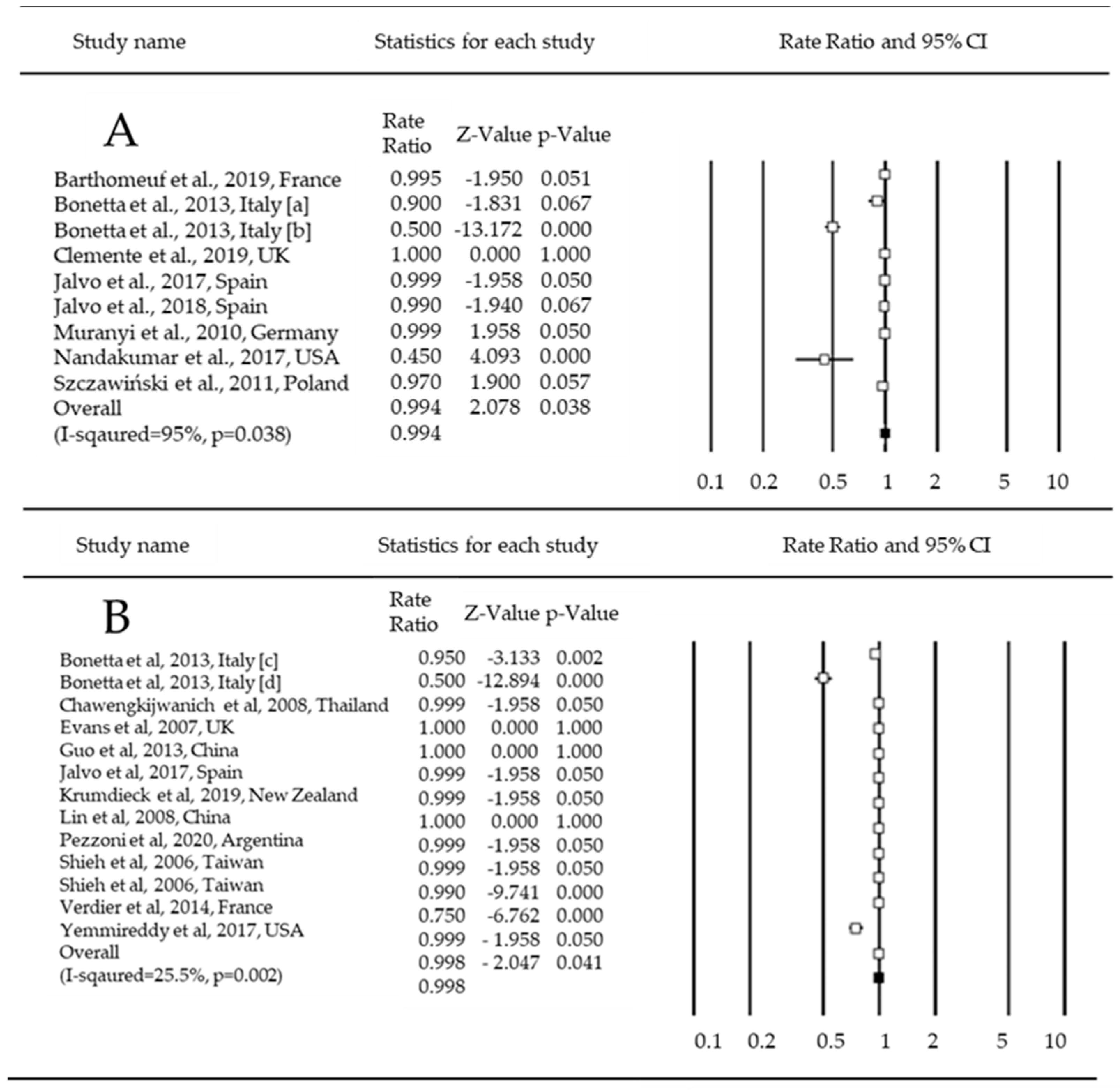
3.3. Antimicrobial Efficacy of Coatings
3.4. Limitations of the Study
4. Conclusions
Author Contributions
Funding
Institutional Review Board Statement
Informed Consent Statement
Data Availability Statement
Acknowledgments
Conflicts of Interest
References
- Wilson, A.M.; Weir, M.H.; King, M.F.; Jones, R.M. Comparing approaches for modelling indirect contact transmission of infectious diseases. J. R. Soc. Interface 2021, 18, 20210281. [Google Scholar] [CrossRef]
- Atkinson, M.P.; Wein, L.M. Quantifying the routes of transmission for pandemic influenza. Bull. Math. Biol. 2008, 70, 820–867. [Google Scholar] [CrossRef]
- Reed, S.E. An investigation of the possible transmission of rhinovirus colds through indirect contact. J. Hyg. 1975, 75, 249–258. [Google Scholar] [CrossRef] [PubMed]
- Mubareka, S.; Lowen, A.C.; Steel, J.; Coates, A.L.; Carcia-Sastre, A.; Palese, P. Transmission of influenza virus via aerosols and fomites in the guinea pig model. J. Infect. Dis. 2009, 199, 858–865. [Google Scholar] [CrossRef] [PubMed]
- Walther, B.A.; Ewald, P.W. Pathogen survival in the external environment and the evolution of virulence. Biol. Rev. 2004, 79, 849–869. [Google Scholar] [CrossRef]
- Sze-To, G.N.; Yang, Y.; Kwan, J.K.; Yu, S.C.; Chao, C.Y. Effects of surface material, ventilation, and human behavior on indirect contact transmission risk of respiratory infection. Risk. Anal. 2014, 34, 818–830. [Google Scholar] [CrossRef]
- Tuson, H.H.; Weibel, D.B. Bacteria–surface interactions. Soft. Matter. 2013, 9, 4368. [Google Scholar] [CrossRef] [PubMed]
- Dancer, S.J. Controlling hospital-acquired infection: Focus on the role of the environment and new technologies for decontamination. Clin. Microbiol. Rev. 2014, 27, 665–690. [Google Scholar] [CrossRef]
- Kumar, A.; Alam, A.; Rani, M.; Ehtesham, N.Z.; Hasnain, S.E. Biofilms: Survival and defense strategy for pathogens. Int. J. Med. Microbiol. 2017, 307, 481–489. [Google Scholar] [CrossRef]
- Maillard, J.Y.; Centeleghe, I. How biofilm changes our understanding of cleaning and disinfection. Antimicrob. Resist. Infect. Control. 2023, 12, 95. [Google Scholar] [CrossRef]
- Krukiewicz, K.; Kazek-Kęsik, A.; Brzychczy-Włoch, M.; Łos, M.J.; Ateba, C.N.; Mehrbod, P.; Ghavami, S.; Shyntum, D.Y. Recent Advances in the Control of Clinically Important Biofilms. Int. J. Mol. Sci. 2022, 23, 9526. [Google Scholar] [CrossRef] [PubMed]
- Protano, C.; Cammalleri, V.; Romano Spica, V.; Valeriani, F.; Vitali, M. Hospital environment as a reservoir for cross transmission: Cleaning and disinfection procedures. Ann. Ig. 2019, 31, 436–448. [Google Scholar]
- Cloutier, M.; Mantovani, D.; Rosei, F. Antibacterial Coatings: Challenges, Perspectives, and Opportunities. Trends Biotechnol. 2015, 33, 637–652. [Google Scholar] [CrossRef] [PubMed]
- Banu, R.; Salvi, N.; Gupta, S.; Ameta, C.; Ameta, R.; Punjabi, P.B. A facile synthesis of GO/CuO nanocomposite with enhancing photocatalytic activity for the degradation of azure-B dye and its antimicrobial behavior. Arab. J. Sci. Eng. 2021, 47, 365–378. [Google Scholar] [CrossRef]
- Kong, X.; Liu, X.; Zheng, Y.; Chu, P.K.; Zhang, Y.; Wu, S. Graphitic carbon nitride-based materials for photocatalytic antibacterial application. Mater. Sci. Eng. R. 2021, 145, 100610. [Google Scholar] [CrossRef]
- Atacan, K.; Güy, N.; Özacar, M. Recent advances in photocatalytic coatings for antimicrobial surfaces. Curr. Opin. Chem. Eng. 2022, 36, 100777. [Google Scholar] [CrossRef]
- Ganguly, P.; Byrne, C.; Breen, A.; Pillai, S.C. Antimicrobial activity of photocatalysts: Fundamentals, mechanisms, kinetics and recent advances. Appl. Catal. B 2018, 225, 51–75. [Google Scholar] [CrossRef]
- Shahsavandi, F.; Amirjani, A.; Hosseini, H.R.M. Plasmon-enhanced photocatalytic activity in the visible range using AgNPs/polydopamine/graphitic carbon nitride nanocomposite. Appl. Surf. Sci. 2022, 585, 152728. [Google Scholar] [CrossRef]
- Amirjani, A.; Amlashi, N.B.; Ahmadiani, Z.R. Plasmon-Enhanced Photocatalysis Based on Plasmonic Nanoparticles for Energy and Environmental Solutions: A Review. ACS Appl. Nano Mater. 2023, 6, 9085–9123. [Google Scholar] [CrossRef]
- Liu, L.; Shen, Z.; Wang, C. Highly efficient visible-light-driven photocatalytic disinfection of flowing bioaerosol using mono/multilayer MXene based catalyst. Chem. Eng. J. 2023, 457, 141327. [Google Scholar] [CrossRef]
- Zhang, P.; Lin, L.; Zang, D.; Guo, X.; Liu, M. Designing bioinspired anti-biofouling surfaces based on a superwettability strategy. Small 2017, 13, 1503334. [Google Scholar] [CrossRef] [PubMed]
- Valenzuela, L.; Faraldos, M.; Bahamonde, A.; Rosal, R. Critical review on the use of photocatalysis and photoelectrocatalysis to create antimicrobial surfaces. Curr. Opin. Chem. Eng. 2021, 34, 100762. [Google Scholar] [CrossRef]
- Schutte-Smith, M.; Erasmus, E.; Mogale, R.; Marogoa, N.; Jayiya, A.; Visser, H.G. Using visible light to activate antiviral and antimicrobial properties of TiO2 nanoparticles in paints and coatings: Focus on new developments for frequent-touch surfaces in hospitals. J. Coat. Technol. Res. 2023, 20, 789–817. [Google Scholar] [CrossRef]
- Rabajczyk, A.; Zielecka, M.; Klapsa, W.; Dziechciarz, A. Self-Cleaning Coatings and Surfaces of Modern Building Materials for the Removal of Some Air Pollutants. Materials 2021, 14, 2161. [Google Scholar] [CrossRef] [PubMed]
- Margarucci, L.M.; Romano Spica, V.; Protano, C.; Gianfranceschi, G.; Giuliano, M.; Di Onofrio, V.; Mucci, N.; Valeriani, F.; Vitali, M.; Romano, F. Potential antimicrobial effects of photocatalytic nanothecnologies in hospital settings. Ann. Ig. 2019, 31, 461–473. [Google Scholar] [PubMed]
- Byrne, J.A.; Dunlop, P.S.; Hamilton, J.W.; Fernández-Ibáñez, P.; Polo-López, I.; Sharma, P.K.; Vennard, A.S. A review of heterogeneous photocatalysis for water and surface disinfection. Molecules 2015, 20, 5574–5615. [Google Scholar] [CrossRef] [PubMed]
- Page, M.J.; McKenzie, J.E. The PRISMA 2020 statement: An updated guideline for reporting systematic reviews. BMJ 2021, 372, 71. [Google Scholar] [CrossRef]
- Covidence-Better Systematic Review Management. Available online: https://www.covidence.org/ (accessed on 31 July 2023).
- Bender, R.; Friede, T.; Koch, A.; Kuss, O.; Schlattmann, P.; Schwarzer, G.; Skipka, G. Methods for evidence synthesis in the case of very few studies. Res. Synth. Methods 2018, 9, 382–392. [Google Scholar] [CrossRef] [PubMed]
- Santabárbara, J.; Olaya, B.; Bueno-Notivol, J.; Pérez-Moreno, M.; Gracia-García, P.; Ozamiz-Etxebarria, N.; Idoiaga-Mondragon, N. Prevalence of depression among medical students during the COVID-19 pandemic. A systematic review and meta-analysis. Rev. Med. Chil 2021, 149, 1579–1588. [Google Scholar] [CrossRef] [PubMed]
- Santabárbara, J.; Ozamiz-Etxebarria, N.; Idoiaga, N.; Olaya, B.; Bueno-Novitol, J. Meta-Analysis of Prevalence of Depression in Dental Students during COVID-19 Pandemic. Medicina 2021, 57, 1278. [Google Scholar] [CrossRef]
- Oldenkamp, R.; Schultsz, C.; Mancini, E.; Cappuccio, A. Filling the gaps in the global prevalence map of clinical antimicrobial resistance. Proc. Natl. Acad. Sci. USA 2021, 118, e2013515118. [Google Scholar] [CrossRef]
- Shieh, K.J.; Li, M.; Lee, Y.H.; Sheu, S.D.; Liu, Y.T.; Wang, Y.C. Antibacterial performance of photocatalyst thin film fabricated by defection effect in visible light. Nanomedicine 2006, 2, 121–126. [Google Scholar] [CrossRef] [PubMed]
- Bletsa, E.; Merkl, P.; Thersleff, T.; Normark, S.; Henriques-Normark, B.; Sotiriou, G.A. Highly durable photocatalytic titanium suboxide–polymer nanocomposite films with visible light-triggered antibiofilm activity. Chem. Eng. J. 2023, 454, 139971. [Google Scholar] [CrossRef]
- Cuadra, J.G.; Molina-Prados, S.; Mínguez-Vega, G. Multifunctional silver-coated transparent TiO2 thin films for photocatalytic and antimicrobial applications. Appl. Surf. Sci. 2023, 617, 156519. [Google Scholar] [CrossRef]
- Fu, H.; Yaniv, V.; Betzalel, Y.; Mamane, H.; Gray, K.A. Creating anti-viral high-touch surfaces using photocatalytic transparent films. Chemosphere 2023, 323, 138280. [Google Scholar] [CrossRef]
- Todorova, N.; Minev, N.; Marinova, V.; Buchkov, K.; Videva, V.; Todorov, R.; Rafailov, P.; Strijkova, V.; Psycharis, V.; Giannakopoulou, T.; et al. Two-dimensional PtSe2 coatings with antibacterial activity. Appl. Surf. Sci. 2023, 611, 155534. [Google Scholar] [CrossRef]
- Evans, P.; Sheel, D.W. Photoactive and antibacterial TiO2 thin films on stainless steel. Surf. Coat. Technol. 2007, 201, 9319–9324. [Google Scholar] [CrossRef]
- Page, K.; Palgrave, R.G.; Parkin, I.P.; Wilson, M.; Savin, S.L.; Chadwick, A.V. Titania and silver–titania composite films on glass—Potent antimicrobial coatings. J. Mater. Chem. 2007, 17, 95–104. [Google Scholar] [CrossRef]
- Chawengkijwanich, C.; Hayata, Y. Development of TiO2 powder-coated food packaging film and its ability to inactivate Escherichia coli in vitro and in actual tests. Int. J. Food. Microbiol. 2008, 123, 288–292. [Google Scholar] [CrossRef]
- Lin, H.; Xu, Z.; Wang, X.; Long, J.; Su, W.; Fu, X.; Lin, Q. Photocatalytic and antibacterial properties of medical-grade PVC material coated with TiO2 film. J. Biomed. Mater. Res. B Appl. Biomater. 2008, 87, 425–431. [Google Scholar] [CrossRef]
- Yao, Y.; Ohko, Y.; Sekiguchi, Y.; Fujishima, A.; Kubota, Y. Self-sterilization using silicone catheters coated with Ag and TiO2 nanocomposite thin film. J. Biomed. Mater. Res. B Appl. Biomater. 2008, 85, 453–460. [Google Scholar] [CrossRef] [PubMed]
- Dunnill, C.W.H.; Aiken, Z.A.; Pratten, J. Enhanced photocatalytic activity under visible light in N-doped TiO2 thin films produced by APCVD preparations using t-butylamine as a nitrogen source and their potential for antibacterial films. J. Photochem. Photobiol. A Chem. 2009, 207, 244–253. [Google Scholar] [CrossRef]
- Sayilkan, F.; Asiltürk, M.; Kiraz, N.; Burunkaya, E.; Arpaç, E.; Sayilkan, H. Photocatalytic antibacterial performance of Sn4+-doped TiO2 thin films on glass substrate. J. Hazard Mater. 2009, 162, 1309–1316. [Google Scholar] [CrossRef] [PubMed]
- Muranyi, P.; Schraml, C.; Wunderlich, J. Antimicrobial efficiency of titanium dioxide-coated surfaces. J. Appl. Microbiol. 2010, 108, 1966–1973. [Google Scholar] [CrossRef] [PubMed]
- Szczawiński, J.; Tomaszewski, H.; Jackowska-Tracz, A.; Szczawińska, M.E. Survival of Staphylococcus aureus exposed to UV radiation on the surface of ceramic tiles coated with TiO2. Pol. J. Vet. Sci. 2011, 14, 41–46. [Google Scholar] [CrossRef]
- Akgun, B.A.; Wren, A.W.; Durucan, C.; Towler, M.R.; Mellott, N.P. Sol–gel derived silver-incorporated titania thin films on glass: Bactericidal and photocatalytic activity. J. Sol.-Gel. Sci. Technol. 2011, 59, 228–238. [Google Scholar] [CrossRef]
- Chien, D.M.; Dung, D.T.M.; Dam, L.D. Preparation of nitrogen co-doped SiO2/TiO2 thin films on ceramic with enhanced photocatalytic activity under visible-light irradiation. J. Exp. Nanosci. 2012, 7, 254–262. [Google Scholar] [CrossRef]
- Thongsuriwong, K.; Amornpitoksuk, P.; Suwanboon, S. Structure, morphology, photocatalytic and antibacterial activities of ZnO thin films prepared by sol–gel dip-coating method. Adv. Powder Technol. 2013, 24, 275–280. [Google Scholar] [CrossRef]
- Bonetta, S.; Bonetta, S.; Motta, F.; Strini, A.; Carraro, E. Photocatalytic bacterial inactivation by TiO2-coated surfaces. AMB Express 2013, 3, 59. [Google Scholar] [CrossRef]
- Guo, M.Z.; Ling, T.C.; Poon, C.S. Nano-TiO2-based architectural mortar for NO removal and bacteria inactivation: Influence of coating and weathering conditions. Cem. Concr. Compos. 2013, 36, 101–108. [Google Scholar] [CrossRef]
- Roldán, M.V.; de Oña, P.; Castro, Y.; Durán, A.; Faccendini, P.; Lagier, C.; Grau, R.; Pellegri, N.S. Photocatalytic and biocidal activities of novel coating systems of mesoporous and dense TiO2-anatase containing silver nanoparticles. Mater. Sci. Eng. C 2014, 43, 630–640. [Google Scholar] [CrossRef] [PubMed]
- Tallósy, S.P.; Janovák, L.; Ménesi, J. LED-light Activated Antibacterial Surfaces Using Silver-modified TiO2 Embedded in Polymer Matrix. J. Adv. Oxid. Technol. 2014, 17, 9–16. [Google Scholar] [CrossRef]
- Verdier, T.; Coutand, M.; Bertron, A.; Roques, C. Antibacterial Activity of TiO2 Photocatalyst Alone or in Coatings on E. coli: The Influence of Methodological Aspects. Coatings 2014, 4, 670–686. [Google Scholar] [CrossRef]
- Xiao, G.; Zhang, X.; Zhao, Y.; Su, H.; Tan, T. The behavior of active bactericidal and antifungal coating under visible light irradiation. Appl. Surf. Sci. 2014, 292, 756–763. [Google Scholar] [CrossRef]
- Deng, W.; Ning, S.; Lin, Q.; Zhang, H.; Zhou, T.; Lin, H.; Long, J.; Lin, Q.; Wang, X. I-TiO2/PVC film with highly photocatalytic antibacterial activity under visible light. Colloids Surf. B Biointerfaces 2016, 144, 196–202. [Google Scholar] [CrossRef]
- Leyland, N.S.; Podporska-Carroll, J.; Browne, J.; Hinder, S.J.; Quilty, B.; Pillai, S.C. Highly Efficient F, Cu doped TiO2 anti-bacterial visible light active photocatalytic coatings to combat hospital-acquired infections. Sci. Rep. 2016, 6, 24770. [Google Scholar] [CrossRef]
- Chuang, K.T.; Abdullah, H.; Leu, S.J.; Cheng, K.B.; Kuo, D.H.; Chen, H.C.; Chien, J.H.; Hu, W.T. Metal oxide composite thin films made by magnetron sputtering for bactericidal application. J. Photochem. Photobiol. A Chem. 2017, 337, 151–164. [Google Scholar] [CrossRef]
- Jalvo, B.; Faraldos, M.; Bahamonde, A.; Rosal, R. Antimicrobial and antibiofilm efficacy of self-cleaning surfaces functionalized by TiO2 photocatalytic nanoparticles against Staphylococcus aureus and Pseudomonas putida. J. Hazard. Mater. 2017, 340, 160–170. [Google Scholar] [CrossRef]
- Nandakumar, V.; Han, Z.; Fritz, Z.; Krishna, V.; Koopman, B.; Moudgil, B. Visible Light Photocatalytic Bacterial Inactivation on Titanium Dioxide Coatings. KONA Powder Part. J. 2017, 34, 234–240. [Google Scholar] [CrossRef]
- Pessoa, R.S.; dos Santos, V.P.; Cardoso, S.B.; Doria, A.C.; Figueira, F.R.; Rodrigues, B.V.; Testoni, G.E.; Fraga, M.A.; Marciano, F.R.; Lobo, A.O.; et al. TiO2 coatings via atomic layer deposition on polyurethane and polydimethylsiloxane substrates: Properties and effects on C. albicans growth and inactivation process. Appl. Surf. Sci. 2017, 422, 73–84. [Google Scholar] [CrossRef]
- Yemmireddy, V.K.; Hung, Y.C. Photocatalytic TiO2 coating of plastic cutting board to prevent microbial cross-contamination. Food. Control 2017, 77, 88–95. [Google Scholar] [CrossRef]
- Hossain, M.A.; Elias, M.; Sarker, D.R.; Diba, Z.R.; Mithun, J.M.; Azad, M.A.; Siddiquey, I.A.; Rahman, M.M.; Uddin, J.; Uddin, M.N. Synthesis of Fe- or Ag-doped TiO2–MWCNT nanocomposite thin films and their visible-light-induced catalysis of dye degradation and antibacterial activity. Res. Chem. Intermed. 2018, 44, 2667–2683. [Google Scholar] [CrossRef]
- Jalvo, B.; Faraldos, M.; Bahamonde, A.; Rosal, R. Antibacterial surfaces prepared by electrospray coating of photocatalytic nanoparticles. Chem. Eng. J. 2018, 334, 1108–1118. [Google Scholar] [CrossRef]
- Won, Y.; Schwartzenberg, K.; Gray, K.A. TiO2-based transparent coatings create self-cleaning surfaces. Chemosphere 2018, 208, 899–906. [Google Scholar] [CrossRef] [PubMed]
- Barthomeuf, M.; Castel, X.; Le Gendre, L.; Louis, J.; Denis, M.; Pissavin, C. Effect of Titanium Dioxide Film Thickness on Photocatalytic and Bactericidal Activities Against Listeria monocytogenes. Photochem. Photobiol. 2019, 95, 1035–1044. [Google Scholar] [CrossRef]
- Clemente, A.; Ramsden, J.J.; Wright, A.; Iza, F.; Morrissey, J.A.; Li Puma, G.; Malik, D.J. Staphylococcus aureus resists UVA at low irradiance but succumbs in the presence of TiO2 photocatalytic coatings. J. Photochem. Photobiol. B 2019, 193, 131–139. [Google Scholar] [CrossRef]
- Krumdieck, S.P.; Boichot, R.; Gorthy, R.; Land, J.G.; Lay, S.; Gardecka, A.J.; Polson, M.I.; Wasa, A.; Aitken, J.E.; Heinemann, J.A.; et al. Nanostructured TiO2 anatase-rutile-carbon solid coating with visible light antimicrobial activity. Sci. Rep. 2019, 9, 1883. [Google Scholar] [CrossRef]
- Valenzuela, L.; Iglesias, A.; Faraldos, M.; Bahamonde, A.; Rosal, R. Antimicrobial surfaces with self-cleaning properties functionalized by photocatalytic ZnO electrosprayed coatings. J. Hazard. Mater. 2019, 369, 665–673. [Google Scholar] [CrossRef]
- Oder, M.; Koklič, T.; Umek, P.; Podlipec, R.; Štrancar, J.; Dobeic, M. Photocatalytic biocidal effect of copper doped TiO2 nanotube coated surfaces under laminar flow, illuminated with UVA light on Legionella pneumophila. PLoS ONE 2020, 15, e0227574. [Google Scholar] [CrossRef]
- Pezzoni, M.; Catalano, P.N.; Delgado, D.C.; Pizarro, R.A.; Bellino, M.G.; Costa, C.S. Antibiofilm effect of mesoporous titania coatings on Pseudomonas aeruginosa biofilms. J. Photochem. Photobiol. B Biol. 2020, 203, 111762. [Google Scholar] [CrossRef]
- Zhao, Y.; Xu, J.; Li, Z.; Fu, T.; Jiang, S. In vitro antibacterial properties of MoO3/SiO2/Ag2O nanocomposite coating prepared by double cathode glow discharge technique. Surf. Coat. Technol. 2020, 397, 125992. [Google Scholar] [CrossRef]
- Álvarez, Á.L.; Dalton, K.P.; Nicieza, I.; Abade Dos Santos, F.A.; de la Peña, P.; Domínguez, P.; Martin-Alonso, J.M.; Parra, F. Virucidal Properties of Photocatalytic Coating on Glass against a Model Human Coronavirus. Microbiol. Spectr. 2022, 10, e0026922. [Google Scholar] [CrossRef] [PubMed]
- Du, J.; Li, Z.; Guo, H.; Zhu, E.; Liu, C.; Ren, B.; Che, G. The facile preparation and antibacterial performance of a conductive polymer-PU coating under visible light. Prog. Org. Coat. 2022, 165, 106755. [Google Scholar] [CrossRef]
- Li, Y.; Liu, Y.; Yao, B.; Narasimalu, S.; Dong, Z. Rapid preparation and antimicrobial activity of polyurea coatings with RE-Doped nano-ZnO. Microb. Biotechnol. 2022, 15, 548–560. [Google Scholar] [CrossRef] [PubMed]
- Vihodceva, S.; Šutka, A.; Otsus, M.; Vija, H.; Grase, L.; Kahru, A.; Kasemets, K. Visible-Light Active Flexible and Durable Photocatalytic Antibacterial Ethylene-co-vinyl Acetate—Ag/AgCl/α-Fe2O3 Composite Coating. Nanomaterials 2022, 12, 1984. [Google Scholar] [CrossRef] [PubMed]
- Xu, X.; Wang, Y.; Zhang, D.; Wang, J.; Yang, Z. In situ growth of photocatalytic Ag-decorated β-Bi2O3/Bi2O2.7 heterostructure film on PVC polymer matrices with self-cleaning and antibacterial properties. Chem. Eng. J. 2022, 429, 131058. [Google Scholar] [CrossRef]
- Serrano-Aroca, Á.; Takayama, K.; Tuñón-Molina, A.; Seyran, M.; Hassan, S.S.; Pal Choudhury, P.; Uversky, V.N.; Lundstrom, K.; Adadi, P.; Palù, G.; et al. Carbon-based nanomaterials: Promising antiviral agents to combat COVID-19 in the microbial-resistant era. ACS Nano 2021, 15, 8069–8086. [Google Scholar] [CrossRef]
- Chong, Y.; Ge, C.; Fang, G.; Wu, R.; Zhang, H.; Chai, Z.; Chen, C.; Yin, J.-J. Light-enhanced antibacterial activity of graphene oxide, mainly via accelerated electron transfer. Environ. Sci. Technol. 2017, 51, 10154–10161. [Google Scholar] [CrossRef]
- Elias, L.; Taengua, R.; Frígols, B.; Salesa, B.; Serrano-Aroca, Á. Carbon nanomaterials and LED irradiation as antibacterial strategies against gram-positive multidrug-resistant pathogens. Int. J. Mol. Sci. 2019, 20, 3603. [Google Scholar] [CrossRef]
- Endo-Kimura, M.; Kowalska, E. Plasmonic photocatalysts for microbiological applications. Catalysts 2020, 10, 824. [Google Scholar] [CrossRef]
- Endo, M.; Wei, Z.S.; Wang, K.L.; Karabiyik, B.; Yoshiiri, K.; Rokicka, P.; Ohtani, B.; Markowska-Szczupak, A.; Kowalska, E. Noble metalmodified titania with visible-light activity for the decomposition of microorganisms. Beilstein J. Nanotechnol. 2018, 9, 829–841. [Google Scholar] [CrossRef] [PubMed]
- Kowalska, E.; Wei, Z.; Karabiyik, B.; Herissan, A.; Janczarek, M.; Endo, M.; Markowska-Szczupak, A.; Remita, H.; Ohtani, B. Silvermodified titania with enhanced photocatalytic and antimicrobial properties under UV and visible light irradiation. Catal. Today 2015, 252, 136–142. [Google Scholar] [CrossRef]
- Janczarek, M.; Endo-Kimura, M.; Wei, Z.; Bielan, Z.; Mogan, T.R.; Khedr, T.M.; Wang, K.; Markowska-Szczupak, A.; Kowalska, E. Novel structures and applications of graphene-based semiconductor photocatalysts: Faceted particles, photonic crystals, antimicrobial and magnetic properties. Appl. Sci. 2021, 11, 1982. [Google Scholar] [CrossRef]
- Herrmann, J.M. Heterogeneous photocatalysis: State of the art and present applications. Top. Catal. 2005, 34, 49–65. [Google Scholar] [CrossRef]
- Kong, D.B.; Ma, C.C.; Wang, W.; Liu, C.; Tian, Y.; Wang, T.; Zhao, Z.P.; Zhang, C.Y.; Feng, H.M.; Chen, S.G. Two birds with one stone: Interfacial controls and pH response for long-term and high efficiency Cu2O antibacterial materials. Chem. Eng. J. 2022, 427, 131734. [Google Scholar] [CrossRef]
- Salvadores, F.; Reli, M.; Alfano, O.M.; Kocí, K.; Ballari, M.D. Efficiencies evaluation of photocatalytic paints under indoor and outdoor air conditions. Front. Chem. 2020, 8, 551710. [Google Scholar] [CrossRef]
- Maulidiyah, M.; Susilowati, P.E.; Mudhafar, N.K.; Salim, L.A.; Wibowo, D.; Muzakkar, M.Z.; Irwan, I.; Arham, Z.; Nurdin, M. Photo-inactivation Staphylococcus aureus by using formulation of Mn-N-TiO2 composite coated wall paint. Biointerface Res. Appl. Chem. 2022, 12, 1628–1637. [Google Scholar]
- Abdulagatov, I.M.; Ragimov, R.M.; Khamidov, M.A.; Maksumova, A.M.; Abdullaeva, N.M. ALD coated polypropylene hernia meshes for prevention of mesh-related post-surgery complications: An experimental study in animals. Biomed. Mater. 2022, 17, 1. [Google Scholar] [CrossRef]
- Ma, J.Z.; Liu, C.Y.; Yan, K. CQDs-MoS2 QDs loaded on Dendritic fibrous Nanosilica/Hydrophobic waterborne polyurethane acrylate for antibacterial coatings. Chem. Eng. J. 2022, 429, 132170. [Google Scholar] [CrossRef]
- Tuñón-Molina, A.; Takayama, K.; Redwan, E.M.; Uversky, V.N.; Andrés, J.; Serrano-Aroca, Á. Protective face masks: Current status and future trends. ACS Appl. Mater. Interfaces 2021, 13, 56725–56751. [Google Scholar] [CrossRef]
- Lu, Y.; Guan, S.; Hao, L.; Yoshida, H.; Nakada, S.; Takisawa, T.; Itoi, T. Inactivation of SARS-CoV-2 and photocatalytic degradation by TiO2 photocatalyst coatings. Sci. Rep. 2022, 12, 16038. [Google Scholar] [CrossRef] [PubMed]
- Margarucci, L.M.; Gianfranceschi, G.; Romano Spica, V.; D’Ermo, G.; Refi, C.; Podico, M.; Vitali, M.; Romano, F.; Valeriani, F. Photocatalytic Treatments for Personal Protective Equipment: Experimental Microbiological Investigations and Perspectives for the Enhancement of Antimicrobial Activity by Micrometric TiO2. Int. J. Environ. Res. Public Health 2021, 18, 8662. [Google Scholar] [CrossRef] [PubMed]
- Guillard, C.; Lachheb, H.; Houas, A.; Ksibi, M.; Elaloui, E.; Herrmann, J.-M. Influence of chemical structure of dyes, of pH and of inorganic salts on their photocatalytic degradation by TiO2 comparison of the efficiency of powder and supported TiO2. J. Photochem. Photobiol. A Chem. 2003, 158, 27–36. [Google Scholar] [CrossRef]
- Yang, Y.; Yan, Z.; Yang, S.; Tang, Z.; Li, W.; Yang, B.; Su, W.; Ji, T. Effect of substrate roughness on NOx removal of poly heptazine imides coated cement pastes exposed to washing and weathering. J. Clean. Prod. 2022, 377, 134397. [Google Scholar] [CrossRef]
- Zhang, J.; Tan, H.; Deng, X. NOx removal ability of photocatalytic cement-based materials with porous structure. J. Clean. Prod. 2022, 377, 134396. [Google Scholar] [CrossRef]
- Li, F.; Liu, G.; Liu, F.; Yang, S. A review of self-cleaning photocatalytic surface: Effect of surface characteristics on photocatalytic activity for NO. Environ. Pollut. 2023, 327, 121580. [Google Scholar] [CrossRef]
- Ahlawat, K.; Jangra, R.; Ish, A.; Dixit, A.; Fulwani, D.; Jain, N.; Prakash, R. Analysis of a UV photocatalytic oxidation-based disinfection system for hydroxyl radicals, negative air ions generation and their impact on inactivation of pathogenic micro-organisms. Rev. Sci. Instrum. 2023, 94, 104103. [Google Scholar] [CrossRef]
- Sizar, O.; Leslie, S.W.; Unakal, C.G. Gram-Positive Bacteria. In StatPearls [Internet]; StatPearls Publishing: Treasure Island, FL, USA, 2023; Available online: https://www.ncbi.nlm.nih.gov/books/NBK470553/ (accessed on 31 December 2023).
- Karaman, R.; Jubeh, B.; Breijyeh, Z. Resistance of Gram-Positive Bacteria to Current Antibacterial Agents and Overcoming Approaches. Molecules 2020, 25, 2888. [Google Scholar]
- Wohlgemuth, S.; Kämpfer, P. BACTERIA|Bacterial Endospores. In Encyclopedia of Food Microbiology, 2nd ed.; Batt, C.A., Tortorello, M.L., Eds.; Academic Press: Cambridge, MA, USA, 2014; pp. 160–168. [Google Scholar]
- Sawada, T.; Yoshino, F.; Kimoto, K.; Takahashi, Y.; Shibata, T.; Hamada, N.; Sawada, T.; Toyoda, M.; Lee, M.C. ESR detection of ROS Generated by TiO2 coated with fluoridated apatite. J. Dent. Res. 2010, 89, 848–853. [Google Scholar] [CrossRef]
- Kado, D.; Sakurai, K.; Sugiyama, T.; Ueda, T. Evaluation of cleanability of a titanium dioxide (TiO2)-coated acrylic resin denture base. Prosthodont. Res. Pract. 2005, 4, 69–76. [Google Scholar] [CrossRef]
- Alrahlah, A.; Fouad, H.; Hashem, M.; Niazy, A.A.; AlBadah, A. Titanium oxide (TiO2)/polymethylmethacrylate (PMMA) denture base nanocomposites: Mechanical, viscoelastic and antibacterial behavior. Materials 2018, 11, 1096. [Google Scholar] [CrossRef] [PubMed]
- Skallevold, H.E.; Rokaya, D.; Khurshid, Z.; Zafar, M.S. Bioactive Glass Applications in Dentistry. Int. J. Mol. Sci. 2019, 20, 5960. [Google Scholar] [CrossRef] [PubMed]
- Khan, K.A.; Ghatak, H.R.; Ahuja, S.M. Photocatalytic technology: A review of environmental protection and renewable energy application for sustainable development. Environ. Technol. Innov. 2020, 19, 100893. [Google Scholar]
- Ray, S.K.; Hur, J. A critical review on modulation of NiMoO4-based materials for photocatalytic applications. J. Environ. Manag. 2021, 278, 111562. [Google Scholar] [CrossRef]
- Kumaravel, V.; Nair, K.M.; Mathew, S.; Bartlett, J.; Kennedy, J.E.; Manning, H.G.; Whelan, B.J.; Leyland, N.S.; Pillai, S.C. Antimicrobial TiO2 nanocomposite coatings for surfaces, dental and orthopaedic implants. Chem. Eng. J. 2021, 416, 129071. [Google Scholar] [CrossRef] [PubMed]
- Rahimi, M.; Noruzi, E.; Sheyksaran, E.; Ebadi, B.; Kariminezhad, Z.; Molaparast, M.; Mehrabani, M.G.; Mehramouz, B.; Yousefi, M.; Ahmadi, R.; et al. Carbohydrate polymer-based silver nanocomposites: Recent progress in the antimicrobial wound dressings. Carbohydr. Polym. 2020, 231, 115696. [Google Scholar] [CrossRef]
- Huang, Z.; Maness, P.C.; Blake, D.M.; Wolfrum, E.J.; Smolinski, S.L.; Jacoby, W.A. Bactericidal mode of titanium dioxide photocatalysis. J. Photochem. Photobiol. A 2000, 130, 163. [Google Scholar] [CrossRef]
- Lu, Z.X.; Zhou, L.; Zhang, Z.L.; Shi, W.L.; Xie, Z.X.; Xie, H.Y.; Pang, D.W.; Shen, P. Cell Damage Induced by Photocatalysis of TiO2 Thin Films. Langmuir 2003, 19, 8765. [Google Scholar] [CrossRef]
- Sunada, K.; Watanabe, T.; Hashimoto, K. Studies on photokilling of bacteria on TiO2 thin film. J. Photochem. Photobiol. A 2003, 156, 227. [Google Scholar] [CrossRef]
- Dell’Edera, M.; Lo Porto, C.; De Pasquale, I.; Petronella, F.; Curri, M.L.; Agostiano, A.; Comparelli, R. Photocatalytic TiO2-based coatings for environmental applications. Catal. Today 2021, 380, 62–83. [Google Scholar] [CrossRef]
- Nabi, I.; Bacha, A.U.; Li, K.; Cheng, H.; Wang, T.; Liu, Y.; Ajmal, S.; Yang, Y.; Feng, Y.; Zhang, L. Complete Photocatalytic Mineralization of Microplastic on TiO2 Nanoparticle Film. IScience 2020, 23, 101326. [Google Scholar] [CrossRef] [PubMed]
- Kim, J.Y.; Youn, D.H. Nanomaterials for Advanced Photocatalytic Plastic Conversion. Molecules 2023, 28, 6502. [Google Scholar] [CrossRef] [PubMed]
- Kouchehbaghi, N.H.; Sohrabi, M.; Razbin, M.; Daryakenari, A.A.; Abbasi, M.; Bahrami, S.A. Soft computing procedure to optimize the electrospinning parameters of polyacrylonitrile nanofibrous air filter. J. Text. Inst. 2023. [Google Scholar] [CrossRef]
- Lv, H.; Liu, Y.; Bai, Y.; Shi, H.; Zhou, W.; Chen, Y.; Liu, Y.; Yu, D.-G. Recent Combinations of Electrospinning with Photocatalytic Technology for Treating Polluted Water. Catalysts 2023, 13, 758. [Google Scholar] [CrossRef]


| Author, Year, Country | Type of Surface | Type of Photocatalyst | Dose of Photocatalyst | Type of Coating Method | Details of Coating Method | Main Results | Reference |
|---|---|---|---|---|---|---|---|
| Akgun et al., 2011, Turkey | Glass | Ag-TiO2 | 6 mL of Ti[O(CH2)3CH3]4; 0.2 g AgNO3 | Spin coating | The cleaned substrate was coated with Ag-TiO2 using a spin coater at 2300 rpm for 30 s. The coating process was repeated three times, and the resulting films were dried at 100 °C for 1 h. Subsequently, the films were calcined in air at 250 °C, 450 °C, and 650 °C for 6 h and then cooled to room temperature. | Under any given illumination condition, the Ag-doped films had increased bactericidal and photocatalytic activity compared to TiO2 thin films. | [47] |
| Álvarez et al., 2022, Spain | Glass | TiO2 | NA | NA | Glass pre-exposed to UVA for 4 h, placed in a sterile dish. HCoV-229E was applied dropwise to the surface and covered with transparent PVC film. | The TiO2-coated glass inactivates coronaviruses in a time-dependent manner on contact under daylight illumination. | [73] |
| Barthomeuf et al., 2019, France | Glass | TiO2 | NA | Sputtering deposition | A glass substrate was loaded into the deposition chamber after pre-sputtering the target in pure argon (Ar) for 10 min. Then, a mixture of argon (Ar) and oxygen (O2) gas was injected into the sputtering chamber. | After photoactivation with UVA radiation for 20 min, TiO2 coatings had a strong bactericidal effect. | [66] |
| Bletsa et al., 2023, Sweden | Glass | Ag/TiOx | NA | Spin coating | The substrate holder was placed 20 cm above the burner for 15 s to deposit nanoparticles. The flame annealing process was conducted 20 cm above the burner using a xylene flame under cooling conditions. Stabilization was achieved by spin-coating at 100–500 rpm for 10 s and at 1000–4000 rpm for 50 s. | The compound was photocatalytically active with the visible light exposition. | [34] |
| Bonetta et al., 2013, Italy | Ceramics | TiO2 | 1 mg cm2 | Chemical process | No details. | Bacterial concentration was reduced for all the microbes exposed to UV irradiation. | [50] |
| Chawengkijwanich et al., 2008, Thailand | Polypropylene | TiO2 | NA | Manual coating | TiO2 was manually coated onto one side of the oriented polypropylene (OPP) film using a bar coater at room temperature. | There was a synergetic effect of TiO2-coated packaging film with UVA light. | [40] |
| Chien et al., 2012, Vietnam | Ceramic | SiO2/TiO2 | NA | Dip coating | Films were dip-coated onto ceramic tile substrates and annealed on a hot plate at 300 °C for 5 min, after which the substrates were calcined at 500 °C for 2 h. This process was repeated three times. | The films had high antibacterial activity by removing E. coli. | [48] |
| Chuang et al., 2017, Taiwan | Glass | ZnO/Ag2O | NA | Sputtering deposition | Deposition was carried out using an RF magnetron sputtering system with a gas flow rate of 40 sccm. The sputtering times were 5 min (Ag2O) and 30 min (ZnO/Ag2O), with a power of 30 W and a working pressure of 2 × 10⁻³ torr at room temperature. | Ag2O also has a great ability to kill bacteria, which may be due to the release of Ag+ ions and the formation of photoelectrons and holes to generate active species to destroy bacteria. | [58] |
| Clemente et al., 2019, UK | Glass | TiO2 | 0.5 ± 0.05 mg | Dip coating | Slides were fixed to a motor-driven bar, which allowed an immersion and withdrawal rate of 3 cm min−1. Then, they were immersed in the TiO2 suspension. | There were increased intracellular levels of oxidative stress, which over 24 h were lethal for S. aureus. | [67] |
| Cuadra et al., 2023, Spain | Glass | TiO2-Ag | 2.6 mL of titanium (IV) bis(acetylacetonate) diisopropoxide (75 wt % in isopropanol) | Sputtering deposition; spray coating | Titanium (IV) bis(acetylacetonate) diisopropoxide and EtOH were mixed for 30 min. The solution was applied to a soda-lime glass substrate heated to 450 °C and then heated to 550 °C on a hot plate. | The films had strong antibacterial activities after irradiation under UV-light for 4 h. | [35] |
| Deng et al., 2016, China | Poly vinyl chloride (PVC) | I-TiO2 | 200 mL TiO2 sol, 5 mL HI | Dip coating | PVA was dissolved in boiling water, then cooled. PVC pieces were dipped in the solution and removed. Then, the PVC pieces were immersed in I-TiO2 solution to obtain I-TiO2/PVC. | I-TiO2/PVC had an excellent photocatalytic antibacterial activity, which can limit the propagation of the E. coli | [56] |
| Du et al., 2022, China | Polyurethane | Photocatalytic conductor polymer (PTET-T-COOH) | 14 mg | Drop coating | 4,4′-Diphenylmethane diisocyanate (MDI) and polycarbonate diol (PCDL) were each placed in a vacuum dryer at 80 °C for 30 min to melt prior to the reaction. After stirring the liquid mixture for 1 h at 80 °C, the pre-polyurethane was ready. 1,4-Butanediol was added to the pre-polyurethane and stirred for 30 min at 80 °C, and the polyurethane (PU) was prepared. The mixture was dropped onto a glass slide. | Under visible light irradiation, (PTET-T-COOH)-PU coating demonstrated an inactivation of S. epidermidis concentration in 6 h. | [74] |
| Dunnill et al., 2009, UK | Glass | N-doped TiO2 | NA | Atmospheric pressure chemical vapor deposition (APCVD) | Depositions were performed on SiO2-coated glass slides after cleaning with water, acetone, petroleum ether, and propan-2-ol. The slides were then placed in an APCVD reactor and heated from room temperature to 500 °C at a rate of 10 °C/min. | The compound killed 99.9% of an E. coli suspension containing more than 104 viable bacteria, when exposed under white light for 24 h. | [43] |
| Evans et al., 2007, UK | Stainless steel | TiO2 | NA | Flame-assisted CVD (FACVD) (for silica); atmospheric pressure chemical vapor deposition (APCVD) (for titania) | The titania deposition was carried out using a horizontal cold wall APCVD quartz reactor, and precursors were supplied via bubblers. The steel substrates were cleaned with warm water and detergent before air drying. The silicon dioxide films were grown in a FACVD reactor. | The TiO2 film is bio-active and that the timescale for 100% kill (6 log reduction) was between 120 and 180 min. | [38] |
| Fu et al., 2023, Israel | Glass | nAg/nTiO2 | NA | Dip coating; Spray coating | The glass substrate was dipped into TiO2 gel four times, air-dried for 5 min between each immersion, and then calcined. An airbrush was used to spray TiO2 suspension above the glass substrate, which was then calcined for 2 h at 200 °C. | The nAg/nTiO2-coated sample reached 5.36 log virus reduction after 90 min under light source. | [36] |
| Guo et al., 2013, China | Glass | TiO2 | 79.87 g/mol | Dip coating | The substrate was dip-coated with a TiO2 film. The TiO2 suspension was prepared from ethanol and glycerol. Then, it was stirred for 15 min, before the substrate was dipped into it (for 5 min). TiO2-coated glass was calcinated at 450 °C for 120 min. | There was a total inactivation of E. coli within a relatively short time. | [51] |
| Hossain et al., 2018, Bangladesh | Glass | Fe-doped TiO2–MWCNT (multiwalled carbon nanotubes) | NA | Drop coating | Soda lime silica glass was rinsed with alcohol and distilled water, then dried at 100 °C. TiO2 gel films were obtained by coating a precursor solution onto the glass. The coated substrates were pretreated and annealed for 20 min at 200 °C. The coating process was repeated two times, followed by annealing at 500 °C for 2 h. | The nanocomposite could be used as an effective growth inhibitor of E. coli. | [63] |
| Jalvo et al., 2017, Spain | Glass | TiO2 | 2 mL | Smearing (glass slides); impregnation (glass filters) | TiO2 suspension applied to glass slides by smearing and to glass filters by impregnation. Substrates were dried at 110 °C before and after deposition and weighed to evaluate photocatalyst. | There was an antibacterial effect due to extensive membrane damage and significant production of ROS. | [59] |
| Jalvo et al., 2018, Spain | Glass | TiO2 | 16.5 mL | Spray coating | Electrosprayed drops were deposited on round glass coverslips, attached to a flat collector that was horizontally arranged. | Light exposition caused membrane damage, with no cell regrowth. | [64] |
| Krumdieck et al., 2019, New Zealand | Steel | TiO2 | NA | Pulsed-pressure metalorganic chemical vapor deposition (pp-MOCVD) | Steel substrates were cleaned by abrading followed by ultrasonication in a silicon-free detergent/water solution, rinsed, and dried prior to loading into the pp-MOCVD chamber for a 30 min bake. | The pp-MOCVD approach could represent a strategy to support catalysts. | [68] |
| Leyland et al., 2016, Ireland | Glass | F, Cu-doped TiO2 | NA | Dip coating | Substrates were immersed in sol and then drawn vertically. The coated glass was dried and heated at 550 °C for 90 min. | There was a bacterial reduction of log10 = 4.2 (visible light) and log10 = 1.8 in darkness. | [57] |
| Li et al., 2022, Singapore | Polyurea | La-, Ce-, Pr-, and Gd (RE-dopants)-doped nano-ZnO | NA | NA | All chemicals were heated and placed in the mixer for 180 s. Then, the polyurea was poured into Teflon molds and placed in an oven at 70 °C to cure for 48 h. | These polyurea coatings had a high bactericidal rate over 85%. | [75] |
| Lin et al., 2008, China | Poly vinyl chloride (PVC) | TiO2 | NA | Dip coating | The PVC sheets were immersed in the precursor suspension (THF) and then pulled out at a speed of 1200 mm/h and dried in air for 1 h. | The pre-irradiated TiO2/PVC had an excellent antibacterial adhesion and sterilization activity. | [41] |
| Muranyi et al., 2010, Germany | Glass | TiO2 | NA | Dip coating | Sol was made by controlled hydrolysis and condensation. Ethanol was split into two beakers. Part A had water and nitric acid, and Part B had TPOT. Part A was slowly added to Part B while stirring with a magnetic stirrer for 30 min. | The titanium dioxide layers can very effectively decompose K. rhizophila cells. | [45] |
| Nandakumar et al., 2017, USA | Ceramics | TiO2 | NA | Multiple coating | The dispersions were applied as uniform coatings on ceramic tiles. A second coat of anatase was applied after the tiles were dried. Coatings of silica were similarly prepared. | The S. aureus reduction under visible light gradually decreased with increasing cut off limits up to 550 nm. | [60] |
| Oder et al., 2020, Slovenia | Polystyrene | Cu-TiO2 | 400 mg of the H2Ti3O7 nanotube; 100 mL of 0.5 mM solution of Cu2+ | Smearing | Petri dishes were treated with compressed air and smeared evenly. After the deposition, they were rinsed with water and put in the oven at 60 °C overnight. | There is a short term microbiocidal effectiveness of TiO2 nanotube coatings irradiated with UVA on L. pneumophila. | [70] |
| Page et al., 2007, UK | Glass | Ag-TiO2 | 17.02 g Titanium n-butoxide; 0.8510 g silver nitrate | Dip coating | A dip-coating apparatus was used to eliminate the slide from the sol (speed of 120 cm min−1). | Ag-doped titania coatings were more photocatalytically and antimicrobially active than a titania coating. | [39] |
| Pessoa et al., 2017, Brazil | Polyurethane; Polydimethylsiloxane | TiO2 | NA | Atomic layer deposition (ALD) | ALD consisted of the different steps: 1. TiCl4 pulse of 0.25 s; 2. purge of 2 s; 3. H2O pulse of 0.25 s; 4. purge of 2 s. During the deposition, the base pressure of the reactor was lower than 10−2 mbar, and the working pressure was kept around of 1.0 mbar through the insertion of 300 sccm of N2. | A reduction was observed in comparison to control. | [61] |
| Pezzoni et al., 2020, Argentina | Glass | TiO2 | NA | Spin coating | The films were prepared by spin-coating (3100 rpm for 30 s) on glass slides at 35 °C solution temperature and 30% relative humidity. | There was a high percentage of cell membrane disruption, compared to non-treated biofilms. | [71] |
| Roldán et al., 2014, Argentina | Glass | Ag-SiO2/TiO2 | NA | Dip coating | SiO2 and Ag-doped SiO2 layers were deposited and heat-treated at 450 °C for 30 min. The TiO2 coating was heat-treated at 450 °C for 1 h and all the slides were coated on both sides. | It was important that Ag NPs and TiO2 are enclosed together because SiO2/Ag–TiO2 has a higher bactericidal effect than Ag–SiO2/TiO2. | [52] |
| Sayilkan et al., 2009, Turkey | Glass | TiO2-Sn4+ | 8.4 g (w/w = 10); 18.8 g (w/w = 20); 32.2 g (w/w = 30); 50.1 g (w/w = 40); 75.2 g (w/w = 50) | Spin coating | The glass surface was pre-coated with a solution consisting of 3-aminopropyltrimethoxy silane (AMMO), distilled water and isopropyl alcohol. Free hydroxyl groups, which are composed of hydrolysis of AMMO, behave as bridge between the film and the glass surface. | The films had higher antibacterial effect than undoped TiO2. | [44] |
| Shieh et al., 2006, Taiwan | Glass; steel | TiO2 | NA | Sputtering deposition | Ar and O2 were introduced to the RF sputter chamber. The substrate of sputter was loaded and after 120 s of deposition, the thickness of the TiOx thin film was about 120 nm. | The coating technology can be applied effectively to surfaces with different degrees of roughness. | [33] |
| Szczawiński et al., 2011, Poland | Ceramics | TiO2 | NA | Sputtering deposition; atmospheric pressure chemical vapor deposition (APCVD); spray coating | Sputtering was performed in pure argon at a pressure of 4.4 Pa. TiO2 targets of 100 mm diameter. For APCVD, titanium tetraisopropoxide (TTIP) was used as a precursor and stored in a glass Dreschler bubbler and maintained at 210 °C. Argon carrier gas was used to transport the TTIP through silicon and quartz lines to the vertical tube furnace. For spray coating, the same technique was used as for APCVD. | The strongest bactericidal effect of UV radiation was observed on the surfaces of tiles coated with TiO2 by APCVD. | [46] |
| Tallósy et al., 2014, Hungary | Glass | Ag-TiO2 | 0.6 mg/cm2 | Spray coating | No details. | There was an antibacterial effect against methicillin-resistant S. aureus under visible light. | [53] |
| Thongsuriwong et al., 2013, Thailand | Glass | ZnO | NA | Dip coating | ZnO thin films were deposited on soda lime glass substrates by the dip-coating method at a withdrawal speed of 1 cm/min at room temperature. | There was a complete inactivation of E. coli after 60 min of irradiation. | [49] |
| Todorova et al., 2023, Bulgaria | Glass | PtSe2 | NA | Sputtering deposition | The Pt pre-deposited glass substrates were placed in a three zone Chemical Vapor Deposition (CVD) reactor for the selenization procedure. Pt/glass samples were positioned in the thermal plateau of the central temperature zone (∼500 °C). | PtSe2 coatings exhibited antibacterial behavior against E. coli in dark and UV irradiation conditions. | [37] |
| Valenzuela et al., 2019, Spain | Glass | ZnO | NA | Spray coating | The electrospray operated in a stable cone-jet mode at room temperature. The dry particles were deposited onto prewashed round glass coverslips attached to the collector. A bacterial suspension was loaded into a nebulizer, which generated an aerosol of 7 μL cm−2, which was then applied on ZnO coated and uncoated glass surfaces. | There was >99.5% (2-log) of bacterial reduction. | [69] |
| Verdier et al., 2014, France | Glass | TiO2 | 13.9 g/L | Drop coating | The cover-glasses were covered with coatings and placed under a sterile flow hood for air drying. Then, the semi-transparent coatings were sanded with fine sandpaper. | There was a difference in antibacterial activity between simple drop-deposited inoculum and inoculum spread under a plastic film. | [54] |
| Vihodceva et al., 2022, Latvia | Glass | Ag/AgCl/α-Fe2O3 | 0.200 g of AgNO3; 0.200 mL of CH2Cl2; 0.400 g α-Fe2O | Spin coating | Ethylene-vinyl acetate (EVA) polymer granules were dissolved in hexane by vigorous stirring at 40 °C temperature for 3 h. The suspension was deposited on cover glasses using the spin-coating technique (4000 rpm, 20 s). Then, the surfaces were heated at 40 °C for 2 h. | After 30 min of visible-light illumination, there was a >7-log reduction of S. aureus, even after 3 cycles of use. | [76] |
| Won et al., 2018, USA | Glass | Ag/TiO2 | NA | Dip coating | Glass substrate was etched in HCl for 30 min, rinsed, and dipped 10 times in TiO2-ethanol solution that was prepared by suspending TiO2 in ethanol for 20 min in sonicator. | N-Ag/anatase-TiO2 <100 nm coated sample had the lowest post-UV bacterial attachment. | [65] |
| Xiao et al., 2014, China | Glass | Fe-doped TiO2 with chitosan | 0.05 g | Drop coating | Chitosan was dissolved in acetic acid, and then Fe-TiO2 powder and Epichlorohydrin were added. Then, the suspension was spread on a slide glass, and the novel anti-fungal coating (ABAC) was prepared. | The ABAC is a promising antibacterial coating, useful for domestic, medical, and industrial applications. | [55] |
| Xu et al., 2022, China | Poly vinyl chloride (PVC) | Ag-decorated β-Bi2O3/Bi2O2.7 | NA | Dip coating | The β-Bi2O3/Bi2O2.7 film was immersed horizontally in AgNO3 solution for 30 min and then washed. The films were immersed horizontally in ascorbic acid solution for 20 min, and they were rinsed and dried at 60 °C in air. | The film was able to significantly reduce E. coli (>99.99%). | [77] |
| Yao et al., 2008, Japan | Silicone | Ag/TiO2 | NA | Dip coating | The catheters were dipped into an ethanol-ethyl acetate solution of modified silicone resin and into an ethanol–water solution of TiO2 sol and silicon oxide compounds. After each dip-coating, the samples were heated and then cooled. | The coating could be useful and reusable as an antimicrobial coating for medical devices against nosocomial infections. | [42] |
| Yemmireddy et al., 2017, USA | Polyethylene | TiO2 | 0.0625 mg/cm2 | Spray coating | Spray TiO2 in ethanol on a steel surface. Put a plastic cutting board on the TiO2-coated SS plate. Compress with Carver® press to transfer TiO2 onto plastic cutting board. | Even after repeated use up to 5 times, the coating showed high durability and strong photocatalytic bactericidal properties. | [62] |
| Zhao et al., 2020, China | Titanium alloy rods (Ti-6Al-4V) | MoO3-SiO2-Ag2O | NA | Sputtering deposition | Prior to the sputter deposition, the chamber was pumped down to a residual gas pressure of 5 × 10⁻⁴ Pa. The substrate samples were etched by Ar ion bombardment at a potential of −650 V for 20 min. | The coating had stronger bactericidal properties to Gram-negative and Gram-positive bacteria and fungi. | [72] |
| Author, Year, Country | Microbial Target | Initial CFU (CFU/mL) | Microbial Reduction | Light Source | Wavelength of Light (nm) | Distance of Light Source from Surface (cm) | Characteristics of Light Source | Time of Light Exposition | Test for Evaluation of Antimicrobial Activity | Reference |
|---|---|---|---|---|---|---|---|---|---|---|
| Akgun et al., 2011, Turkey | S. epidermidis | NA | 100% | UV | 365 | 3 | 0.2 W/m2 | 3 h; 6 h; 12 h | Disk diffusion assay; UV-induced bactericidal test; qualitative Ag ion release in bacteria inoculated agar media; surface topographical examination by laserscan profilometry | [47] |
| Álvarez et al., 2022, Spain | Human coronavirus 229E (HCoV-229E) | NA | 99% of virus titer | D65 (radiation that emu-lates day-light) | 380–750 | 25 | 1.8 W | 234 min | Endpoint titration method | [73] |
| Barthomeuf et al., 2019, France | L. monocytogenes | 108 | 2.5 log | UV | 400 | 2 | 14 W/m2 | 20 min | Plate counting | [66] |
| Bletsa et al., 2023, Sweden | E. coli; S. aureus; P. aeruginosa | 108 | 1.4 log (E. coli after 15 min); 1.8 log (E. coli after 90 min); 1.2 (P. aeruginosa after 15 min); 2.7 (P. aeruginosa after 90 min); 1.3 (S. aureus after 15 min); 1.5 (S. aureus after 90 min) | Visible | 400–600 | NA | 300 W | 15 min; 30 min; 1 h; 90 min | Plate counting | [34] |
| Bonetta et al., 2013, Italy | E. coli; S. aureus; P. putida; L. innocua | 104 | E. coli: 1.5 log (180 min); S. aureus: 1 log (60 min); P. putida: 0.5 log (30 min); L. innocua: 0.5 log (20 min) | UV | 350–380 | 10.3 | 9 W/m2 | 20 min; 30 min 1 h, 180 min | Plate counting | [50] |
| Chawengkijwanich et al., 2008, Thailand | E. coli | 107 | 3 log | UV | 300–400 | NA | 20 W × 2 LED lamps | 3 h | Plate counting | [40] |
| Chien et al., 2012, Vietnam | E. coli | 109 | 70% | Visible | NA | NA | 18 W | 24 h | Plate counting | [48] |
| Chuang et al., 2017, Taiwan | E. coli; S. aureus | 104 | 100% | Visible | NA | 30 | 20 W | 3 h | Plate counting | [58] |
| Clemente et al., 2019, UK | S. aureus | 2.5 × 10⁶ | 100% | UV | 360 | 4 | 10 W/m2 | 24 h | Plate counting | [67] |
| Cuadra et al., 2023, Spain | E. coli | 4 × 10⁵ | 93% | UV | NA | NA | 0.5 W/m2 | 4 h | Plate counting | [35] |
| Deng et al., 2016, China | E. coli | 107 | 100% | Visible | 420 | NA | 300 W | 30 min | Plate counting | [56] |
| Du et al., 2022, China | S. epidermidis | 108 | 100% | Visible | NA | NA | 0.08 W/m2 | 6 h | Plate counting | [74] |
| Dunnill et al., 2009, UK | E. coli | 107 | 99,9% | UV | 254 | 20 | 28 W | 24 h (only the surface) + 24 h (surface + E. coli) | Plate counting | [43] |
| Evans et al., 2007, UK | E. coli | 106 | 100% | UV | NA | NA | NA | 3 h | Plate counting | [38] |
| Fu et al., 2023, Israel | MS2 virus | 106 | 0.5 log reduction | UV | 365 | 15 | NA | 90 min | Plate counting | [36] |
| Guo et al., 2013, China | E. coli | 1 × 10⁵ | 100% | UV | NA | NA | 10 W/m2 | 1 h | Plate counting | [51] |
| Hossain et al., 2018, Bangladesh | E. coli | NA | >80% (Fe-TiO2); >90% (Ag-TiO2) | Visible | NA | 20 | 200 W | 1 h | Plate counting | [63] |
| Jalvo et al., 2017, Spain | S. aureus; P. putida | NA | 99.9% | UV | 290–400 | 20 | 11.2 W/m2 | 2 h | LIVE/DEAD Biofilm Viability Kit | [59] |
| Jalvo et al., 2018, Spain | S. aureus | 108 | 99% | UV | NA | NA | 11.2 W/m2 | 18 h | LIVE/DEAD Biofilm Viability Kit | [64] |
| Krumdieck et al., 2019, New Zealand | E. coli | 107 | 99.9% (UV); 3 log (visible) | UV | 365 | NA | NA | 4 h | Plate counting | [68] |
| Leyland et al., 2016, Ireland | S. aureus | 1 × 10⁵ | log10 = 4.2 | Visible | NA | NA | NA | 24 h | Plate counting | [57] |
| Li et al., 2022, Singapore | E. coli; P. aeruginosa | NA | 3.20 log mL−1 (E. coli); 3.92 log mL−1 (P. aeruginosa) | UV | NA | NA | NA | 25 min | LIVE/DEAD Biofilm Viability Kit | [75] |
| Lin et al., 2008, China | E. coli | 104 | 100% | UV | 365 | NA | 8 W | 90 min | Plate counting | [41] |
| Muranyi et al., 2010, Germany | K. rhizophila; spores of A. niger and B. atrophaeus | 105 | 3 log10 (k. rhizophila), 0 (spores of A. niger and B. atrophaeus) | UVA | NA | NA | 0.027 W/m2 | 4 h | Plate counting | [45] |
| Nandakumar et al., 2017, USA | S. aureus | 2 × 10⁶ | 45% | UV; Visible | 300–450 | NA | 1.8 W/m2 | 18 h | Plate counting | [60] |
| Oder et al., 2020, Slovenia | L. pneumophila | 300 | 90% | UV | 365 | 23 | 15 W/m2 | 24 h | Plate counting | [70] |
| Page et al., 2007, UK | S. aureus; E. coli; B. cereus | 109 (S. aureus; E. coli); 108 (B. cereus) | 99.9% (S. aureus; B. cereus), 69% (E. coli) | UV | 365 | NA | 8 W | 6 h | Plate counting | [39] |
| Pessoa et al., 2017, Brazil | C. albicans | 106 | 70.4% (Polyurethane); 80% (Polydimethylsiloxane) | UV | 365 | NA | 10 W/m2 | 1 h | Plate counting | [61] |
| Pezzoni et al., 2020, Argentina | P. aeruginosa | NA | 99.9% | UV | 365 | NA | 18 W | 3 h | Plate counting; membrane integrity evaluation | [71] |
| Roldán et al., 2014, Argentina | E. coli; L. monocytogenes; spores of B. anthracis and C. perfringens | 1 × 10⁶ (E. coli; L. monocytogenes); 1 × 10⁹ (B. anthracis and C. perfringens) | 85% | UV | 365 | NA | 6 W | 45 min (E. coli; L. monocytogenes); 2 h (B. anthracis and C. perfrigens) | LIVE/DEAD Biofilm Viability Kit | [52] |
| Sayilkan et al., 2009, Turkey | E. coli; S. aureus | 107 | E. coli: 58.8% (w/w = 10); 66% (w/w = 20); 95.1% (w/w = 30); 98.6% (w/w = 40); 99.9% (w/w = 50); S. aureus: 68.2% (w/w = 10); 78.3% (w/w = 20); 96.9% (w/w = 30); 99.9% (w/w = 40); 99.9% (w/w = 50). | UV (only the surface was irradiated) | NA | 20 | 1100 W/m2 (prior to bacterial treatment, the surface with TiO2–Sn4+ was irradiated) | 1 h (prior to bacterial treatment, the surface with TiO2–Sn4+ was irradiated) | Plate counting | [44] |
| Shieh et al., 2006, Taiwan | E. coli | 105 | 99.9% | Visible (steel); UV (glass) | NA | NA | 15 W × 4 lamps | 5 h | Plate counting | [33] |
| Szczawiński et al., 2011, Poland | S. aureus | 2.5 × 10⁸ | 5.48–7.17 log | UV | 254 | 0,57 | 16 W × 4 lamps | 120 s | Plate counting | [46] |
| Tallósy et al., 2014, Hungary | S. aureus | 104 | 99.9% | Visible | 405 | NA | NA | 2 h | Plate counting | [53] |
| Thongsuriwong et al., 2013, Thailand | E. coli | 2.3 × 10⁵ | 100% | UV | NA | NA | NA | 1 h | Plate counting | [49] |
| Todorova et al., 2023, Bulgaria | E. coli | 2 × 10⁶ | Bacteria viability 7.3% (Pt 8 s = thickness of 9 nm) and 1.2% (Pt 10 s = thickness of 12 nm) | UV | NA | NA | 0.1 W/m2 | 6 h | LIVE/DEAD Biofilm Viability Kit | [37] |
| Valenzuela et al., 2019, Spain | S. aureus | 108 | >99.5% | UV | 365 | 20 | 27 ± 3 W/m2 | 2 h | Plate counting; LIVE/DEAD Biofilm Viability Kit | [69] |
| Verdier et al., 2014, France | E. coli | 108 | −0.91 ± 0.14 log | UV | NA | NA | 2.5 W/m2 | 6 h | Plate counting | [54] |
| Vihodceva et al., 2022, Latvia | S. aureus | 107 | 100% | Visible | NA | NA | 20 W × 2 LED lamp | 30 min | Plate counting | [76] |
| Won et al., 2018, USA | E. coli | 108 | 80% | UV | NA | 20 | 1000 W | 45 min | LIVE/DEAD Biofilm Viability Kit | [65] |
| Xiao et al., 2014, China | E. coli; C. albicans; A. niger | 9.6 × 10⁴ | 99.9% (E. coli); 97% (C. albicans); 95% (A. niger) | Visible | NA | NA | 100 W | 2 h | Plate counting | [55] |
| Xu et al., 2022, China | E. coli | 107 | >99.9% | Visible | NA | NA | 5 W | 18 h | Plate counting | [77] |
| Yao et al., 2008, Japan | E. coli; S. aureus; P. aeruginosa | 106 | 99% | UV | NA | NA | 10 W/m2 | 20 min (E. coli); 90 min (S. aureus); 1 h (P. aeruginosa) | Cell attachment method | [42] |
| Yemmireddy et al., 2017, USA | E. coli | 107 | 5.71 log | UV | 254 | NA | 5 ± 0.05 W/m2 | 3 h | Plate counting | [62] |
| Zhao et al., 2020, China | E. coli; S. typhimurium; S. aureus; C. albicans | 1 × 10⁵ | 100% (E. coli; S. typhimurium; S. aureus); 95,5% (C. albicans) | Visible | NA | NA | NA | 1 h | Plate counting | [72] |
| Author, Year, Country | Gram Stain | Type of Surface | Type of Coating Method | Light Source | Time of Light Exposition | Microbial Reduction | Reference |
|---|---|---|---|---|---|---|---|
| Barthomeuf et al., 2019, France | Gram-positive | Glass | Sputtering deposition | UVA | 2–30 min | 99.5% | [66] |
| Bonetta et al., 2013, Italy | Gram-positive; Gram-negative | Ceramics | Chemical process | UVA | 2–30 min; 31–90 min; 91–180 min | 50%; 90%; 95% | [50] |
| Chawengkijwanich et al., 2008, Thailand | Gram-negative | Plastic | Manual coating | UVA | 91–180 min | 99.9% | [40] |
| Clemente et al., 2019, UK | Gram-positive | Glass | Dip coating | UVA | >180 min | 100% | [67] |
| Evans et al., 2007, UK | Gram-negative | Steel | Chemical process | UV all | 91–180 min | 100% | [38] |
| Guo et al., 2013, China | Gram-negative | Glass | Dip coating | UV all | 31–90 min | 100% | [51] |
| Jalvo et al., 2017, Spain | Gram-positive; Gram-negative | Glass | Multiple coating | UVA; UVB | 91–180 min | 99.9% | [59] |
| Jalvo et al., 2018, Spain | Gram-positive | Glass | Spray coating | UV all | >180 min | 99% | [64] |
| Krumdieck et al., 2019, New Zealand | Gram-negative | Steel | Chemical process | UVA | >180 min | 99.9% | [68] |
| Lin et al., 2008, China | Gram-negative | Plastic | Dip coating | UVA | 31–90 min | 100% | [41] |
| Muranyi et al., 2010, Germany | Gram-positive | Glass | Dip coating | UVA | >180 min | 99.9% | [45] |
| Nandakumar et al., 2017, USA | Gram-positive | Ceramics | Multiple coating | UVA; Visible | >180 min | 45% | [60] |
| Pezzoni et al., 2020, Argentina | Gram-negative | Glass | Spin coating | UVA | 91–180 min | 99.9% | [71] |
| Shieh et al., 2006, Taiwan | Gram-negative | Glass; Steel | Sputtering deposition | UV all; Visible | >180 min | 99.9% | [33] |
| Szczawiński et al., 2011, Poland | Gram-positive | Ceramics | Multiple coating | UVC | 2–30 min | 97% | [46] |
| Verdier et al., 2014, France | Gram-negative | Glass | Drop coating | UV all | >180 min | 75% | [54] |
| Yemmireddy et al., 2017, USA | Gram-negative | Plastic | Spray coating | UVC | 91–180 min | 99.9% | [62] |
Disclaimer/Publisher’s Note: The statements, opinions and data contained in all publications are solely those of the individual author(s) and contributor(s) and not of MDPI and/or the editor(s). MDPI and/or the editor(s) disclaim responsibility for any injury to people or property resulting from any ideas, methods, instructions or products referred to in the content. |
© 2024 by the authors. Licensee MDPI, Basel, Switzerland. This article is an open access article distributed under the terms and conditions of the Creative Commons Attribution (CC BY) license (https://creativecommons.org/licenses/by/4.0/).
Share and Cite
Ubaldi, F.; Valeriani, F.; Volpini, V.; Lofrano, G.; Romano Spica, V. Antimicrobial Activity of Photocatalytic Coatings on Surfaces: A Systematic Review and Meta-Analysis. Coatings 2024, 14, 92. https://doi.org/10.3390/coatings14010092
Ubaldi F, Valeriani F, Volpini V, Lofrano G, Romano Spica V. Antimicrobial Activity of Photocatalytic Coatings on Surfaces: A Systematic Review and Meta-Analysis. Coatings. 2024; 14(1):92. https://doi.org/10.3390/coatings14010092
Chicago/Turabian StyleUbaldi, Francesca, Federica Valeriani, Veronica Volpini, Giusy Lofrano, and Vincenzo Romano Spica. 2024. "Antimicrobial Activity of Photocatalytic Coatings on Surfaces: A Systematic Review and Meta-Analysis" Coatings 14, no. 1: 92. https://doi.org/10.3390/coatings14010092