Article

Photonic Applications for Restoration and Conservation of 19th Century Polychrome Religious Wooden Artworks

by
Victoria Atanassova
1,
Monica Dinu
1,*,
Sultana-Ruxandra Polizu
2,3 and
Roxana Radvan
1
1
National Institute of Research and Development for Optoelectronics—INOE 2000, 077125 Măgurele, Romania
2
Department of Systematic Theology, Practice and Sacred Art, Faculty of Orthodox Theology “Justinian the Patriarch”, University of Bucharest, 040155 Bucharest, Romania
3
IORUX Restorations, 040155 Bucharest, Romania
*
Author to whom correspondence should be addressed.
Coatings 2023, 13(7), 1235; https://doi.org/10.3390/coatings13071235
Submission received: 17 May 2023 / Revised: 16 June 2023 / Accepted: 7 July 2023 / Published: 11 July 2023

Abstract

:
The present paper reports the multi-analytical approach for the removal of thick layers of metallic overpaints from a Brancovan iconostasis of the “Holy Trinity” church in Măgureni, România, which was built in 1694. After a restoration procedure at the beginning of the 20th century, the polychrome sculpture of the frame, which was initially gilded with a thin silver foil, was covered with a thick metallic overpaint layer imitating silver and gold. Currently, the conservation project of the church is focused on restoring the original aspect; thus, the overpainting that presented strong oxidation and soiling was removed. The adopted conservation methodology involved physicochemical characterization of the pictorial layers via optical microscopy, laser-induced breakdown spectroscopy, and Fourier-transform infrared spectroscopy, followed by the removal of the overpaints. The cleaning tests were performed by evaluating several methods in order to find the proper regime that would help preserve as much of the underlying polychrome layers as possible. Based on the tests, it was decided that the best solution was to use laser cleaning for the rough removal of the metallic paint overlayers and finalizing with chemical cleaning.

1. Introduction

The “Holy Trinity” church (also called the Cantacuzini church) is placed in the locality of Măgureni, which is a part of Prahova County in Romania. It was built during 1671–1674 as a chapel on the east side of the Cantacuzini estate in Măgureni. The painting of the church was executed in 1694. In 1838, a strong earthquake destroyed the vaults and the upper parts of the church, which were rebuilt again in 1839, but only in 1925, it was painted and the icons of the iconostasis were replaced [1].
The iconostasis, with a width of 549 cm and a height of 520 cm reaching the upper limit of the molenia icons, has four registers of icons arranged in a classical form, as can be observed in Figure 1. The iconostasis was manufactured in a manner specific to the end of the 17th century, the era of Constantin Brâncoveanu, but influenced by the Baroque style as well, considering the oval shape of the icons from the register of the prophets and the abundance of polychrome decorations with plant ornaments. To achieve more special chromatic effects, the areas in the background of the sculpted decorations were painted in vivid shades of orange and blue.
The metallic leaf is usually very thin (0.1–10 µm) and the embellished surfaces can be easily damaged, resulting in losses of the metal or cracking, which is due to environmental factors and aging of the underlying materials [2,3]. Probably due to the fact that it appeared degraded, the polychrome sculpture of the iconostasis was covered with paint imitating gilding (liquid bronze), hiding the details of the sculpture and affecting the overall aesthetic. The first coat of metallic paint was applied most probably during the conservation procedures in 1925, and the second was applied later, also using gold metallic paint. Such practice was well known and adopted not only in the Eastern Orthodox churches [4], but all around Europe, as reported in similar cases [2,5,6,7,8]. Since the last intervention, the appearance of the iconostasis has changed over time, manifested by the accumulation of adherent and non-adherent deposits on the surface, strong oxidation of the silvery paint applied to the sculpture, cracks, dislocations, and loss of sculptural elements. Considering all the facts, it was decided that the best approach, from a conservation point of view, is to remove the degraded overpainting and reveal the original polychrome pictorial layer. The methodology involved a multi-analytical approach consisting of documentation and examination of the morphology of the surfaces, stratigraphic layers, and constituent materials, followed by the overpaint removal by combining the action of a laser and chemical solvents.
Photonic technology has become an important part of the conservation and restoration of cultural heritage. Lasers have gained popularity in the field, especially as polychrome multi-layered artworks pose many challenges for conservation and restoration due to their complex composition, structure, and degradation mechanisms. Comprehensive analytical techniques, such as laser-induced breakdown spectroscopy (LIBS), complemented by Fourier transform infrared spectroscopy (FTIR), are particularly useful for studying and characterizing these materials. The combination of FTIR and LIBS can provide complementary information about the composition and structure of the artwork. FTIR can provide information on the organic components of the materials, such as binders, whereas LIBS can provide information on inorganic elements present in the materials, such as pigments and fillers. The use of these techniques can help determine the original paint composition, identify areas of restoration or overpainting, and provide information on the materials and methods used in the artwork’s structure.
Laser cleaning, on the other hand, is a well-established technique for the restoration of artworks, becoming a reliable alternative to traditional cleaning methods since it enables a selective and controlled removal of unwanted materials with immediate feedback [9,10,11]. The most commonly used lasers are solid-state Q-switched Nd:YAG lasers, that generate pulses with a duration of several nanoseconds. They provide high efficiency in the removal of surface-adherent deposits due to the pronounced photothermal and photomechanical effects confined within a certain depth of the surface layer. There are many reports in the literature on the use of QS YAG:Nd lasers for cleaning gilded wooden artworks [2,12,13], but SFR YAG:Nd [14] and Er:YAG lasers emitting at 2940 nm wavelength have also been reported as alternative cases [8,15,16].
A combined methodology of applying solvents first and then laser cleaning was reported as well [17]. Striber et al. found that the laser irradiation weakened the bond between the overpaint and the gold leaf due to the induced photomechanical stress, and the coating could be easily removed mechanically after that [18]. On the other hand, Gaspar et al. observed that the brass-based overpaint could not be removed with a Q-switched Nd:YAG laser generating 1064 nm without damaging the gold leaf [7].

2. Materials and Methods

The altarpiece. The investigation focused on a part of the 4th register of the iconostasis representing the Mother of God in the Oranta position, placed in the centre and surrounded by 7 round-shaped icons on the left and right sides (Figure 1). The register has a rectangular shape with dimensions of 550 cm × 35 cm. Each medallion depicts a prophet and has a diameter of 27 cm. The register is made of two panels of linden wood, positioned horizontally. The medallions are separated from each other by a vast space decorated with carved ornamental plant motifs. Each medallion is surrounded by laurel garlands and the remaining space is decorated with wine motifs (branches, leaves, and grapes).
The preliminary characterization of the object was undertaken by employing a multi-analytical approach including several techniques, as described below.
Microscopic observations. The surface morphology and the stratigraphy of the pictorial layers of the frame were observed via optical microscopy (OM) using a Leica M205FA fluorescence stereomicroscope. This was performed on 3 samples (some of the micrographs are shown in Figure 2), which were taken from areas S1–S3 (pointed out in Figure 1), where the pictorial layer was detached from the wooden frame, so as to limit the invasiveness of the object. The photomicrographs were acquired with the 0.63 × PlanApo objective at different magnifications, ranging from 9× to 101×. Laser cleaning was evaluated using a portable Leica DMS 300 microscope with an integrated digital camera.
Elemental analyses. The characterization of the elemental composition of the materials used for the decoration was performed directly on the iconostasis via laser-induced breakdown spectroscopy (LIBS), providing compositional depth profiles as well. The LIBS spectra were recorded using a handheld spectrometer from SciAps operating in an Argon purge environment with 1–20 pulses. The laser used for irradiation is a Q-switched Nd:YAG emitting at 1064 nm, with an energy of 5 mJ and a laser spot of 50 μm. The system is equipped with three spectrometers that provide a spectral range of 190 nm to 950 nm. The spectra were processed in OriginLab, and the chemical lines were identified using the SciAps software and the NIST database [19].
Molecular characterization. Fourier-transform infrared spectroscopy (FTIR) in the attenuated total reflectance (ATR) mode was applied to 3 samples (S1–3, areas shown in Figure 1) containing both the pictorial and ground layers. The equipment used was a Perkin Elmer Spectrum Two FTIR spectrometer equipped with a PIKE GladiATR accessory. The spectra were acquired in the 4000–380 cm−1 spectral region at a resolution of 4 cm−1, averaging 32 scans. They are presented in transmission (%T), with automatic correction of the CO2 lines and the baseline. Data processing was performed using OriginPro 2021b and interpreted based on the relevant literature.
Laser cleaning. The laser removal of the overpainting was performed on the frame of part of the register shown in the red rectangle in Figure 1. It was carried out with a Q-switched Nd:YAG laser (Palladio by Quanta System) using a wavelength of 1064 nm, pulse duration of 8 ns, pulse repetition rate of 10–20 Hz, and an optimal working energy per pulse of 350 mJ. The beam delivery system consists of an articulated arm that closely follows the surface relief. The handpiece was kept at an approximate distance of 20 cm from the surface, thus delivering a beam with a spot size of ~0.4 cm2. Under these conditions, the working laser fluence was about 0.8 J/cm2.

3. Results and Discussion

Following the methodology adopted from other research teams dealing with similar cases [13], the altarpiece was first examined using the outlined techniques, after which the cleaning tests were performed. The results are described below.

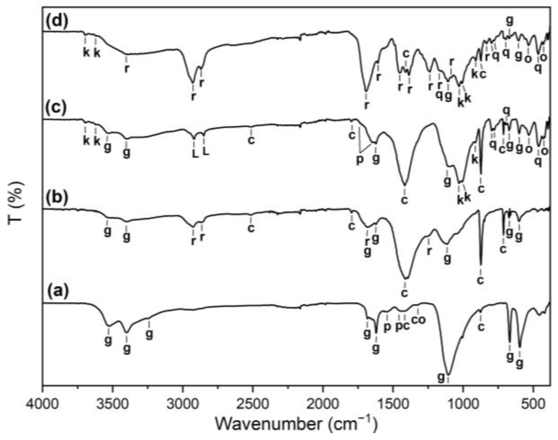
3.1. Preliminary Physico-Chemical Analysis

In order to decide the proper conservation approach, the altarpiece was analysed and the fundamental information concerning the morphology and chemical composition of the treated surfaces was gathered. Preliminary characterization is essential because it supports the conservation–restoration procedures and allows proper work in accordance with the original materials.
Microscopic images were acquired on some small detached fragments, and it was observed that the surface of the frame had suffered severe discoloration due to dirt deposits and the oxidation of the overpainted layers (Figure 2A,B). In addition to the yellowish color and porous structure in its base (Figure 2C), the ground seemed to be composed of several layers with a thickness ranging from 100 microns to several hundreds of microns divided by thin intermediate layers with an approximate thickness of several microns (Figure 2D). Such a multilayered structure of the ground is a common technique applied by post-Byzantine iconographers [20]. The stratigraphy showed a notable red layer after the ground, most probably a red bole, with varying thicknesses between 10 and 50 µm, which is a traditional base for the metallic leaf. The leaf itself is not so visible in the stratigraphic microscopic pictures. The thickness of the metallic overpaint was not uniform and was measured to be from several hundred microns to several milimetres. The preparation layer varied between 2 and 5 mm in thickness, covering both the elements of the relief and those in the background. The gilding of the sculptural elements was performed with silver leaf applied in the traditional manner on a layer of bole.
The FTIR analysis of the paint and the ground layers is presented in Figure 3. Since the spectra of the paint and the ground of S1 and S2 are similar, only S1 is presented in the figure. The infrared analysis indicated the presence of inorganic compounds characteristic of earth and clay minerals [21,22]. Kaolinite was identified via the O–H stretching vibrations at 3694, 3651, and 3621 cm−1; the vibrations of the Si–O–Si and Si–O–Al groups at 1029 and 1008 cm−1, respectively; and the Al–O–H vibration at 913 cm−1. Quartz was indicated by the characteristic doublet at 797–777 cm−1, and the bands at 1163, 695, and 464 cm−1. The bands at 534 and 430 cm−1 were ascribed to iron oxides [23,24]. The presence of calcite (CaCO3) was observed in all the spectra, as it was much more pronounced in the ground of S3 than in the grounds of the other two samples. This was detected through the weak combination modes at 2512 and 1795 cm−1 and the prominent stretching and bending modes of the carbonate group at 1412, 872, and 713 cm−1 [25]. Gypsum (CaSO4·2H2O) was found in all the spectra via the characteristic stretching (3526 and 3401 cm−1) and bending (1682 and 1623 cm−1) bands of the O–H bond, and the vibration modes of the sulphate group at 1106, 673, and 600 cm−1 [24]. The origin of the calcite and the gypsum in the paint could be from their possible use as mineral fillers or from the ground layers [26]. Given the observed multilayered structure of the ground, it is possible that the separate constituent layers could have been prepared using different recipes. It was not uncommon for the ground to consist of calcite-rich and gypsum-rich sub-layers in different ratios [20].
Some organic compounds were found as well. Proteins were present in both the paint and the grounds of S1 and S2, as determined by the emerging bands at 1646, 1544, and 1446 cm−1 assigned to C=O stretching (amide I), the combination of NH bending and CN stretching (amide II), and C-H bending (amide III) vibrations, respectively [27]. In the spectra of the paint in S1 and S2, only the bands associated with amide I and partly amide II appeared since amide III overlapped with the strong broad absorption of the carbonate group around 1410 cm−1. On the other hand, the amide I overlapped with the bending vibration modes of the hydroxyl bond in gypsum. The vibrations emerging at 2920 and 2850 cm−1 in the spectra of the paint in S1 and S2, respectively, are ascribed to the stretching mode of the CH bonds. Together with the weak absorption (appearing as a shoulder) at 1736 cm−1, this indicates the presence of lipids. Their occurrence is a clear indicator of an organic binder that can be associated with the saturated fatty acids of the proteinaceous medium, which in this case is most likely the egg yolk [28,29]. The identified proteinaceous species in the ground could be linked with animal glue, which is a common gluing ingredient in traditional recipes for the preparation layers in iconographic art [20]. It is highly likely that the intermediate thin layers observed in the multilayered ground are indeed animal glue.
The spectra of S3 show strong features of natural tree (pine) resin associated with the strong CH stretching modes at 2929 and 2870 cm−1, the strongly broadened C=O stretching at 1693 cm−1, the shoulder at 1609 cm−1, the aliphatic C–H bending bands at 1483 and 1387 cm−1, the C–O mode of the ester groups at 1240 cm−1, the C–O stretching bands of the acid and alcohol groups at 1163 and 1081 cm−1, and the band at 834 cm−1 [29,30]. The ground layer of S3 showed the presence of the resin, too. Since the bands were much weaker than those in the paint, it was suggested that they could come from the upper layer. The appearance of pine resin in such paintings is not a single case reported in the literature [31,32]. However, due to the lack of spectral features of the resin in the other samples, it cannot be concluded whether the resin was used as a finish layer against humidity or something else.
Calcium oxalate was also identified in the ground layers of S1 and S2 via the C=O stretching at 1326 cm−1. Its occurrence could be linked to the degradation of organic layers or biodeterioration by the activity of microorganisms [33].
The LIBS results obtained from averaging the spectra from 20 pulses confirmed the presence of the following chemical elements: Ca, Al, Fe, Na, K, Si, Sr, H, and C, yet no S lines were detected probably due to the fact that sulphur is present in low concentration, below the spectrometer’s limit of detection (Z300 SLOD = 1%–2%). The LIBS spectra are presented in Figure 4a,b for the main elements identified. Traces of Ti, Mg, Mn, and Li were detected as well, which were linked to the earth and clay minerals in the composition of the materials. Potassium was also found as a trace element and might be associated with soiling deposits or the formation of salts on the surface [34]. These findings are in accordance with the FTIR interpretation, showing the presence of gypsum, calcite, and iron oxides.
LIBS stratigraphy was performed in order to obtain a glimpse of the distribution of the chemical elements by analysing each of the 20 pulses (Figure 5a–d) [35,36]. It was observed that the most analysed elements started to count only from the 5 to 6th pulse, with a peak between the 7th and the 11th pulses, corresponding to the metallic overpaint. This can be an indication that the metallic overpaint layer is covered with another layer of organic or synthetic nature, as LIBS detects only trace elements. Starting with pulse 12 till the end of the series, the distribution tends to have a quasi-constant shape for most of the elements pictured, which can be attributed to reaching the ground, as can be observed in Figure 5c,d that presents the Si and Sr distribution.

3.2. Laser Removal of the Metallic Overpaint

The laser cleaning regime was established empirically as a result of preliminary tests conducted on an area of approximately 4 cm2. A low starting laser energy was set, and it was increased gradually until the ablation of the overpaint layers was observed using OM and LIBS. The ablation thresholds determined using the D2 method are presented in Table 1. In Figure 6, microscopic images of the laser-cleaned areas are shown. The different testing areas for optimal and overcleaning are noted. It was noticed that the first overpaint layer and the adherent deposits were removed efficiently at an energy of 250 mJ (fluence of 0.62 J/cm2), as can be seen in Figure 6C,G. As the energy was increased, at energies around 350 mJ (fluence of 0.8 J/cm2), the thick metallic overpaint layer started ablating and the traces of the silver leaf became noticeable, as can be seen in Figure 6E. Any further increase in the energy would result in overcleaning; thus, the ablation threshold was set at 0.8 J/cm2.
Since the overpainting layer was not uniform, nor was its thickness, the nonuniformity of the cleaning was clearly visible, and in some areas, the red bole was reached. Therefore, it was decided to use several pulses in each area (20–30 pulses) rather than using higher energy.
Laser cleaning provided good results for the rough removal of the thick layers of the metallic paints applied on top of the highly degraded pictorial substrate in a relatively short time compared to traditional cleaning techniques. Taking into consideration the thin and fragile remains of the original materials under the overpaints, and also the fact that the high fluence necessary for removal of the overpaints is over the ablation threshold of the pictorial composition reported in the literature [11,37,38], the best conservation strategy is unquestionably a hybrid approach that combines photonics with traditional methods, using the laser for the rough removal of the thick overpaint and then applying solvents to reveal the original religious motifs.
The pictorial layer was revealed by chemical cleaning using a solution composed of 85% dichloromethane, 10% methyl alcohol, and Deck 3000 with a gel-like consistency. This solution was applied using moistened pads, with the gel serving the purpose of retaining a mixture of highly volatile solvents in the treated areas for an adequate duration.

4. Conclusions

The current paper presents the interdisciplinary approach undertaken for the conservation of a 19th Century iconostasis belonging to the “Holy Trinity” church from Măgureni, România. The polychrome religious paintings were covered with what was initially thought to be a thick layer of metallic paint that was applied on top of previous restorations.
Advanced photonic methods were selected for the investigation of the chemical composition and characterization of the substrates, and laser-cleaning efficiency was tested. The stratigraphic analyses determined that the overpaint actually consisted of two layers, one of synthetic nature and the other of a metallic paint based on Fe and Al. Also, elemental analysis detected traces of many elements resulting from the terrigenous origin of the raw materials. Earth and clay minerals, such as kaolinite, calcite, gypsum, iron oxides, and quartz, were identified in a binding medium of egg yolk. The ground consisted of several sublayers of gypsum and/or calcite mixed with animal glue, which was a typical preparation layer following the conservative iconographic traditions. In one of the samples, a prominent presence of a natural tree (pine) resin was observed. The information obtained from the spectroscopic analysis was essential for establishing a conservation approach and for understanding the historical and cultural value of the polychrome wooden iconostasis.
The cleaning methodology discussed in this paper involved the use of a laser to roughly remove the overpaint layers. A Q-switched Nd:YAG laser emitting at a wavelength of 1064 nm, a pulse duration of 8 ns, and a pulse repetition rate of 10–20 Hz was used. The optimal working fluence for removing the thick metallic overpaint layers was found to be 0.8 J/cm2, which achieved good cleaning efficiency after 5–6 passes of the beam on the surface. Taking into consideration the spectroscopic analysis and the laser cleaning tests, the best conservation strategy was decided to be a hybrid approach, combining photonic with traditional methods: using laser for the rough removal of the thick overpaint layers and solvents to reveal the original religious motifs.
Photonics plays a dynamic role in most fields and, as technology advances, so do the applications developed within the Heritage Sciences field, enabling better understanding and valorization. By providing detailed information about their composition, structure, and condition, photonic techniques can guide conservation interventions and help preserve these valuable cultural treasures for future generations.

Author Contributions

Conceptualization, V.A.; methodology, R.R. and S.-R.P.; FTIR, V.A., LIBS, M.D.; laser cleaning, S.-R.P. and M.D.; microscopy, V.A.; writing—original draft preparation, V.A.; writing—review and editing, M.D.; funding acquisition, R.R. and M.D. All authors have read and agreed to the published version of the manuscript.

Funding

This research was funded by the Romanian Ministry of Research, Innovation, and Digitalization, under Program 1—Development of the National Research-Development System, Subprogram 1.2—Institutional Performance: Projects to finance the excellence in RDI, SUPECONEX, grant no. 18PFE/30.12.2021, PNCDI 2022-2027—Core Programme 11N/03.01.2023, project no. PN 23 05 and under CNCS - UEFISCDI, project number PN-III-P4-PCE-2021-1605, within PNCDI III.

Institutional Review Board Statement

Not applicable.

Informed Consent Statement

Not applicable.

Data Availability Statement

The data that support the findings of this study are available upon request from the corresponding authors.

Conflicts of Interest

The authors declare no conflict of interest. The funders had no role in the design of the study; in the collection, analyses, or interpretation of data; in the writing of the manuscript; or in the decision to publish the results.

References

  1. Draghiceanu, V.N. Casa Cantacuzinilor din Magureni; Ed. Ramuri: Craiova, Romania, 1924. [Google Scholar]
  2. Boonrat, P.; Dickinson, M.; Cooper, M. Initial investigation into the effect of varying parameters in using an Er:YAG laser for the removal of brass-based overpaint from an oil-gilded frame. J. Inst. Conserv. 2020, 43, 94–106. [Google Scholar] [CrossRef] [Green Version]
  3. Panzner, M.; Wiedemann, G.; Meier, M.; Conrad, W.; Kempe, A.; Hutsch, T. Laser Cleaning of Gildings. Lasers Conserv. Artworks 2007, 116, 21–28. [Google Scholar] [CrossRef]
  4. Ungurean, B. Notes on the iconostas of St. Theodore’s church from Iasi. Technique of execution, stylistic description and state of conservation. Anastasis 2018, 5, 86–109. [Google Scholar]
  5. Vuga, M.; Semion, M.M. Typical conservation problems of polychrome wooden sculptures in Slovenia. Conserv. Patrim. 2015, 22, 17–27. [Google Scholar] [CrossRef] [Green Version]
  6. Strzelec, M.; Marczak, J.; Koss, A.; Szambelan, R. Overpaint Removal on a Gilded Wooden Bas-Relief Using a Nd:YAG Laser at 1.064 µm. Laser Conserv. Artworks 2005, 100, 133–138. [Google Scholar] [CrossRef]
  7. Gaspar, P.; Rocha, M.; Kearns, A.; Watkins, K.; Vilar, R. A study of the effect of the wavelength in the Q-switched Nd:YAG laser cleaning of gilded wood. J. Cult. Herit. 2000, 1, 133–144. [Google Scholar] [CrossRef]
  8. Alabone, G.; Carvajal, M.S. The removal of bronze paint repairs from overgilded picture frames using an Erbium:YAG laser. J. Inst. Conserv. 2020, 43, 107–121. [Google Scholar] [CrossRef]
  9. Pouli, P.; Selimis, A.; Georgiou, S.; Fotakis, C. Recent studies of laser science in paintings conservation and research. Acc. Chem. Res. 2010, 43, 771–781. [Google Scholar] [CrossRef]
  10. Asmus, J.F. Light for Art Conservation. Interdiscip. Sci. Rev. 2012, 12, 171–179. [Google Scholar] [CrossRef]
  11. Fotakis, C.; Kautek, W.; Castillejo, M. Lasers in the Preservation of Cultural Heritage. Laser Chem. 2006, 2006, 1. [Google Scholar] [CrossRef] [Green Version]
  12. Acquaviva, S.; D’anna, E.; De Giorgi, M.; Della Patria, A.; Pezzati, L.; Pasca, D.; Vicari, L.; Bloisi, F.; Califano, V. Laser cleaning of gilded wood: A comparative study of colour variations induced by irradiation at different wavelengths. Appl. Surf. Sci. 2007, 253, 7715–7718. [Google Scholar] [CrossRef]
  13. Siano, S.; Grazzi, F.; Parfenov, V.A. Laser cleaning of gilded bronze surfaces. J. Opt. Technol. 2008, 75, 419–427. [Google Scholar] [CrossRef]
  14. Andreotti, A.; Bracco, P.; Colombini, M.P.; Decruz, A.; Lanterna, G.; Nakahara, K.; Penaglia, F. Novel Applications of the Er:YAG Laser Cleaning of Old Paintings. Lasers Conserv. Artworks 2007, 116, 239–247. [Google Scholar] [CrossRef]
  15. Brunetto, A.; Bono, G.; Frezzato, F. Er:YAG laser cleaning of ‘San Marziale in Gloria’ by Jacopo Tintoretto in the Church of San Marziale, Venice. J. Inst. Conserv. 2020, 43, 44–58. [Google Scholar] [CrossRef]
  16. Wiedemann, G.; Pueschner, K.; Wust, H.; Kempe, A. The Capability of the Laser Application for Selective Cleaning and the Removal of Different Layers on Wooden Artworks. Lasers Conserv. Artworks 2006, 100, 179–190. [Google Scholar] [CrossRef]
  17. Striber, J.; Jovanović, V.; Jovanović, M. Easel paintings on canvas and panel: Application of Nd:YAG laser at 355 nm, 1064 nm and UV, IR and visible light for the development of new methodologies in conservation. In Proceedings of the LACONA XI; Elsevier: Amsterdam, The Netherlands, 2017; pp. 279–292. [Google Scholar] [CrossRef]
  18. Kramida, A.; Ralchenko, Y.; Reader, J.; NIST Atomic Spectra Database Team. Atomic Spectra Database (Version 5.3); National Institute of Standards and Technology: Gaithersburg, MD, USA, 2015.
  19. Sawicki, M.; Bramwell–Davis, V.; Dabrowa, B. Laser cleaning from a practical perspective: Cleaning tests of varied gilded-wood surfaces using Nd:YAG Compact Phoenix laser system. AICCM Bull. 2011, 32, 44–53. [Google Scholar] [CrossRef]
  20. Mastrotheodoros, G.P.; Beltsios, K.G.; Bassiakos, Y.; Papadopoulou, V. On The Grounds of Post-Byzantine Greek Icons. Archaeometry 2016, 58, 830–847. [Google Scholar] [CrossRef]
  21. Cortea, I.M.; Ghervase, L.; Ratoiu, L.; Dinu, M.; Rădvan, R. Uncovering hidden jewels: An investigation of the pictorial layers of an 18th-century Taskin harpsichord. Herit. Sci. 2020, 8, 55. [Google Scholar] [CrossRef]
  22. Cortea, I.M.; Ghervase, L.; Rădvan, R.; Serițan, G. Assessment of Easily Accessible Spectroscopic Techniques Coupled with Multivariate Analysis for the Qualitative Characterization and Differentiation of Earth Pigments of Various Provenance. Minerals 2022, 12, 755. [Google Scholar] [CrossRef]
  23. Chukanov, N.V.; Chervonnyi, A.D. Some General Aspects of the Application of IR Spectroscopy to the Investigation of Minerals. In Infrared Spectroscopy of Minerals and Related Compounds; Springer: Berlin/Heidelberg, Germany, 2016; pp. 1–49. [Google Scholar] [CrossRef]
  24. Genestar, C.; Pons, C. Earth pigments in painting: Characterisation and differentiation by means of FTIR spectroscopy and SEM-EDS microanalysis. Anal. Bioanal. Chem. 2005, 382, 269–274. [Google Scholar] [CrossRef]
  25. Gunasekaran, S.; Anbalagan, G.; Pandi, S. Raman and infrared spectra of carbonates of calcite structure. J. Raman Spectrosc. 2006, 37, 892–899. [Google Scholar] [CrossRef]
  26. Carlyle, L.; Roy, A. Artists’ Pigments: A Handbook of Their History and Characteristics, Volume 2. J. Am. Inst. Conserv. 1996, 37, 892–899. [Google Scholar] [CrossRef]
  27. Derrick, M.; Stulik, D.; Landry, J. Infrared Spectroscopy in Conservation Science. The Effects of Brief Mindfulness Intervention on Acute Pain Experience: An Examination of Individual Difference; Getty Publications: Los Angeles, CA, USA, 2015. [Google Scholar]
  28. Cortea, I.M.; Ratoiu, L.; Chelmuș, A.; Mureșan, T. Unveiling the original layers and color palette of 18th century overpainted Transylvanian icons by combined X-ray radiography, hyperspectral imaging, and spectroscopic spot analysis. X-ray Spectrom. 2022, 51, 26–42. [Google Scholar] [CrossRef]
  29. Lazidou, D.; Lampakis, D.; Karapanagiotis, I.; Panayiotou, C. Investigation of the Cross-Section Stratifications of Icons Using Micro-Raman and Micro-Fourier Transform Infrared (FT-IR) Spectroscopy. Appl. Spectrosc. 2018, 72, 1258–1271. [Google Scholar] [CrossRef] [PubMed]
  30. Vahur, S.; Teearu, A.; Peets, P.; Joosu, L.; Leito, I. ATR-FT-IR spectral collection of conservation materials in the extended region of 4000-80 cm–1. Anal. Bioanal. Chem. 2016, 408, 3373–3379. [Google Scholar] [CrossRef]
  31. Bretz, S.; Baumer, U.; Stege, H.; Von Miller, J.; Von Kerssenbrock-Krosigk, D. A German house altar from the sixteenth century: Conservation and research of reverse paintings on glass. Stud. Conserv. 2008, 408, 3373–3379. [Google Scholar] [CrossRef]
  32. Guttmann, M.J. Transylvanian glass icons: A GC/MS study on the binding media. J. Cult. Herit. 2013, 14, 439–447. [Google Scholar] [CrossRef]
  33. Antunes, V.; Candeias, A.; Mirão, J.; Carvalho, M.L.; Dias, C.B.; Manhita, A.; Cardoso, A.; Francisco, M.J.; Lauw, A.; Manso, M. Analytical characterization of the palette and painting techniques of Jorge Afonso, the great 16th century Master of Lisbon painting workshop. Spectrochim. Acta-Part A Mol. Biomol. Spectrosc. 2018, 193, 264–275. [Google Scholar] [CrossRef]
  34. Dinu, M.; Cortea, I.M.; Cortea, L.; Stancu, M.C.; Mohanu, I.; Cristea, N. Optoelectronic investigation of the mural paintings from Drăguțești wooden church, Argeș County, Romania. J. Optoelectron. Adv. Mater. 2020, 22, 303–309. [Google Scholar]
  35. Pacher, U.; Dinu, M.; Nagy, T.O.; Radvan, R.; Kautek, W. Multiple wavelength stratigraphy by laser-induced breakdown spectroscopy of Ni-Co alloy coatings on steel. Spectrochim. Acta-Part B At. Spectrosc. 2018, 146, 36–40. [Google Scholar] [CrossRef]
  36. Simileanu, M. Libs quantitative analyses of bronze objects for cultural heritage applications. Rom. Reports Phys. 2016, 68, 203–209. [Google Scholar]
  37. Dascalu, G.R.; Stancu, M.C.; Dinu, M.; Puscas, N. Laser cleaning of polychrome artworks. Case study on graffiti. UPB Sci. Bull. Ser. A Appl. Math. Phys. 2020, 82, 307–316. [Google Scholar]
  38. Kautek, W. Lasers in Cultural Heritage: The Non-Contact Intervention. Springer Ser. Mater. Sci. 2010, 130, 331–349. [Google Scholar] [CrossRef]
Figure 1. The iconostasis of the “Holy Trinity” church (left). In the red rectangle, the fragment of the 4th register cleaned with a laser is shown. Closer details and the analysed areas of the altarpiece (right).
Coatings 13 01235 g001
Figure 2. Microscopic images (A) S2, (B) S3, (C) S2—ground, and (D) S1—stratigraphy.
Coatings 13 01235 g002
Figure 3. ATR-FTIR analysis of the paint and the ground layers of part of the samples: (a) S1—ground layer, (b) S3—ground layer, (c) S1—paint layer, and (d) S3—paint layer. Legend: k—kaolinite; g—gypsum; c—calcite; q—quartz; o—iron oxides; p—proteins; L—lipids; r—resin; co—calcium oxalate.
Coatings 13 01235 g003
Figure 4. LIBS elemental analysis for (a) Ca, Al, and Fe; and (b) C, Mg, and Si.
Coatings 13 01235 g004
Figure 5. LIBS elemental depth distribution through 20 pulses for (a) Fe, (b) Al, (c) Si, and (d) Sr.
Coatings 13 01235 g005
Figure 6. Micro details of the laser-cleaned surfaces. (A) test areas 1-3, (B) test area 4, (C) test area 5, (D) test area 6, (E) test area 7, (F) test area 8, (G) test area 9.
Coatings 13 01235 g006
Table 1. Laser cleaning ablation thresholds for the adherent deposits and the overpainted substrates.
SubstrateAblation Threshold
Surface adherent deposits0.37 J/cm2
First overpaint layer0.62 J/cm2
Second overpaint layer0.8 J/cm2
Disclaimer/Publisher’s Note: The statements, opinions and data contained in all publications are solely those of the individual author(s) and contributor(s) and not of MDPI and/or the editor(s). MDPI and/or the editor(s) disclaim responsibility for any injury to people or property resulting from any ideas, methods, instructions or products referred to in the content.

