Article

Sensitive Fingerprint Detection Using Biocompatible Mesoporous Silica Nanoparticle Coating on Non-Porous Surfaces

1
Division of Forensic Science, School of Basic and Applied Sciences, Galgotias University, Greater Noida 201301, India
2
International Centre for Research on Innovative Biobased Materials (ICRI-BioM)-International Research Agenda, Lodz University of Technology, Żeromskiego 116, 90-924 Lodz, Poland
3
Department of Biotechnology, School of Liberal Arts & Science, Sree Vidyanikethan Engineering College, Mohan Babu University, Tirupati 517102, India
4
Department of Biotechnology, Yeungnam University, Gyeongsan 38541, Republic of Korea
5
Division of Sustainable Agriculture, The Energy and Resources Institute (TERI), New Delhi 110003, India
*
Authors to whom correspondence should be addressed.
These authors contributed equally to this work.
Coatings 2023, 13(2), 268; https://doi.org/10.3390/coatings13020268
Submission received: 8 December 2022 / Revised: 8 January 2023 / Accepted: 18 January 2023 / Published: 23 January 2023

Abstract

:
In recent years, the development and application of biocompatible nanomaterials in the detection of fingerprints have become a major focus for the forensic sector and crime investigators. This study aims to synthesize biocompatible silica nanoparticles (Si NPs) through cost-effective green methods and will be used to detect a latent fingerprint on a non-porous surface. As a type of environmentally friendly nanomaterial, Si NPs were prepared via an oil–water mixed micro-emulsion templating (MET) approach. Their characteristics and optical properties were measured using EDX-SEM, HR-TEM, FTIR, XRD, and UV–visible absorption. The biocompatibility of the synthesized Si NPs in terms of cell viability was observed, even at high concentrations (83.46% and 75.28% at 20 and 50 mg mL−1, respectively). The developed Si NPs were tested on different surfaces, including plastic, glass, silicon, steel, and soft plastic for the detection of crime scene fingerprints. In this research, it was found that the Si NPs were of the size of 100–150 nm. Results confirmed that synthesized mesoporous Si NPs can be used to detect latent fingerprints on multiple non-porous surfaces and were easy to detect under a UV lamp at 395 nm. These findings reinforce the suggestion that the developed Si NP coating has a high potential to increase sensitive and stable crime traces for forensic latent fingerprint detection, even in packaged food with different packaging surfaces.

1. Introduction

Identification has always been a problem associated with the criminal justice system (CJS); fingerprint identification is itself an already proven magical boon to the CJS. The science of fingerprints is one of the most specialized sciences, which plays an utmost authoritative role in various investigative processes that come across the forensic world. Fingerprints are the impressions made through ridge patterns, mainly on the tips of fingers [1]. Along with the advancements, the term “fingerprints” is not merely limited to finger impressions but also encompasses the impressions obtained by the palm and sole impressions of the human body. Fingerprints are also associated with various other sciences such as forensic chemistry, anthropology, anthropometry, and biometric sciences. It has been known since scientific times that forensic scientists generally approach genetic profiles through blood, semen, hair, bone, urine, etc. [2].
Latent fingerprints are special kind of fingerprints which are not visible to the naked eye and need to be procured and analyzed with more sophistication. There were various traditional methods, such as powder dusting, iodine fuming, cyanoacrylate fuming, etc., which are used as preferred techniques at the scene of the crime for the development of latent fingerprints. These methods have some drawbacks in terms of toxicity, less sensitivity, less contrast, high autofluorescence intrusion, interference of background, and unclear ridges, and not all levels of fingerprints have been developed [3,4]. New approaches have been introduced to improve the quality of hidden finger impressions, making them more affordable, and providing higher efficacy to help in the investigation by identifying the perpetrator in order to overcome these shortcomings. Nowadays, nanoscience is also playing a vital role in the personal identification and characterization of suspects, and offenders being related to each other or to the scene of crime [5,6].
Due to the precise and sensitive analysis in a criminal investigation, nanoparticles are gaining more and more consideration in the field of latent fingerprinting. Some of the specific characteristics of nanoparticles that recommend their exploitation in latent fingerprints are their reduced size and good chemical characteristics. Nanomaterials that are currently playing a major role in latent fingerprinting include carbon dots, quantum dots, rare earth, etc. [7]. High sensitivity, minimal toxicity, and high contrast images are only a few benefits of nanoparticles, some of which are also cost-effective [7,8]. By chemically reacting with the amino acids, fatty acids, and other water-soluble compounds found in the imprint, nanoparticles can physically bond to them. Nanomaterials also have photoluminescence characteristics. These attributes allow for the creation of clear imprints of finger impressions. Nanoparticles made of gold, silver, titanium dioxide, zinc oxide, silica etc. are used to detect fingerprints [9,10,11].
Fluorescent nanoparticles (NPs) have been claimed to be superior in this class due to their properties of enhancing contrast in latent fingerprints due to their nano-level size and specific optical characteristics, strong fluorescence intensity, and photostability [12]. The fluorescent NPs emphasize the finer features of the buried finger impressions and improve all three levels of fingerprints. Fluorescent NPs can achieve the best contrast quality, enhancement, sensitivity, and selectivity, as compared to other NPs, as well as possess reduced toxicity, less autofluorescence interference, and lower background inference [13]. Because people typically touch their faces and hair, oily substances can be found in dormant finger impressions. Therefore, a fluorescent reagent might be considered a flexible probe for latent finger impression fluorescence imaging if it diffused into the oily substances without rubbing out the hidden impressions with the substrate.
Due to their optical transparency, tiny particle size, large surface area, high fluorescence, robust photo-stability, outstanding biocompatibility, advantageous low toxicity, and high surface absorbency, mesoporous silica nanoparticles (MSNPs) are discovered to be potential fluorescent NPs for utilization in latent fingerprinting. To improve adsorption, silica materials frequently increase surface functionalization with hydrophobic and/or hydrophilic groups. Following that, they offer fingerprint visualization using fluorescence markers. MSNPs have significant use prospects in fluorescence sensors, bio-labeling, and medical imaging because of their unique characteristics. The surface controllability of MSNPs is another appealing quality of them as compared to other semiconductor materials. The emission of fluorescence is significantly impacted by changes in surface conditions [14].
By adding silane-modified organic dye molecules, the Stober technique integrates fluorescent NPs. The allylation and hydrolyzation processes can be used to synthesize these changed compounds. Similar optical characteristics with exceptional photostability have been observed in the produced fluorescent NPs and PR254A (allylated pigment red 254). In order to increase the binding affinity of poly (N-vinyl-2-pyrrolidone) (PVP)-coated Si NPs for fingerprint detection, PVP is added to their surface. Fluorescent Si NPs have the potential to improve fingerprint detection on hydrophobic and hydrophilic surfaces for individual identification due to their simple fabrication and controllable surface qualities [15].
A unique emerging agent for fingerprint detection can be made from mesoporous MSNPs with a particle size as small as 50 nm. A form of dye, methylene blue molecules, can improve the contrast of produced fingerprints when combined with nanomaterials. The two techniques for generating fingerprints are suspension and powder. Additionally, the powder approach exhibits a better effect on fingerprint growth than the other method and is successful in detecting the sweat pores of fingerprints [16]. The inventions and advances made possible by nanotechnology have had a huge influence on how fingerprint development procedures are now more sensitive and selective. Nanomaterials can be used to improve the development of fingerprint ridges, luminous fingerprints, and chemical probing. As a result, it is shown that the spherical mono-dispersed method is extremely effective in leaving fingerprints on both semi-porous and non-porous surfaces [17,18].
Although attempts have been made to increase the detection limit of latent fingerprints (LFPs) by using fluorescent nanomaterials, such as quantum dots, carbon dots, and up-conversion nanoparticles, issues still persist due to their poor detection performance, difficult manufacturing process, photobleaching, and toxicity [15,19,20]. There is a need to develop more efficient and cost-effective Si NPs using easy methods. Therefore, in this study, to overcome the aforementioned limitations, this approach has been synthesized for the development of latent finger impressions.

2. Materials and Methods

2.1. Chemicals and Reagents

Cetyltrimethylammonium bromide (CTAB), ammonium hydroxide, and tetraethyl orthosilicate (TEOS), silica, fumed (SiO2) (MW 60.08, 0.2–0.3 µm), and Whatman® Grade 1 filter paper were purchased from Sigma-Aldrich (St. Louis, MO, USA). Hydrochloric acid (HCl), methanol, ethyl acetate, mercuric chloride, and ethanol were of analytical grade (AR) and purchased from Merck (Mumbai, India).

2.2. Sample Collection

Three individuals provided the samples for this work (three female and one male). To eliminate any type of filth, each volunteer cleansed their hands with soap and water. In order to remove any extra sweat or oil, the fingers were rubbed on the nose and forehead before being placed on various surfaces. On these surfaces, full and partial prints of each participant were obtained. The surfaces taken included both porous and non-porous ones.

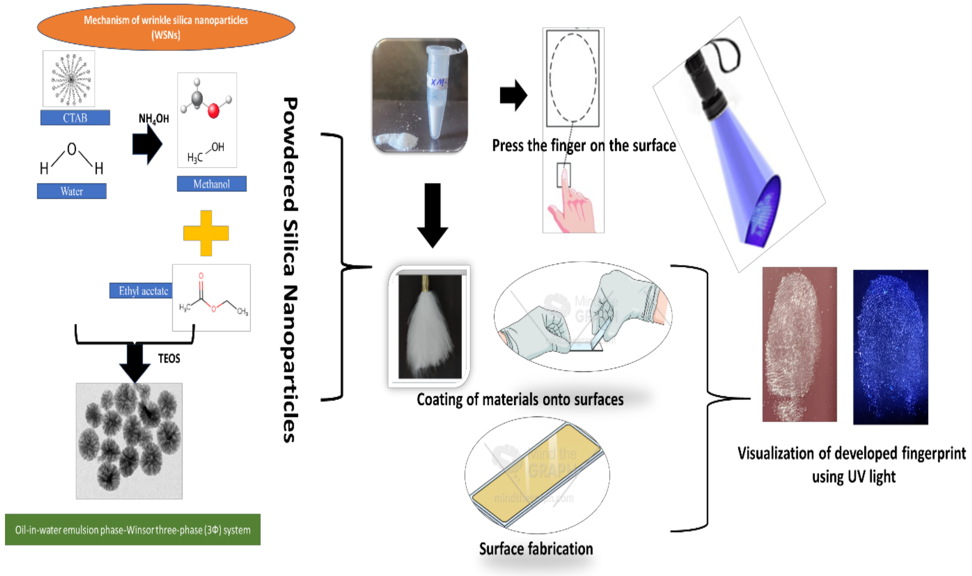
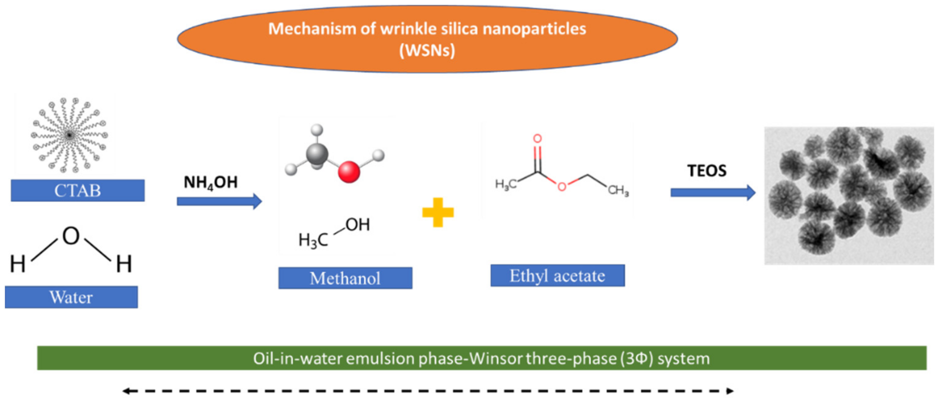
2.3. Synthesis of Mesoporous Silica Nanoparticles (MSNPs)

MSNPs were synthesized using some previously reported methods with slight modifications [21,22,23]. In particular, for the synthesis of MSNPs, 0.055 M n-hexadecyl-trimethylammonium bromide (CTAB) aqueous solution was prepared in 10 mL Milli-Q water under vigorous stirring for 30 min, followed by heating at 60 °C for 15 min. The mixture was cooled to room temperature and further added to a solvent mixture containing 5 mL of methanol, 95 mL of MilliQ water, 3 mL of ammonium hydroxide (NH4OH), and 20 mL of ethyl acetate (CH3COOC2H5) under stirring conditions and then 300 μL of tetraethyl orthosilicate (TEOS) was added to the reaction solution. The resulting solution was stirred for 12 h. The synthesized MSNPs were washed with excess ethanol and stored in 40 mL ethanol until further use. The CTAB was extracted by adjusting the pH to 1.6 and stirring for 3 h at 60 °C. After washing three times with ethanol, the pellet was dispersed in 20 mL of ethanol and stored at room temperature for further characterization. The aliquots were sealed tightly.

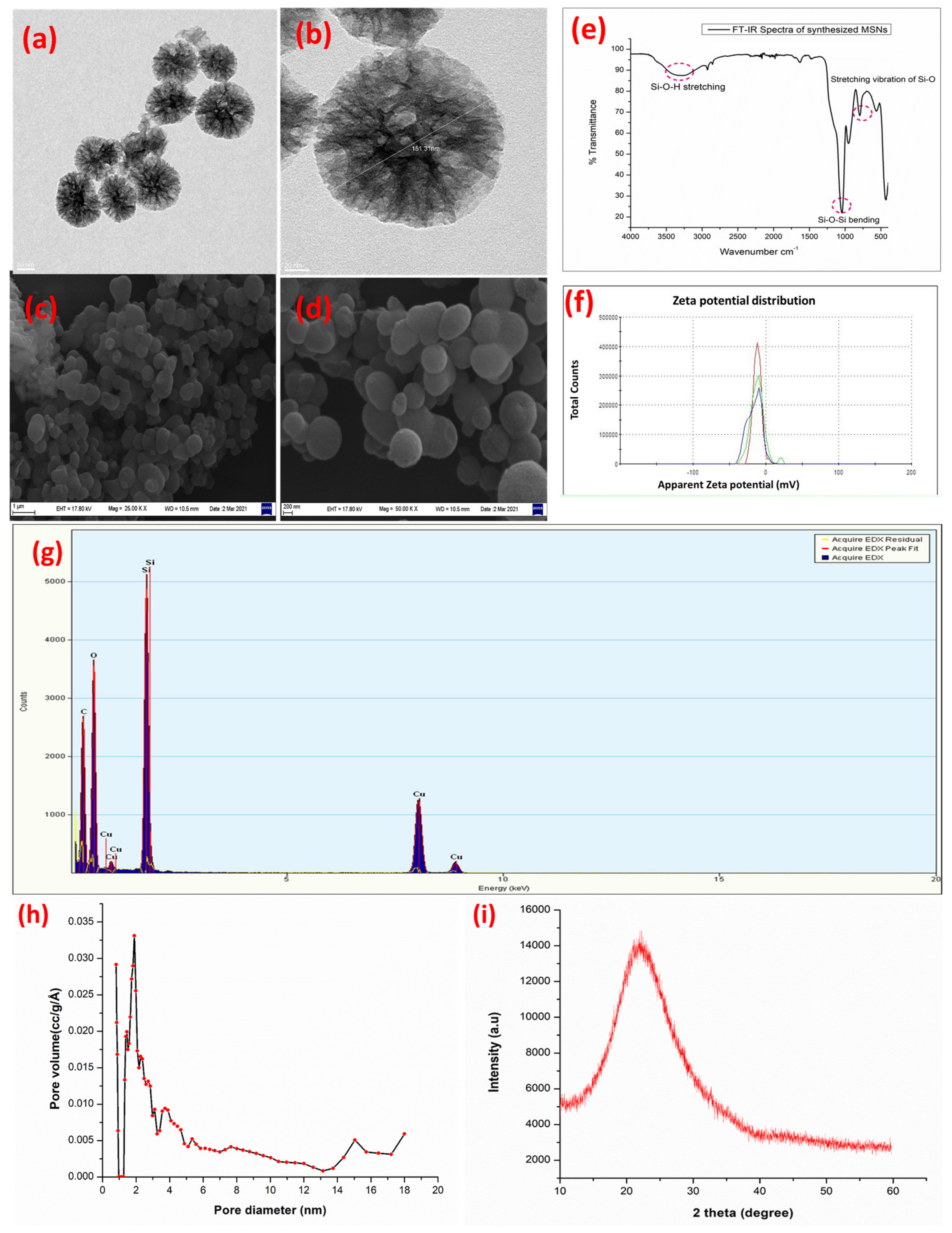
2.4. Characterization of Synthesized Nanoparticles

The Fourier transform infrared spectroscopy (FTIR) spectra were recorded using a PerkinElmer FTIR spectrometer at room temperature in the 400–4000 cm−1 spectral range. The pore size and pore volume were measured following the Barrett–Joyner–Halenda (BJH) method using an Autosorb automated gas desorption analyzer. Brunauer–Emmett–Teller (BET) measurements were done to measure the surface area of the synthesized Si NPs.
The morphological characterization of the synthesized MSNPs was observed via scanning electron microscope (SEM) and transmission electron microscope (TEM). In brief, for SEM, dried samples were coated in a sample holder and coated with gold-palladium sputtering, followed by SEM analysis at different magnifications. Similarly, TEM samples were prepared in ethanol and sonicated for 10 min, followed by dropping on a copper grid and dried under a hygroscopic chamber at room temperature.
The morphology of the synthesized MSNPs was examined using a JEOL 2100F transmission electron microscope (TEM) at 200 kV connected to an energy-dispersive analysis.

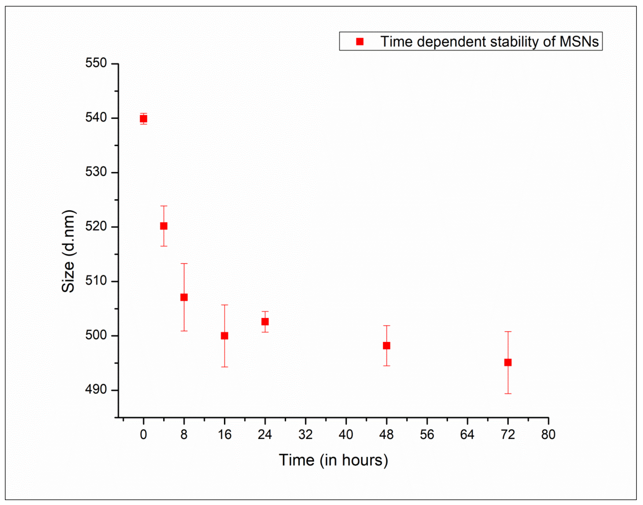
2.5. Assessment of Post-Synthesis Stability of MSNPs

The stability of these MSNPs in terms of aggregation and nanoparticle size, prior to soil and foliar application, was assessed using a quantitative non-plasmonic characterization technique (DLS: dynamic light scattering) at different time intervals (0–72 h).

2.6. Cytotoxicity of the Synthesized MSNPs via Cell Viability Analysis

Cell Lines and Culturing

Lung epithelial cells of human origin (beas-2) were purchased from the American Type Cell Culture Collection Center (ATCC) and maintained in RPMI cell culture medium supplemented with fetal calf serum and penicillin−streptomycin (1%), under a CO2 atmosphere at 37 °C. The growth of cells in the culture medium was confirmed after every second day, followed by refreshing the culture medium.
The biocompatible nature of the synthesized highly porous MSNPs was measured via a 3-(4,5-dimethylthiazol-2-yl)-2,5-diphenylshytetrazolium bromide (MTT) assay. Two days prior to the assay, cells (Hela) were cultured at the density of 5 × 10⁴ cells/well, followed by incubation for 24 h until confluency of 80%, after which the cells were treated with different concentrations of MSNPs (5, 10, and 15 µg mL−1), followed by incubation for 18−24 h at 37 °C. Cells were then treated with 5 mg mL−1 MTT reagent to achieve blue crystals that were dissolved in dimethyl sulfoxide; the absorbance was then measured at 540 nm. Results were calculated based on initial and final absorbance measurements taken at 490 and 680 nm, respectively. The cells were also checked for microscopic morphological alterations after the treatments.

2.7. Application of MSNPs for Detection of Fingerprints

The synthesized MSNPs were tested for their ability for detecting fingerprints on different types of surfaces (porous, semi-porous, and non-porous) for crime diagnosis.

Detection Procedure for Fingerprints

A camel hairbrush was used to apply nano-powder to the surface. To develop the prints on the surface, strokes were applied. The substances secreted by the sebaceous, eccrine, and apocrine glands that result in a finger impression were being left on the surface. The fluids were discovered in finger impressions when suggested on the surface after wiping fingertips across the nose and forehead.
By using a hairbrush, the nano-powder of MSNPs was dispersed to the multiple substrates that retained latent finger impressions. The white color of the nano-powder contrasted well with both colored and dark surfaces. The latent fingermarks of nano-powder produced ridge features with little background disturbance.

3. Results

3.1. Synthesis and Characterization of the Synthesized MSNPs

The MSNPs were prepared using ethyl acetate which acted as a pore expansion agent in the presence of CTAB in a silica sol-gel. The overall scheme of the current work is presented in Figure 1. The characterization of the synthesized MSNPs using TEM (Figure 2a,b) and SEM (Figure 2c,d) showed deep, furrow-like mesopores. The functional groups present in the synthesized MSNPs were analyzed with FT-IR spectroscopy in the spectral range of 400–4000 cm−1 to identify the presence of silica (Figure 2e). Typical absorption bands of the silicate at 784.71, 1057.45, 1609.33, and 3308.95 cm−1 were observed in the MSNPs and were assigned to a siloxane bond, Si-O-Si bending, and silanol (Si-OH) symmetric stretching, [24,25] and bending vibrations [25,26]. The pore size and volume of MSNPs were determined by N2 sorption (Figure 2h). The MSNPs showed bimodal pores that peaked at 7.2 nm in the pore size distribution. The BET surface area and pore volume of the synthesized MSNPs were found to be 941.88 m2 g−1 and 1.30 cm3 g−1, respectively gb (Figure 2h).
The XRD pattern of the amorphous silica powder is shown in Figure 2g. The XRD pattern reveals the amorphous nature of the material. The strong broad peak at 22.17°(2θ) (Figure 2i) indicates that the MSNPs that were synthesized chemically were amorphous and no crystalline structure appeared. Hence, the sample is considered as amorphous MSNs. Similar XRD patterns of silica nanoparticles have been reported by other research groups [27,28]. The EDX analysis (Figure 2g) confirmed the presence of major constituting elements, i.e., Si, O, and C in the MSNPs. Moreover, the zeta potential of the Si NPs was determined to be −27 mV, confirming the colloidal stability of the synthesized MSNPs with a reduced potential for aggregation (Figure 2f).

3.2. Stability Studies of MSNPs via Transformative Changes in Particles during Storage

The stability of the synthesized MSNPs was analyzed using DLS over a time period of 72 h at room temperature (25 ± 2 °C) (Figure 3). The MSNPs displayed stability over 72 h and no changes were detected from the DLS data under different time points, ensuring that MSNPs remained stable until 72 h. It also indicates that these MSNPs can be easily formulated for foliar spray applications. The chemical linking and scheme of wrinkled silica nanoparticles with enlarged pores are presented in Figure 4.

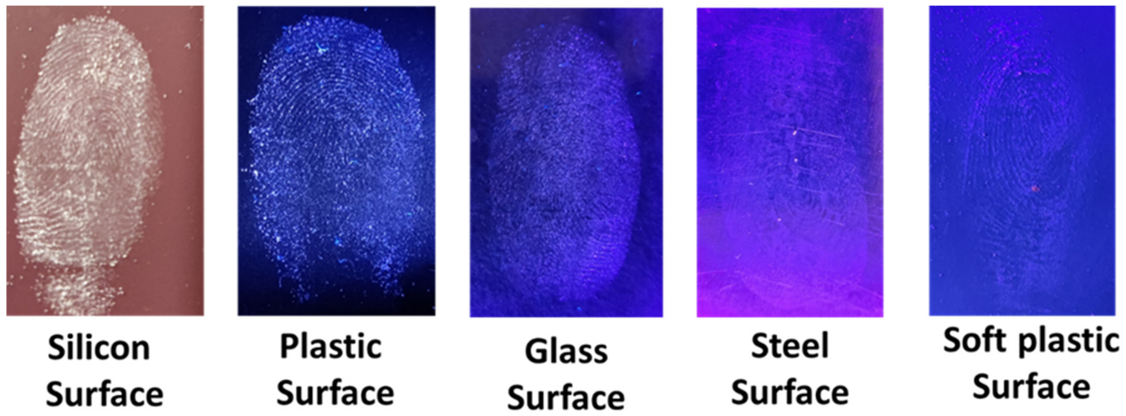
3.3. Development of Quiescent Finger Impressions Using MSNPs

To adhere the powder particles to perspiration and greasy elements, such as grease and oil in the fingerprint residue, powder brushing is a fundamental physical approach. MSNPs were physically adsorbed into the ridges created on the surface during printing at a faster rate due to the presence of sweat and oil in fingerprint residue. The fluorescent MSNPs in the fingerprints were then exposed to 365 nm UV light, which caused them to emit a blue light, thereby revealing the fingerprint imprint.

3.4. Development of Latent Fingerprints by Using the MSNPs

The dormant finger impressions were developed on five different porous and non- porous surfaces, including glass, paper, wooden surface, steel cup, and plastic lid using the MSNPs (Table 1). After developing the dormant finger impressions, they were visualized under a UV torch light (395–400 nm). The fingerprint patterns and ridge characteristics were observed by the naked eye. The plastic, glass, and metal surfaces showed good results based on the size of the nanoparticles and also due to the suspension of the fingermark residue on the surfaces (Figure 5), whereas the prints were not developed on the wooden and paper surfaces (porous surface). The developed finger impressions showed the level 1 (pattern) and level 2 (individual characteristics—bifurcation, trifurcation, dot, etc.) details in them, whereas level 3 (sweat pores) details were not clearly visible.
In addition, the stability of the shelf life of the developed fingerprints on different surfaces, including the silicon phone cover, plastic phone case, glass slide, stainless steel, plastic calculator cover, and transparent poly bag, was evaluated for 15 days and 30 days. It was observed that the silicon phone cover surface and glass surface showed stable fingerprints for <30 days, whereas the plastic calculator showed the stability of the developed fingerprints for <20 days (Table 2).

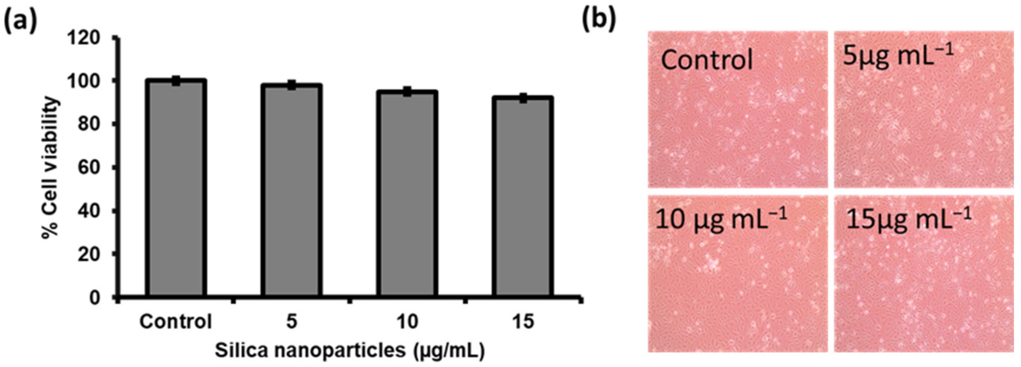
3.5. Biocompatibility/Toxicity Measurements

The biocompatibility and nontoxicity of the highly porous MSNPs were examined by measuring their effect on the cell viability (MTT and cellular morphology) of normal epithelial cells. Different concentrations of MSNPs ranging from 5 to 15 µg mL−1 showed no cytotoxicity on epithelial cells when tested with an MTT assay (Figure 6a,b, respectively). In brief, the percentages of viable cells at higher concentrations of the MSNPs (10 and 15 µg mL−1) were revealed as 89.46% and 96.28%, respectively (Figure 6). Similar morphological changes were observed in Hela cells after exposure to MSNPs at high concentrations (10 and 15 µg mL−1) compared to untreated cells (control).

4. Discussion

The MSNPs can be used on porous and non-porous surfaces as they give distinct results on both surfaces. MSNPs developed latent fingerprints on non-porous surfaces (glass, metal, plastic, and steel) with a clear appearance of class and individual characteristics, as well as pores, whereas fingerprints were not developed on porous surfaces. (Figure 5). This may be due to the transparency of polybags and the lower Rf index difference of polybag with the light in the UV-Visible spectrum. In the case of porous surfaces, surface porosity increases the surface area, thus resulting in the absorption of nanoparticles. The absorption of NPs leads to the distortion of the fingerprints in the form of agglomerates [29,30].
Numerous scientists have carried out similar investigations to demonstrate the value of MSNPs in the formation of latent fingerprint impressions. When compared to porous surfaces, the research by Rajan et al., Zhang et al., and Huang et al. demonstrated that non-porous surfaces produced superior results [17,18,31]. Rajan et al. and Bogeshwaran et al. reported the synthesis of monodispersed and spherical MSNPs from rice husks for possible use in high-definition latent finger-mark development with comparable outcomes [18,28]. On the majority of non-porous surfaces, such as glass, metal, and tiles, and semi-porous surfaces, such as painted wood, the silica nanoparticle powder was found to provide clear and precise images of latent finger impressions with little background discoloration, exposing outstanding ridge characteristics. Using the synthesized silica nanoparticle powder, excellent visibility of the minute details or ridges was seen [18].
In recent years, researchers have claimed the biocompatible nature of MSNPs when tested on various cells lines and also observed the potential usages of rode-like morphology in cancer theranostics [29]. Hence, MSNPs have shown their advantages in multiple applications, including as adsorption agents and sensors. Similarly, Liu et al., developed silica-based nanostructures of graphene/SiO2-Ag for increasing the adsorption of biomolecules [30]. In line of sensing or detecting anything on any surface related to food or human direct contact, it is very important to consider the biocompatibility, no toxicity, sensitivity, and cost-effectivity. In order to achieve these major criteria, several researchers have developed a variety of nanomaterials and nanocomposites for developing sensitive sensors. Roostaee et al., developed a nanosensor based on Co-MOF and graphene oxide for detecting the dopamine and uric acids in biological samples with the limit of detection (LOD) of 0.04 μM [32].
On similar grounds, our synthesized MSNPs could be used for the development of sensors against multiple environmental analytes due to their biocompatible nature and cost-effectivity. Most of the micro-sized metal oxide powders blended with the color additives, including dyes, pigments, and luminescent materials, have been used to enhance fingerprint images. Nowadays, nano-sized powders are becoming increasingly more popular in forensic science for fingerprint detection on various surfaces and can also be utilized in food packaging. There are various nanomaterials which have been used for fingerprint detections in porous and non-porous surfaces. Recently, Verma et al., developed the ZnO nano-powder with suitable fluorescent properties and utilized this powder for fingerprint development for surfaces with significant visual detections under UV lights [33]. The most important advantage of all nano-sized materials for fingerprint detection purposes is that almost all nanomaterials are kept in contact with the ridges of the finger to improve the visualization of latent fingerprints. Similarly, gold nanoparticle powders have also been used in mass spectrometry for the identification and imaging of latent fingerprints on non-porous and porous surfaces because of their sensitivity, good selectivity, and inert nature [34]. Richardson [35] reported that industrial titanium dioxide powder could be applied in forensic science for fingerprint detection and showed the best performance on only black adhesive tape surfaces.
On the other hand, MSNP powder has also been tested by several researchers after little surface functionalization. Huang et al., verified that 4-(chloromethyl) phenyltrichlorosilane can be used to modify MSNPs for the detection of aged fingerprints and fingerprints on glass substrates [31]. The developed MSNPs were prepared using different mass ratios of silica and 4-(chloromethyl) phenyltrichlorosilane with 700 nm of Si NPs. These nanoparticles have shown the best LFP detection on only non-porous substrates. Such results have led to further investigations on new types of MSNPs without further surface functionalization for fingerprint detections. Therefore, here we have synthesized green Si NP powder with the ability to detect pre-fingerprints on both porous and non-porous surfaces for versatile surfaces. Although the visuality of these fingerprints is low, they can be utilized in multiple food packaging surfaces due to their easy coating efficiency on various surfaces as pre-fingerprint detection tools.

5. Conclusions

A fingerprint is a unique identifier of an individual since it is an unchangeable feature that differs from person to person. Conventional techniques sometimes fail to detect old fingerprints and are less stable. Therefore, here we developed biocompatible MSNPs that produce blue color under a UV lamp. The application of the powdered silica nano-material was carried out with experiments on various non-porous materials (glass, plastic, silicon, steel, soft plastic, etc.) by keeping in mind that the developed fingerprints on various surfaces will also be helpful in food packaging sectors where crime prints could be detected on food packaging surfaces. Interestingly, the shelf life of the developed fingerprints was also significantly longer (up to 30 days). Therefore, these MSNPs can be applicative for identifying fingerprints on different surfaces used in food packaging.

Author Contributions

K.B. and D.B.T. performed the experimentation for the application part of the material and contributed to writing the manuscript; R.C. synthesized and characterized the nanomaterials and contributed to the writing of the manuscript; V.K., S.A. and H.P.K.S. contributed to the scientific review of the manuscript and revision of the manuscript; S.S. conceptualized the theme of the manuscript and edited and reviewed the manuscript. All authors have read and agreed to the published version of the manuscript.

Funding

This research received a part of funding from DBT-Ramalingaswamy Grant (BT/RLF/Re-entry/20/2017) and International Research Agendas PLUS program of the Foundation for Polish Science, co-financed by the European Union under the European Regional Development Fund (MABPLUS/2019/11).

Institutional Review Board Statement

Not applicable.

Informed Consent Statement

Not applicable.

Data Availability Statement

Not applicable.

Acknowledgments

The authors are thankful to the DBT-Ramalingaswamy research grant (BT/RLF/Re-entry/20/2017) for providing financial and academic support. Authors are also thankful to The Energy and Resources Institute (TERI) and Galgotias University for providing research support. The author VK would like to thank the International Research Agendas PLUS program of the Foundation for Polish Science, co-financed by the European Union under the European Regional Development Fund (MABPLUS/2019/11).

Conflicts of Interest

The authors declare no conflict of interest.

References

  1. Crispino, F.; Houck, M.M. Principles of Forensic Science; Elsevier Ltd.: Amsterdam, The Netherlands, 2013. [Google Scholar]
  2. Kücken, M.; Newell, A.C. Fingerprint Formation. J. Theor. Biol. 2005, 235, 71–83. [Google Scholar] [CrossRef] [PubMed]
  3. Bhati, K. Role of Nanoparticles in Latent Fingerprinting: An Update. Lett. Appl. NanoBioSci. 2020, 9, 1427–1443. [Google Scholar] [CrossRef]
  4. Kim, B.-I.; AfsaráUddin, M.; YoungáWoo, H. Others Surfactant Chemistry for Fluorescence Imaging of Latent Fingerprints using Conjugated Polyelectrolyte Nanoparticles. Chem. Commun. 2015, 51, 13634–13637. [Google Scholar]
  5. Budrikis, Z. Getting a Grip on Fingerprints. Nat. Rev. Phys. 2021, 3, 5. [Google Scholar] [CrossRef]
  6. Gupta, A. Evaluation of Correlation between Dental Caries and Fingerprints in Known Subjects. J. Adv. Med. Dent. Sci. Res. 2019, 7, 236–239. [Google Scholar]
  7. Wang, M.; Li, M.; Yu, A.; Zhu, Y.; Yang, M.; Mao, C. Fluorescent Nanomaterials for the Development of Latent Fingerprints in Forensic Sciences. Adv. Funct. Mater. 2017, 27, 1606243. [Google Scholar] [CrossRef]
  8. Gao, F.; Lv, C.; Han, J.; Li, X.; Wang, Q.; Zhang, J.; Chen, C.; Li, Q.; Sun, X.; Zheng, J.; et al. CdTe-Montmorillonite Nanocomposites: Control Synthesis, UV Radiation-Dependent Photoluminescence, and Enhanced Latent Fingerprint Detection. J. Phys. Chem. C 2011, 115, 21574–21583. [Google Scholar] [CrossRef]
  9. Lee, J.; Kim, J.; Heo, I.; Kim, S.J.; Jang, S.; Lee, H.-Y.; Jang, K.-S.; Yang, C.-S.; Lee, Y.; Yoo, W.C.; et al. One-Pot Bifunctionalization of Silica Nanoparticles Conjugated with Bioorthogonal Linkers: Application in Dual-Modal Imaging. Biomater. Sci. 2022, 10, 3540–3546. [Google Scholar] [CrossRef]
  10. Çetli, E.; Özkoçak, V.; Tatar, D. The Role of Silica Nanoparticle in Fingerprint Visualization Studies. Aksaray Univ. J. Sci. Eng. 2022, 6, 27–41. [Google Scholar] [CrossRef]
  11. Rajan, R.; Zakaria, Y.; Shamsuddin, S.; Nik Hassan, N.F. Fluorescent Variant of Silica Nanoparticle Powder Synthesised from Rice Husk for Latent Fingerprint Development. Egypt. J. Forensic Sci. 2019, 9, 50. [Google Scholar] [CrossRef] [Green Version]
  12. Bhati, K.; Tripathy, D.; Chhabra, P. Role of Fluorescent Substances in Development of Latent Fingerprints: A Review. Indian J. Forensic Med. Pathol. 2021, 14, 351–358. [Google Scholar] [CrossRef]
  13. Lee, P.L.T.; Kanodarwala, F.K.; Lennard, C.; Spindler, X.; Spikmans, V.; Roux, C.; Moret, S. Latent Fingermark Detection Using Functionalised Silicon Oxide Nanoparticles: Investigation into Novel Application Procedures. Forensic Sci. Int. 2022, 335, 111275. [Google Scholar] [CrossRef] [PubMed]
  14. Leśniewski, A. Hybrid Organic–Inorganic Silica Based Particles for Latent Fingermarks Development: A Review. Synth. Met. 2016, 222, 124–131. [Google Scholar] [CrossRef]
  15. Kim, Y.J.; Jung, H.S.; Lim, J.; Ryu, S.J.; Lee, J.K. Rapid Imaging of Latent Fingerprints Using Biocompatible Fluorescent Silica Nanoparticles. Langmuir 2016, 32, 8077–8083. [Google Scholar] [CrossRef] [PubMed]
  16. Sawhney, S.; Bhati, K.; Chhabra, P.; Tripathy, D. Role of Nanotechnology in Techniques in Fingerprints Enhancement. Indian J. Forensic Med. Pathol. 2021, 14, 288–294. [Google Scholar] [CrossRef]
  17. Zhang, M.; Ou, Y.; Du, X.; Li, X.; Huang, H.; Wen, Y.; Zhang, X. Systematic Study of Dye Loaded Small Mesoporous Silica Nanoparticles for Detecting Latent Fingerprints on Various Substrates. J. Porous Mater. 2017, 24, 13–20. [Google Scholar] [CrossRef]
  18. Rajan, R.; Zakaria, Y.; Shamsuddin, S.; Nik Hassan, N.F. Robust Synthesis of Mono-Dispersed Spherical Silica Nanoparticle from Rice Husk for High-Definition Latent Fingermark Development. Arab. J. Chem. 2020, 13, 8119–8132. [Google Scholar] [CrossRef]
  19. Wang, M.; Li, M.; Yu, A.; Wu, J.; Mao, C. Rare Earth Fluorescent Nanomaterials for Enhanced Development of Latent Fingerprints. ACS Appl. Mater. Interfaces 2015, 7, 28110–28115. [Google Scholar] [CrossRef] [Green Version]
  20. Yang, Y.; Liu, R.; Cui, Q.; Xu, W.; Peng, R.; Wang, J.; Li, L. Red-Emissive Conjugated Oligomer/Silica Hybrid Nanoparticles with High Affinity and Application for Latent Fingerprint Detection. Colloids Surf. A Physicochem. Eng. Asp. 2019, 565, 118–130. [Google Scholar] [CrossRef]
  21. Kwon, D.; Cha, B.G.; Cho, Y.; Min, J.; Park, E.-B.; Kang, S.-J.; Kim, J. Extra-Large Pore Mesoporous Silica Nanoparticles for Directing in Vivo M2 Macrophage Polarization by Delivering IL-4. Nano Lett. 2017, 17, 2747–2756. [Google Scholar] [CrossRef]
  22. Kim, J.; Lee, J.E.; Lee, J.; Yu, J.H.; Kim, B.C.; An, K.; Hwang, Y.; Shin, C.-H.; Park, J.-G.; Kim, J.; et al. Magnetic Fluorescent Delivery Vehicle Using Uniform Mesoporous Silica Spheres Embedded with Monodisperse Magnetic and Semiconductor Nanocrystals. J. Am. Chem. Soc. 2006, 128, 688–689. [Google Scholar] [CrossRef] [PubMed]
  23. Cha, B.G.; Jeong, J.H.; Kim, J. Extra-Large Pore Mesoporous Silica Nanoparticles Enabling Co-Delivery of High Amounts of Protein Antigen and Toll-like Receptor 9 Agonist for Enhanced Cancer Vaccine Efficacy. ACS Cent. Sci. 2018, 4, 484–492. [Google Scholar] [CrossRef] [PubMed]
  24. Sevimli, F.; Yilmaz, A. Surface Functionalization of SBA-15 Particles for Amoxicillin Delivery. Microporous Mesoporous Mater. 2012, 158, 281–291. [Google Scholar] [CrossRef] [Green Version]
  25. Liu, F.; Wang, J.; Huang, P.; Zhang, Q.; Deng, J.; Cao, Q.; Jia, J.; Cheng, J.; Fang, Y.; Deng, D.Y.B.; et al. Outside-in Stepwise Functionalization of Mesoporous Silica Nanocarriers for Matrix Type Sustained Release of Fluoroquinolone Drugs. J. Mater. Chem. B 2015, 3, 2206–2214. [Google Scholar] [CrossRef] [PubMed]
  26. Wardhani, G.A.P.K.; Nurlela, N.; Azizah, M. Silica Content and Structure from Corncob Ash with Various Acid Treatment (HCl, HBr, and Citric Acid). Molekul 2017, 12, 174–181. [Google Scholar] [CrossRef] [Green Version]
  27. Thuadaij, N.; Nuntiya, A. Preparation of Nanosilica Powder from Rice Husk Ash by Precipitation Method. Chiang Mai J. Sci. 2008, 35, 206–211. [Google Scholar]
  28. Bogeshwaran, K.; Kalaivani, R.; Ashraf, S.; Manikandan, G.N.; Prabhu, G.E. Production of Silica from Rice Husk. Int. J. ChemTech Res. 2014, 6, 4337–4345. [Google Scholar]
  29. Sargazi, S.; Laraib, U.; Barani, M.; Rahdar, A.; Fatima, I.; Bilal, M.; Pandey, S.; Sharma, R.K.; Kyzas, G.Z. Recent Trends in Mesoporous Silica Nanoparticles of Rode-like Morphology for Cancer Theranostics: A review. J. Mol. Struct. 2022, 1261, 132922. [Google Scholar] [CrossRef]
  30. Liu, X.L.; Liu, J.; Yang, H.M.; Huang, B.; Zeng, G.H. Design of a High-performance Graphene/SiO2-Ag Periodic Grating/MoS2 Surface Plasmon Resonance Sensor. Appl. Opt. 2022, 61, 6752–6760. [Google Scholar] [CrossRef]
  31. Huang, W.; Li, X.; Wang, H.; Xu, X.; Liu, H.; Wang, G. Synthesis of Amphiphilic Silica Nanoparticles for Latent Fingerprint Detection. Anal. Lett. 2015, 48, 1524–1535. [Google Scholar] [CrossRef]
  32. Roostaee, M.; Beitollahi, H.; Sheikhshoaie, I. Simultaneous Determination of Dopamine and Uric Acid in Real Samples Using a Voltammetric Nanosensor Based on Co-MOF, Graphene Oxide, and 1-Methyl-3-butylimidazolium Bromide. Micromachines 2022, 13, 1834. [Google Scholar] [CrossRef] [PubMed]
  33. Verma, R.K.; Nagar, V.; Aseri, V.; Mavry, B.; Pandit, P.P.; Chopade, R.L.; Singh, A.; Singh, A.; Yadav, V.L.; Pandey, K.; et al. Zinc oxide (ZnO) nanoparticles: Synthesis Properties and their Forensic Applications in Latent Fingerprints Development. Mater. Today Proceed. 2022, 69, 36–41. [Google Scholar] [CrossRef]
  34. Choi, M.J.; McDonagh, A.M.; Maynard, P.J.; Wuhrer, R. Preparation and evaluation of metal nanopowders for the detection of fingermarks on nonporous surfaces. J. Forensic Ident. 2006, 56, 756–768. [Google Scholar]
  35. Richardson, M. Comparison of the Effectiveness of Different Commercial Titanium Dioxides (TiO2) for White Powder Suspension on Rubber-based Adhesive Tapes. In Home Office Internal Report; HOSDB: St Albans, UK, 2006. [Google Scholar]
Figure 1. Method development for the application of MSNPs in detecting criminal fingerprints.
Coatings 13 00268 g001
Figure 2. Characterization of synthesized MSNPs. (a,b): TEM morphological characterization; (c,d): SEM morphology; (e): FTIR measurements; (f): Zeta potential; (g): EDX confirmation for elemental composition; (h): XRD analysis; (i): BET measurements.
Coatings 13 00268 g002
Figure 3. The stability of the synthesized MSNPs analyzed using DLS over a time period of 72 h at room temperature (25 ± 2 °C).
Coatings 13 00268 g003
Figure 4. Schematic of wrinkled MSNPs with enlarged pores.
Coatings 13 00268 g004
Figure 5. Developed fingerprints on various non-porous surfaces using MSNPs.
Coatings 13 00268 g005
Figure 6. Biocompatibility/cytotoxicity of the synthesized MSNPs.
Coatings 13 00268 g006
Table 1. Latent finger impressions developed using MSNs nanoparticles.
SNNanoparticlesSurfaceMaterialLevel-I (Pattern)Level-II (Ridge Characteristics)
1.MSNPsNON-POROUSSilica phone coverVisibleVisible
2.MSNPsNON-POROUSPlastic phone caseVisibleVisible
3.MSNPsNON-POROUSGlass slideVisibleVisible
4.MSNPsNON-POROUSStainless steelVisibleVisible
5.MSNPsNON-POROUSPlastic calculator coverVisibleVisible
6.MSNPsNON-POROUSTransparent poly bagNot-VisibleNot-Visible
7.MSNPsPOROUSBlack paperNot-VisibleNot-Visible
8.MSNPsPOROUSPaper bagNot-VisibleNot-Visible
Table 2. Stability shelf life of developed fingerprints.
SNSurface MaterialStability/Shelf Life
1Silica phone cover<30 days
2Plastic phone case<15 days
3Glass slide<30 days
4Stainless steel<15 days
5Plastic calculator cover<20 days
6Transparent poly bag<30 days
Disclaimer/Publisher’s Note: The statements, opinions and data contained in all publications are solely those of the individual author(s) and contributor(s) and not of MDPI and/or the editor(s). MDPI and/or the editor(s) disclaim responsibility for any injury to people or property resulting from any ideas, methods, instructions or products referred to in the content.

