
Study of Al–SiO2 Aesthetic Composite Coating on Orthodontic Metal Archwire


Abstract
:1. Introduction
2. Materials and Methods
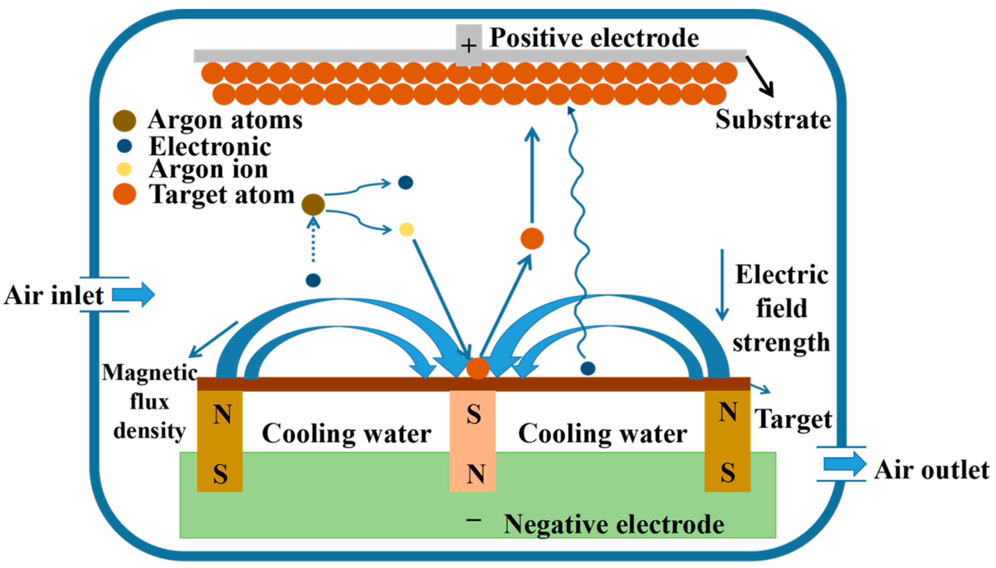
2.1. Preparation and Characterization of Al–SiO2-Coated Samples
2.2. Cytotoxicity Tests
2.3. Electrochemical Corrosion Experiments
2.4. Wear Test
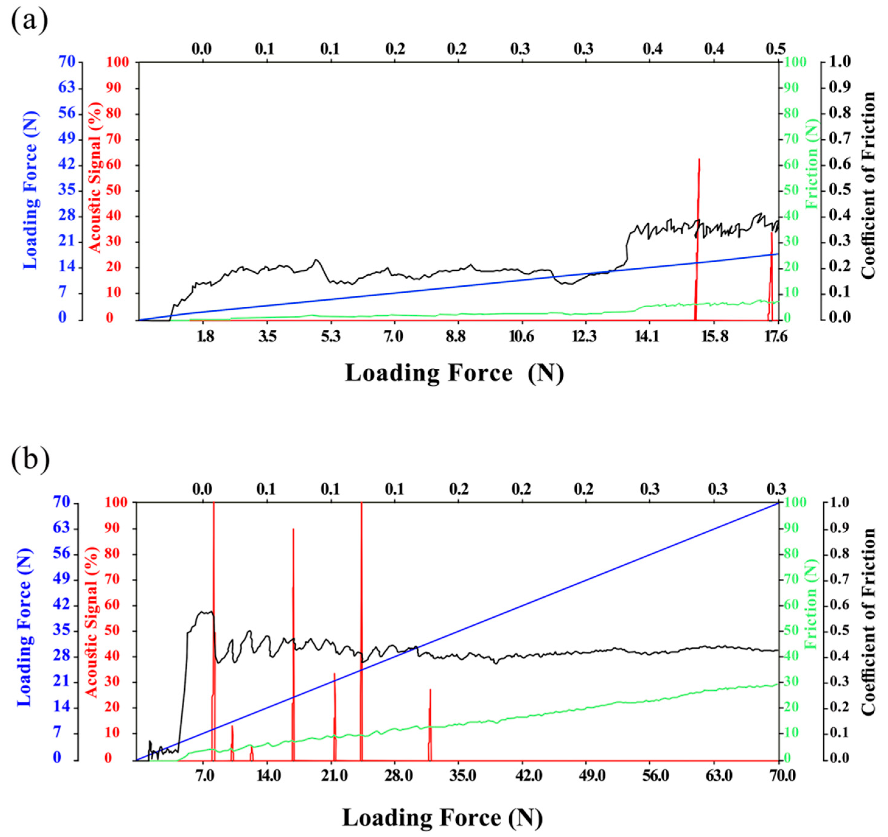
2.5. Scratch Test
2.6. Statistical Analysis
3. Results
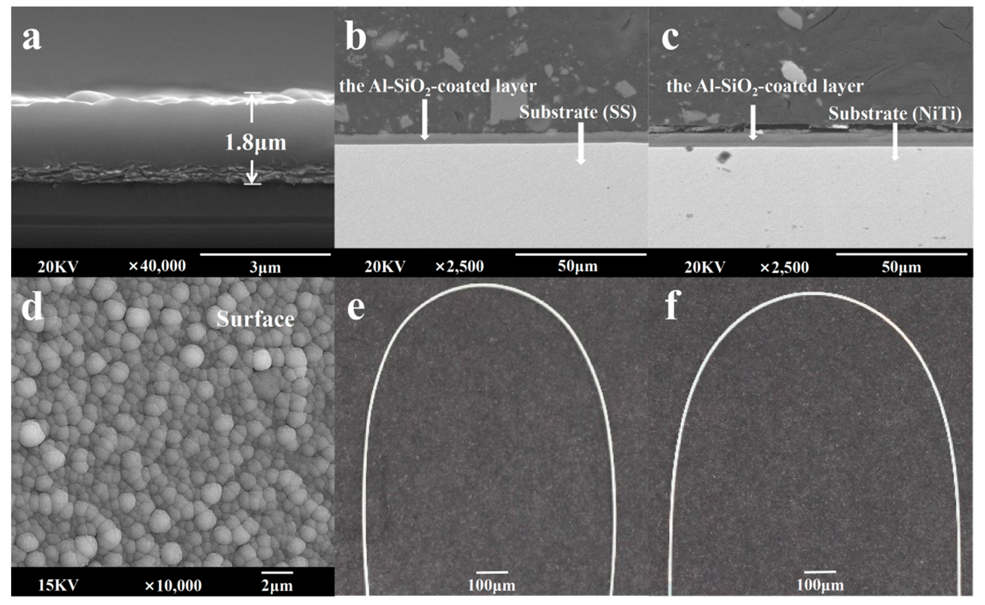
3.1. Coating Morphology Analysis
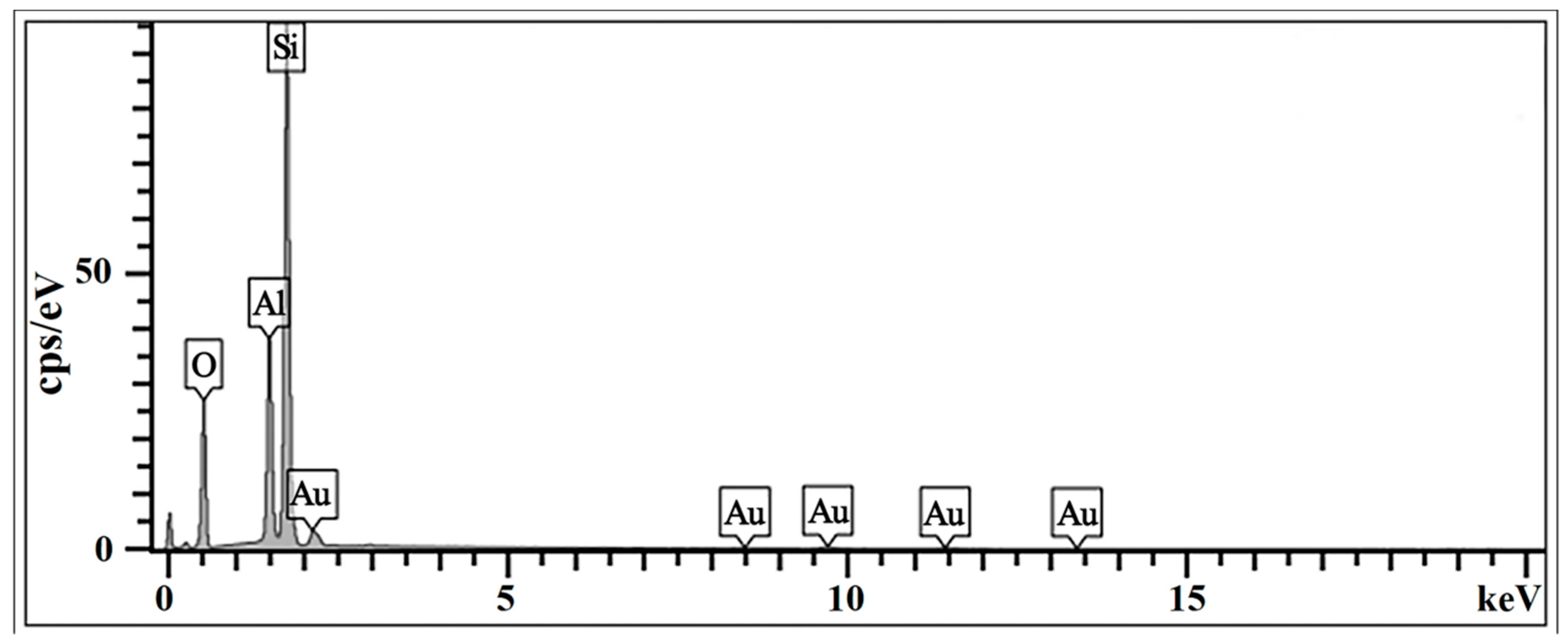
3.2. EDS Analysis
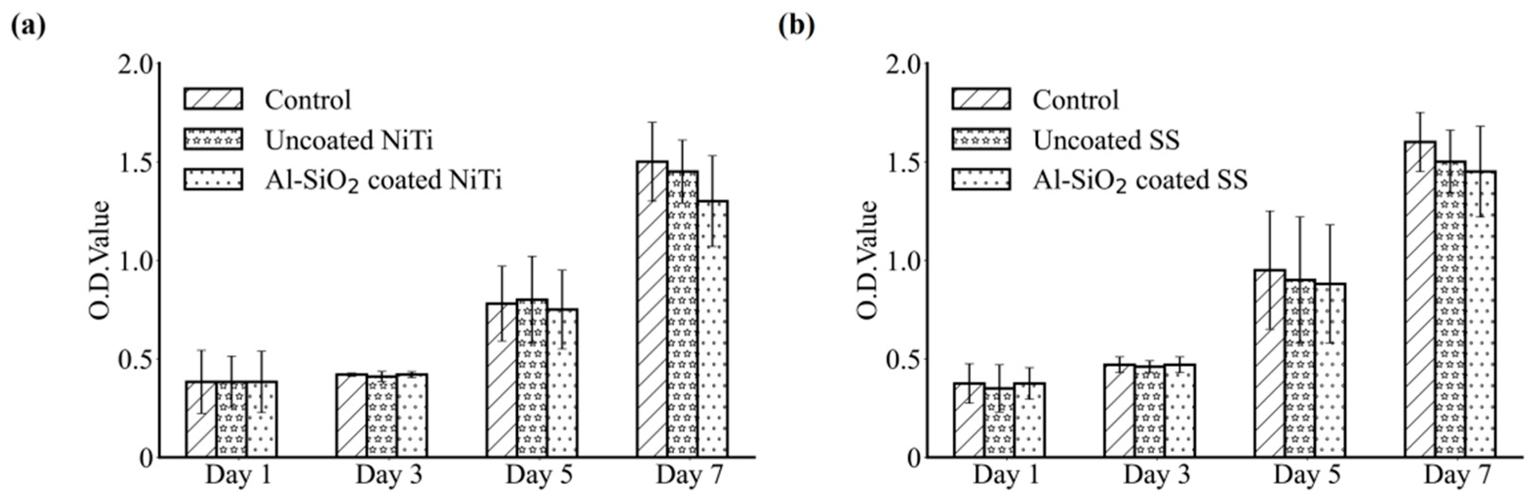
3.3. Cytotoxicity
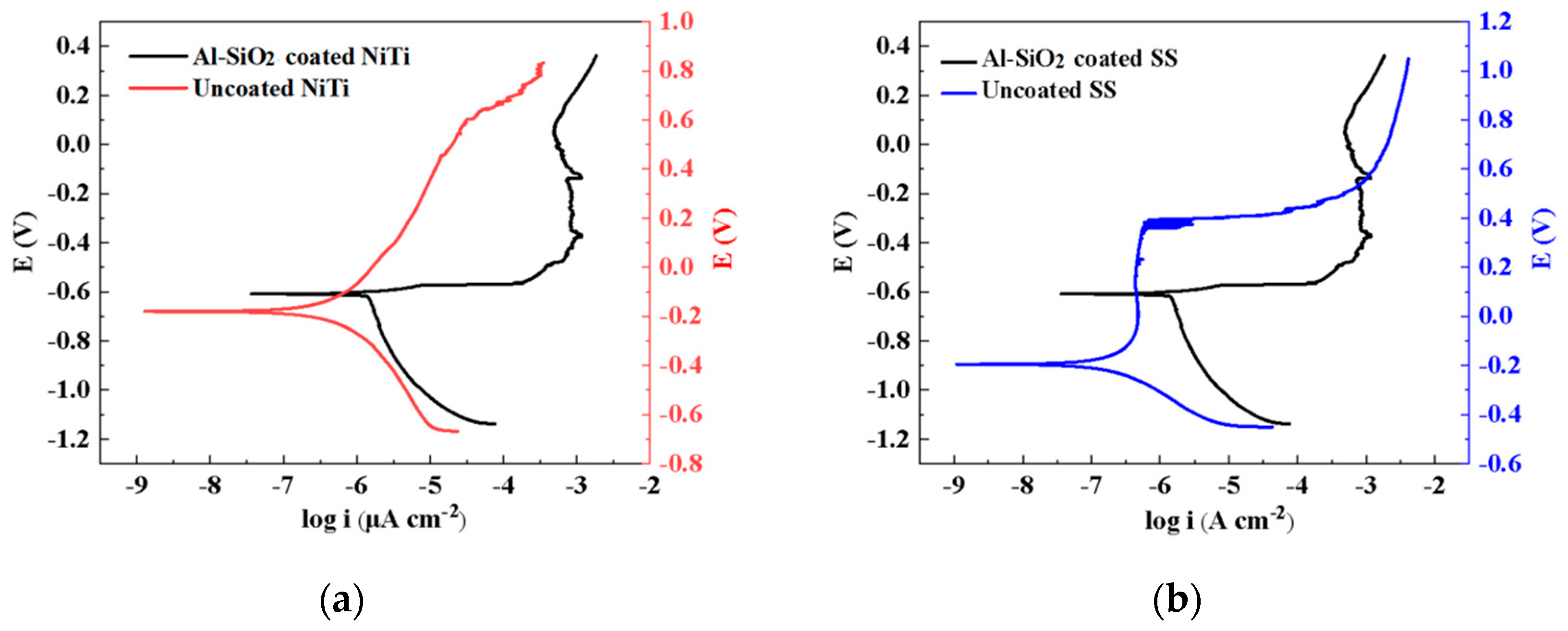
3.4. Electrochemical Corrosion Experiment
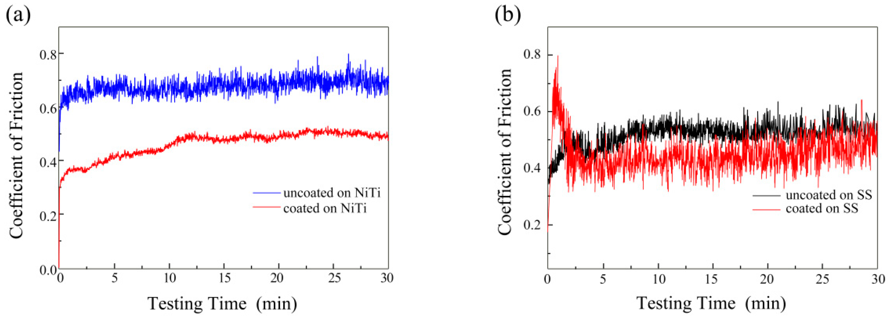
3.5. Friction and Wear Properties
3.6. Bonding Strength
4. Discussion
5. Conclusions
Author Contributions
Funding
Institutional Review Board Statement
Informed Consent Statement
Data Availability Statement
Acknowledgments
Conflicts of Interest
References
- Močnik, P.; Kosec, T. A Critical Appraisal of the Use and Properties of Nickel-Titanium Dental Alloys. Materials 2021, 14, 7859. [Google Scholar] [CrossRef] [PubMed]
- Dias, V.; Maciel, H.; Fraga, M.; Lobo, A.O.; Pessoa, R.; Marciano, F.R. Atomic Layer Deposited TiO2 and Al2O3 Thin Films as Coatings for Aluminum Food Packaging Application. Materials 2019, 12, 682. [Google Scholar] [CrossRef] [PubMed] [Green Version]
- Chen, L.; Yan, X.; Tan, L.; Zheng, B.; Muhammed, F.K.; Yang, K.; Liu, Y. In vitro and in vivo characterization of novel calcium phosphate and magnesium (CaP-Mg) bilayer coated titanium for implantation. Surf. Coat. Technol. 2019, 374, 784–796. [Google Scholar] [CrossRef]
- Hosseinzadeh Nik, T.; Hooshmand, T.; Farazdaghi, H.; Mehrabi, A.; Razavi, E.S.E. Effect of chlorhexidine-containing prophylactic agent on the surface characterization and frictional resistance between orthodontic brackets and archwires: An in vitro study. Prog. Orthod. 2013, 14, 48–55. [Google Scholar] [CrossRef] [Green Version]
- Alsanea, J.A.; Al Shehri, H. Evaluation of Nanomechanical Properties, Surface Roughness, and Color Stability of Esthetic Nickel-Titanium Orthodontic Archwires. J. Int. Soc. Prev. Community Dent. 2019, 9, 33–39. [Google Scholar] [CrossRef]
- Zhao, Y.; Liu, J.; Zhang, M. Use of silver nanoparticle–gelatin/alginate scaffold to repair skull defects. Coatings 2020, 10, 948. [Google Scholar] [CrossRef]
- Shirakawa, N.; Iwata, T.; Miyake, S.; Otuka, T.; Koizumi, S.; Kawata, T. Mechanical properties of orthodontic wires covered with a polyether ether ketone tube. Angle Orthod. 2018, 88, 442–449. [Google Scholar] [CrossRef]
- Doshi, U.H.; Bhad-Patil, W.A. Static frictional force and surface roughness of various bracket and wire combinations. Am. J. Orthod. Dentofac. Orthop. 2011, 139, 74–79. [Google Scholar] [CrossRef]
- Pezzato, L.; Vranescu, D.; Sinico, M.; Gennari, C.; Settimi, A.G.; Pranovi, P.; Brunelli, K.; Dabalà, M. Tribocorrosion Properties of PEO Coatings Produced on AZ91 Magnesium Alloy with Silicate- or Phosphate-Based Electrolytes. Coatings 2018, 8, 202. [Google Scholar] [CrossRef] [Green Version]
- Rachele Bertolini, S.B.; Bordin, A.; Ghiotti, A.; Pezzato, L.; Dabala, M. Fretting Corrosion Behavior of Additive Manufactured and Cryogenic-Machined Ti6Al4V for Biomedical Applications. Adv. Eng Mater. 2016, 19, 1500629. [Google Scholar] [CrossRef] [Green Version]
- Abdullah, A.O.; Hui, Y.; Pollington, S.; Muhammed, F.K.; Sun, X.; Liu, Y. Comparative Effectiveness of Multiple Laser Scanning and Conventional Techniques on Zirconia Shear Bond Strength. Coatings 2019, 9, 422. [Google Scholar] [CrossRef] [Green Version]
- Li, C.-J.; Li, J.-L. Evaporated-gas-induced splashing model for splat formation during plasma spraying. Surf. Coat. Technol. 2004, 184, 13–23. [Google Scholar] [CrossRef]
- Baxamusa, S.H.; Im, S.G.; Gleason, K.K. Initiated and oxidative chemical vapor deposition: A scalable method for conformal and functional polymer films on real substrates. Phys. Chem. Chem. Phys. 2009, 11, 5227–5240. [Google Scholar] [CrossRef] [PubMed]
- Sioshansi, P. Tailoring surface properties by ion implantation. Mater. Eng. 1987, 104, 19–23. [Google Scholar]
- Qadir, M.; Li, Y.; Wen, C. Ion-substituted calcium phosphate coatings by physical vapor deposition magnetron sputtering for biomedical applications: A review. Acta Biomater. 2019, 89, 14–32. [Google Scholar] [CrossRef] [PubMed]
- Kao, W.-H.; Su, Y.-L.; Horng, J.-H.; Hsieh, Y.-T. Improved tribological properties, electrochemical resistance and biocompatibility of AISI 316L stainless steel through duplex plasma nitriding and TiN coating treatment. J. Biomater. Appl. 2017, 32, 12–27. [Google Scholar] [CrossRef]
- Arunkumar, P.; Aarthi, U.; Sribalaji, M.; Mukherjee, B.; Keshri, A.K.; Tanveer, W.H.; Cha, S.-W.; Babu, K.S. Deposition rate dependent phase/mechanical property evolution in zirconia and ceria-zirconia thin film by EB-PVD technique. J. Alloy. Compd. 2018, 76, 418–427. [Google Scholar] [CrossRef]
- Krishnan, V.; Krishnan, A.; Remya, R.; Ravikumar, K.K.; Nair, S.A.; Shibli, S.M.A.; Varma, H.K.; Sukumaran, K.; Kumar, K.J. Development and evaluation of two PVD-coated β-titanium orthodontic archwires for fluoride-induced corrosion protection. Acta Biomater. 2011, 7, 1913–1927. [Google Scholar] [CrossRef]
- Harlin, P.; Bexell, U.; Olsson, M. Influence of surface topography of arc-deposited TiN and sputter-deposited WC/C coatings on the initial material transfer tendency and friction characteristics under dry sliding contact conditions. Surf. Coat. Technol. 2009, 203, 1748–1755. [Google Scholar] [CrossRef]
- Goudarzi, M.; Saviz, S.; Ghoranneviss, M.; Salar Elahi, A. Antibacterial characteristics of thermal plasma spray system. J. X-ray Sci. Technol. 2018, 26, 509–521. [Google Scholar] [CrossRef]
- Monteiro, O.R. Thin film synthesis by energetic condensation. Annu. Rev. Mater. Res. 2000, 31, 111–137. [Google Scholar] [CrossRef] [Green Version]
- Kula, K.; Phillips, C.; Gibilaro, A.; Proffit, W.R. Effect of ion implantation of TMA archwires on the rate of orthodontic sliding space closure. Am. J. Orthod. Dentofac. Orthop. 1998, 114, 577–580. [Google Scholar] [CrossRef]
- Heinrichs, J. Laboratory test simulation of aluminium cold forming: Influence from PVD tool coatings on the tendency to galling. Surf. Coat. Technol. 2010, 204, 3606–3613. [Google Scholar] [CrossRef]
- Schiller, S.; Goedicke, K.; Reschke, J.; Kirchhoff, V.; Schneider, S.; Milde, F. Pulsed magnetron sputter technology. Surf. Coat. Technol. 1993, 61, 331–337. [Google Scholar] [CrossRef]
- Ozeki, K.; Yuhta, T.; Aoki, H.; Asaoka, T.; Daisaku, T.; Fukui, Y. Deterioration in the superelasticity of Ti sputter coated on NiTi orthodontic wire. Bio-Med. Mater. Eng. 2003, 13, 355–362. [Google Scholar]
- Krishnan, M.; Saraswathy, S.; Sukumaran, K.; Abraham, K.M. Effect of ion-implantation on surface characteristics of nickel titanium and titanium molybdenum alloy arch wires. Indian J. Dent. Res. 2013, 24, 411–417. [Google Scholar] [CrossRef]
- Palei, B.B.; Dash, T.; Biswal, S.K. Graphene reinforced aluminum nanocomposites: Synthesis, characterization and properties. J. Mater. Sci. 2022, 57, 8544–8556. [Google Scholar] [CrossRef]
- Yan, Y.; Zheng, B.; Yassir, L.; Liu, X. D-leucine enhances antibiofilm activity of chlorhexidine against caries-causing streptococcus mutans biofilm. Int. Biodeterior Biodegradation. 2021, 157, 105135. [Google Scholar] [CrossRef]
- Amaya, S.; Pérez, A.; Guzmán, H.; Espinosa, A.; Motta, G.; Mojica, J.; Plaza-Ruiz, S.P. Changes in the mechanical properties of two nickel-titanium archwires after 3 months of clinical usage. J. World Fed. Orthod. 2020, 9, 175–180. [Google Scholar] [CrossRef]
- Prioteasa, P.; Ibris, N.; Visan, T. The influence of chemical nature on the corrosion behaviour of some dental alloys in Fusayama-Meyer artificial saliva. J. Optoelectron. Adv. Mater. 2007, 9, 3405–3410. [Google Scholar]
- D’Antò, V.; Rongo, R.; Ametrano, G.; Spagnuolo, G.; Manzo, P.; Martina, R.; Paduano, S.; Valletta, R. Evaluation of surface roughness of orthodontic wires by means of atomic force microscopy. Angle Orthod. 2012, 82, 922–928. [Google Scholar] [CrossRef] [PubMed]
- Abdullah, A.O.; Yu, H.; Xudong, S.; Muhammed, F.K.; Pollington, S.; Liu, Y. Comparative in Vitro Evaluation between Zirconia and Veneer Ceramic Materials Using Different Techniques. J. Mater. Eng. Perform. 2019, 28, 6656–6668. [Google Scholar] [CrossRef]
- Zhang, H.; Guo, S.; Wang, D.; Zhou, T.; Wang, L.; Ma, J. Effects of nanostructured, diamondlike, carbon coating and nitrocarburizing on the frictional properties and biocompatibility of orthodontic stainless steel wires. Angle Orthod. 2016, 86, 782–788. [Google Scholar] [CrossRef] [PubMed] [Green Version]
- Mystkowska, J.; Niemirowicz-Laskowska, K.; Łysik, D.; Tokajuk, G.; Dąbrowski, J.R.; Bucki, R. The Role of Oral Cavity Biofilm on Metallic Biomaterial Surface Destruction-Corrosion and Friction Aspects. Int. J. Mol. Sci. 2018, 19, 743. [Google Scholar] [CrossRef] [PubMed] [Green Version]
- Meyer, R.D.; Meyer, J.; Taloumis, L.J. Intraoral galvanic corrosion: Literature review and case report. J. Prosthet. Dent. 1993, 69, 141–143. [Google Scholar] [CrossRef]
- Song, F.; Koo, H.; Ren, D. Effects of Material Properties on Bacterial Adhesion and Biofilm Formation. J. Dent. Res. 2015, 94, 1027–1034. [Google Scholar] [CrossRef] [PubMed]
- Katić, V.; Curković, H.O.; Semenski, D.; Baršić, G.; Marušić, K.; Spalj, S. Influence of surface layer on mechanical and corrosion properties of nickel-titanium orthodontic wires. Angle Orthod. 2014, 84, 1041–1048. [Google Scholar] [CrossRef] [Green Version]
- Kim, H.; Johnson, J.W. Corrosion of stainless steel, nickel-titanium, coated nickel-titanium, and titanium orthodontic wires. Angle Orthod. 1999, 69, 39–44. [Google Scholar]
- Kusy, R.P.; Whitley, J.Q. Friction between different wire-bracket configurations and materials. Semin. Orthod. 1997, 3, 166–177. [Google Scholar] [CrossRef]
- Rincic Mlinaric, M.; Karlovic, S.; Ciganj, Z.; Acev, D.P.; Pavlic, A.; Spalj, S. Oral antiseptics and nickel-titanium alloys: Mechanical and chemical effects of interaction. Odontology 2019, 107, 150–157. [Google Scholar] [CrossRef]
- De Albuquerque, C.G.; Correr, A.B.; Venezian, G.C.; Santamaria, M.; Tubel, C.A.; Vedovello, S.A.S. Deflection and Flexural Strength Effects on the Roughness of Aesthetic-Coated Orthodontic Wires. Braz. Dent. J. 2017, 28, 40–45. [Google Scholar] [CrossRef] [PubMed] [Green Version]
- Anuradha, P.; Varma, N.K.S.; Balakrishnan, A. Reliability performance of titanium sputter coated Ni-Ti arch wires: Mechanical performance and nickel release evaluation. Bio-Med. Mater. Eng. 2015, 26, 67–77. [Google Scholar] [CrossRef] [PubMed]
- Alavi, S.; Hosseini, N. Load-deflection and surface properties of coated and conventional superelastic orthodontic archwires in conventional and metal-insert ceramic brackets. Dent. Res. J. 2012, 9, 133–138. [Google Scholar]
- Pan, X.; Li, Y.; Abdullah, A.O.; Wang, W.; Qi, M.; Liu, Y. Micro/nano-hierarchical structured TiO coating on titanium by micro-arc oxidation enhances osteoblast adhesion and differentiation. R. Soc. Open Sci. 2019, 6, 182031. [Google Scholar] [CrossRef] [PubMed] [Green Version]


| Friction Pairs | Load (N) | Turning Radius (mm) | Speed (r/min) | Test Time (min) |
|---|---|---|---|---|
| Alumina ball (Ø = 4 mm) | 1 | 3 | 200 | 30 |
| Sample | Ecorr/V vs. SCE | icorr/μA cm−2 |
|---|---|---|
| Uncoated NiTi | −0.18 ± 0.02 | 23.72 ± 4.99 |
| Al–SiO2-coated NiTi | −0.60 ± 0.08 | 1.21 ± 0.48 |
| Uncoated SS | −0.20 ± 0.03 | 0.22 ± 0.09 |
| Al–SiO2-coated SS | −0.61 ± 0.10 | 0.06 ± 0.01 |
Publisher’s Note: MDPI stays neutral with regard to jurisdictional claims in published maps and institutional affiliations. |
© 2022 by the authors. Licensee MDPI, Basel, Switzerland. This article is an open access article distributed under the terms and conditions of the Creative Commons Attribution (CC BY) license (https://creativecommons.org/licenses/by/4.0/).
Share and Cite
Wu, H.; Yang, J.; Yan, Y.; Zheng, B.; Algahefi, A.L.; Ma, S.; Liu, Y. Study of Al–SiO2 Aesthetic Composite Coating on Orthodontic Metal Archwire. Coatings 2022, 12, 746. https://doi.org/10.3390/coatings12060746
Wu H, Yang J, Yan Y, Zheng B, Algahefi AL, Ma S, Liu Y. Study of Al–SiO2 Aesthetic Composite Coating on Orthodontic Metal Archwire. Coatings. 2022; 12(6):746. https://doi.org/10.3390/coatings12060746
Chicago/Turabian StyleWu, Haopeng, Jie Yang, Yuwen Yan, Bowen Zheng, Ahmed Lotf Algahefi, Song Ma, and Yi Liu. 2022. "Study of Al–SiO2 Aesthetic Composite Coating on Orthodontic Metal Archwire" Coatings 12, no. 6: 746. https://doi.org/10.3390/coatings12060746