Review

Electrospinning as a Promising Process to Preserve the Quality and Safety of Meat and Meat Products

1
Food Quality and Sensory Science Department, Teagasc Food Research Centre, Dublin 15, D15 KN3K Ashtown, Ireland
2
Graduate Program of Food Science, and Technology (PPGCTAL), Universidade Federal da Fronteira Sul (UFFS), Campus Laranjeiras do Sul, Laranjeiras do Sul 85301-970, PR, Brazil
3
Department of Food Technology, Vocational School of Technical Siences at Mersin, Tarsus Organized Industrial Zone, Tarsus University, 33100 Mersin, Turkey
4
Chemistry Interdisciplinary Project (CHIP), School of Pharmacy, University of Camerino, Via Madonna delle Carceri, 62032 Camerino, Italy
5
Department of Food Science, Mouloud Mammery University, Tizi-Ouzou 15000, Algeria
6
Gestion Santé et Productions Animales Research Laboratory, Institut des Sciences Vétérinaires El-Khroub, Université Frères Mentouri Constantine 1, Constantine 25000, Algeria
7
ClonBio Group LTD, 6 Fitzwilliam Pl, D02 XE61 Dublin, Ireland
*
Author to whom correspondence should be addressed.
These authors contributed equally to this work.
Coatings 2022, 12(5), 644; https://doi.org/10.3390/coatings12050644
Submission received: 17 April 2022 / Revised: 1 May 2022 / Accepted: 6 May 2022 / Published: 8 May 2022

Abstract

:
Fresh and processed meat products are staple foods worldwide. However, these products are considered perishable foods and their deterioration depends partly on the inner and external properties of meat. Beyond conventional meat preservation approaches, electrospinning has emerged as a novel effective alternative to develop active and intelligent packaging. Thus, this review aims to discuss the advantages and shortcomings of electrospinning application for quality and safety preservation of meat and processed meat products. Electrospun fibres are very versatile, and their features can be modulated to deliver functional properties such as antioxidant and antimicrobial effects resulting in shelf-life extension and in some cases product quality improvement. Compared to conventional processes, electrospun fibres provide advantages such as casting and coating in the fabrication of active systems, indicators, and sensors. The approaches for improving, stabilizing, and controlling the release of active compounds and highly sensitive, rapid, and reliable responsiveness, under changes in real-time are still challenging for innovative packaging development. Despite their advantages, the active and intelligent electrospun fibres for meat packaging are still restricted to research and not yet widely used for commercial products. Industrial validation of lab-scale achievements of electrospinning might boost their commercialisation. Safety must be addressed by evaluating the impact of electrospun fibres migration from package to foods on human health. This information will contribute into filling knowledge gaps and sustain clear regulations.

1. Introduction

Fresh meat and processed meat products are of high quality for human nutrition, but they are perishable foods that need to be properly processed and handled to preserve their sensory and safety qualities and extend their shelf life to avoid wastage [1,2,3,4]. The oxidation of lipids and proteins of muscle foods (fresh and processed meat products) is a major issue, also related to chemical degradation. For example, oxidation alters colour, odour, and texture of meat products, as well as decreases the nutritional quality [5,6]. Meat quality deterioration is the consequence of a multitude of factors and depends partly on the inner properties of meat products such as composition and ingredients and external properties such as oxygen, light exposure, and temperature [7]. Phospholipids and triglycerides as major components of muscle foods contain high amounts of polyunsaturated fatty acids, which are highly susceptible to oxidation in the presence of pro-oxidant components such as oxygen, enzymes, or metals [6]. The primary products of lipid oxidation, hydroperoxides, do not have odour or aroma, but they are unstable and can result in aldehydes, ketones, alcohols, esters, and acids, as secondary by-products, which are responsible for the development of off-odours and off-flavours [6]. Aldehydes also react with proteins resulting in changes in the sensory and nutritional features of meat [8,9].
Meat and meat products are further susceptible to microbial contamination due to their high-water activity/content favouring physical, chemical, and sensorial changes along the production chain (processing, storage, and distribution). As such, the growth and proliferation of harmful microorganisms result in product spoilage and quality loss [5]. Listeria monocytogenes, Escherichia coli, Salmonella typhimurium, Bacillus cereus, Salmonella enteritidis, Staphylococcus aureus, and Yersinia enterocolitica are among the frequent microorganisms involved with foodborne illnesses and, in extreme cases, death [2,10]. Due to cross-contamination caused by spoilage food microorganisms, much emphasis has been placed on the safety aspect of foods in recent years. Consequently, innovative technologies and strategies continuously seek solutions to ensure fresh meat and meat products safety [11].
Meat packaging plays different and important roles where its primary functions are to prevent product spoilage and quality loss resulting in increasing the shelf life, reducing wastage, increasing consumer acceptability, ensuring information transmission, and facilitating storage and transport. Changes in dietary habits and lifestyles boosted packaging to put more emphasis on convenience and sustainability besides safety. Innovative packaging emerged as solutions not only to extend product quality and shelf life but also to monitor its quality during transportation and storage. Active and intelligent packaging are technologies adopted in recent years to ensure product traceability, safety, and quality [12]. The active packaging is designed to protect and preserve packed food through interacting and/or modifying the package headspace, hence extending the shelf life of foods, and consequently improving its safety [13]. Intelligent packaging is designed to provide consumers feedback after interacting and/or monitoring the packed product quality during storage and transport up to consumption [1,14,15].
Among the innovative packaging approaches, electrospinning has gained a lot of interest in the biomedical sector and food industry including meat packaging [14,15,16,17]. The incorporation of electrospun nanofibres as nanocarriers demonstrated vast advantages in drugs formulation and delivery due to their stability and improved bioavailability [18]. For instance, electrospinning enabled the formation of nano-drug delivery systems for the treatment of different diseases such as cancer [18]. These nanofibres have also been applied in bone tissue engineering to enhance encapsulated bone mineral release due to the resemblance between electrospun fibres and the native tissues [19,20]. In food packaging, electrospun nanofibres can improve the barrier and antimicrobial properties of materials due to their functional properties. These nanofibres may also be used as nanosensors for the detection and monitoring of food conditions during transport and storage [21]. This opens doors to their use for the packaging of dried foods such as wheat flour, rice, grains, and dried fruits, and fresh products such as fruits, vegetables, meats, and ready-to-eat meals [22].
Electrospinning technology is designed to develop fibres between 100 nm and a few microns in size, each with its own set of unique features using an eletrohydrodinamic approach [23]. It has been reported that nano-sized materials are more effective at low concentrations in a wide range of applications due to their high surface area to volume ratio [24,25]. A variety of natural and synthetic materials, such as polymers and their blends can be used in electrospinning processing. Some non-polymer structures (phospholipids, cyclodextrins, peptides, and oligosaccharides) can also be electrospun into nanofibres, but it is still at limited extent [26].
Natural polymers, particularly polysaccharides and proteins, are frequently used to produce nanofibres, owing to their biocompatibility, non-toxic, food-grade, and biodegradability [27]. Additionally, their functional group diversity enables the binding or entrapping of a wide range of active ingredients through molecular interactions [28]. A variety of polysaccharides are being used to manufacture nanofibres deriving from plant-based (starch, cellulose derivates, and pectin), animal-based (chitosan), and algae-based (alginate or carrageenan) sources [27,28,29,30]. The molecular configurations and degree of chain entanglements in these polymers have an impact on the self-assembly of nanofibres [31], and sometimes electrospun fibres production is difficult or not possible to achieve. Plant-based (soy proteins and zein), animal-based (egg white proteins, whey proteins, gelatin, collagen, and casein) or insect-based (silk) proteins are electrospun into fibres, showing different degrees of success to achieve acceptable results due to their secondary and tertiary folds. However, functionalization, appropriate solvents, and rearranging of polysaccharides and protein conformation can improve their potential fibres production, as well as using a carrier polymer to enable the processing [32]. Microbial-based and biomass fermentation derivate polymers such pullulan, xanthan gum, and polyhydroxyalkanoates (PHAs) and its derivates (especially the poly(3-hydroxybutyrate) (PHB), and the synthetic bio-based monomer polylactic acid (PLA) are also reported for electrospun applications [33]. Other synthetic biopolymers such as polycaprolactone (PCL) and polyglycolic acid (PGA), both petroleum-based materials, and polyvinyl alcohol (PVA) and polyethylene oxide (PEO) are among the most used synthetic biopolymers as they have features for electrospun fibres [22,34]. These fibres have a high capacity for loading active agents, and their large surface area allows a fast response to intrinsic and/or extrinsic factors by releasing/activating the entrapped compounds at the appropriate timing [23]. Thus, electrospinning as a novel technology can contribute in the improvement of the overall quality and increase the shelf life of fresh or packaged meat products [27], including (i) the preservation of products against microbial contamination [16,35], (ii) preventing lipids and proteins oxidation [6,36], (iii) the development of sensory properties [15,37], and (iv) the improvement of nutritional and functional features of meat products [14,38].
The commercial benefit of implementing electrospinning in the meat processing sector is highly dependent on its economic relevance, accuracy, customer acceptance, and consideration of particular legislation governing the implementation of this technology as recently discussed for nanotechnology [27]. In this light, the present review intends to discuss the application of electrospinning process in the meat industry by (i) providing a comprehensive overview on processing, formulation, characterization, and types of electrospun fibres currently developed, (ii) critically discussing the role of electrospun fibres in meat processing, preservation and safety, and (iii) addressing research gaps and challenges that could result in a new paradigm for the meat industry.

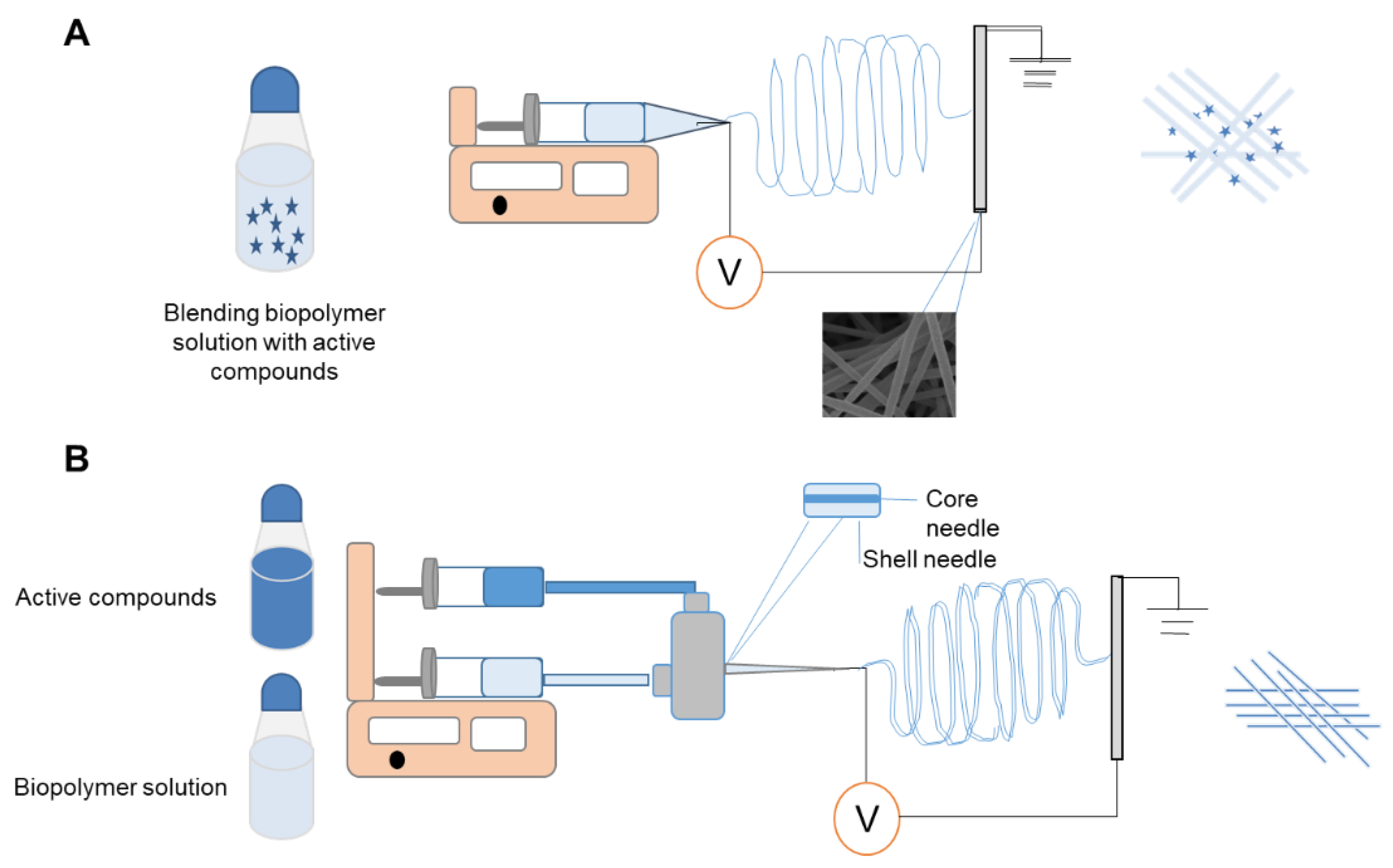
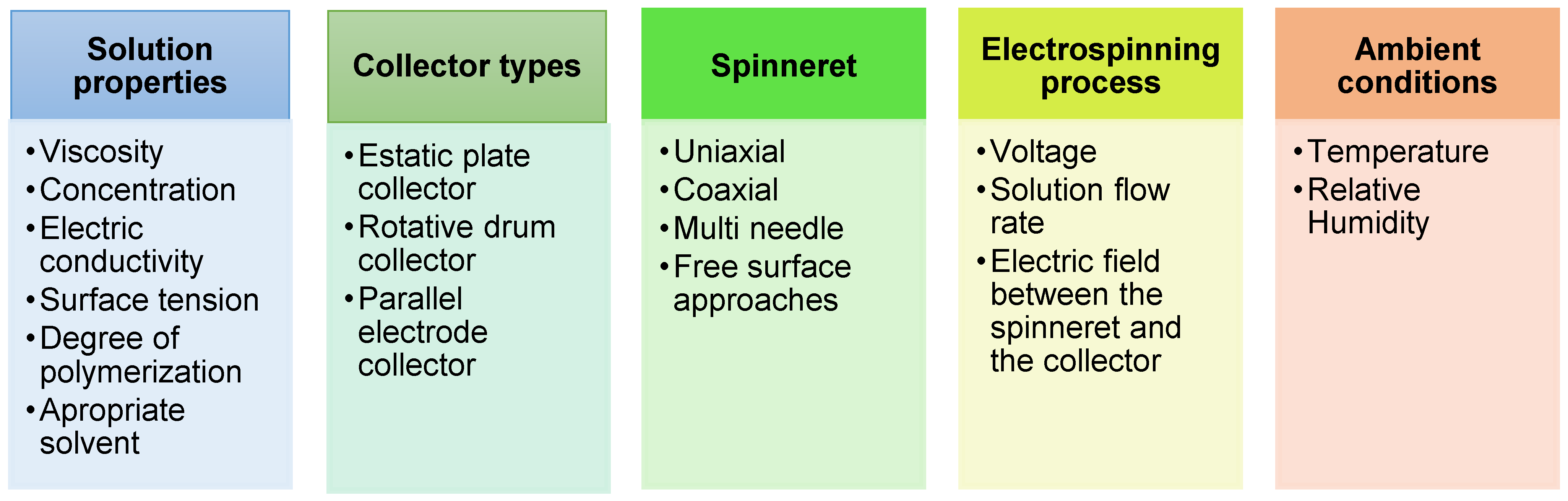
2. Strategies of Electrospinning Process

Electrospinning is a low-cost and high-efficiency electrohydrodynamic processing for producing micron, submicron, or ultrathin and nanoscale electrospun polymers [28]. Electrospun fibres have a low density, large porosity, and variable pore size as mats [17,39]. Furthermore, the flexibility to change their thickness, length, area-to-volume ratio, surface chemistry, and composition allows us to tailor their properties to fit specific purposes. Electrospinning has been utilized for a range of food applications through playing different roles such as (i) food processing; (ii) transport and controlled release of bioactive substances; (iii) detecting pathogens and increasing food safety; and (iv) the development of packaging systems able to increase the shelf life and improve food safety and quality.
An electrospinning process consists of a grounded collector (to collect the electrospun fibres) positioned at 10 to 30 cm of a spinneret tip, such as a metallic needle or capillary tube (to withdraw the polymer solution), connected to a high-voltage power supply (5–30 kV), which generates a high electric field (typically 1–5 kV cm−1) between the needle and collector. The polymer solution is infused through the tip by an infusion pump pressing a syringe piston or tubing to hold the solubilized or melted polymer [28]. Electrospun fibres are formed by the high-voltage electric field on the surface of polymer solution droplets, causing a liquid jet to be ejected through a spinneret [40]. A high voltage induces free charges in the polymer solution in the capillary distorting the hemispherical surface of the droplet into a conical shape (i.e., Taylor cone) near the tip of the capillary. This occurs mainly thanks to two significant electrostatic forces (the electrostatic repulsion of similar charges and the Coulombic force of the external electric field) [41]. A charged polymer jet is released from the tip of the Taylor cone once the electrostatic force has counteracted the surface tension. The unevenly distributed charges generate whipping or bending motion of the jet as it is propelled towards the collector. As a result of the elongation of the jet and the quick evaporation of the solvent, solid polymer fibres are collected as a randomly or oriented electrospun mat on the grounded collector [42,43].
Different approaches of electrospinning, such as single (uniaxial) and coaxial spinnerets are used to produce nanofibres (Figure 1). In the uniaxial output (Figure 1A), a simple spinneret is used to eject the solution toward the electric field [44]. Solubilized polymer can be blended or mixed with a compound of interest that has active activity. This device enables the optimization of the functional performance of antioxidant or antimicrobial electrospun mats by combining natural or synthetic polymers with active compounds [44,45,46]. In this setup, sensitive bioactive compounds and enzymes are preserved by a non-thermal process, whereas they might show low solubility or be inactive due to their denaturation by some organic solvents [28,47].
Coaxial electrospinning (Figure 1B) was developed by modifying the classic axial setup through the arrangement of multiple solution feed systems to simultaneously electrospun two or more polymer solutions from coaxial spinnerets [48,49]. The coaxial electrospun fibres can load bioactive ingredients in the inner capillary offering versatile encapsulation process [42]. The concentric or dual needle spinneret allows the fabrication of core–shell fibres for probiotics, enzymes, vitamins, polyphenols, and peptides, among other sensitive active compounds, due to a greater ability to control the flow system [24]. The core–shell structure of coaxial electrospinning nanofibres can be used to offer sustained release profiles of encapsulated compounds without diminishing their bioavailability and bioaccessibility properties [39,40]. Furthermore, two or more immiscible or with desired degradation rate polymers can be blended in the same fibre structure by core–shell assembly [50]. Coaxial electrospinning also provides functionality of electrospun fibres that is mainly improved by producing tubular nanofibres using both the inner solvent (or vapor gas) and outer polymer solution [51]. These methods depend on the pre-setting of a series of parameters to define the morphology, diameter, and surface characteristic of electrospun fibres, which are summarized in Figure 2.
Key parameters such as polymer concentration, molecular weight, and solvent type have a significant influence on fibre production and morphology, partly due to the viscosity relationship [54,55]. Droplet formation and non-homogeneous and beaded fibres are formed when the solution viscosity is low, whereas high-viscosity solutions increase the diameter of fibres and prevent fibre formation due to aggregation at the spinneret tip [56]. Higher molecular weight of polymer increases viscosity and results in uniform fibres, while low molecular weight results in the formation of beaded fibres or an unstable jet [57]. Very low molecular weight polymers might struggle to be electrospun into fibres, but some researchers are looking for polymer-free electrospun fibres. Certain polymer-free electrospinning fibres are successfully produced by phospholipids, cyclodextrins, peptides, oligosaccharides under specific concentrations, solvents, and processing [26]. To produce smooth fibres, the viscosity and concentration of the polymer should be set at optimum conditions [30,35,57,58]. Furthermore, it has been established that polymer solutions with reduced surface tension are able to promote the formation of beaded fibres [17]. Additionally, appropriate solvent selection considering its evaporation rate is quite challenging during the electrospinning process and bead-free fibres recovery [55].
The electrical conductivity is one of the most important properties of a solution, influenced by the nature of the polymer, other solutes, and different solvents [58]. The charges migration in the solution surface is responsible for the charge repulsion surmounting the surface tension and establishing a stable jet. Absence or low electrical conductivity results in reducing charges in the solution surface; the increased solution conductivity typically results in greater elongation of the polymer jet, and thus in the formation of beads and homogeneous and fine diameter fibres. Excessive electrical conductivity reduces the surface charge density and the electrostatic force by a quick charge transfer [59]; therefore, the formation of nanofibres from the solution prepared from different polymers relates to its electrical conductivity [53,60].
It is notable that each polymer material requires different voltage for fibre formation, usually >10 kV (Table 1). Increasing the applied voltage causes the diameter of the fibres to shrink until it reaches the optimum point, which varies depending on the polymer solution [23]. This is due to the stretching of the polymer solution caused by the repulsive electrostatic forces in the jet. Non-uniform fibres with beads can be formed at low voltages [61]. However, high voltages (above the critical voltage) reduce the length of the single jet and increase the apex angle of the Taylor cone, resulting in thick and non-homogeneous fibres. Additionally, greater amounts of charge at high voltage result in a faster and larger volume of solution being drawn from the spinneret, due to a smaller and less stable Taylor cone [35,59,60].
The polymer flow rate is also an important process variable because it affects the jet velocity and mass transfer performance (Table 1). Low flow rate is preferable, as it allows the solvent to have sufficient time to evaporate. The spinning solution should always have a minimum flow rate because a high rate increases the fibres’ diameter which might alter the morphological structure and the surface area [17,60,90]. The diameter and morphology of the fibres are also affected by the electric field between the spinneret and the collector, where a short distance might result in a high electrical field and moist, thick, and non-uniform fibres [91]. The fibre formation requires time to stretch and elongate before the solvent evaporates and its aggregating in the collector in ultrafine and nanosized diameters [53]. On the other hand, low viscosity solutions require short distances to be spun into fibres, or even beads or nanocapsules [42,92,93].
Despite the various potential uses of uniaxial and coaxial electrospinning, they are still used on a lab-scale due to their low production rates [94]. The “multi-jet electrospinning” was introduced by increased number of needles. As needles are used as spinnerets, a large working surface, needle spacing control, and a frequent cleaning system for each needle are required [95]. Free-surface or needle-less electrospinning has recently received attention, showing the ability of large-scale manufacturing by erupting large numbers of jets from an open solution surface. Needle-less setup can address some syringe blockage and remove solvents from the free surface under the influence of an electrical field [37,96,97].
The type of collector is important for the electrospun process as it serves as a grounded target which allows electric field formation and the gathering of nanofibres [98]. A static plate results in unwoven and randomly oriented nanofibre formation [99]. On the other hand, the use of advanced collectors such as a rotating drum, a rotating wheel-like bobbin, or a metal frame result in aligned electrospun fibre mats [60,100].
The morphology of fibres and the productivity of the electrospinning process can be influenced by environmental factors such as relative humidity and temperature, which have rarely been examined. Indeed, solvent evaporation and temperature, as well as the conductivity of the solvent, are closely related to environmental conditions and might affect the electrospinnability of the polymer solution [101]. Temperature influences the solvent evaporation rate, viscosity, and surface tension of the polymer solution [102]. Relative humidity has an effect on the average fibres diameter of electrospun fibres [103], and in some cases, at high relative humidity, the solution electrospinnability is compromised [97]. This behaviour is partly related to the electrostatic charges dissipating under high relative humidity, and the water on the solvent system cannot evaporate into the humid air, which results in a wet and fused material in the collector surface [97].

3. Overview of the Biopolymers Used for Electrospun Fibres

To reduce the toxicity issues with packaging materials, researchers used a multitude of biopolymers, likely polysaccharides and their derivatives such as cellulose acetate, dextroses, and proteins, such as gelatin and zein. These have often been electrospun into fibres due to their high safety and biodegradability. However, few natural polymers are electrospinnable, as shown in Table 1. In fact, each polymer material requires specific solvent and processing parameters to result in micro, submicron, and nanosized fibres. Additionally, different polymers blends are often used to widen the fibres’ characteristics and properties and, in some cases, synthetic polymers are used as a starting carrier, or to aid polymers for fibre assembly. It is worth mentioning that biomass-derivates and synthetic biopolymers are easily electrospun into nanofibres.
Polysaccharide electrospun feasibility depends on their molecular weight, degree of ramification, functional groups, charges, type, and degree of modification. These chemical properties orient the selection of the appropriate solvent and its evaporation rate for the spinning solution. Some solutions’ properties, such as polymer concentration, viscosity, electrical conductivity, surface tension, and vapor pressure, determines if it can be electrospun into nanofibres and plays an important role in the fibres’ morphology [30,44]. Some successful electrospun fibres from polysaccharides are given in Table 1. The main uses of polysaccharides are to encapsulate bioactive compounds for food and packaging incorporation. For example, it is known that material size and quantity play a major role in protecting highly perishable fish and fish products. In fact, rancidity and generation of free fatty acids are usually common drawbacks of fish oil and other fish products [104]. Thus, to overcome such problems, microencapsulation using marine polysaccharides played an important part. It was also observed that microencapsulated materials had a larger contact area than macroscale materials, affecting oil properties [105]. Accordingly, nanotechnologies gained interest in developing lab or pilot-scale material in the form of surface coatings to better preserve the quality of food products, including those from muscle foods [1,27].
Starch electrospun fibres are dependent of amylose and amylopectin ratios, with high-amylose starches (>50% of amylose content) being recommended for fibre production. Starch fibres are made from dimethyl sulfoxide (DMF) as a solvent-modified electrospinning setup using anethanol coagulation bath [106]. This solvent has a high boiling point and does not evaporate at room temperature. On other hand, ultrafine fibres from soluble potato starch (32.5% of amylose content) were achieved using formic acid as a solvent in a regular setup (Table 1), via low solution viscosity after the solution aging, which improves the spinning processing and fibres morphology. The starch nanofibres retained up to 50% of the phenolic compounds from thyme essential oil, after exposure to thermal treatment [107]. Both processes allow starch–guest inclusion complexes formation and fibres use on active packaging development. Several other polymers, that are not all described in this report, can be used, and some examples are given in Table 1.
Chitosan is a versatile biopolymer, generally of marine crustaceans origin, obtained through chemical or enzymatic deacetylation of chitin and exhibiting powerful antimicrobial potential against many microorganisms [108,109]. Despite the extensive range of applications of chitosan nanofibres, the pure chitosan has low spinnability and is deemed a challenge [110], with few reports about its use. To achieve such a challenge, coaxial steps are required, as well as the use of cross-linkers and neutralization processes to stabilize the nanofibres [64]. Chitosan starter polymer blends with PEO (chitosan/PEO) (1:4), PLA (chitosan/PLA), (1:6), and PVA (chitosan/PVA) (3:7 to 7:3) were successfully used to electrospin chitosan into fibres and nanofibres [111,112,113]. Further details and examples are given in Table 1.
In the objective of successfully electrospinning carbohydrate gums into fibres, several electrospinning process parameters should be considered depending on gum features, including molecular structure and solubility in water or organic solvents, polymer–polymer conjugation, supramolecular polymer, and other small molecules [114]. There is a well-established knowledge about the use of polymer chain entanglement, high conductivity, adequate viscosity, and surface tension in spinning solutions. Therefore, to meet these requirements, blends, crosslinking, and carrier materials to help in electrospinning are the main strategies for successful gum-derived electrospunfibre production through weakening shear-thinning to favour the liquid jet elongation when propelled towards the collector by the electrical field [115]. Xanthan and pullulan are two microbial-based polysaccharides very often used as biomaterials for electrospun fibres. Xanthan gum is an extracellular heteropolysaccharide produced by the bacterium Xanthomonas campestris; using water as solvent, it was not possible to electrospin into fibres. Moreover, using formic acid as a solvent, few reports describe the concentration effect of xanthan alone in nanofibres’ morphology and size distribution [115].
Pullulan is an extracellular microbial water-soluble polysaccharide produced by the fungus Aureobasidium pullulans and insoluble in organic solvents and reported as efficient in reducing meat perishability [116]. Additionally, pullulan-carboxymethylcellulose sodium-tea polyphenols nanofibres decreased weight loss and maintained the firmness of strawberries, and improved the fruit quality during storage [87,117]. Blending pullulan with hydroxypropyl-β-cyclodextrin was efficient to produce electrospun fibres and uses in encapsulation applications [87,117]. Pullulan–tetraethoxysilane (TEOS) hybrids were used on poly(ethylene terephthalate) (PET) films as a coating for the oxygen barrier. The hybrid coatings deposition decreased the oxygen transmission rate of the PET substrate by up to 2 orders of magnitude under dry conditions with low concentration of silica [118].
Zein and alcohol solution is a challenge overcome by using ethanol 70–80% (w/w) and widely used for zein nano- and submicron fibre production. It has been used to avoid fish oil oxidation in comparison to non-encapsulated solutions and polyphenols from plant-extract preservation and controlled releasing [66,119]. Electrospun zein nanofibres ensure the stable release of compounds such as cinnamaldehyde (CD) essential oils. They also inhibited the growth of microorganisms and extended the shelf life of mushrooms, proving that these fibres can be used in an active antibacterial package [68]. Zein/IC-β-CD electrospun ultrafine fibres were used to encapsulate eucalyptus essential oils and resulted in 28.5% of L. monocytogenes and 24.3% of S. aureus growth reduction by volatile micro-atmosphere releasing [120].
To overcome the unstable morphology, even with adjustment by post-electrospun process, of gelatin nanofibrous mats in aqueous solutions, physical (e.g., irradiation), biological (e.g., transglutaminase) and chemical (e.g., genipin, diisocyanate, glutaraldehyde, and carbodiimide) crosslinking approaches have been proposed in several reports [121,122,123]. For example, some chemical treatments on gelatin might cause fibre flattening and cytotoxicity. Citric acid is a natural acid that showed crosslinking ability to electrospun fish gelatin nanofibres, resulting in morphological stability under heat treatment [72]. Several biodegradable nanocomposite electrospun fibres are emerging, such as fish scales extract [84] and fish sarcoplasmic protein [74], which are showing great potential for the food and biomedical fields (Table 1).
Gelatin from cold-water fish nanofibres in different concentrations (23.7 to 35.5% (w/v)) resulted in fibre diameter from 91.42 nm to 200.48 nm, while at 4.8 and 9.1% (w/v), spherical beads with some fibres and from 12.9 to 20.5% (w/v) beaded fibres were observed. Different solvent systems (acetic acid/water, 2, 2, 2-trifluoroethanol) also result in fibres with average diameter of ~200 nm [71]. Furthermore, when compared to their non-encapsulated counterparts, the shelf stability of the encapsulated extract, in the case of zein/gelatin shell core fibres via coaxial electrospinning, at both refrigerated and ambient conditions was significantly extended [70].
PLA is one of the most important synthetic biocompatible, bioavailable, and biodegradable polymers that is also considered “generally recognised as safe (GRAS)” by the United States Food and Drug Administration (FDA) [22,27,124]. There are several reports using PLA for nanofibres production. The PLA electrospun fibres have a continuous and smooth surface. In some cases, porous fibres can be achieved by a binary solvent system, therefore enabling the encapsulation of bioactive compounds and their slow release [77,97]. For example, multi-coated electrospun PLA nanohybrids mats incorporated by α-tocopherol (α-TC) and α-TC/cyclodextrin (CD)–inclusion complex (IC) resulted in better solubility and antioxidant activity, which are expected to be sufficient to inhibit lipid oxidation in meat products [77]. With PLA/tea polyphenols composite nanofibres, the tea polyphenols improved the antioxidant and antimicrobial activities against E. coli and S. aureus [76]. These suggest great potential for these multicoated fibres in active packaging applications to increase food shelf life.
Polyvinyl alcohol (PVA) is biocompatible, nontoxic, water soluble, biodegradable, and easy to transform into electrospun fibres. Cross-linked PVA/β-CD electrospun nanofibres were, for example, reported to release cinnamon essential oil in a sustained manner to retard peroxidation to pertain the freshness of mushrooms, as well as improving the antibacterial properties against S. aureus and E. coli [79]. There are also reports of novel approaches to preserve fresh rainbow trout fish fillets with sodium alginate electrospun mats of 60.09–522.1 nm loaded with Lactobacillus rhamnosus in PVA [125]. The results indicated retention stability of polyunsaturated fatty acids such as eicosapentaenoic acid, and docosahexaenoic acid. The inhibition in predominant change of monounsaturated acid, such as oleic acid, was further observed. Consistently, electrospun nanofibres with dimension of 200–288 nm developed from blend of PVA with chitosan loaded with antibacterial agent nisin resulted in the retardation of total viable bacteria, psychrophilic bacteria, yeast, and mould growth in the flesh, thereby retaining freshness in fish and fish products [126].

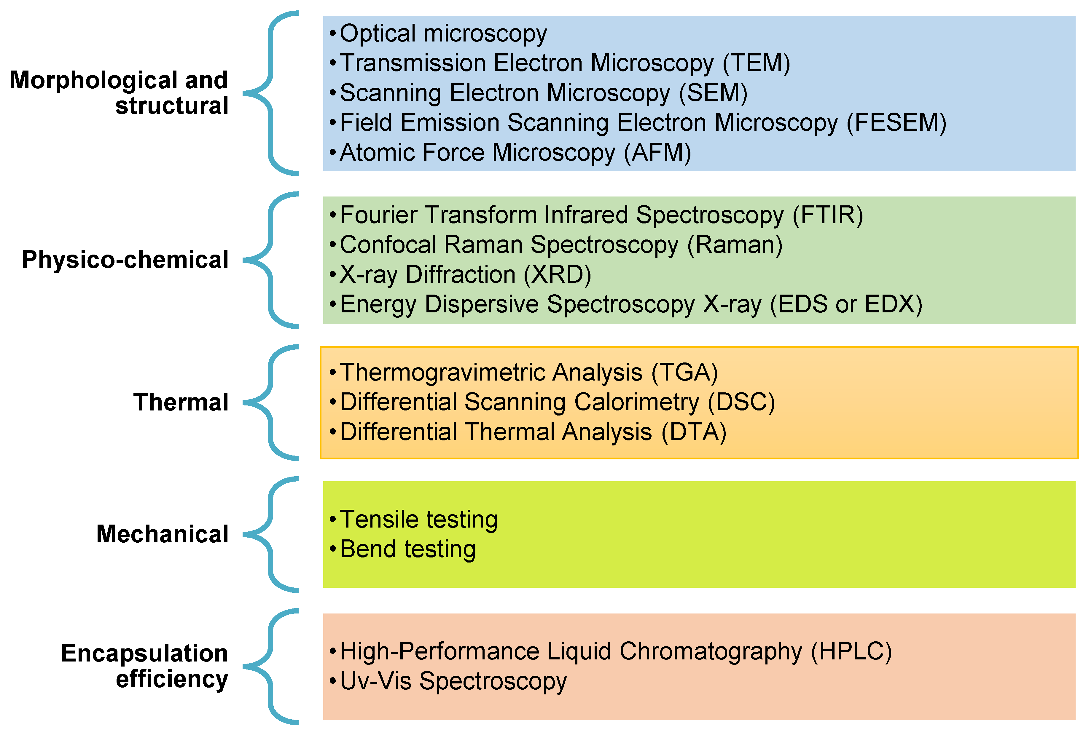
4. Characterization of Electrospun Structures

The fundamental characterisation of morphological, structural, physicochemical, thermal, and mechanical properties are of high relevance for determining the suitable application of electrospun fibres [110,127]. Figure 3 illustrates the most used methods for the characterization of electrospun fibre mats. The morphological and structural properties of nanofibres that are characterized are mainly the fibres’ diameter, diameter distribution, fibres’ orientation, porosity, and morphology [128,129]. Fibre membrane or mat porosity are also often characterized. Scanning electron microscopy (SEM), field emission scanning electron microscopy (FE-SEM), transmission electron microscopy (TEM), light microscopy (LM), confocal fluorescence microscopy (CFM), and atomic force microscopy (AFM) are among the widely used techniques. SEM, in particular, was reported as an efficient tool to obtain a quite complete characterisation of dry nanofibers [11,52,110,130]. For sample preparation, a sputtering coating with gold or carbon is used to improve the electrical conductivity and allows visualization at high magnification [131,132]. If the sample is not dry, TEM can be used to determine fibre diameter below 300 nm, after spreading the sample into a coated grid support and dyeing it when required [133]. On the other hand, AFM gives an exact image of fibre topography and description, as well as the roughness [133]. LM is often used as a preliminary assay of the fibres formation, as well as surface characterization, orientation, and diameter; while CFM is based on emission signal of fluorescence from some polymer structure or active compound, or even from an external dye bind with fluorescence signal [134,135]. To determine the average fibres diameter from SEM, LM, or TEM, the open source software for analysis mainly used to analyse the graphs is ImageJ [130].
Attenuated total reflectance-Fourier transform spectroscopy (ATR-FTIR), Raman spectroscopy, X-ray diffraction (XRD) and energy dispersive spectroscopy X-ray (EDX or EDS) are techniques used for chemical characterization of electrospinning structures and their interaction with bioactive agents [34,127,136].
FTIR analysis was found efficient in accessing the stabilization of the encapsulated bioactive agent and understanding the interaction between polymers and bioactive agents, such as oil and essential oils oxidation [119,133,137]. For ATR-FTIR, minimal or no sample preparation is required prior to spectral measurements because the penetration depth of infrared light is independent of sample thickness. Raman spectroscopy allows the determination of molecular and crystal symmetry and the strength of chemical bonds, while it also detects structural abnormalities and defects [110]. XRD is another method able to access structural changes related to a crystalline structure [130]. These same post-electrospun treatments may change the fibres’ molecular organization and result in an improvement of the fibres’ crystallinity accessed by XRD and FTIR [138,139]. EDX is a surface analytical method enabling the elemental composition owing to the interaction between X-ray and samples components [140]. This elemental composition analysis is conventionally used in SEM to give a qualitative microanalytical technique SEM-EDS for all SEM-suitable samples.
Thermal properties can be determined using various methods aiming to establish a connection between the temperature of a target material and its physical features [34]. The differential scanning calorimetry (DSC), thermogravimetric analysis (TGA), and differential thermal analysis (DTA) are the most prominent techniques for analysing electrospun fibres. DSC provides melting (Tm), glass transition (Tg), crystallisation (Tc), and dissociation temperatures, and process enthalpy (H). This method enables us to effectively understand and measure the melting and crystallization behaviours, polymorphism, and purity of the materials [141]. Thermal degradation and mass loss of electrospun fibres may be measured by TGA, and complement DTA (measuring the temperatures of reactions and identifying if the reaction is endothermic or exothermic); both are extremely useful to see and check the biodegradability of packaging materials [142]. DSC and TGA enable complementary information about the polymer and electrospun fibres to assess the thermal characteristics of the carrier and the encapsulated compounds, as well as its stability, to predict its structure’s behaviour and characteristics if used as packaging materials [130,143].
Tensile testing is used to analyse some mechanical properties of electrospun fibres and membranes using a texture machine or an universal testing tool [144]. It is achieved by applying loads to specimens prepared from the electrospun mats [145], which results in a stress × strain curve where tensile strength and strain at break are set [133]. Young’s modulus or elastic modulus (ε) is computed by the slope of the straight-line from two points from the stress × strain curve. It is important to note the difficulty to achieve mechanical properties from a single electrospun fibre by tensile testing. Usually, single fibre approach tensile test is provided by AFM cantilever assisted protocols; additionally, a bending test can be achieved by the same AFM [138]. On other hand, encapsulation efficiency is defined by the concentration of the incorporated material over the initial concentration used to prepare the spinning solution for determining if the bioactive agent has been effectively loaded into the electrospun fibres [146]. UV–vis spectroscopy and high-performance liquid chromatography (HPLC or LC-MS) are mostly used to identify and quantify the active compounds and allows encapsulation efficiency computing [34]. Each individual active compound or some group of compounds need a specific standard and analytic method for proper quantification, with standardized and accurate LC and UV–vis protocols.

5. Active Compounds for Meat Processing and Preservation

Electrospun fibres have a high capacity for loading active agents, and their large surface area allows a fast response to intrinsic and/or extrinsic factors by releasing/activating the entrapped compounds at the appropriate timing [23,27,147]. Furthermore, the narrow gaps between the fibres can be considered a barrier against viruses and microorganisms [148]. Unlike intelligent casting film versions, electrospun mats have several structural and functional advantages [149,150]. Beyond their high surface-to-volume ratio structural advantage, entangled fibrous formation porosity, customized surface roughness, and other adjustable physical properties are also reported [148]. The functional benefits include a high loading capacity, improved stability of sensitive bioactive compounds, sustained and targeted delivery of the embedded compounds, and the availability of food-grade polymers and solvents [47]. In addition, the fillers and active components for processing and preservation can improve the overall quality and increase the shelf life of fresh meat or packaged meat via electrospinning as recently discussed for nanotechnology [27].
Antioxidant and antimicrobial compounds used for food and packaging can be natural, such as essential oils, nisin, curcumin, α tocopherol and vitamins, phenolic-rich extracts from plants and pomaces, allyl isothiocyanate, and chitosan, among others; or synthetic antioxidants, such as BHT (butyl-hydroxytoluene) and its analogues, BHA (butyl-hydroxyanisol) and TBHQ (t-butyl-hydroxyquinone); or antimicrobial, such as organic acids (acetic, sorbic and ascorbic, benzoic, and propanoic), nitrites and nitrates, and others [150,151,152]. Additionally, nanofillers such as nanometals and nanoclays are often reported as alternatives for food packaging development [153]. Thus, researchers are continuously exploring ways to arrest its microbial activity on food matrices leading to adverse effects. In recent years, the development of antimicrobial agents from various natural products such as essential oils and plant extracts proved to be the most promising [154]. Natural compounds are preferable for new packaging approaches, due to consumers concerns over the safety of additives and synthetics material and their potential effect on health [155,156]. Natural bioactive compounds can be added to the electrospun nanomats to improve their bioavailability and solubility as they are effective antibacterial agents and promising preservatives for meat and meat products [157]. Zein/gelatin nano electrospun composites were efficiently used against S. aureus and S. enteritidis [158]. An antioxidant activity, reduced drip/purge loss, better colour stability, and antibacterial activity against the aerobic mesophilic and psychrophilic bacteria in pork chops were noticed when they were wrapped with electrospun mats of blended chitosan and low density polyethylene incorporated with propolis extract [159]. Similar antibacterial effects were observed when thyme and Chrysanthemum essential oils were loaded in the electrospun fibres to preserve chicken [160] and beef [161], respectively.
Essential oils have a wide spectrum of usage as antimicrobial agents. Nevertheless, their poor water solubility and chemical instability restricts their uses. Thus, advanced nanotechnology such as nano liposomes were implemented to enhance their physic chemical properties [162]. Nevertheless, their encapsulation efficacy percentage is still a challenge due to their high hydrophobicity rate. To overcome such drawback, the role of introducing β-cyclodextrin moieties play an important role in developing the aqueous stability of essential oils [163]. Still, research gaps exist as the complex moiety consisting of essential oil, nanoliposomes, and cyclodextrin would agglomerate on the food surface leading to reduced bioavailability [164]. Avoiding such difficulties, electrospinning comes into action to provide a support to proteoliposomes for a better encapsulation efficacy, hence enhancing the mechanical strength of packaging material.
Polyphenol-rich plant extracts are a mix of complex bioactive compounds, such as phenolic acids, flavonoids, and other polar compounds. Phlorotannin is a type of highly hydrophilic tannin from brown algae formed by the polymerization of phloroglucinol monomer units and biosynthesized through the acetate–malonate pathway, also known as the polyketide pathway [165]. For example, Ilex paraguariensis extract chlorogenic acids-rich entrapped into zein nanofibres improved the thermal stability of biocompounds and moderated the antioxidants release [66]. Tea polyphenols are bioactive catechins and may interact with carbohydrates polymers, likely pullulan and carboxymethylcellulose sodium. Hydrogen bonds are formed between hydroxyl groups of polyphenols and oxygen atoms of the glycosidic linkages of polysaccharides, and it has been shown that these interactions have a significant influence on fibres’ structure and morphology [87] (Table 1).
Anthocyanin-rich extract is often used as a pH indicator due to its acid–alkali structure changing, being flavylium cation (red) and quinoidal anhydrase (purple) in acid conditions and to quinoidal (green) and chalcone (yellow) structures in alkaline solutions [67,166]. pH indication using red cabbage anthocyanin-rich extract into zein ultrafine electrospun fibres resulted in colour changes in a board pH range. Purple sweet potato anthocyanin-based pH indicator had high colorimetric response sensitivity for pork freshness monitoring and preservation [167]. Anthocyanin from blueberries is able to detect total volatile basic nitrogen (TVB-N) in package headspace for food [168]
Nisin is a bacteriocin produced by Lactococcus lactis ssp. Lactis is often anchored in polymer membranes, which display good antimicrobial activity. An extruded composite film of pectin/PLA blend and nisin was efficient to inhibit Listeria monocytogenes in an in vitro assay and in food samples [169]. Additionally, a biodegradable membrane from oxidised cellulose incorporated with nisin showed a long-term antimicrobial activity against Alicyclobacillus acidoterrestris DSM 3922T [170]. Curcumin is a polyphenol compound from turmeric spice, with antioxidant, antimicrobial, and anti-inflammatory activity, and is highly sensitive to acid–base reactions [171,172]. Antibacterial activity of curcumin/nisin loaded nanoliposomes incorporated in PVA electrospun nanomats were actively used as packaging material of trout fillets [173] and fish (Oncorhynchus mykiss) flesh [174]. For example, real-time shrimp spoilage monitoring was developed with a curcumin-based sensor visible to the naked eye, to detect volatile amines (TVB-N) in packaging headspace by increasing basic pH. The sensor response had a correlation with bacterial growth patterns in shrimp samples, useful for monitoring shrimp spoilage in ambient and chilling conditions [172]. The tara gum/PVA blended films containing curcumin, a safe biosensor, exhibited a visible colour change in an NH3 environment to monitor shrimp spoilage, being high in relative humidity favourable for this colour response [171]. Electrospun fibre can be used to improve these sensors sensibility and response speeding against food spoilage microorganisms due to its high surface area.
Despite the fact that the antimicrobial properties of chitosan solutions have been widely reported, the antibacterial activity of chitosan has, to date, received less attention and has only been investigated superficially [175]. There is considerable controversy about mechanism actions of Gram-positive and Gram-negative bacteria with chitosan, and thus the aspect of antimicrobial efficacy of chitosan is a streamline. Chitosan/PEO nanofibres (40.6–75.5 nm in diameter) were developed as packaging material for shelf life enhancement of meat cubes [176]. When the microorganisms (E. coli, S. Typhymurium, L. innocua, S. aureus) were grown in presence of chitosan nanofibres, a bactericidal effect on E. coli was detected (the growth arrested completely), whereas S. Typhimurium growth was significantly affected due to the inhibitory effect of free amino acids of chitosan. No significant changes were observed in L. innocua and S. aureus. Thus, it can be concluded that Gram-positive bacteria are more susceptible than Gram-negative bacteria to chitosan nanofibre activity, which can be a component of active food packaging for meat industry to enhance shelf life.
Silver nanoparticles (AgNPs) are an inorganic nanofiller with antibacterial properties [27,177]. AgNPs have been widely used in the preparation of antimicrobial nanofibres for food and non-food applications. Electrospun nanofibres from chicken feather keratin and polyvinyl alcohol (PVA) blend embedded with AgNPs resulted in improved antimicrobial properties and thermal stability. It can be efficiently used as a promising material for active packaging [88]. AgNPs were also incorporated in the PVA, and the nanocomposite electrospun mats resulted with a stable porosity and nanofibrous structure as well antibacterial activity against S. aureus [178]. Coating polycaprolactone (PCL) nanofibre membranes using AgNPs embedded in gelatin showed an improved antibacterial effect in comparison with single coating nanofibre against S. aureus and P. aeruginosa [179].

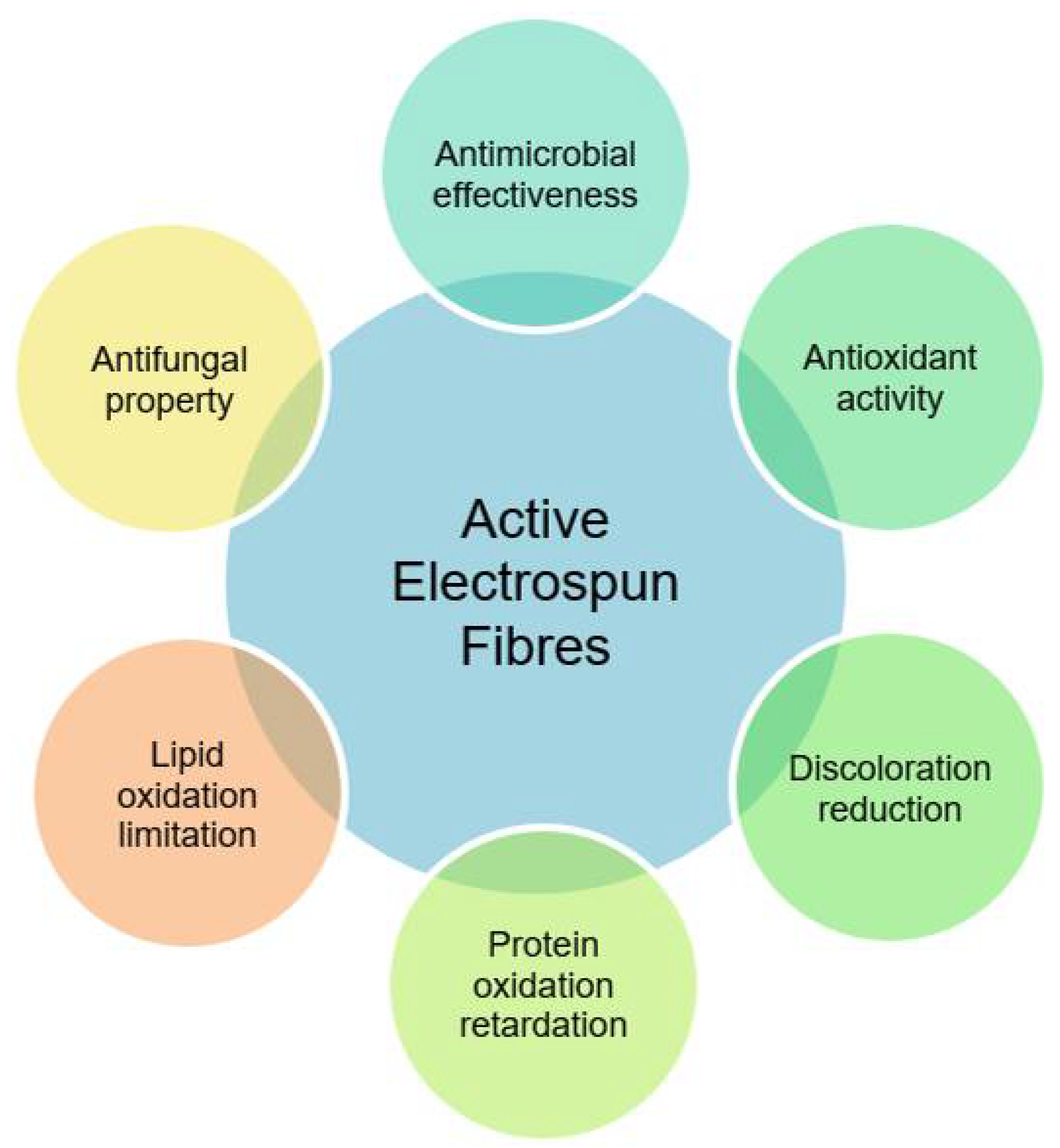
6. Role of Electrospun Fibres in Meat Processing and Preservation

Food poisoning and wastage are two major issues in the food industry, both caused by microbial contamination and lipids/proteins oxidation. Among all food goods, meat products are highly perishable with short shelf life where quality and safety are highly dependent on the processing, storage, and packaging [1,2,36,180,181]. Accordingly, consumers are becoming more concerned about pursing health with diversification of diets such as fresh foods, fruits, and vegetables and their products as living standards improve. Fresh foods tend to deteriorate fast by microbial deterioration and enzyme degradation in the farm to fork chain process, resulting in shorter shelf life [2,182]. Therefore, the use of active and intelligent packaging to prevent microbial contamination, spoilage of meat products, and oxidative degradation, all avoiding food waste, is of great interest to both the food industry and consumers. These packaging systems allow food products to extend shelf life, maintaining quality and safety. The electrospinning process has been reported as an efficient meat packaging approach [105]. This is further highlighted in Table 2 and Table 3 which summarize, respectively, the active electrospun fibres roles in meat processing and preservation through active (Figure 4) and intelligent (Figure 5) packaging systems.

6.1. Active Packaging

Active packaging interacts and/or modifies the packaging headspace, hence extending the shelf life of foods and consequently improving its safety [13]. These systems are embedded by active compounds, which will remove gases and vapours (O2, CO2, water, humidity, and microbial metabolites) by absorbing and adsorbing or via chemical reactions. On the other hand, the active compounds may be released from the package matrix as vapor or target compounds (essential oils, antimicrobial and antioxidant agents, volatile and fumigant agents, or metal nanoparticles). This enhances food protection and preservation against oxidation, microbial proliferation, and other mechanisms that shorten the products shelf life [152,204].
Electrospun nanofibres from polymers containing βCD and loaded cinnamon essential oil with (317–364 nm) were used for beef packaging [191]. The results indicate antimicrobial activity against B. cereus, preservation of the beef colour, with no impact on texture during storage. The electrospun fibres increased the essential oil water solubility and addressed sensitivity towards enzymes, temperature, light, and oxygen. Antibacterial activity against P. fluorescens P07 was observed when electrospun fibres from blend of propyl gallate and PLA were wrapped around salmon slices [184]. Poor insolubility of natural antimicrobial agent nanofibre derived from oranges was achieved by loading them in the core of PLA electrospun nanofibres. These electrospun packaging materials were used to maintain the freshness of salmon slices. The electrospun nanofibres (206–243 nm) were effective to retard the growth of P. fluorescens [183]. Novel electrospun nanofibres based packaging material were used to store raw shrimp for 7 days [188]. It was reported that PVA electrospun nanofibres embedded with cinnamon essential oils, nanophytosomes, enhanced the antimicrobial activity of cinnamon essential oil against S. aureus, P. aeruginosa, and E. coli. Poly(hexamethylene biguanide) hydrochloride (PHMB) loaded PVA electrospun nanomats were found effective as active packaging material in preserving Japanese Sea Bass [185]. The results indicated a delay in TVB-N production, inhibition of total viable count, Pseudomonas spp. and hydrogen sulphide producing bacteria. Electrospun fibres of European eel gelatin and European eel oil resulted in an eco-friendly encapsulation material for synthetic polymer replacement which can have a promising application in the food, nutraceuticals, and medical fields [193]. Zein nanofibres were also efficient to achieve oxidative stability of fish oil by encapsulation [119].
To delay food spoilage, continuous efforts are being made in exploring advanced techniques leading to rapid detection of quality deterioration changes by introducing smart nanoparticles as packaging materials [27]. Nanotechnology already plays relevant roles in microbiological infestation control, sensory deterioration, and chemical alteration in food matrices during the storage period. Currently, the implantation of biogenic techniques is gaining interest to reduce metal nanoparticles such as silver nanoparticles and gold nanoparticles. Recently, it was reported that zein/gold electrospun nanomats with zein nanoparticles (161 ± 45 nm) embedded with gold nanoparticles limited the total microbial amount in Japanese sea bass (to 1 log CFU/g) with improvements in the sensory quality of the meat for an extended period of storage [195].
Silk fibroin nanofibres (SF-nanofibres) were fabricated and embedded by cinnamaldehyde (CA)/hyaluronic acid (HA)/carboxymethyl chitosan nanoparticles (CA/HA/CMCS-NPs). When concentration of CA/HA/CMCS-NPs was set at 5 mg/mL, the mechanical characteristics of the nanofibres showed a double effect in the controlled release of CA from CA/HA/CMCS-NPs SF-nanofibres. CA/HA/CMCS-NPs SF-nanofibres were used to store beef for 5 days at 4 °C and 25 °C, and this decreased E. coli O157:H7 in beef samples up to ~99.9%. In addition, the CA/HA/CMCS-NPs SF-nanofibres demonstrated a moderated effect on the sensory quality of beef and delay the spoilage [205].
Novel bioactive coatings were prepared to extend the quality and shelf life of fresh fish fillets of rainbow trout (Oncorhynchus mykiss). For this, whey protein isolate (WPI, 96% of protein) was used as the coating-forming with 50 and 70 g 100 g−1 of poly(ɛ-caprolactone) (PCL) electrospun nanofibres containing extract of stinging nettle (Urtica dioica L.) at 60:40 (g:g). Fish fillets protected by means of the functionalized coatings were stored for 15 days at 4 °C. Active coating protected the fish fillets against mesophilic, psychrophilic, and lactic acid bacteria as well as Enterobacteriaceae with higher antimicrobial efficiency than equivalent coatings containing Urtica dioica L. water extract. In particular, the incorporation of 70 g 100 g−1 PCL nanofibres exhibited the lowest counts in bacterial growth, TVB-N, and thiobarbituric acid concentrations during storage. Additionally, the coatings successfully provided an antioxidant activity that could help to increase fish fillets’ quality and favour their preservation [187].
Electrospun chitosan/PEO-based nanofibres (CNF) was developed to keep the shelf life and safety of red meat, and it extended the shelf life of fresh meat up to a week. The CNF had antibacterial activity against E. coli, S. typhyimurium, S. aureus, and L. inocua [176]. Additionally, in both in vitro and in situ studies, the efficiency of CNF as inner part of a multilayer packaging in maintaining the quality of fresh red meat was reported [186]. Activated CNF-based packaging (CNFP) was obtained by direct electrospinning chitosan/PEO solutions on top of a conventional multilayer food packaging. The in vitro antibacterial activity of CNF was achieved against E. coli, S. enterica serovar Typhimurium, S. aureus, and L. innocua, bacteria commonly incriminated in the alteration of food products. The in situ bioactivity against E. coli showed the potential of CNFP as bioactive nanomaterial barriers to meat contamination by extending the shelf life of fresh meat by up to one week.

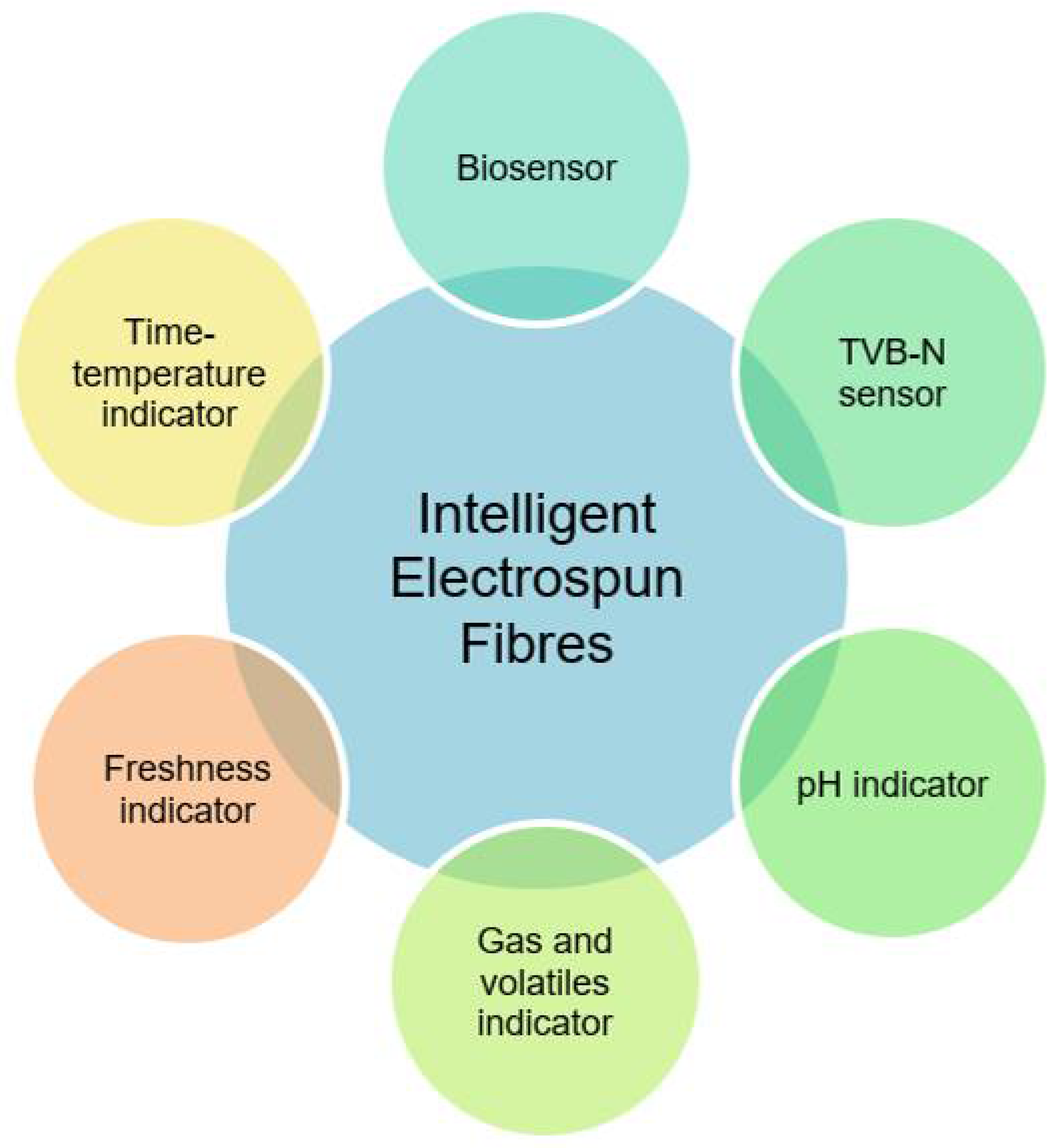
6.2. Intelligent Packaging

Intelligent packaging is continuously exploring mechanisms of various advanced techniques leading to rapid detection and warning of quality deterioration and changes of food goods. Thus, there are two basic approaches of quality indicators: direct (moisture, temperature, freshness and leakage, and biosensor) and indirect (monitoring and tracking) devices [206]. Both systems ensure food security [47,207] and offer visual feedback and information about the spoilage or food quality to be judged by the consumers [53,208], with no misleading.
These systems entail integrating packaging material with improved functional qualities and sensors for including monitoring, detecting, sensing, recording, tracing, and communicating the status of food in both internal and external environments during transport and storage (Figure 5). It assists customers or manufacturers in showing and reporting the packaged food’s quality [30,53]. Various intelligent tools, such as time–temperature, freshness, leakage, pH and gas indicators, and biosensors have been investigated to accomplish real-time tracking of food throughout the supply chain [53,209]. Food safety is a greatest concern, where consumers may face adverse effects on their health such as hemopoiesis or allergy if antimicrobial agent drug residues are accidentally ingested.
Over the past few years, the electrospinning process has been used for designing and producing colorimetric pH indicator packaging materials by incorporating pH-sensitive dyes in a variety of combinations to polymer formulations, allowing for the detection of a wide range of pH values [30,199]. Depending on the indicator’s purpose, a number of electrospun polymers with various morphologies can be conceived, including normal, helical, porous, and coaxial [47]. Table 3 summarizes the electrospun fibres in intelligent packaging mechanisms of meat processing and preservation.
Solid-phase extraction (SPE) is a sample preparation for analytical archiving which can be used for different types of samples. This is not an intelligent packaging; however, it can contribute into new intelligent system development. An SPE adsorbent membrane was developed based on graphene oxide-doped poly(acrylonitrile-co-maleic acid) (PANCMA) nanofibres to further access tetracycline antibiotics and volatile nitrogen on chicken breast by liquid chromatography (LC) [196]. Furthermore, to identify the presence of harmful sulphonamide residues in pork, an intelligent recovery SPE system was developed using poly styrene sulfonic acid (PSSA) electrospun fibres (407–469 nm) for LC quantification [197].
To date, electrospun fibres achieved valuable findings on meat freshness/spoilage monitoring, yet it is still restricted to the developing area [5,24,203]. Ammonia and amine molecules are generated as a result of meat and meat product deterioration due to microbiological and enzyme degradation [12]. The increase in total volatile base nitrogen (TVB-N) during storage is an indirect explanation of the colour change of the indicator, because it causes the pH of the meat to rise. In non-fermented foodstuffs, TVB-N is a collection of biogenic amines such as ammonia, dimethylamine (DMA), and trimethylamine (TMA) that form during storage. This indicator is commonly used as a measure for the freshness of meat products and the overall quality of meat products. TVB-N production is mostly caused by the proliferation of microorganisms, more specifically by bacteria decarboxylation of proteins. The major stage in this process is the microbiological breakdown of trimethylamine N-oxide (TMAO) to trimethylamine and ammonia, which is carried out by bacteria. All of this volatile base nitrogen contributes to the formation of alkaline environments [53], which can be used to trigger some dye sensitivity in the sensor.
A porous structure is preferred as the principle for freshness indicator manufacturing because the migration of volatile chemicals occurs readily and fast, and the interaction between the volatile substances proceeds more effectively and quickly. Food freshness can be monitored by employing colour-based pH indicators, which are easy to use and inexpensive indicators [5,22]. Moreover, the porosity structure of the electrospun nanofibres that have indicator dyes added boosts the number of active sites for interacting with chemically reactive compounds. The high sensitivity and rapid responsiveness of nanofibre-based fibres can therefore be envisaged [209].
Intelligent packaging systems for food deterioration were developed by blending chitosan/PEO loaded with halochromic curcumin to ascertain surface pH changes, TVB-N release, and an active antibacterial action against Enterobacteriaceae sp. [199]. Curcumin-loaded electrospun mats were efficient to indicate packaged chicken breast freshness stored at 4 °C for eight days. During deterioration, the colour of the freshness indicator changed from a bright yellow to reddish colour, according to the results of colorimetric analysis. The pH values and TVB-N concentration of the meat increased from 6.2 and 7.01 mg N 100 g−1 in the fresh samples to 6.53 and 23.45 mg N 100 g−1, respectively, at the end of day 5, showing that the samples had reached the marginal acceptability threshold. On the eighth day of storage, the pH values reached 6.75, the TVB-N concentration reached 50 mg 100 g−1, and the intensity of the colour of the pH sensing indicator decreased at the same time [199].
Halochromic nanofibres based on Poly-L-Lactic acid (PLLA) integrated with blueberry anthocyanins was developed to detect the freshness of mutton during storage time at 4–25 °C. SEM images of nanofibres revealed that the net PLLA fibres had more beads, but that introducing anthocyanin into fibres changed their structure and resulted in more uniform and thick fibres. The colour of the electrospun polymers changed from the original pink to a pale pink after the first day of storage and became colourless after three days of storage, and the TVB-N content of fresh mutton (7.2 mg 100 g−1) increased to 11.6 mg 100 g−1 and 34 mg 100 g−1 due to the deterioration of the meat [168].
An active-intelligent device was designed using pullulan nanofibres mat with purple sweet potato extract as a colorimetric layer and an antibacterial layer by zein-glycerol-carvacrol to monitor pork pH changes and improve pork shelf life. The distinctive colour changes of the package system at pH values ranging from 2 to 12 provided the mats a broad pH–sensing range. Initially, the colour of electrospun fibres was purple and blue, changed to green, and subsequently was reversed. The fibres demonstrated good antibacterial and antioxidant activities in addition to highly sensitive colour change, allowing shelf life extension of pork by one day at 25 °C [167].
An intelligent oxygen leakage indicator was designed by electrospun hydrolysed PVA (PVOH) carrier nanofibres of the colour changing system-based on methylene blue (MB) oxidation-reduction reaction (MBOx is blue, MBrd is colourless) catalysed by TiO2 and using glycerol as a sacrificial electron donor. A polystyrene (PS) microfibre coating layer was applied to address the indicator shortcomings. Both PS coated and uncoated oxygen indicators might be UV activated in a short period of time as an innovative design. The oxygen indicators were tested on packaged meatball samples and exhibited substantial colour changes in the presence or absence of oxygen. Colour changes can deliver direct information to the customers about the quality of the food prior to making their purchase decision [200].
Zein electrospun nanofibres were designed by incorporating alizarin as a halochromic indicator based on TVB-N to control the freshness of rainbow trout fillets during storage time. The average diameter of the fibres produced ranged from 79 to 619 nm. Initially, the mats were yellow in colour, indicating that the TVB-N concentration was nearly 17 mg 100 g−1. On day 6, the mats were light purple in colour and finally magenta in colour on day 12 (TVB-N concentration approximately 33 mg 100 g−1). These colour changes from light to darker suggested that the newly constructed system was capable of successfully monitoring the freshness of fillets through evaluating TVB-N concentrations [201].
A halochromic sensor of cellulose acetate nanofibres incorporated with alizarin as a fish spoilage indicator was produced. Rainbow trout fillets were maintained at 4 °C in the refrigerator for a total of 12 days. Colorimetric and TVB-N were determined, as well as pH and total viable count (TVC) analyses. The pH, the TVB-N, and the TVC all increased with time, according to the results. Within 48 h, no colour changes were seen on the electrospun sensor; nevertheless, by the fourth day, a very light brick colour was observed on the mat. On the sixth day, the colour turned darker, indicating that the pH of the fillet had changed. During the 12th day, the colour of the sensor began to shift towards violet; the colorimetric results confirmed that the electrospun fibres had undergone the expected visual colour change as a result of the use of alizarin as a halochromic dye [202].
Active-intelligent food packaging systems were produced by electrospun nanofibres based on pullulan/chitin (PCN) that contained curcumin (CR) and anthocyanins (ATH) as natural dyes. The average diameters of the nanofibres were range 176.81 to 379.07 nm. The antioxidant and antibacterial activity of the nanofibres combining ATH and CR (PCN/CR/ATH) were significantly higher than those of the nanofibres having only CR (PCN/CR) or only ATH (PCN/ATH). The pH-sensitive colour of the PCN/ATH and PCN/CR/ATH nanofibres did alter dramatically with the change in pH, indicating that they were highly sensitive. Furthermore, at ambient temperature, the colour of the PCN/CR/ATH nanofibres was altered in response to the increasing Plectorhinchus cinctus spoilage. The electrospun PCN/CR/ATH was found with great potential in active–intelligent food packaging applications [203].

7. Packaging Safety and Risk Assessment

The safety of electrospun nanofibres in the formation of food packaging films is still not fully clarified. Information about their potential risks is still limited and lacking in the literature. Therefore, the safety risk assessment is one research gap that should be examined in new future studies about electrospun food contact fibres and for some gaseous/volatile compounds realising into food packaging systems. Not very much attention was dispensed on electrospun fibres; however, there are a lot of studies about the safety of the most used materials and solvents, which can be a source of information on some consequent safety in their use in food packaging development, regardless of the used technology. Industrial validation of lab-scale achievements of electrospinning might boost their commercialisation. Furthermore, the evaluation of the toxicity of each substance/additive used is necessary to ensure customer safety. Due to their extremely small size, nanofibres can easily migrate by digestion, skin, and inhalation from packaging to the human body [210]. Even though silver and copper nanoparticles are found to be efficient in meat product packaging due to their antibacterial effects, their potential migration to food can be an issue [27]. It was reported that small-sized nanoparticles (e.g., silver) may easily migrate from packaging, enter the blood stream, and cause considerable harm to liver cells, brain tissue, and the immune system [211,212]. Copper was reported to induce liver injury, oxidative stress, and inflammatory reactions [213]. Nevertheless, it seems that their migration amounts are lower than the actual dietary intake level and thus may be considered as potential safe antimicrobial agents [27]. To overcome this bottleneck, laws and clear strategies for the risk assessment of nanoparticles must be implemented to assist decision making for regulatory authorities [22].
For now, there are no specific strategies to safely create packaging containing electrospun fibres. In Europe, migration assays of substances in contact with food are performed according to the Commission Regulation (EU) No 10/2011. This regulation focuses on the safety and risk assessment of plastic materials and articles intended to come into contact with food. Accordingly, the risk assessment of a substance should cover the substance itself, relevant impurities, and foreseeable reaction and degradation products in the intended use. Efficient and accurate methods of quantification of low amounts of nanoparticles are crucial to facilitate the risk assessment evaluation and enable future standard methods. The risk assessment also takes into consideration the potential migration of substances under worst foreseeable conditions of use and the toxicity. The European Union Reference Laboratory for Food Contact Materials (EURL-FCM) have agreed on a set of test conditions (technical guidance) to ensure the comparability of measurement results according to the Regulation (EU) 2017/625.

8. Conclusions

The electrohydrodynamic processing is a cost-effective technology to produce micron, submicron, ultrathin, and nanoscale continuous fibres. As a result of unique properties such as high surface area to volume ratio, high porosity, small size, uniformity, and homogenous distribution of functional chemicals, electrospun fibres provide advantages over conventional processes, such as casting and coating in the fabrication of active systems, indicators, and sensors [47,214] for food packaging, including meat and meat processing.
This paper summarized the electrospinning process and its applications with a focus on meat and meat products processing and preservation. There are many advantages of electrospun fibres boosting their use for different objectives. Electrospinning can be considered a promising and efficient meat packaging approach where the properties of electrospun fibres might induce antioxidant and antimicrobial effects among other functionalities depending on their features. Thus, their application in packaging systems of meat products can result in extending their shelf life, preserving their quality and safety. To date, electrospinning has already been applied to real food systems and achieved valuable findings on meat freshness/spoilage monitoring; however, the number of published papers is limited, probably due to limited funding in this research area. All the reviewed papers focused on the electrospinning process and characterization of electrospun fibres made at lab level, and none considered commercial products. The electrospinning process scale up is still in progress, and the best use for the industries are the free-surface setups which achieve the production of high-throughput fibres [37,96]. The first commercially electrospun products were reported in 2017, which are the encapsulation of DHA enriched fish oils and probiotics [96].
When upscaling the production of electrospun active and intelligent fibres, the meat freshness needs to be carefully considered to ensure colour and taste stability by avoiding fat and protein oxidation, and to limit microbial growth during storage. Fibre development by natural or GRAS solvents, polymers, and active agents, highly sensitive, triggered, and controlled release systems or new functional properties are also mandatory [215]. The key parameters affecting the electrospinning process, as well as the characterization techniques discussed in this review, should be kept in mind to face the challenges of each individual packaging device and meat product features.
To achieve multi-functionality for nanofibre-based food packaging materials, various additives have been incorporated into polymer substance. Nevertheless, additive manufacturers still have a challenging task to produce non-toxic and efficient material at low cost and with limited impact on the environment to be used in food packaging. Among the commercially available polymers used in packaging industry, some of them can only be dissolved in toxic solvents. Therefore, it is required to find non-toxic solvents to replace the conventional toxic solvents with non-toxic or low-toxic solvents, such as the emulsion electrospinning method [24].
Natural active compounds are a hot topic of interest due to their non-toxic effects on health. However, they are often inaccurate and sensitive to spoilage or to trigger compounds in low concentrations. The sensing, stable, and non-toxic dyes and packaging systems must be efficient to detect target changes in meat products. Various approaches for improving, stabilizing, and controlling the release of active compounds were designed to develop active packaging, including polymer blends and structure modifications, processing conditions optimization, encapsulation, and immobilization [22]. To achieve reliable naked eye detection changes, a food packaging sensor must be highly sensitive with rapid responsiveness, therefore with the ability to disclose a uniform and clear response and the ability to detect any changes in real-time [17].
The considerable obstacles that hamper the marketing of active and intelligent food packaging, not only for meat products, are the legislative and regulative aspects. There are only a few compounds certified and approved for use in food by the U.S. Food and Drug Administration and European Food Safety Authority. The non-food grade active agents can migrate to the food as intentionally and non-intentionally added substances, which can be present in a food contact material or food contact product. Thus, a careful examination is required for each particular material and nanostructure, especially when nanosilver, nanocopper, nanoclays, zinc oxide nanopowders, and recently titanium dioxide are used in the packaging development. The discovery, identification, and risk assessment of these nanomaterials must be elucidated and regulated. A new advanced packaging function seems to be emerging based on electrospun fibres for meat products. A wrapping system was developed using vitamin B1 immobilized into nanofibres to enhance the bioaccessibility of refrigerated raw and cooked salmon and red meat [38]. This approach can be a new view to develop personalized diets through the modulation of food components using conventional ingredients and no extra processing steps.

Author Contributions

Conceptualization, M.G.; methodology, M.G.; data curation, M.G., V.Z.P., G.G. and F.B.; writing—original draft preparation, M.G., V.Z.P., G.G. and F.B.; writing—review and editing, M.G., V.Z.P., G.G., L.A., M.L., A.L.D. and F.B. All authors have read and agreed to the published version of the manuscript.

Funding

This research received no external funding.

Institutional Review Board Statement

Not applicable.

Informed Consent Statement

Not applicable.

Data Availability Statement

Not applicable.

Acknowledgments

The authors convey special thanks to Tanima Bhattacharya for the earlier scientific discussion on this review. Vânia Zanella Pinto acknowledges the support of the project Universal CNPq Brazil grant # 432181/2018-0.

Conflicts of Interest

The authors declare no conflict of interest.

References

  1. Gagaoua, M.; Bhattacharya, T.; Lamri, M.; Oz, F.; Dib, A.L.; Oz, E.; Uysal-Unalan, I.; Tomasevic, I. Green Coating Polymers in Meat Preservation. Coatings 2021, 11, 1379. [Google Scholar] [CrossRef]
  2. Gagaoua, M.; Duffy, G.; Álvarez García, C.; Burgess, C.; Hamill, R.; Crofton, E.C.; Botinestean, C.; Ferragina, A.; Cafferky, J.; Mullen, A.M.; et al. Current research and emerging tools to improve fresh red meat quality. Ir. J. Agric. Food Res. 2022. [Google Scholar] [CrossRef]
  3. Gagaoua, M.; Picard, B. Current Advances in Meat Nutritional, Sensory and Physical Quality Improvement. Foods 2020, 9, 321. [Google Scholar] [CrossRef] [Green Version]
  4. Gagaoua, M.; Monteils, V.; Picard, B. Data from the farmgate-to-meat continuum including omics-based biomarkers to better understand the variability of beef tenderness: An integromics approach. J. Agric. Food Chem. 2018, 66, 13552–13563. [Google Scholar] [CrossRef]
  5. Ahmed, I.; Lin, H.; Zou, L.; Li, Z.; Brody, A.L.; Qazi, I.M.; Lv, L.; Pavase, T.R.; Khan, M.U.; Khan, S.; et al. An overview of smart packaging technologies for monitoring safety and quality of meat and meat products. Packag. Technol. Sci. 2018, 31, 449–471. [Google Scholar] [CrossRef]
  6. Domínguez, R.; Pateiro, M.; Gagaoua, M.; Barba, F.J.; Zhang, W.; Lorenzo, J.M. A Comprehensive Review on Lipid Oxidation in Meat and Meat Products. Antioxidants 2019, 8, 429. [Google Scholar] [CrossRef] [PubMed] [Green Version]
  7. Umaraw, P.; Munekata, P.E.S.; Verma, A.K.; Barba, F.J.; Singh, V.P.; Kumar, P.; Lorenzo, J.M. Edible films/coating with tailored properties for active packaging of meat, fish and derived products. Trends Food Sci. Technol. 2020, 98, 10–24. [Google Scholar] [CrossRef]
  8. Gagaoua, M.; Durand, D.; Micol, D.; Santé-Lhoutellier, V.; Terlouw, C.; Ellies-Oury, M.P.; Boudjellal, A.; Hocquette, J.F.; Picard, B. Biomarkers of meat sensory qualities of Angus beef breed: Towards the development of prediction equations. In Proceedings of the 15èmes Journées Sciences du Muscle et Technologies des Viandes, Clermont-Ferrand, France, 4–5 November 2014; pp. 137–138. [Google Scholar]
  9. Gagaoua, M.; Terlouw, E.M.; Micol, D.; Boudjellal, A.; Hocquette, J.F.; Picard, B. Understanding Early Post-Mortem Biochemical Processes Underlying Meat Color and pH Decline in the Longissimus thoracis Muscle of Young Blond d’Aquitaine Bulls Using Protein Biomarkers. J. Agric. Food Chem. 2015, 63, 6799–6809. [Google Scholar] [CrossRef]
  10. Sengun, I.Y.; Kilic, G.; Ozturk, B. The effects of koruk products used as marination liquids against foodborne pathogens (Escherichia coli O157:H7, Listeria monocytogenes and Salmonella Typhimurium) inoculated on poultry meat. LWT 2020, 133, 110148. [Google Scholar] [CrossRef]
  11. Amna, T.; Yang, J.; Ryu, K.-S.; Hwang, I.H. Electrospun antimicrobial hybrid mats: Innovative packaging material for meat and meat-products. J. Food Sci. Technol. 2015, 52, 4600–4606. [Google Scholar] [CrossRef]
  12. Bhargava, N.; Sharanagat, V.S.; Mor, R.S.; Kumar, K. Active and intelligent biodegradable packaging films using food and food waste-derived bioactive compounds: A review. Trends Food Sci. Technol. 2020, 105, 385–401. [Google Scholar] [CrossRef]
  13. Ahmed, I.; Lin, H.; Zou, L.; Brody, A.L.; Li, Z.; Qazi, I.M.; Pavase, T.R.; Lv, L. A comprehensive review on the application of active packaging technologies to muscle foods. Food Control 2017, 82, 163–178. [Google Scholar] [CrossRef]
  14. Göksen, G.; Fabra, M.J.; Pérez-Cataluña, A.; Ekiz, H.I.; Sanchez, G.; López-Rubio, A. Biodegradable active food packaging structures based on hybrid cross-linked electrospun polyvinyl alcohol fibers containing essential oils and their application in the preservation of chicken breast fillets. Food Packag. Shelf Life 2021, 27, 100613. [Google Scholar] [CrossRef]
  15. Lin, L.; Liao, X.; Cui, H. Cold plasma treated thyme essential oil/silk fibroin nanofibers against Salmonella Typhimurium in poultry meat. Food Packag. Shelf Life 2019, 21, 100337. [Google Scholar] [CrossRef]
  16. Surendhiran, D.; Cui, H.; Lin, L. Encapsulation of Phlorotannin in Alginate/PEO blended nanofibers to preserve chicken meat from Salmonella contaminations. Food Packag. Shelf Life 2019, 21, 100346. [Google Scholar] [CrossRef]
  17. Forghani, S.; Almasi, H.; Moradi, M. Electrospun nanofibers as food freshness and time-temperature indicators: A new approach in food intelligent packaging. Innov. Food Sci. Emerg. Technol. 2021, 73, 102804. [Google Scholar] [CrossRef]
  18. Mohammadinejad, R.; Madamsetty, V.S.; Kumar, A.; Varzandeh, M.; Dehshahri, A.; Zarrabi, A.; Sharififar, F.; Mohammadi, M.; Fahimipour, A.; Ramakrishna, S. Electrospun nanocarriers for delivering natural products for cancer therapy. Trends Food Sci. Technol. 2021, 118, 887–904. [Google Scholar] [CrossRef]
  19. Rahmati, M.; Mills, D.K.; Urbanska, A.M.; Saeb, M.R.; Venugopal, J.R.; Ramakrishna, S.; Mozafari, M. Electrospinning for tissue engineering applications. Prog. Mater. Sci. 2021, 117, 100721. [Google Scholar] [CrossRef]
  20. Nikmaram, N.; Roohinejad, S.; Hashemi, S.; Koubaa, M.; Barba, F.J.; Abbaspourrad, A.; Greiner, R. Emulsion-based systems for fabrication of electrospun nanofibers: Food, pharmaceutical and biomedical applications. RSC Adv. 2017, 7, 28951–28964. [Google Scholar] [CrossRef] [Green Version]
  21. Moreira, J.B.; Morais, M.G.d.; Morais, E.G.d.; Vaz, B.d.S.; Costa, J.A.V. Chapter 14—Electrospun Polymeric Nanofibers in Food Packaging. In Impact of Nanoscience in the Food Industry; Grumezescu, A.M., Holban, A.M., Eds.; Academic Press: Cambridge, MA, USA, 2018; pp. 387–417. [Google Scholar] [CrossRef]
  22. Sameen, D.E.; Ahmed, S.; Lu, R.; Li, R.; Dai, J.; Qin, W.; Zhang, Q.; Li, S.; Liu, Y. Electrospun nanofibers food packaging: Trends and applications in food systems. Crit. Rev. Food Sci. Nutr. 2021, 1–14. [Google Scholar] [CrossRef]
  23. Xue, J.; Wu, T.; Dai, Y.; Xia, Y. Electrospinning and Electrospun Nanofibers: Methods, Materials, and Applications. Chem. Rev. 2019, 119, 5298–5415. [Google Scholar] [CrossRef] [PubMed]
  24. Zhao, L.; Duan, G.; Zhang, G.; Yang, H.; He, S.; Jiang, S. Electrospun Functional Materials toward Food Packaging Applications: A Review. Nanomaterials 2020, 10, 150. [Google Scholar] [CrossRef] [PubMed] [Green Version]
  25. Patiño Vidal, C.; López de Dicastillo, C.; Rodríguez-Mercado, F.; Guarda, A.; Galotto, M.J.; Muñoz-Shugulí, C. Electrospinning and cyclodextrin inclusion complexes: An emerging technological combination for developing novel active food packaging materials. Crit. Rev. Food Sci. Nutr. 2021, 1–16. [Google Scholar] [CrossRef] [PubMed]
  26. Che, H.; Yuan, J. Recent advances in electrospinning supramolecular systems. J. Mater. Chem. B 2022, 10, 8–19. [Google Scholar] [CrossRef]
  27. Lamri, M.; Bhattacharya, T.; Boukid, F.; Chentir, I.; Dib, A.L.; Das, D.; Djenane, D.; Gagaoua, M. Nanotechnology as a Processing and Packaging Tool to Improve Meat Quality and Safety. Foods 2021, 10, 2633. [Google Scholar] [CrossRef]
  28. Hemmati, F.; Bahrami, A.; Esfanjani, A.F.; Hosseini, H.; McClements, D.J.; Williams, L. Electrospun antimicrobial materials: Advanced packaging materials for food applications. Trends Food Sci. Technol. 2021, 111, 520–533. [Google Scholar] [CrossRef]
  29. Mendes, A.C.; Stephansen, K.; Chronakis, I.S. Electrospinning of food proteins and polysaccharides. Food Hydrocoll. 2017, 68, 53–68. [Google Scholar] [CrossRef]
  30. Senthil Muthu Kumar, T.; Senthil Kumar, K.; Rajini, N.; Siengchin, S.; Ayrilmis, N.; Varada Rajulu, A. A comprehensive review of electrospun nanofibers: Food and packaging perspective. Compos. Part B Eng. 2019, 175, 107074. [Google Scholar] [CrossRef]
  31. Iacob, A.-T.; Drăgan, M.; Ionescu, O.-M.; Profire, L.; Ficai, A.; Andronescu, E.; Confederat, L.G.; Lupașcu, D. An Overview of Biopolymeric Electrospun Nanofibers Based on Polysaccharides for Wound Healing Management. Pharmaceutics 2020, 12, 983. [Google Scholar] [CrossRef]
  32. Niemczyk-Soczynska, B.; Gradys, A.; Sajkiewicz, P. Hydrophilic Surface Functionalization of Electrospun Nanofibrous Scaffolds in Tissue Engineering. Polymers 2020, 12, 2636. [Google Scholar] [CrossRef]
  33. Sanhueza, C.; Acevedo, F.; Rocha, S.; Villegas, P.; Seeger, M.; Navia, R. Polyhydroxyalkanoates as biomaterial for electrospun scaffolds. Int. J. Biol. Macromol. 2019, 124, 102–110. [Google Scholar] [CrossRef] [PubMed]
  34. Castro Coelho, S.; Nogueiro Estevinho, B.; Rocha, F. Encapsulation in food industry with emerging electrohydrodynamic techniques: Electrospinning and electrospraying—A review. Food Chem 2021, 339, 127850. [Google Scholar] [CrossRef] [PubMed]
  35. Surendhiran, D.; Li, C.; Cui, H.; Lin, L. Fabrication of high stability active nanofibers encapsulated with pomegranate peel extract using chitosan/PEO for meat preservation. Food Packag. Shelf Life 2020, 23, 100439. [Google Scholar] [CrossRef]
  36. Domínguez, R.; Barba, F.J.; Gómez, B.; Putnik, P.; Bursać Kovačević, D.; Pateiro, M.; Santos, E.M.; Lorenzo, J.M. Active packaging films with natural antioxidants to be used in meat industry: A review. Food Res. Int. 2018, 113, 93–101. [Google Scholar] [CrossRef]
  37. Karim, M.; Fathi, M.; Soleimanian-Zad, S. Nanoencapsulation of cinnamic aldehyde using zein nanofibers by novel needle-less electrospinning: Production, characterization and their application to reduce nitrite in sausages. J. Food Eng. 2021, 288, 110140. [Google Scholar] [CrossRef]
  38. Yaman, M.; Sar, M.; Ceylan, Z. A nanofiber application for thiamine stability and enhancement of bioaccessibility of raw, cooked salmon and red meat samples stored at 4 °C. Food Chem. 2022, 373, 131447. [Google Scholar] [CrossRef]
  39. Fadil, F.; Affandi, N.D.N.; Misnon, M.I.; Bonnia, N.N.; Harun, A.M.; Alam, M.K. Review on Electrospun Nanofiber-Applied Products. Polymers 2021, 13, 2087. [Google Scholar] [CrossRef]
  40. Jacobsen, C.; García-Moreno, P.J.; Mendes, A.C.; Mateiu, R.V.; Chronakis, I.S. Use of electrohydrodynamic processing for encapsulation of sensitive bioactive compounds and applications in food. Annu. Rev. Food Sci. Technol. 2018, 9, 525–549. [Google Scholar] [CrossRef]
  41. Rosell-Llompart, J.; Grifoll, J.; Loscertales, I.G. Electrosprays in the cone-jet mode: From Taylor cone formation to spray development. J. Aerosol Sci. 2018, 125, 2–31. [Google Scholar] [CrossRef]
  42. Anu Bhushani, J.; Anandharamakrishnan, C. Electrospinning and electrospraying techniques: Potential food based applications. Trends Food Sci. Technol. 2014, 38, 21–33. [Google Scholar] [CrossRef]
  43. Wen, P.; Zong, M.-H.; Linhardt, R.J.; Feng, K.; Wu, H. Electrospinning: A novel nano-encapsulation approach for bioactive compounds. Trends Food Sci. Technol. 2017, 70, 56–68. [Google Scholar] [CrossRef]
  44. Lim, L.-T. 7-Electrospinning and electrospraying technologies for food and packaging applications. In Electrospun Polymers and Composites; Dong, Y., Baji, A., Ramakrishna, S., Eds.; Woodhead Publishing: Sawston, UK, 2021; pp. 217–259. [Google Scholar] [CrossRef]
  45. Haider, A.; Haider, S.; Kang, I.-K. A comprehensive review summarizing the effect of electrospinning parameters and potential applications of nanofibers in biomedical and biotechnology. Arab. J. Chem. 2018, 11, 1165–1188. [Google Scholar] [CrossRef]
  46. Dierings de Souza, E.J.; Kringel, D.H.; Guerra Dias, A.R.; da Rosa Zavareze, E. Polysaccharides as wall material for the encapsulation of essential oils by electrospun technique. Carbohydr. Polym. 2021, 265, 118068. [Google Scholar] [CrossRef] [PubMed]
  47. Topuz, F.; Uyar, T. Antioxidant, antibacterial and antifungal electrospun nanofibers for food packaging applications. Food Res. Int. 2020, 130, 108927. [Google Scholar] [CrossRef]
  48. Qin, X. 3-Coaxial electrospinning of nanofibers. In Electrospun Nanofibers; Afshari, M., Ed.; Woodhead Publishing: Sawston, UK, 2017; pp. 41–71. [Google Scholar] [CrossRef]
  49. Han, D.; Steckl, A.J. Coaxial Electrospinning Formation of Complex Polymer Fibers and their Applications. ChemPlusChem 2019, 84, 1453–1497. [Google Scholar] [CrossRef] [PubMed]
  50. Abdullah, M.F.; Nuge, T.; Andriyana, A.; Ang, B.C.; Muhamad, F. Core–Shell Fibers: Design, Roles, and Controllable Release Strategies in Tissue Engineering and Drug Delivery. Polymers 2019, 11, 2008. [Google Scholar] [CrossRef] [PubMed] [Green Version]
  51. Yoon, J.; Yang, H.-S.; Lee, B.-S.; Yu, W.-R. Recent Progress in Coaxial Electrospinning: New Parameters, Various Structures, and Wide Applications. Adv. Mater. 2018, 30, 1704765. [Google Scholar] [CrossRef] [PubMed]
  52. Ghorani, B.; Tucker, N. Fundamentals of electrospinning as a novel delivery vehicle for bioactive compounds in food nanotechnology. Food Hydrocoll. 2015, 51, 227–240. [Google Scholar] [CrossRef]
  53. Aman Mohammadi, M.; Hosseini, S.M.; Yousefi, M. Application of electrospinning technique in development of intelligent food packaging: A short review of recent trends. Food Sci. Nutr. 2020, 8, 4656–4665. [Google Scholar] [CrossRef]
  54. Tarus, B.; Fadel, N.; Al-Oufy, A.; El-Messiry, M. Effect of polymer concentration on the morphology and mechanical characteristics of electrospun cellulose acetate and poly (vinyl chloride) nanofiber mats. Alex. Eng. J. 2016, 55, 2975–2984. [Google Scholar] [CrossRef] [Green Version]
  55. Ahmadian, A.; Shafiee, A.; Aliahmad, N.; Agarwal, M. Overview of Nano-Fiber Mats Fabrication via Electrospinning and Morphology Analysis. Textiles 2021, 1, 206–226. [Google Scholar] [CrossRef]
  56. Korycka, P.; Mirek, A.; Kramek-Romanowska, K.; Grzeczkowicz, M.; Lewińska, D. Effect of electrospinning process variables on the size of polymer fibers and bead-on-string structures established with a 23 factorial design. Beilstein J. Nanotechnol. 2018, 9, 2466–2478. [Google Scholar] [CrossRef] [PubMed] [Green Version]
  57. Akkoyun, S.; Öktem, N. Effect of viscoelasticity in polymer nanofiber electrospinning: Simulation using FENE-CR model. Eng. Sci. Technol. Int. J. 2021, 24, 620–630. [Google Scholar] [CrossRef]
  58. Zhang, S.; Campagne, C.; Salaün, F. Influence of Solvent Selection in the Electrospraying Process of Polycaprolactone. Appl. Sci. 2019, 9, 402. [Google Scholar] [CrossRef] [Green Version]
  59. Feng, J.J. The stretching of an electrified non-Newtonian jet: A model for electrospinning. Phys. Fluids 2002, 14, 3912–3926. [Google Scholar] [CrossRef] [Green Version]
  60. Bhardwaj, N.; Kundu, S.C. Electrospinning: A fascinating fiber fabrication technique. Biotechnol. Adv. 2010, 28, 325–347. [Google Scholar] [CrossRef]
  61. Suresh, S.; Becker, A.; Glasmacher, B. Impact of Apparatus Orientation and Gravity in Electrospinning—A Review of Empirical Evidence. Polymers 2020, 12, 2448. [Google Scholar] [CrossRef]
  62. Fonseca, L.M.; Bona, N.P.; Crizel, R.L.; Pedra, N.S.; Stefanello, F.M.; Lim, L.-T.; Carreño, N.L.V.; Dias, A.R.G.; Zavareze, E.d.R. Electrospun Starch Nanofibers as a Delivery Carrier for Carvacrol as Anti-Glioma Agent. Starch-Stärke 2022, 74, 2100115. [Google Scholar] [CrossRef]
  63. Beikzadeh, S.; Akbarinejad, A.; Swift, S.; Perera, J.; Kilmartin, P.A.; Travas-Sejdic, J. Cellulose acetate electrospun nanofibers encapsulating Lemon Myrtle essential oil as active agent with potent and sustainable antimicrobial activity. React. Funct. Polym. 2020, 157, 104769. [Google Scholar] [CrossRef]
  64. Pérez-Nava, A.; Reyes-Mercado, E.; González-Campos, J.B. Production of chitosan nanofibers using the HFIP/acetic acid mixture as electrospinning solvent. Chem. Eng. Processing Process Intensif. 2022, 173, 108849. [Google Scholar] [CrossRef]
  65. Moomand, K.; Lim, L.-T. Properties of Encapsulated Fish Oil in Electrospun Zein Fibres Under Simulated In Vitro Conditions. Food Bioprocess Technol. 2015, 8, 431–444. [Google Scholar] [CrossRef]
  66. Pinheiro Bruni, G.; dos Santos Acunha, T.; de Oliveira, J.P.; Martins Fonseca, L.; Tavares da Silva, F.; Martins Guimarães, V.; da Rosa Zavareze, E. Electrospun protein fibers loaded with yerba mate extract for bioactive release in food packaging. J. Sci. Food Agric. 2020, 100, 3341–3350. [Google Scholar] [CrossRef] [PubMed]
  67. Prietto, L.; Pinto, V.Z.; El Halal, S.L.M.; de Morais, M.G.; Costa, J.A.V.; Lim, L.-T.; Dias, A.R.G.; Zavareze, E.d.R. Ultrafine fibers of zein and anthocyanins as natural pH indicator. J. Sci. Food Agric. 2018, 98, 2735–2741. [Google Scholar] [CrossRef] [PubMed]
  68. Shao, P.; Liu, Y.; Ritzoulis, C.; Niu, B. Preparation of zein nanofibers with cinnamaldehyde encapsulated in surfactants at critical micelle concentration for active food packaging. Food Packag. Shelf Life 2019, 22, 100385. [Google Scholar] [CrossRef]
  69. Maria Leena, M.; Yoha, K.S.; Moses, J.A.; Anandharamakrishnan, C. Edible coating with resveratrol loaded electrospun zein nanofibers with enhanced bioaccessibility. Food Biosci. 2020, 36, 100669. [Google Scholar] [CrossRef]
  70. Torkamani, A.E.; Syahariza, Z.A.; Norziah, M.H.; Wan, A.K.M.; Juliano, P. Encapsulation of polyphenolic antioxidants obtained from Momordica charantia fruit within zein/gelatin shell core fibers via coaxial electrospinning. Food Biosci. 2018, 21, 60–71. [Google Scholar] [CrossRef]
  71. Kwak, H.W.; Shin, M.; Lee, J.Y.; Yun, H.; Song, D.W.; Yang, Y.; Shin, B.-S.; Park, Y.H.; Lee, K.H. Fabrication of an ultrafine fish gelatin nanofibrous web from an aqueous solution by electrospinning. Int. J. Biol. Macromol. 2017, 102, 1092–1103. [Google Scholar] [CrossRef]
  72. Liguori, A.; Uranga, J.; Panzavolta, S.; Guerrero, P.; de la Caba, K.; Focarete, M.L. Electrospinning of Fish Gelatin Solution Containing Citric Acid: An Environmentally Friendly Approach to Prepare Crosslinked Gelatin Fibers. Materials 2019, 12, 2808. [Google Scholar] [CrossRef] [Green Version]
  73. Selvaraj, S.; Ramalingam, S.; Parida, S.; Rao, J.R.; Nishter, N.F. Chromium containing leather trimmings valorization: Sustainable sound absorber from collagen hydrolysate intercalated electrospun nanofibers. J. Hazard. Mater. 2021, 405, 124231. [Google Scholar] [CrossRef]
  74. Zhou, W.; Deng, H.; Xiong, S.; Huang, Q. Fabrication and characterization of electrospun nanofibers of Hypophthalmichthys molitrix sarcoplasmic protein recovered by acid-chitosan flocculation coupling treatment. J. Appl. Polym. Sci. 2021, 138, 51472. [Google Scholar] [CrossRef]
  75. Correia, D.M.; Ribeiro, C.; Ferreira, J.C.C.; Botelho, G.; Ribelles, J.L.G.; Lanceros-Méndez, S.; Sencadas, V. Influence of electrospinning parameters on poly(hydroxybutyrate) electrospun membranes fiber size and distribution. Polym. Eng. Sci. 2014, 54, 1608–1617. [Google Scholar] [CrossRef]
  76. Liu, Y.; Liang, X.; Wang, S.; Qin, W.; Zhang, Q. Electrospun Antimicrobial Polylactic Acid/Tea Polyphenol Nanofibers for Food-Packaging Applications. Polymers 2018, 10, 561. [Google Scholar] [CrossRef] [PubMed] [Green Version]
  77. Aytac, Z.; Keskin, N.O.S.; Tekinay, T.; Uyar, T. Antioxidant α-tocopherol/γ-cyclodextrin–inclusion complex encapsulated poly(lactic acid) electrospun nanofibrous web for food packaging. J. Appl. Polym. Sci. 2017, 134. [Google Scholar] [CrossRef]
  78. Kriegel, C.; Kit, K.M.; McClements, D.J.; Weiss, J. Nanofibers as Carrier Systems for Antimicrobial Microemulsions. Part I: Fabrication and Characterization. Langmuir 2009, 25, 1154–1161. [Google Scholar] [CrossRef]
  79. Pan, J.; Ai, F.; Shao, P.; Chen, H.; Gao, H. Development of polyvinyl alcohol/β-cyclodextrin antimicrobial nanofibers for fresh mushroom packaging. Food Chem. 2019, 300, 125249. [Google Scholar] [CrossRef]
  80. Solaberrieta, I.; Jiménez, A.; Cacciotti, I.; Garrigós, M.C. Encapsulation of Bioactive Compounds from Aloe Vera Agrowastes in Electrospun Poly (Ethylene Oxide) Nanofibers. Polymers 2020, 12, 1323. [Google Scholar] [CrossRef]
  81. Qin, X.; Wu, D. Effect of different solvents on poly(caprolactone) (PCL) electrospun nonwoven membranes. J. Therm. Anal. Calorim. 2012, 107, 1007–1013. [Google Scholar] [CrossRef]
  82. Rashidi, M.; Seyyedi Mansour, S.; Mostashari, P.; Ramezani, S.; Mohammadi, M.; Ghorbani, M. Electrospun nanofiber based on Ethyl cellulose/Soy protein isolated integrated with bitter orange peel extract for antimicrobial and antioxidant active food packaging. Int. J. Biol. Macromol. 2021, 193, 1313–1323. [Google Scholar] [CrossRef]
  83. Ahmad, B.; Stoyanov, S.; Pelan, E.; Stride, E.; Edirisinghe, M. Electrospinning of ethyl cellulose fibres with glass and steel needle configurations. Food Res. Int. 2013, 54, 1761–1772. [Google Scholar] [CrossRef] [Green Version]
  84. Wu, C.-S.; Wu, D.-Y.; Wang, S.-S. Biodegradable Composite Nanofiber Containing Fish-Scale Extracts. ACS Appl. Bio Mater. 2021, 4, 462–469. [Google Scholar] [CrossRef]
  85. Liu, Y.; Wang, D.; Sun, Z.; Liu, F.; Du, L.; Wang, D. Preparation and characterization of gelatin/chitosan/3-phenylacetic acid food-packaging nanofiber antibacterial films by electrospinning. Int. J. Biol. Macromol. 2021, 169, 161–170. [Google Scholar] [CrossRef] [PubMed]
  86. Esparza, Y.; Ullah, A.; Boluk, Y.; Wu, J. Preparation and characterization of thermally crosslinked poly(vinyl alcohol)/feather keratin nanofiber scaffolds. Mater. Des. 2017, 133, 1–9. [Google Scholar] [CrossRef]
  87. Shao, P.; Niu, B.; Chen, H.; Sun, P. Fabrication and characterization of tea polyphenols loaded pullulan-CMC electrospun nanofiber for fruit preservation. Int. J. Biol. Macromol. 2018, 107, 1908–1914. [Google Scholar] [CrossRef] [PubMed]
  88. He, M.; Chen, M.; Dou, Y.; Ding, J.; Yue, H.; Yin, G.; Chen, X.; Cui, Y. Electrospun Silver Nanoparticles-Embedded Feather Keratin/Poly(vinyl alcohol)/Poly(ethylene oxide) Antibacterial Composite Nanofibers. Polymers 2020, 12, 305. [Google Scholar] [CrossRef] [Green Version]
  89. de Souza, S.O.L.; Guerra, M.C.A.; Heneine, L.G.D.; de Oliveira, C.R.; Cunha Junior, A.d.S.; Fialho, S.L.; Oréfice, R.L. Biodegradable core-shell electrospun nanofibers containing bevacizumab to treat age-related macular degeneration. J. Mater. Sci. Mater. Med. 2018, 29, 173. [Google Scholar] [CrossRef]
  90. Fathi, M.; Martín, Á.; McClements, D.J. Nanoencapsulation of food ingredients using carbohydrate based delivery systems. Trends Food Sci. Technol. 2014, 39, 18–39. [Google Scholar] [CrossRef]
  91. Cetinkaya, T.; Wijaya, W.; Altay, F.; Ceylan, Z. Fabrication and characterization of zein nanofibers integrated with gold nanospheres. LWT 2022, 155, 112976. [Google Scholar] [CrossRef]
  92. do Evangelho, J.A.; Crizel, R.L.; Chaves, F.C.; Prietto, L.; Pinto, V.Z.; Miranda, M.Z.d.; Dias, A.R.G.; Zavareze, E.d.R. Thermal and irradiation resistance of folic acid encapsulated in zein ultrafine fibers or nanocapsules produced by electrospinning and electrospraying. Food Res. Int. 2019, 124, 137–146. [Google Scholar] [CrossRef] [Green Version]
  93. Li, D.; Wang, M.; Song, W.-L.; Yu, D.-G.; Bligh, S.W.A. Electrospun Janus Beads-On-A-String Structures for Different Types of Controlled Release Profiles of Double Drugs. Biomolecules 2021, 11, 635. [Google Scholar] [CrossRef]
  94. Yan, T.; Tian, L.; Pan, Z. Structures and mechanical properties of plied and twisted polyacrylonitrile nanofiber yarns fabricated by a multi-needle electrospinning device. Fibers Polym. 2016, 17, 1627–1633. [Google Scholar] [CrossRef]
  95. Omer, S.; Forgách, L.; Zelkó, R.; Sebe, I. Scale-up of Electrospinning: Market Overview of Products and Devices for Pharmaceutical and Biomedical Purposes. Pharmaceutics 2021, 13, 286. [Google Scholar] [CrossRef] [PubMed]
  96. Echegoyen, Y.; Fabra, M.J.; Castro-Mayorga, J.L.; Cherpinski, A.; Lagaron, J.M. High throughput electro-hydrodynamic processing in food encapsulation and food packaging applications. Trends Food Sci. Technol. 2017, 60, 71–79. [Google Scholar] [CrossRef]
  97. Lim, L.-T.; Mendes, A.C.; Chronakis, I.S. Chapter Five-Electrospinning and electrospraying technologies for food applications. In Advances in Food and Nutrition Research; Lim, L.-T., Rogers, M., Eds.; Academic Press: Cambridge, MA, USA, 2019; Volume 88, pp. 167–234. [Google Scholar]
  98. Li, Y.; Zhu, J.; Cheng, H.; Li, G.; Cho, H.; Jiang, M.; Gao, Q.; Zhang, X. Developments of Advanced Electrospinning Techniques: A Critical Review. Adv. Mater. Technol. 2021, 6, 2100410. [Google Scholar] [CrossRef]
  99. Khamforoush, M.; Mahjob, M. Modification of the rotating jet method to generate highly aligned electrospun nanofibers. Mater. Lett. 2011, 65, 453–455. [Google Scholar] [CrossRef]
  100. Li, D.; Wang, Y.; Xia, Y. Electrospinning of Polymeric and Ceramic Nanofibers as Uniaxially Aligned Arrays. Nano Letters 2003, 3, 1167–1171. [Google Scholar] [CrossRef]
  101. Nezarati, R.M.; Eifert, M.B.; Cosgriff-Hernandez, E. Effects of humidity and solution viscosity on electrospun fiber morphology. Tissue Eng Part C Methods 2013, 19, 810–819. [Google Scholar] [CrossRef] [Green Version]
  102. Yang, G.-Z.; Li, H.-P.; Yang, J.-H.; Wan, J.; Yu, D.-G. Influence of Working Temperature on The Formation of Electrospun Polymer Nanofibers. Nanoscale Res. Lett. 2017, 12, 55. [Google Scholar] [CrossRef] [Green Version]
  103. Ghorani, B.; Tucker, N.; Yoshikawa, M. Approaches for the assembly of molecularly imprinted electrospun nanofibre membranes and consequent use in selected target recognition. Food Res. Int. 2015, 78, 448–464. [Google Scholar] [CrossRef]
  104. Marques, C.; Lise, C.C.; Bonadimann, F.S.; Mitterer-Daltoé, M.L. Flash Profile as an effective method for assessment of odor profile in three different fishes. J. Sci. Food Agric. 2019, 56, 4036–4044. [Google Scholar] [CrossRef]
  105. Lavanya, M.N.; Kathiravan, T.; Moses, J.A.; Anandharamakrishnan, C. Influence of spray-drying conditions on microencapsulation of fish oil and chia oil. Dry. Technol. 2020, 38, 279–292. [Google Scholar] [CrossRef]
  106. Kong, L.; Ziegler, G.R. Fabrication of pure starch fibers by electrospinning. Food Hydrocoll. 2014, 36, 20–25. [Google Scholar] [CrossRef]
  107. Fonseca, L.M.; Radünz, M.; dos Santos Hackbart, H.C.; da Silva, F.T.; Camargo, T.M.; Bruni, G.P.; Monks, J.L.; da Rosa Zavareze, E.; Dias, A.R. Electrospun potato starch nanofibers for thyme essential oil encapsulation: Antioxidant activity and thermal resistance. J. Sci. Food Agric. 2020, 100, 4263–4271. [Google Scholar] [CrossRef] [PubMed]
  108. Nanda, P.K.; Das, A.K.; Dandapat, P.; Dhar, P.; Bandyopadhyay, S.; Dib, A.L.; Lorenzo, J.M.; Gagaoua, M. Nutritional aspects, flavour profile and health benefits of crab meat based novel food products and valorisation of processing waste to wealth: A review. Trends Food Sci. Technol. 2021. [Google Scholar] [CrossRef]
  109. Wang, H.; Qian, J.; Ding, F. Emerging Chitosan-Based Films for Food Packaging Applications. J. Agric. Food Chem. 2018, 66, 395–413. [Google Scholar] [CrossRef]
  110. de Farias, B.S.; Sant’Anna Cadaval Junior, T.R.; de Almeida Pinto, L.A. Chitosan-functionalized nanofibers: A comprehensive review on challenges and prospects for food applications. Int. J. Biol. Macromol. 2019, 123, 210–220. [Google Scholar] [CrossRef]
  111. Abid, S.; Hussain, T.; Nazir, A.; Zahir, A.; Ramakrishna, S.; Hameed, M.; Khenoussi, N. Enhanced antibacterial activity of PEO-chitosan nanofibers with potential application in burn infection management. Int. J. Biol. Macromol. 2019, 135, 1222–1236. [Google Scholar] [CrossRef]
  112. Hardiansyah, A.; Tanadi, H.; Yang, M.-C.; Liu, T.-Y. Electrospinning and antibacterial activity of chitosan-blended poly(lactic acid) nanofibers. J. Polym. Res. 2015, 22, 59. [Google Scholar] [CrossRef]
  113. Koosha, M.; Mirzadeh, H. Electrospinning, mechanical properties, and cell behavior study of chitosan/PVA nanofibers. J. Biomed. Mater. Res. Part A 2015, 103, 3081–3093. [Google Scholar] [CrossRef]
  114. Padil, V.V.T.; Cheong, J.Y.; Kp, A.; Makvandi, P.; Zare, E.N.; Torres-Mendieta, R.; Wacławek, S.; Černík, M.; Kim, I.-D.; Varma, R.S. Electrospun fibers based on carbohydrate gum polymers and their multifaceted applications. Carbohydr. Polym. 2020, 247, 116705. [Google Scholar] [CrossRef]
  115. Shekarforoush, E.; Faralli, A.; Ndoni, S.; Mendes, A.C.; Chronakis, I.S. Electrospinning of Xanthan Polysaccharide. Macromol. Mater. Eng. 2017, 302, 1700067. [Google Scholar] [CrossRef]
  116. Khan, M.J.; Kumari, S.; Selamat, J.; Shameli, K.; Sazili, A.Q. Reducing Meat Perishability through Pullulan Active Packaging. J. Food Qual. 2020, 2020, 8880977. [Google Scholar] [CrossRef]
  117. Poudel, D.; Swilley-Sanchez, S.; O’keefe, S.; Matson, J.; Long, T.; Fernández-Fraguas, C. Novel Electrospun Pullulan Fibers Incorporating Hydroxypropyl-β-Cyclodextrin: Morphology and Relation with Rheological Properties. Polymers 2020, 12, 2558. [Google Scholar] [CrossRef] [PubMed]
  118. Farris, S.; Introzzi, L.; Fuentes-Alventosa, J.M.; Santo, N.; Rocca, R.; Piergiovanni, L. Self-Assembled Pullulan–Silica Oxygen Barrier Hybrid Coatings for Food Packaging Applications. J. Agric. Food Chem. 2012, 60, 782–790. [Google Scholar] [CrossRef] [PubMed] [Green Version]
  119. Moomand, K.; Lim, L.-T. Oxidative stability of encapsulated fish oil in electrospun zein fibres. Food Res. Int. 2014, 62, 523–532. [Google Scholar] [CrossRef]
  120. Dias Antunes, M.; da Silva Dannenberg, G.; Fiorentini, Â.M.; Pinto, V.Z.; Lim, L.-T.; da Rosa Zavareze, E.; Dias, A.R.G. Antimicrobial electrospun ultrafine fibers from zein containing eucalyptus essential oil/cyclodextrin inclusion complex. Int. J. Biol. Macromol. 2017, 104, 874–882. [Google Scholar] [CrossRef] [PubMed]
  121. Terao, K.; Nagasawa, N.; Nishida, H.; Furusawa, K.; Mori, Y.; Yoshii, F.; Dobashi, T. Reagent-free crosslinking of aqueous gelatin: Manufacture and characteristics of gelatin gels irradiated with gamma-ray and electron beam. J. Biomater. Sci. Polym. Ed. 2003, 14, 1197–1208. [Google Scholar] [CrossRef] [PubMed] [Green Version]
  122. Cataldo, F.; Ursini, O.; Lilla, E.; Angelini, G. Radiation-induced crosslinking of collagen gelatin into a stable hydrogel. J. Radioanal. Nucl. Chem. 2008, 275, 125–131. [Google Scholar] [CrossRef]
  123. Prasertsung, I.; Damrongsakkul, S.; Saito, N. Crosslinking of a Gelatin Solutions Induced by Pulsed Electrical Discharges in Solutions. Plasma Processes Polym. 2013, 10, 792–797. [Google Scholar] [CrossRef]
  124. Weiss, J.; Kanjanapongkul, K.; Wongsasulak, S.; Yoovidhya, T. 13-Electrospun fibers: Fabrication, functionalities and potential food industry applications. In Nanotechnology in the Food, Beverage and Nutraceutical Industries; Huang, Q., Ed.; Woodhead Publishing: Sawston, UK, 2012; pp. 362–397. [Google Scholar] [CrossRef]
  125. Ceylan, Z.; Meral, R.; Cavidoglu, I.; Yagmur Karakas, C.; Tahsin Yilmaz, M. A new application on fatty acid stability of fish fillets: Coating with probiotic bacteria-loaded polymer-based characterized nanofibers. J. Food Saf. 2018, 38, e12547. [Google Scholar] [CrossRef]
  126. Ceylan, Z. A new cost-effective process for limitation of microbial growth in fish fleshes: Wrapping by aluminum foil coated with electrospun nanofibers. J. Food Saf. 2019, 39, e12697. [Google Scholar] [CrossRef]
  127. Ibrahim, H.M.; Klingner, A. A review on electrospun polymeric nanofibers: Production parameters and potential applications. Polym. Test. 2020, 90, 106647. [Google Scholar] [CrossRef]
  128. Lanno, G.-M.; Ramos, C.; Preem, L.; Putrinš, M.; Laidmäe, I.; Tenson, T.; Kogermann, K. Antibacterial Porous Electrospun Fibers as Skin Scaffolds for Wound Healing Applications. ACS Omega 2020, 5, 30011–30022. [Google Scholar] [CrossRef] [PubMed]
  129. Nitti, P.; Gallo, N.; Natta, L.; Scalera, F.; Palazzo, B.; Sannino, A.; Gervaso, F. Influence of Nanofiber Orientation on Morphological and Mechanical Properties of Electrospun Chitosan Mats. J. Healthc. Eng. 2018, 2018, 3651480. [Google Scholar] [CrossRef] [PubMed]
  130. Leidy, R.; Maria Ximena, Q.-C. Use of electrospinning technique to produce nanofibres for food industries: A perspective from regulations to characterisations. Trends Food Sci. Technol. 2019, 85, 92–106. [Google Scholar] [CrossRef]
  131. Rydz, J.; Šišková, A.; Andicsová Eckstein, A. Scanning Electron Microscopy and Atomic Force Microscopy: Topographic and Dynamical Surface Studies of Blends, Composites, and Hybrid Functional Materials for Sustainable Future. Adv. Mater. Sci. Eng. 2019, 2019, 6871785. [Google Scholar] [CrossRef] [Green Version]
  132. Sharma, A.; Pathak, D.; Patil, D.S.; Dhiman, N.; Bhullar, V.; Mahajan, A. Electrospun PVP/TiO2 Nanofibers for Filtration and Possible Protection from Various Viruses like COVID-19. Technologies 2021, 9, 89. [Google Scholar] [CrossRef]
  133. Islam, M.S.; Ang, B.C.; Andriyana, A.; Afifi, A.M. A review on fabrication of nanofibers via electrospinning and their applications. SN Appl. Sci. 2019, 1, 1248. [Google Scholar] [CrossRef] [Green Version]
  134. Haru, Y.; Tomioka, A. Luminescent electrospun nanofibers doped with organic dye: Toward a disentangled deposition (Phys. Status Solidi B 6/2017). Phys. Status Solidi (B) 2017, 254, 1770230. [Google Scholar] [CrossRef] [Green Version]
  135. Ahmed, R.M. Surface Characterization and Optical Study on Electrospun Nanofibers of PVDF/PAN Blends. Fiber Integr. Opt. 2017, 36, 78–90. [Google Scholar] [CrossRef]
  136. Richard-Lacroix, M.; Pellerin, C. Raman spectroscopy of individual poly(ethylene oxide) electrospun fibers: Effect of the collector on molecular orientation. Vib. Spectrosc. 2017, 91, 92–98. [Google Scholar] [CrossRef]
  137. Göksen, G.; Fabra, M.J.; Ekiz, H.I.; López-Rubio, A. Phytochemical-loaded electrospun nanofibers as novel active edible films: Characterization and antibacterial efficiency in cheese slices. Food Control 2020, 112, 107133. [Google Scholar] [CrossRef]
  138. Nauman, S.; Lubineau, G.; Alharbi, H.F. Post Processing Strategies for the Enhancement of Mechanical Properties of ENMs (Electrospun Nanofibrous Membranes): A Review. Membranes 2021, 11, 39. [Google Scholar] [CrossRef] [PubMed]
  139. Susanto, H.; Samsudin, A.M.; Faz, M.W.; Rani, M.P.H. Impact of post-treatment on the characteristics of electrospun poly (vinyl alcohol)/chitosan nanofibers. AIP Conf. Proc. 2016, 1725, 020087. [Google Scholar] [CrossRef] [Green Version]
  140. Orasugh, J.T.; Ghosh, S.K.; Chattopadhyay, D. Chapter 10—Nanofiber-reinforced biocomposites. In Fiber-Reinforced Nanocomposites: Fundamentals and Applications; Han, B., Sharma, S., Nguyen, T.A., Longbiao, L., Bhat, K.S., Eds.; Elsevier: Amsterdam, The Netherlands, 2020; pp. 199–233. [Google Scholar] [CrossRef]
  141. Leyva-Porras, C.; Cruz-Alcantar, P.; Espinosa-Solís, V.; Martínez-Guerra, E.; Piñón-Balderrama, C.I.; Compean Martínez, I.; Saavedra-Leos, M.Z. Application of Differential Scanning Calorimetry (DSC) and Modulated Differential Scanning Calorimetry (MDSC) in Food and Drug Industries. Polymers 2020, 12, 5. [Google Scholar] [CrossRef] [PubMed] [Green Version]
  142. Aytac, Z.; Ipek, S.; Durgun, E.; Tekinay, T.; Uyar, T. Antibacterial electrospun zein nanofibrous web encapsulating thymol/cyclodextrin-inclusion complex for food packaging. Food Chem. 2017, 233, 117–124. [Google Scholar] [CrossRef] [PubMed]
  143. Neo, Y.P.; Ray, S.; Jin, J.; Gizdavic-Nikolaidis, M.; Nieuwoudt, M.K.; Liu, D.; Quek, S.Y. Encapsulation of food grade antioxidant in natural biopolymer by electrospinning technique: A physicochemical study based on zein–gallic acid system. Food chem. 2013, 136, 1013–1021. [Google Scholar] [CrossRef]
  144. Liu, X.; Baldursdottir, S.G.; Aho, J.; Qu, H.; Christensen, L.P.; Rantanen, J.; Yang, M. Electrospinnability of Poly Lactic-co-glycolic Acid (PLGA): The Role of Solvent Type and Solvent Composition. Pharm. Res. 2017, 34, 738–749. [Google Scholar] [CrossRef]
  145. Tarus, B.K.; Fadel, N.; Al-Oufy, A.; El-Messiry, M. Investigation of mechanical properties of electrospun poly (vinyl chloride) polymer nanoengineered composite. J. Eng. Fibers Fabr. 2020, 15, 1558925020982569. [Google Scholar] [CrossRef]
  146. Piacentini, E. Encapsulation Efficiency. In Encyclopedia of Membranes; Drioli, E., Giorno, L., Eds.; Springer: Berlin/Heidelberg, Germany, 2016; pp. 706–707. [Google Scholar] [CrossRef]
  147. Wen, P.; Wen, Y.; Zong, M.-H.; Linhardt, R.J.; Wu, H. Encapsulation of Bioactive Compound in Electrospun Fibers and Its Potential Application. J. Agric. Food Chem. 2017, 65, 9161–9179. [Google Scholar] [CrossRef]
  148. Reddy, V.S.; Tian, Y.; Zhang, C.; Ye, Z.; Roy, K.; Chinnappan, A.; Ramakrishna, S.; Liu, W.; Ghosh, R. A Review on Electrospun Nanofibers Based Advanced Applications: From Health Care to Energy Devices. Polymers 2021, 13, 3746. [Google Scholar] [CrossRef]
  149. Antaby, E.; Klinkhammer, K.; Sabantina, L. Electrospinning of Chitosan for Antibacterial Applications—Current Trends. Appl. Sci. 2021, 11, 11937. [Google Scholar] [CrossRef]
  150. Domingues, J.M.; Teixeira, M.O.; Teixeira, M.A.; Freitas, D.; Silva, S.F.d.; Tohidi, S.D.; Fernandes, R.D.V.; Padrão, J.; Zille, A.; Silva, C.; et al. Inhibition of Escherichia Virus MS2, Surrogate of SARS-CoV-2, via Essential Oils-Loaded Electrospun Fibrous Mats: Increasing the Multifunctionality of Antivirus Protection Masks. Pharmaceutics 2022, 14, 303. [Google Scholar] [CrossRef] [PubMed]
  151. Kara, H.H.; Xiao, F.; Sarker, M.; Jin, T.Z.; Sousa, A.M.M.; Liu, C.-K.; Tomasula, P.M.; Liu, L. Antibacterial poly(lactic acid) (PLA) films grafted with electrospun PLA/allyl isothiocyanate fibers for food packaging. J. Appl. Polym. Sci. 2016, 133. [Google Scholar] [CrossRef]
  152. Zaitoon, A.; Lim, L.-T.; Scott-Dupree, C. Activated release of ethyl formate vapor from its precursor encapsulated in ethyl Cellulose/Poly(Ethylene oxide) electrospun nonwovens intended for active packaging of fresh produce. Food Hydrocoll. 2021, 112, 106313. [Google Scholar] [CrossRef]
  153. Ahari, H.; Anvar, A.A.; Ataee, M.; Naeimabadi, M. Employing Nanosilver, Nanocopper, and Nanoclays in Food Packaging Production: A Systematic Review. Coatings 2021, 11, 509. [Google Scholar] [CrossRef]
  154. Cerqueira, M.A.; Fabra, M.J.; Castro-Mayorga, J.L.; Bourbon, A.I.; Pastrana, L.M.; Vicente, A.A.; Lagaron, J.M. Use of Electrospinning to Develop Antimicrobial Biodegradable Multilayer Systems: Encapsulation of Cinnamaldehyde and Their Physicochemical Characterization. Food Bioprocess Technol. 2016, 9, 1874–1884. [Google Scholar] [CrossRef] [Green Version]
  155. Munekata, P.E.S.; Pateiro, M.; Barba, F.J.; Dominguéz, R.; Gagaoua, M.; Lorenzo, J.M. Chapter Three-Development of new food and pharmaceutical products: Nutraceuticals and food additives. In Advances in Food and Nutrition Research; Lorenzo, J.M., Barba, F.J., Eds.; Academic Press: Cambridge, MA, USA, 2020; Volume 92, pp. 53–96. [Google Scholar]
  156. Pateiro, M.; Domínguez, R.; Bermúdez, R.; Munekata, P.E.S.; Zhang, W.; Gagaoua, M.; Lorenzo, J.M. Antioxidant active packaging systems to extend the shelf life of sliced cooked ham. Curr. Res. Food Sci. 2019, 1, 24–30. [Google Scholar] [CrossRef] [PubMed]
  157. Alvarado, N.; Romero, J.; Torres, A.; López de Dicastillo, C.; Rojas, A.; Galotto, M.J.; Guarda, A. Supercritical impregnation of thymol in poly(lactic acid) filled with electrospun poly(vinyl alcohol)-cellulose nanocrystals nanofibers: Development an active food packaging material. J. Food Eng. 2018, 217, 1–10. [Google Scholar] [CrossRef]
  158. Wang, D.; Liu, Y.; Sun, J.; Sun, Z.; Liu, F.; Du, L.; Wang, D. Fabrication and Characterization of Gelatin/Zein Nanofiber Films Loading Perillaldehyde for the Preservation of Chilled Chicken. Foods 2021, 10, 1277. [Google Scholar] [CrossRef]
  159. Vargas Romero, E.; Lim, L.-T.; Suárez Mahecha, H.; Bohrer, B.M. The Effect of Electrospun Polycaprolactone Nonwovens Containing Chitosan and Propolis Extracts on Fresh Pork Packaged in Linear Low-Density Polyethylene Films. Foods 2021, 10, 1110. [Google Scholar] [CrossRef]
  160. Lin, L.; Zhu, Y.; Cui, H. Electrospun thyme essential oil/gelatin nanofibers for active packaging against Campylobacter jejuni in chicken. LWT 2018, 97, 711–718. [Google Scholar] [CrossRef]
  161. Lin, L.; Mao, X.; Sun, Y.; Rajivgandhi, G.; Cui, H. Antibacterial properties of nanofibers containing chrysanthemum essential oil and their application as beef packaging. Int. J. Food Microbiol. 2019, 292, 21–30. [Google Scholar] [CrossRef] [PubMed]
  162. Fattahian, A.; Fazlara, A.; Maktabi, S.; Pourmahdi, M.; Bavarsad, N. The effects of chitosan containing nano-capsulated Cuminum cyminum essential oil on the shelf-life of veal in modified atmosphere packaging. J. Food Meas. Charact. 2022, 16, 920–933. [Google Scholar] [CrossRef]
  163. Huang, H.; Huang, C.; Yin, C.; Khan, M.R.; Zhao, H.; Xu, Y.; Huang, L.; Zheng, D.; Qi, M. Preparation and characterization of β-cyclodextrin–oregano essential oil microcapsule and its effect on storage behavior of purple yam. J. Sci. Food Agric. 2020, 100, 4849–4857. [Google Scholar] [CrossRef] [PubMed]
  164. Wadhwa, G.; Kumar, S.; Chhabra, L.; Mahant, S.; Rao, R. Essential oil–cyclodextrin complexes: An updated review. J. Incl. Phenom. Macrocycl. Chem. 2017, 89, 39–58. [Google Scholar] [CrossRef]
  165. Li, Y.-X.; Wijesekara, I.; Li, Y.; Kim, S.-K. Phlorotannins as bioactive agents from brown algae. Process Biochem. 2011, 46, 2219–2224. [Google Scholar] [CrossRef]
  166. Sohany, M.; Tawakkal, I.S.M.A.; Ariffin, S.H.; Shah, N.N.A.K.; Yusof, Y.A. Characterization of Anthocyanin Associated Purple Sweet Potato Starch and Peel-Based pH Indicator Films. Foods 2021, 10, 2005. [Google Scholar] [CrossRef]
  167. Guo, M.; Wang, H.; Wang, Q.; Chen, M.; Li, L.; Li, X.; Jiang, S. Intelligent double-layer fiber mats with high colorimetric response sensitivity for food freshness monitoring and preservation. Food Hydrocoll. 2020, 101, 105468. [Google Scholar] [CrossRef]
  168. Sun, W.; Liu, Y.; Jia, L.; Saldaña, M.D.A.; Dong, T.; Jin, Y.; Sun, W. A smart nanofibre sensor based on anthocyanin/poly-l-lactic acid for mutton freshness monitoring. Int. J. Food Sci. Technol. 2021, 56, 342–351. [Google Scholar] [CrossRef]
  169. Jin, T.; Liu, L.; Zhang, H.; Hicks, K. Antimicrobial activity of nisin incorporated in pectin and polylactic acid composite films against Listeria monocytogenes. Int. J. Food Sci. Technol. 2009, 44, 322–329. [Google Scholar] [CrossRef]
  170. Wu, H.; Teng, C.; Liu, B.; Tian, H.; Wang, J. Characterization and long term antimicrobial activity of the nisin anchored cellulose films. J. Biol. Macromol. 2018, 113, 487–493. [Google Scholar] [CrossRef] [PubMed]
  171. Ma, Q.; Du, L.; Wang, L. Tara gum/polyvinyl alcohol-based colorimetric NH3 indicator films incorporating curcumin for intelligent packaging. Sens. Actuators B Chem. 2017, 244, 759–766. [Google Scholar] [CrossRef]
  172. Kuswandi, B.; Jayus; Larasati, T.S.; Abdullah, A.; Heng, L.Y. Real-Time Monitoring of Shrimp Spoilage Using On-Package Sticker Sensor Based on Natural Dye of Curcumin. Food Anal. Methods 2012, 5, 881–889. [Google Scholar] [CrossRef]
  173. Meral, R.; Alav, A.; Karakas, C.; Dertli, E.; Yilmaz, M.T.; Ceylan, Z. Effect of electrospun nisin and curcumin loaded nanomats on the microbial quality, hardness and sensory characteristics of rainbow trout fillet. LWT 2019, 113, 108292. [Google Scholar] [CrossRef]
  174. Ceylan, Z. Use of characterized chitosan nanoparticles integrated in poly(vinyl alcohol) nanofibers as an alternative nanoscale material for fish balls. J. Food Saf. 2018, 38, e12551. [Google Scholar] [CrossRef]
  175. Ionescu, O.M.; Iacob, A.-T.; Mignon, A.; Van Vlierberghe, S.; Baican, M.; Danu, M.; Ibănescu, C.; Simionescu, N.; Profire, L. Design, preparation and in vitro characterization of biomimetic and bioactive chitosan/polyethylene oxide based nanofibers as wound dressings. Int. J. Biol. Macromol. 2021, 193, 996–1008. [Google Scholar] [CrossRef] [PubMed]
  176. Arkoun, M.; Daigle, F.; Heuzey, M.-C.; Ajji, A. Mechanism of Action of Electrospun Chitosan-Based Nanofibers against Meat Spoilage and Pathogenic Bacteria. Molecules 2017, 22, 585. [Google Scholar] [CrossRef] [Green Version]
  177. Franci, G.; Falanga, A.; Galdiero, S.; Palomba, L.; Rai, M.; Morelli, G.; Galdiero, M. Silver Nanoparticles as Potential Antibacterial Agents. Molecules 2015, 20, 8856–8874. [Google Scholar] [CrossRef] [Green Version]
  178. Aktürk, A.; Erol Taygun, M.; Karbancıoğlu Güler, F.; Goller, G.; Küçükbayrak, S. Fabrication of antibacterial polyvinylalcohol nanocomposite mats with soluble starch coated silver nanoparticles. Colloids Surf. A Physicochem. Eng. Asp. 2019, 562, 255–262. [Google Scholar] [CrossRef]
  179. Tra Thanh, N.; Ho Hieu, M.; Tran Minh Phuong, N.; Do Bui Thuan, T.; Nguyen Thi Thu, H.; Thai, V.P.; Do Minh, T.; Nguyen Dai, H.; Vo, V.T.; Nguyen Thi, H. Optimization and characterization of electrospun polycaprolactone coated with gelatin-silver nanoparticles for wound healing application. Mater. Sci. Eng. C 2018, 91, 318–329. [Google Scholar] [CrossRef]
  180. Wang, H.H.; Chen, J.; Bai, J.; Lai, J. Meat packaging, preservation, and marketing implications: Consumer preferences in an emerging economy. Meat Sci. 2018, 145, 300–307. [Google Scholar] [CrossRef] [PubMed]
  181. Fang, Z.; Zhao, Y.; Warner, R.D.; Johnson, S.K. Active and intelligent packaging in meat industry. Trends Food Sci. Technol. 2017, 61, 60–71. [Google Scholar] [CrossRef]
  182. Rantsiou, K.; Kathariou, S.; Winkler, A.; Skandamis, P.; Saint-Cyr, M.J.; Rouzeau-Szynalski, K.; Amézquita, A. Next generation microbiological risk assessment: Opportunities of whole genome sequencing (WGS) for foodborne pathogen surveillance, source tracking and risk assessment. Int. J. Food Microbiol. 2018, 287, 3–9. [Google Scholar] [CrossRef] [PubMed]
  183. Li, T.; Wang, D.; Ren, L.; Mei, J.; Xu, Y.; Li, J. Preparation of pH-sensitive polylactic acid-naringin coaxial electrospun fiber membranes for maintaining and monitoring salmon freshness. Int. J. Biol. Macromol. 2021, 188, 708–718. [Google Scholar] [CrossRef]
  184. Ding, T.; Li, T.; Li, J. Preparation of Coaxial Polylactic Acid–Propyl Gallate Electrospun Fibers and the Effect of Their Coating on Salmon Slices during Chilled Storage. ACS Appl. Mater. Interfaces 2019, 11, 6463–6474. [Google Scholar] [CrossRef]
  185. Zhang, Y.; Yang, L.; Dong, Q.; Li, L. Fabrication of antibacterial fibrous films by electrospinning and their application for Japanese sea bass (Lateolabrax japonicus) preservation. LWT 2021, 149, 111870. [Google Scholar] [CrossRef]
  186. Arkoun, M.; Daigle, F.; Holley, R.A.; Heuzey, M.C.; Ajji, A. Chitosan-based nanofibers as bioactive meat packaging materials. Packag. Technol. Sci. 2018, 31, 185–195. [Google Scholar] [CrossRef]
  187. Alp Erbay, E.; Dağtekin, B.B.; Türe, M.; Yeşilsu, A.F.; Torres-Giner, S. Quality improvement of rainbow trout fillets by whey protein isolate coatings containing electrospun poly(ε-caprolactone) nanofibers with Urtica dioica L. extract during storage. LWT 2017, 78, 340–351. [Google Scholar] [CrossRef]
  188. Nazari, M.; Majdi, H.; Milani, M.; Abbaspour-Ravasjani, S.; Hamishehkar, H.; Lim, L.-T. Cinnamon nanophytosomes embedded electrospun nanofiber: Its effects on microbial quality and shelf-life of shrimp as a novel packaging. Food Packag. Shelf Life 2019, 21, 100349. [Google Scholar] [CrossRef]
  189. Ceylan, Z.; Yaman, M.; Sağdıç, O.; Karabulut, E.; Yilmaz, M.T. Effect of electrospun thymol-loaded nanofiber coating on vitamin B profile of gilthead sea bream fillets (Sparus aurata). LWT 2018, 98, 162–169. [Google Scholar] [CrossRef]
  190. Gudjónsdóttir, M.; Gacutan, M.D.; Mendes, A.C.; Chronakis, I.S.; Jespersen, L.; Karlsson, A.H. Effects of electrospun chitosan wrapping for dry-ageing of beef, as studied by microbiological, physicochemical and low-field nuclear magnetic resonance analysis. Food Chem. 2015, 184, 167–175. [Google Scholar] [CrossRef] [PubMed]
  191. Lin, L.; Dai, Y.; Cui, H. Antibacterial poly(ethylene oxide) electrospun nanofibers containing cinnamon essential oil/beta-cyclodextrin proteoliposomes. Carbohydr. Polym 2017, 178, 131–140. [Google Scholar] [CrossRef] [PubMed]
  192. Lin, L.; Gu, Y.; Cui, H. Novel electrospun gelatin-glycerin-ε-Poly-lysine nanofibers for controlling Listeria monocytogenes on beef. Food Packag. Shelf Life 2018, 18, 21–30. [Google Scholar] [CrossRef]
  193. Taktak, W.; Nasri, R.; López-Rubio, A.; Chentir, I.; Gómez-Mascaraque, L.G.; Boughriba, S.; Nasri, M.; Karra-Chaâbouni, M. Design and characterization of novel ecofriendly European fish eel gelatin-based electrospun microfibers applied for fish oil encapsulation. Process Biochem. 2021, 106, 10–19. [Google Scholar] [CrossRef]
  194. Li, T.; Shen, Y.; Chen, H.; Xu, Y.; Wang, D.; Cui, F.; Han, Y.; Li, J. Antibacterial Properties of Coaxial Spinning Membrane of Methyl ferulate/zein and Its Preservation Effect on Sea Bass. Foods 2021, 10, 2385. [Google Scholar] [CrossRef]
  195. Çetinkaya, T.; Ceylan, Z.; Meral, R.; Kılıçer, A.; Altay, F. A novel strategy for Au in food science: Nanoformulation in dielectric, sensory properties, and microbiological quality of fish meat. Food Biosci. 2021, 41, 101024. [Google Scholar] [CrossRef]
  196. Weng, R.; Sun, L.; Jiang, L.; Li, N.; Ruan, G.; Li, J.; Du, F. Electrospun Graphene Oxide–Doped Nanofiber-Based Solid Phase Extraction followed by High-Performance Liquid Chromatography for the Determination of Tetracycline Antibiotic Residues in Food Samples. Food Anal. Methods 2019, 12, 1594–1603. [Google Scholar] [CrossRef]
  197. Chen, R.; Yang, Y.; Qu, B.; Li, Y.; Lu, Y.; Tian, L.; Shen, W.; Ramakrishna, S. Rapid determination of sulfonamide residues in pork by surface-modified hydrophilic electrospun nanofibrous membrane solid-phase extraction combined with ultra-performance liquid chromatography. Anal. Bioanal. Chem. 2016, 408, 5499–5511. [Google Scholar] [CrossRef]
  198. Chen, S.Y.; Harrison, M.; Ng, E.K.; Sauvageau, D.; Elias, A. Immobilized Reporter Phage on Electrospun Polymer Fibers for Improved Capture and Detection of Escherichia coli O157:H7. ACS Food Sci. Technol. 2021, 1, 1085–1094. [Google Scholar] [CrossRef]
  199. Yildiz, E.; Sumnu, G.; Kahyaoglu, L.N. Monitoring freshness of chicken breast by using natural halochromic curcumin loaded chitosan/PEO nanofibers as an intelligent package. Int. J. Biol. Macromol. 2021, 170, 437–446. [Google Scholar] [CrossRef]
  200. Yılmaz, M.; Altan, A. Optimization of functionalized electrospun fibers for the development of colorimetric oxygen indicator as an intelligent food packaging system. Food Packag. Shelf Life 2021, 28, 100651. [Google Scholar] [CrossRef]
  201. Aghaei, Z.; Ghorani, B.; Emadzadeh, B.; Kadkhodaee, R.; Tucker, N. Protein-based halochromic electrospun nanosensor for monitoring trout fish freshness. Food Control 2020, 111, 107065. [Google Scholar] [CrossRef]
  202. Aghaei, Z.; Emadzadeh, B.; Ghorani, B.; Kadkhodaee, R. Cellulose Acetate Nanofibres Containing Alizarin as a Halochromic Sensor for the Qualitative Assessment of Rainbow Trout Fish Spoilage. Food Bioprocess Technol. 2018, 11, 1087–1095. [Google Scholar] [CrossRef]
  203. Duan, M.; Yu, S.; Sun, J.; Jiang, H.; Zhao, J.; Tong, C.; Hu, Y.; Pang, J.; Wu, C. Development and characterization of electrospun nanofibers based on pullulan/chitin nanofibers containing curcumin and anthocyanins for active-intelligent food packaging. Int. J. Biol. Macromol. 2021, 187, 332–340. [Google Scholar] [CrossRef]
  204. Zaitoon, A.; Luo, X.; Lim, L.-T. Triggered and controlled release of active gaseous/volatile compounds for active packaging applications of agri-food products: A review. Compre. Rev. Food Sci. Food Saf. 2022, 21, 541–579. [Google Scholar] [CrossRef]
  205. Lin, L.; Wu, J.; Li, C.; Chen, X.; Cui, H. Fabrication of a dual-response intelligent antibacterial nanofiber and its application in beef preservation. LWT 2022, 154, 112606. [Google Scholar] [CrossRef]
  206. Luo, X.; Zaitoon, A.; Lim, L.-T. A review on colorimetric indicators for monitoring product freshness in intelligent food packaging: Indicator dyes, preparation methods, and applications. Compre. Rev. Food Sci. Food Saf. 2022. [Google Scholar] [CrossRef]
  207. Holman, B.W.B.; Kerry, J.P.; Hopkins, D.L. Meat packaging solutions to current industry challenges: A review. Meat Sci. 2018, 144, 159–168. [Google Scholar] [CrossRef]
  208. Kerry, J.; O’grady, M.; Hogan, S. Past, current and potential utilisation of active and intelligent packaging systems for meat and muscle-based products: A review. Meat Sci. 2006, 74, 113–130. [Google Scholar] [CrossRef]
  209. Kuntzler, S.G.; Costa, J.A.V.; Brizio, A.P.D.R.; Morais, M.G.d. Development of a colorimetric pH indicator using nanofibers containing Spirulina sp. LEB 18. Food Chem. 2020, 328, 126768. [Google Scholar] [CrossRef]
  210. Oberdörster, G.; Castranova, V.; Asgharian, B.; Sayre, P. Inhalation Exposure to Carbon Nanotubes (CNT) and Carbon Nanofibers (CNF): Methodology and Dosimetry. J. Toxicol. Environ. Health Part B 2015, 18, 121–212. [Google Scholar] [CrossRef] [PubMed] [Green Version]
  211. Gaiser, B.K.; Hirn, S.; Kermanizadeh, A.; Kanase, N.; Fytianos, K.; Wenk, A.; Haberl, N.; Brunelli, A.; Kreyling, W.G.; Stone, V. Effects of Silver Nanoparticles on the Liver and Hepatocytes In Vitro. Toxicol. Sci. 2012, 131, 537–547. [Google Scholar] [CrossRef] [PubMed] [Green Version]
  212. Foldbjerg, R.; Dang, D.A.; Autrup, H. Cytotoxicity and genotoxicity of silver nanoparticles in the human lung cancer cell line, A549. Arch. Toxicol. 2011, 85, 743–750. [Google Scholar] [CrossRef] [PubMed]
  213. Tang, H.; Xu, M.; Luo, J.; Zhao, L.; Ye, G.; Shi, F.; Lv, C.; Chen, H.; Wang, Y.; Li, Y. Liver toxicity assessments in rats following sub-chronic oral exposure to copper nanoparticles. Environ. Sci. Eur. 2019, 31, 30. [Google Scholar] [CrossRef] [Green Version]
  214. Hoang, A.T.; Cho, Y.B.; Park, J.-S.; Yang, Y.; Kim, Y.S. Sensitive naked-eye detection of gaseous ammonia based on dye-impregnated nanoporous polyacrylonitrile mats. Sens. Actuators B Chem. 2016, 230, 250–259. [Google Scholar] [CrossRef]
  215. Dainelli, D.; Gontard, N.; Spyropoulos, D.; Zondervan-van den Beuken, E.; Tobback, P. Active and intelligent food packaging: Legal aspects and safety concerns. Trends Food Sci. Technol. 2008, 19, S103–S112. [Google Scholar] [CrossRef]
Figure 1. Schematic illustration of different electrospinning approaches and resulting fibres. (A) Uniaxial, (B) coaxial.
Coatings 12 00644 g001
Figure 2. A summary of key parameters affecting the electrospinning process based on the knowledge described in the following references [25,28,37,39,47,52,53].
Coatings 12 00644 g002
Figure 3. A summary of the main techniques used to characterize electrospun fibres and mats.
Coatings 12 00644 g003
Figure 4. Benefits of active electrospun fibres in processing and preservation of meat and meat products.
Coatings 12 00644 g004
Figure 5. Main indicators of intelligent electrospun fibres for the preservation and processing of meat and products.
Coatings 12 00644 g005
Table 1. Summary of electrospinning process for food processing and preservation.
PolymersSolventActive AgentFeeding Rate (mL h−1)Distance (cm)Electrical Potential (kV)Fibres Diameter (nm/µm)References
Polysaccharides
Starch Formic acid 75%Carvacrol0.6202573–95 nm[62]
Cellulose acetate2:1 v/v acetone/ DMF and 2:1 v/v acetone/DMAcLemon myrtle essential oil~0.51517.5–25440–515 nm[63]
Chitosan HFIP/acetic acid mixture (80:20, v/v)N/A0.31712202.10 ± 9.52[64]
Proteins
ZeinEthanol 70%
Isopropanol 70%
Fish oil preservation12020200–500 nm[65]
ZeinEthanol 70%Ilex paraguariensis polyphenols12023106–158 nm[66]
ZeinEthanol 70%Red cabbage anthocyanins11616444–510 nm[67]
Zein Lecithin loaded Cinnamaldehyde0.31213–15166–198 nm[68]
Zein + glycerolEthanol 80%Resveratrol0.5814378–510 nm[69]
Zein/gelatin blend Bittergoard phenol0.25–0.751715–20160 ± 25 nm[70]
Gelatin from cold water-fishWater, acetic acid/water (50:50, v/v), and 2,2,2-trifluoroethanol (TFE)N/A0.1–0.5151591–200 nm[71]
240 bloom type Fish Gelatin Citric acid0.115232.19 ± 0.07 µm[72]
Leather trimmings N/A12020229 ± 49 nm[73]
Hypophthalmichths molitrix sarcoplasmic protein chitosan0.81420342.8–458.7 nm[74]
Biomass-Derivates and Synthetic Biopolymers
PHBChloroform/DMF (3/7, v/v)N/A5–20-1–1.75 kV cm−11310–2010 nm[75]
PLA Tea phenol201520493 ± 46 nm[76]
PLA α tocopherol/ γ-CD inclusion complex11015430 ± 170 nm[77]
PVAAqueous acetic acid (1%, w/w)Eugenol microemulsions1.2102057–126 nm[78]
PVAWater at 80 °C CD/Cinnamon essential oil (80% trans-cinnamaldehyde)0.6–0.91512–15522.1–751.1 nm[79]
PEOEthanol 80%Aloe vera skin extract0.51516185–250 nm[80]
PCLNMP, acetoneN/A31520100–500 nm[81]
Polymer Blends
Ethyl cellulose/Soy protein isolatedwater/ethanol/acetic acid with volume ratio of 2/2/6 (v/v/v)Bitter orange peel extract11014–16177.6–204 nm[82,83]
Polyhydroxyalkanoate PHA or PHB/fish scale blend N/A0.82425100–500 nm[84]
Gelatin/chitosan/PLA blendsacetic acid 80%N/A0.5102050–70 nm[85]
Chicken feather Keratin/PVA blend Citric acid0.31520353 nm[86]
Pullulan-carboxymethylcellulose sodiumWater Tea polyphenols0.36–0.61519–21100–300 nm[87]
Keratin, PVA & PEO blend AgNPs 0.61520249.76 ± 38.02 nm[88]
PCL & gelatin blend bevacizumab4.215.525175–248 nm[89]
DMF = N,N-dimethylformamide; DMAc = dimethylacetamide; HFIP = 1,1,1,3,3,3-Hexafluoro-2-propanol; PHA = Polyhydroxyalkanoate; CD = cyclodextrin; PLA = Poly(lactic acid), 3-phenylacetic acid; PVA = Polyvinyl alcohol; PEO = Polyethylene oxide; PCL = Poly(ε-caprolactone); NMP = 1-methyl-2-pyrrolidone; AgNPs = Silver nanoparticles; and PHB = Poly(3-hydroxybutyrate).
Table 2. Brief summary of the applications of active electrospun fibres in meat processing and preservation.
PolymersActive AgentFibres Diameter (nm)Muscle Food TypesRole of NanofibresReferences
Synthetic
PLANaringin206–243SalmonpH-monitoring and antibacterial activity against P. fluorescens[183]
PLAAcid-propyl gallate<100Salmon slicesAntibacterial activity against P. fluorescens P07[184]
PVAChitosan200–2500Fish BallsPsychrophilic bacterial count was inhibited thereby retaining freshness in fish balls[174]
PVANisin & curcumin288 ± 63Fish (Oncorhynchus mykiss) fleshRetarded total viable bacteria, psychrophilic bacteria, yeast, and mould growth in the flesh[126]
PVAEssential oil from Laurus nobilis & Rosmarinus officinalis-Chicken breast filletsAntibacterial activity Listeria monocytogenes[14]
PVAPoly(hexamethylene biguanide) hydrochloride (PHMB)<1000Japanese sea bass
Lateolabrax japonicus
Delayed total volatile basic nitrogen production, inhibited total viable count, Pseudomonas spp. and hydrogen sulphide producing bacteria[185]
PVACurcumin, Nisin from Lactococcus lactis172Rainbow Trout fish filletAntibacterial activity against total mesophilic aerobic bacteria and lactic acid bacteria[173]
PEOChitosan40.6–75.5Fresh meatAntibacterial activity against E. coli, S. typhyimurium, S. aureus, L. inocua[176]
PEOChitosan-Fresh red meatIn vitro antibacterial activity against E. coli, S. typhyimurium, S. aureus, L. inocua; in situ bioactivity against E. coli[186]
PEOChrysanthemum essential oil50–250BeefAntibacterial activity against L. monocytogenes; no effect on red colour and texture of meat[161]
PCL(Urtica dioica L.) extract-Rainbow trout filletsAntibacterial activity against mesophilic, psychrophilic, and lactic acid bacteria as well as Enterobacteriaceae, low TVB-N, and TBA concentrations[187]
PCLColombian Propolis extract & Chitosan<5000Boneless pork loin chopsAntioxidant activity; delayed drip/purge loss;
better colour stability; antibacterial activity against aerobic mesophilic and psychrophilic bacteria
[159]
Cross linked PVACinnamon essential oil nanophytosomes66.48ShrimpAntibacterial activity against S. aureus, P. aeruginosa, and E. coli[188]
Natural
ChitosanThymol135.94 ± 35.43Gilthead sea bream fillets (Sparus aurata)Proximate analysis of Vitamin B complex (Riboflavin, thiamin Nictonimadie, pyridoxal, pyridoxine, and pyridoxamine)[189]
ChitosanSodium carbonate446 ± 13Beef M. longissimus dorsi muscleAntibacterial activity against total aerobic bacteria, lactic acid bacteria, yeast, and mould[190]
PEOCinnamon essential oil/βCD loaded proteoliposomes317–364BeefAntimicrobial activity against B. cereus, better colour stability, no impact on texture during storage[191]
Zein + glycerolCarvacrol<5000PorkAntibacterial activities against E. coli and S. aureus and antioxidant activities[167]
GelatinThyme essential oil Β-cyclodextrin150–195ChickenAntibacterial activity against C. jejuni[160]
GelatinGlycerine-Poly lysine154BeefAntibacterial activity against L. monocytogens[192]
GelatinThiamine (vitamin B1)41.51 ±18.64Red meat and salmonProvide thiamine stability and improve the bioaccessibility of raw and cooked meats[14,38]
Gelatin/zeinPerillaldehyde, thymol, ɛ-polylysine52.32–78.54Chilled chickenAntibacterial activity against S. aureus and S. enteritidis[158]
Fish Eel skin gelatin-1125European eel oilEcofriendly encapsulation material for synthetic polymer replacement[193]
Zein-190Fish oilOxidative stability of fish oil was enhanced after zein nanofibres encapsulation[119]
ZeinMethyl ferulate185–342Sea bassAntibacterial activity against S. putrefaciens and antioxidant activity[194]
ZeinGold Nanoparticles161–530Fish
Dicentrarchus labrax
Retained dielectric properties, delayed both microbial infestation and sensory deterioration[195]
ZeinCinnamic aldehyde334.2–383Meat sausagesAntibactericidal against E. coli O157:H7 and S. aureus PTCC 1337, no effect on colour, texture profile, or sensory properties of sausages[37,96]
Alginate /PEOPhlorotannin282ChickenAntibacterial activity against S. enteritidis[16]
Sodium alginate/PVALactobacillus rhamnosas60.09–522.1Fresh rainbow trout filletsStability of polyunsaturated fatty acids such as eicosapentaenoic acid docosa hexaenoic acid retained. Predominant change of Monounsaturated acid such as oleic acid is inhibited[125]
PLA = Poly(lactic acid); PVA = Poly(vinyl alcohol); PEO = Poly(ethylene oxide); and PCL = Poly(ɛ-caprolactone).
Table 3. The application and mechanisms of electrospun fibres in meat processing and preservation.
Supporting MaterialFibres Diameter (nm)Intelligent MechanismMuscle FoodsRole of NanofibresReferences
PANCMA fibres *200–1000SPE adsorbent membrane graphene oxide-dopedChicken muscleRecovery tetracycline antibiotics and volatile nitrogen to analytical determination by LC[196]
PSSA fibres *407–469SPE adsorbent membrane plasma treated surfacePork meatRecovering 13 sulphonamide residues to analytical determination by LC[197]
PHB porous fibres2780Portable biosensor of immobilized bacteriophage-Detecting the bioluminescent protein Nanoluc expression upon infection of E. coli O157:H7[198]
Chitosan/PEO fibres283–383Halochromic curcumin colour changesChicken breastQuality indicator by monitoring surface pH changes based on total volatile basic nitrogen formation by microbial spoilage[199]
PLLA fibres250–500Blueberry anthocyanin colour changesMutton meatFreshness indication by TVB-N content changing colours distinguished by naked eyes from pink to a pale pink and finally colourless[168]
Pullulan Purple sweet potato extract anthocyanin-richPorkNaked eyes visible pH-sensing reversible changes from purple and blue to green[167]
PVOH nanofibres and PS microfibres layers713–1259Methylene blue oxidation (MBox, blue)-reduction (MBrd, colorless) reaction activated by UVMeatballsIntelligent response of oxygen leakage by colour changing of colourless (no oxygen) to blue (oxygen in package headspace)[200]
Zein fibres79–619.37Alizarin halochromic dye colour changesRainbow trout filletsTVB-N detect by pH varying and consequent visual colour change from yellow to magenta monitoring freshness of fillets[201]
Cellulose acetate nanofibres210–304Alizarin halochromic dye colour changesRainbow trout filletsTVB-N detect by pH varying and consequent visual colour change from yellow to magenta monitoring freshness of fillets spoilage[202]
Pullulan/chitin176.81–379.07Curcumin and anthocyanins dyes colour changesFish (Plectorhynchus cinctus)Colour changes from pink (pH of 6–7) to powder blue (pale blue)[203]
PEO = Poly (ethylene oxide); PANCMA = Poly(acrylonitrile-co-maleic acid); PSSA = surface-modified hydrophilic polystyrene sulfonic acid; PHB = oly-3-hydroxybutyrate; PLLA = Poly-L-Lactic acid; SPE = Solid-phase extraction; LC = Liquid chromatography; PVOH = hydrolyzed PVA; and PS = Polystyrene. * Solid-phase extraction (SPE) systems for sample preparation.
Publisher’s Note: MDPI stays neutral with regard to jurisdictional claims in published maps and institutional affiliations.

