1. Introduction
Directly exciting RE
3+ ions upon irradiation for luminescence is not an efficient method, because the f–f electronic transitions of the rare earth ions (RE
3+) are usually forbidden by the spin selection rules [
1]. Therefore, doping RE
3+ ions with a low concentration into an appropriate host lattice is widely used to produce solid-solution phosphor and obtain desirable luminescence [
2]. In recent years, much attention has been paid to RE
3+-doped inorganic materials with a uniform size and specific morphology, because they allow for the attainment of functionalities, not only from the constituent substance but also from the special structure [
3,
4,
5,
6]. For rare earth oxide phosphors, calcining the precursors is widely used to obtain the phosphors with a variety of novel structures, since the precursors and their calcined products tend to exhibit generic relationships [
3,
7,
8,
9,
10,
11]. RE
2(OH)
5NO
3·
nH
2O layered rare earth hydroxide (LRH), RE
4O(OH)
9NO
3 oxy-hydroxyl nitrate, RE(OH)
2.94(NO
3)
0.06·
nH
2O hydroxyl nitrate, and RE(OH)
3 hydroxide are the reported products synthesized from the hydrothermal reaction system [
7,
8,
9,
10], and their morphology and size could be easily regulated by varying the synthesis conditions, including the pH value, reaction temperature, and reaction time. Therefore, they are the desirable precursors for rare earth oxide phosphors. Therefore, there are a lot of investigations on these precursors. However, abundant water molecules or hydroxyls directly coordinate to the rare earth ions, which result in a serious luminescence quenching [
8,
9]. Thus, there is the question of how to enhance the emission intensity of the rare earth phosphors. Of course, the thermal decomposition of the precursors is an efficient method to synthesize rare earth oxides, and it could remove the water molecules/dehydroxyl and thus enhance the luminescence intensity [
3]. However, morphological damage and crystal-structure collapse would take place during the annealing process, which may significantly affect the final performance [
12]. Therefore, developing an efficient approach to improve the luminescence of the phosphors without heating processing is a challenge, but attracts much attention.
Recently, it has been reported that an antenna effect could give rise to an enhanced photoluminescence upon light irradiation, because an effective intramolecular energy transfer from coordinated ligands to the activated RE
3+ ions can enhance the absorption of the optical excitation. This antenna effect effectively promotes the energy transfer from coordinated ligands to the activated RE
3+ ions, and thus enhances the emission intensity [
13]. This light-harvesting ability was successfully applied onto the layered rare earth hydroxide nanosheets [
14,
15], hydroxyl nitrate square nanoplates [
16], and rare earth nanoparticles [
17,
18], through the grafting of organic ligands, including picolinic acid and a rare earth complex, on the surface of the nanocrystals. Indeed, a considerable improvement of luminescence was observed for these rare earth crystals. However, the organic ligands were indirectly linked to the surface of the nanocrystals by hydrogen bonding, indicating that the hybrid phosphors grafted organic ligands were unstable. In addition, the hydrogen bonding connection made the energy transfer from the ligands to the activated RE
3+ ions difficult [
7,
15], so it is not the most effective way for energy transfer, compared to the directly connecting coordination.
In the present work, prismatic microcrystals of RE4O(OH)9NO3 (RE = Y, Eu) were synthesized by the hydrothermal reaction. The surface modification of the microcrystals was successfully achieved by eroding the surface of the microcrystals through vanadate ions. The main means of sample characterization are X-ray diffraction (XRD), transmission electron microscopy (TEM), selected area electron diffraction (SAED), scanning transmission electron microscopy (STEM), photoluminescence (PL)/photoluminescence excitation (PLE) spectroscopy, and fluorescence decay curve analysis. The reaction with VO3− contributed to the formation of amorphous particles containing VO3− on the surface of the prism, which lead to a great enhancement of luminescence. The usage of the energy transfer to the activated RE3+ ions through surface eroding processing, paves a new paradigm for luminescence improvement of the rare earth compounds.
3. Results and Discussion
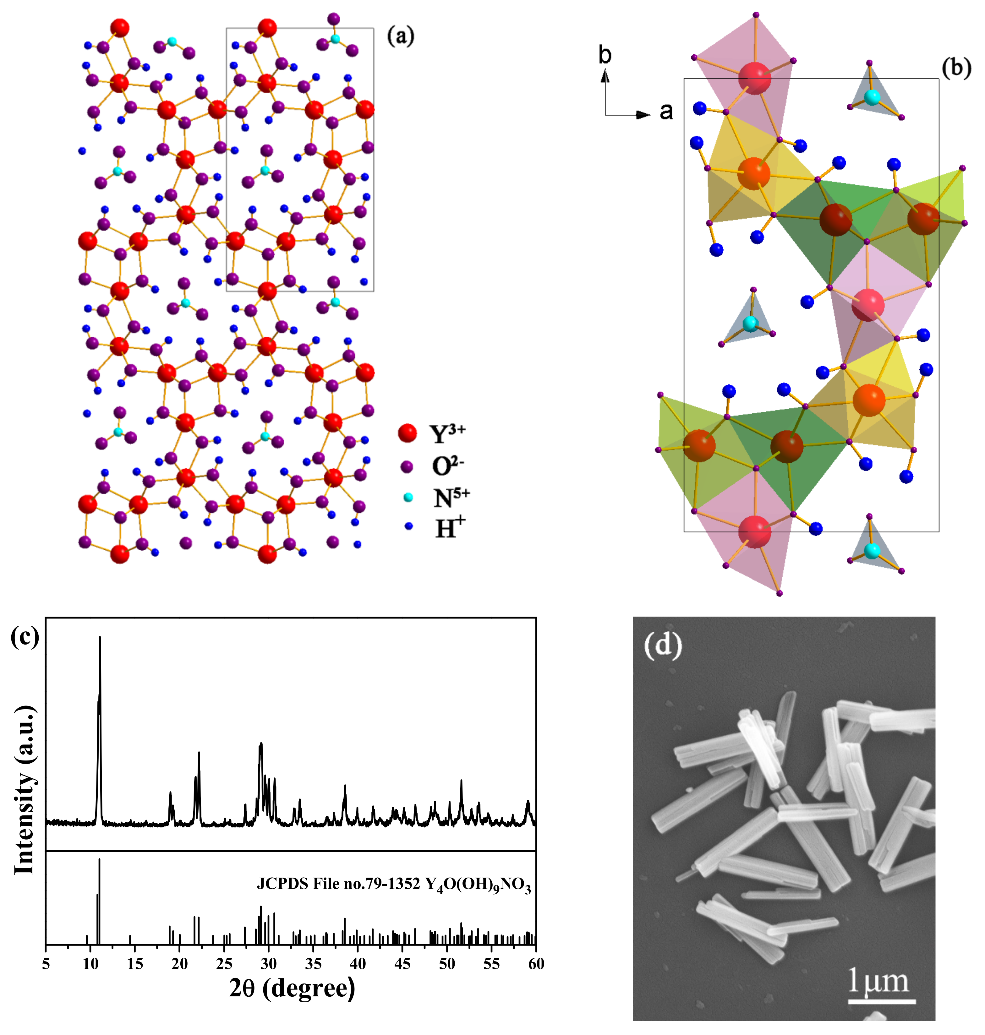
In the present study, the RE
4O(OH)
9NO
3 (RE = Y, Eu) microcrystals (termed as MC) was chosen as an example for surface modification. As reported in the literature [
3], Y
4O(OH)
9NO
3 is a monoclinic crystal structure, which is a three-dimensional framework with one-dimensional channels containing NO
3−. The NO
3− is indirectly linked to Y
3+ rather than forming a direct connection. There are 4 trivalent yttrium ions in the asymmetric unit, with 3 in a 7-coordinated environment with a capped trigonal prismatic geometry and 1 in a 9-coordinated environment with a tricapped trigonal prismatic geometry. The trivalent yttrium ions are linked through hydroxide anions forming the framework around the channels. In the channel, the nitrate ion is indirectly linked to Y
3+ through the hydrogen bonding. As a result of the one-dimensional channels, Y
4O(OH)
9NO
3 always crystallizes into prismatic and wire-like crystallites [
3]. Here, the incorporation of Eu
3+ in Y
4O(OH)
9NO
3 does not significantly affect its crystal structure, because the diffractions of MC are indexed to the monoclinic Y
4O(OH)
9NO
3 (JCPDS File no. 79-1352), except for slight spectral shifts to the lower angle side (
Figure 1c). The replacement of Y
3+ with larger Eu
3+ ions (for 8-fold coordination,
= 0.1019 nm,
= 0.1066 nm) contributed to the lattice expansion, thus resulting in the diffraction shifts [
20].
Figure 1d shows the FE-SEM image of MC, and pure hexagonal prisms with a diameter of ~0.3–0.5 μm and a length of ~1.5–2.5 μm are found in the observation.
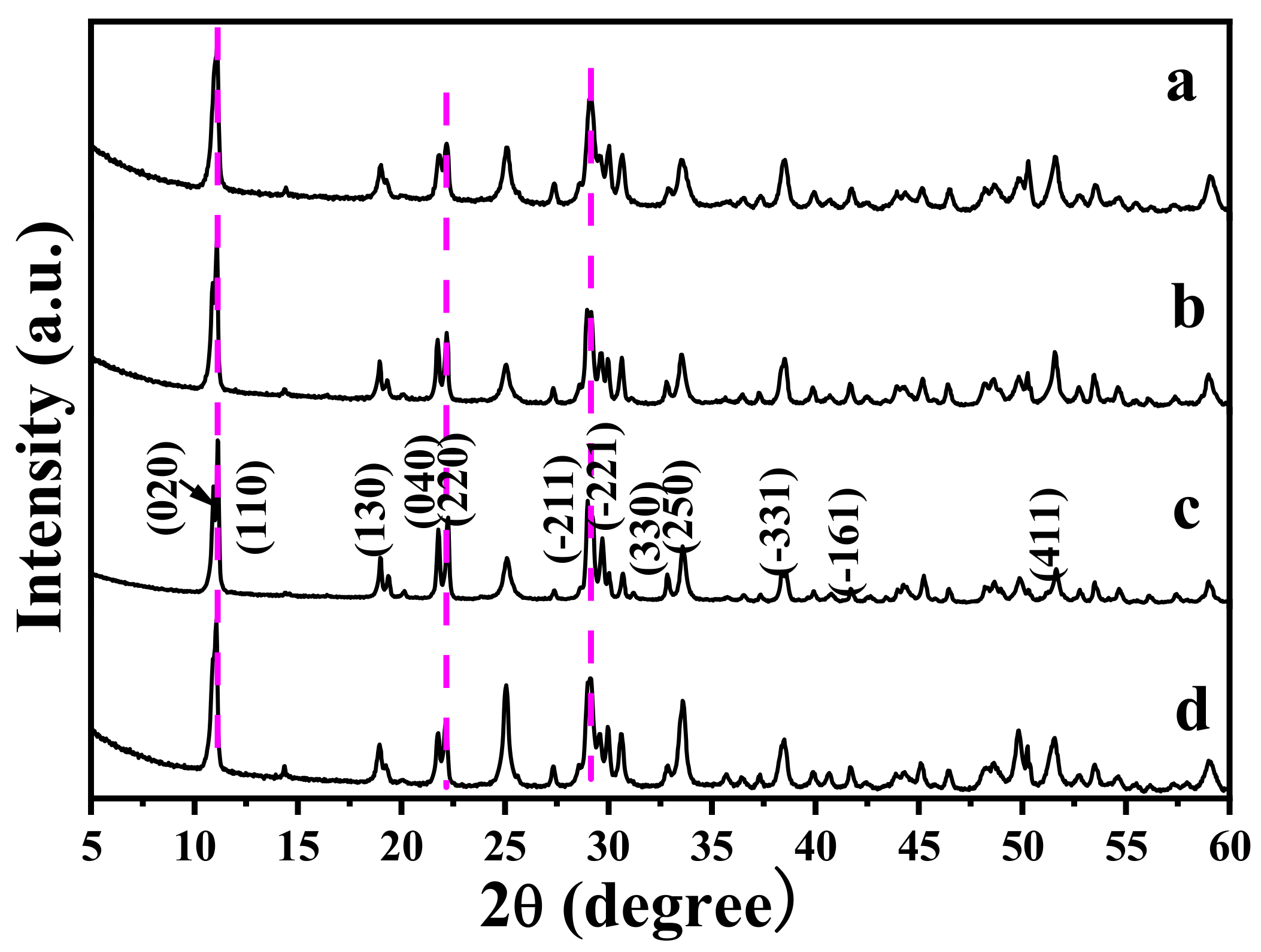
Figure 2 shows the XRD patterns of MC after the reaction with VO
3−. Evidently, the reaction products are the same as MC, because all the diffractions are indexed to the monoclinic Y
4O(OH)
9NO
3 (JCPDS File no. 79-1352), indicating that the reaction with VO
3− did not result in a phase transformation. Increasing the
R value from 0 to 1.5 (
R, the molar ratio of VO
3− to MC) induced a small shift of the diffraction positions. Since VO
3− is smaller than NO
3− [
21], VO
3− substituting for NO
3− anions may induce the diffraction shift to the higher angle side arising from the lattice contraction. Indeed, NO
3− is indirectly coordinated in Y
4O(OH)
9NO
3 rather than the free anion, so it cannot be easily replaced by other anions through ion exchange. After the reaction with VO
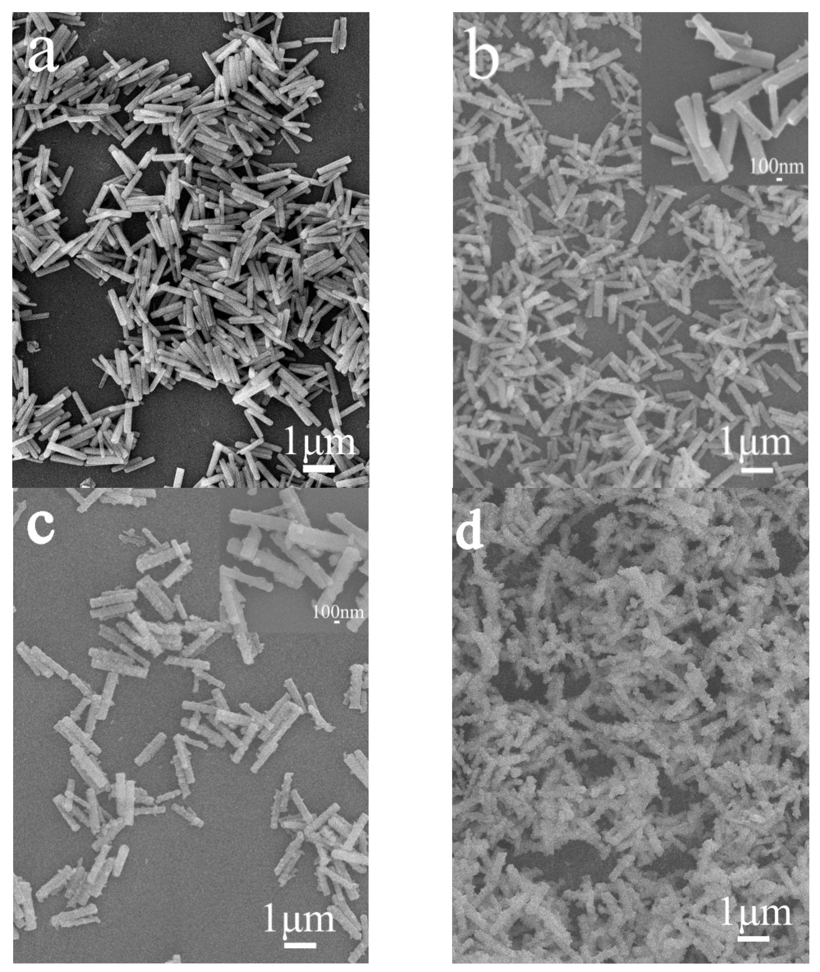
3−, the products mainly remain in the original morphology of MC (
Figure 3). However, the surface of MC-
RV (microcrystals reacted with VO
3−, with
R-fold VO
3− in the reaction system) is not smooth, with nano-sized crystals on the prism surface. Increasing the
R value from 0.25 to 1.5 contributed to a rougher particle surface for MC-
RV. Since the above phenomenon is similar to that for the surface corrosion of metal, in the present paper, it is called “surface eroding” for the reaction with VO
3−. However, there are not any other impurity phases in the XRD patterns, indicating that the nano-sized crystals are amorphous.
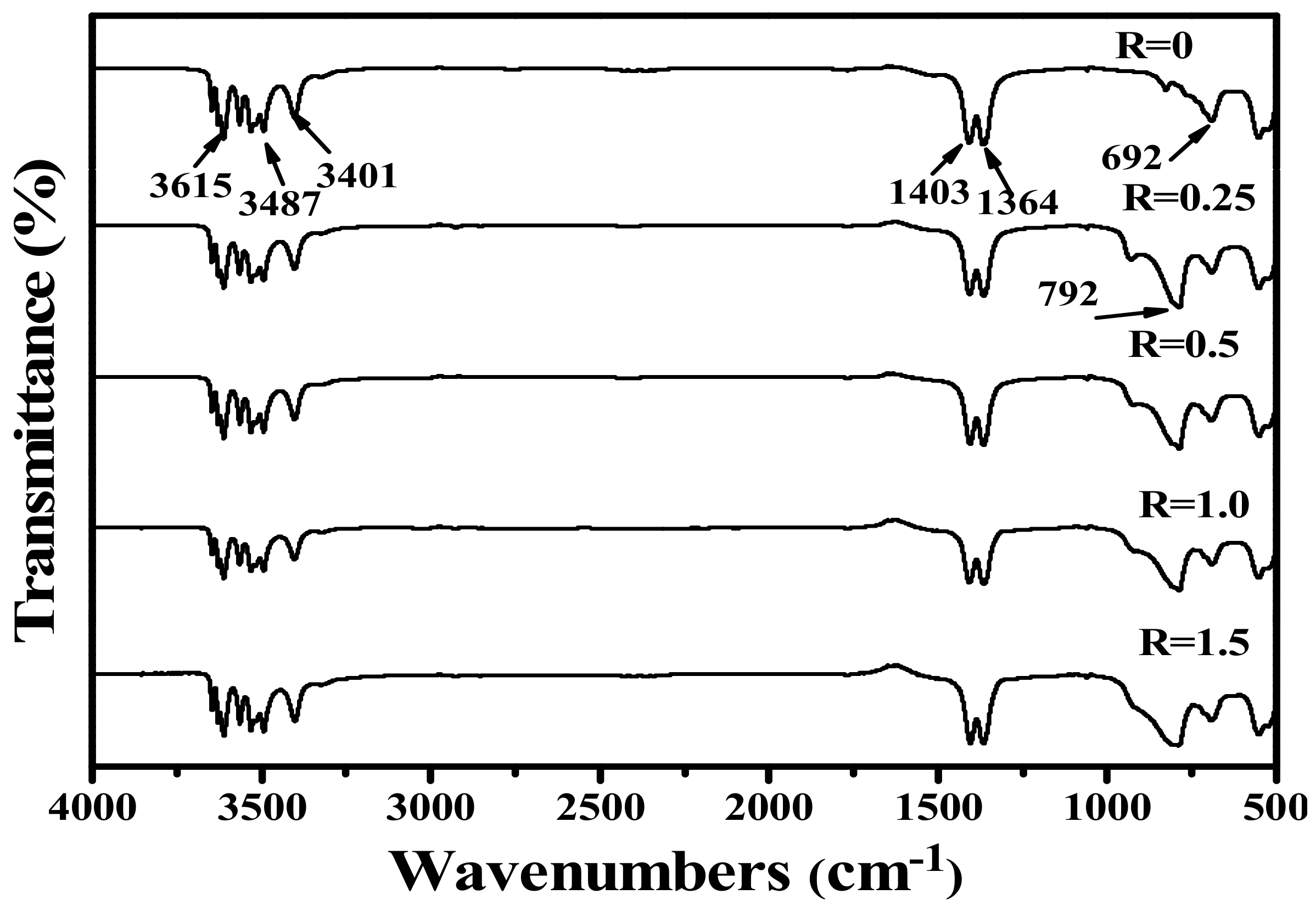
Figure 4 shows the FT-IR spectra for the samples after the reaction with different VO
3− contents. The MC exhibits splitting absorption peaks in the range of 3350–3750 cm
−1, with intense absorptions at 3401, 3487, and 3615 cm
−1, which arise from hydroxyl (OH
−) groups [
22,
23]. This is in compliance with the derived chemical formula of RE
4O(OH)
9NO
3. The absorption peaks around 1364 and 1407 cm
−1 are assignable to the
v3 vibration mode of NO
3− and the
v4 asymmetric stretch of O-NO
2, respectively [
22,
23]. It is clearly seen that the absorptions of NO
3− in MC are different from those of the interlaye- free NO
3− in layered rare earth hydroxide. After the reaction with VO
3−, NO
3− still exists in the FT-IR spectra and is not significantly affected by the reaction. However, the absorption for VO
3− at 792 cm
−1 appeared after the reaction with VO
3− [
22,
23,
24], and more intense absorption is found at a higher
R value.
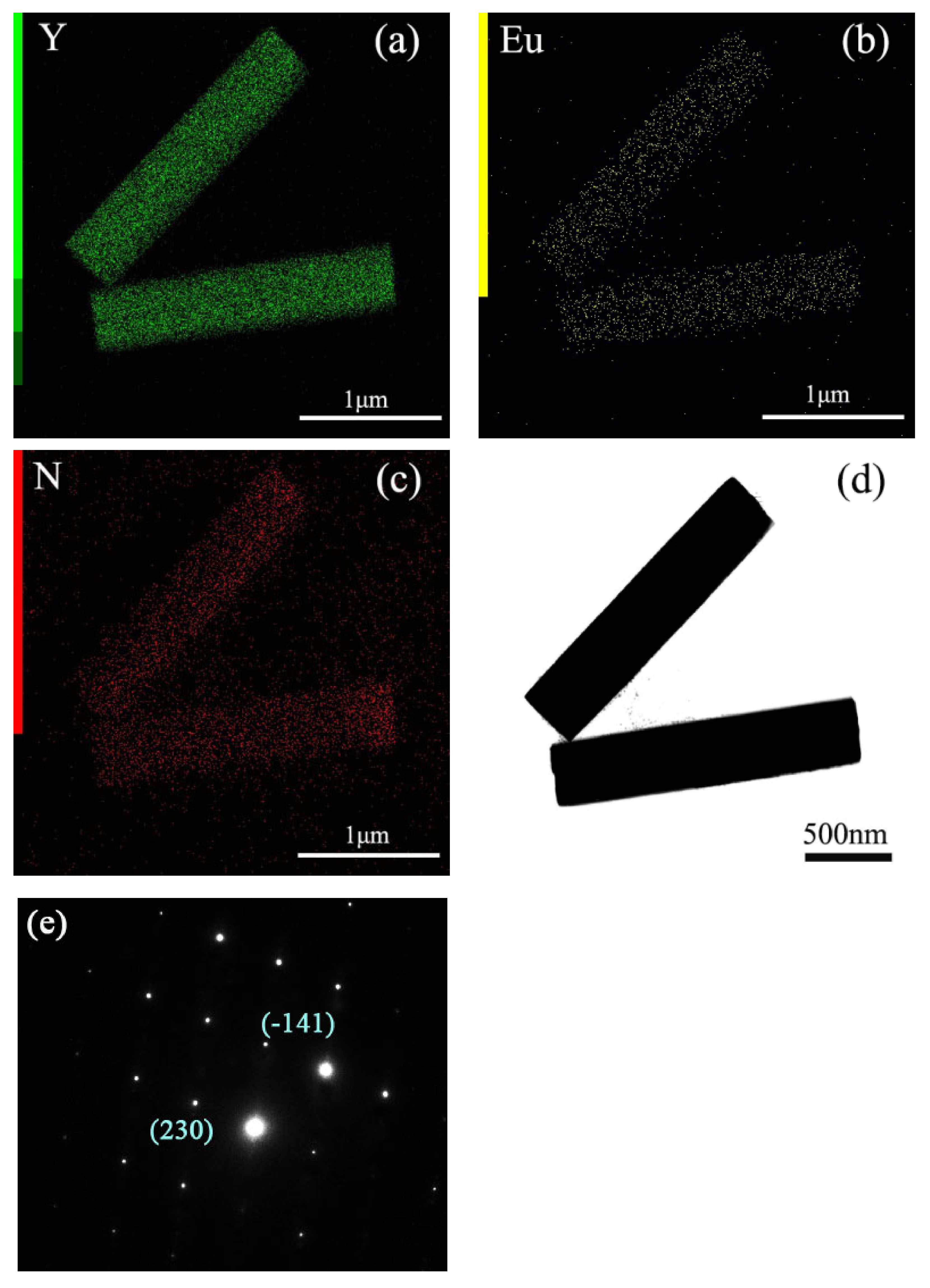
Figure 5 shows the elemental mapping distribution of MC, and the results indicate that MC is a homogeneous solid solution, because all the elements of Y, Eu, and N are distributed among the particles. In addition, the close observation of MC through the TEM image found that MC is well crystallized, with sharp edges and corners, and the surface is smooth. Selected area electron diffraction (SAED) found that the MC is a single crystalline. The calculated planar spacings of ~0.352 nm and ~0.268 nm correspond well with the (230) and (-141) planes of Y
4O(OH)
9NO
3. After the reaction with VO
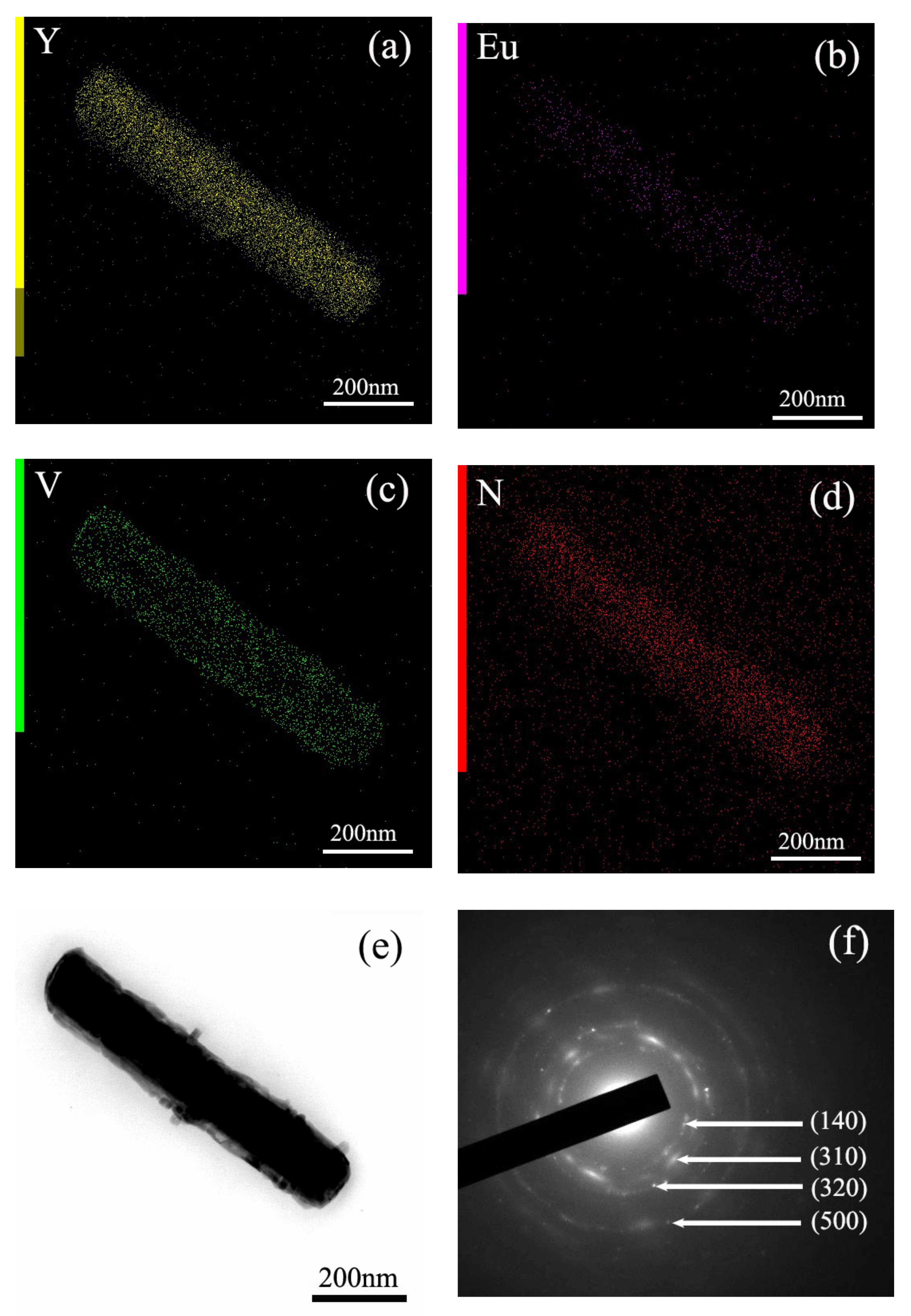
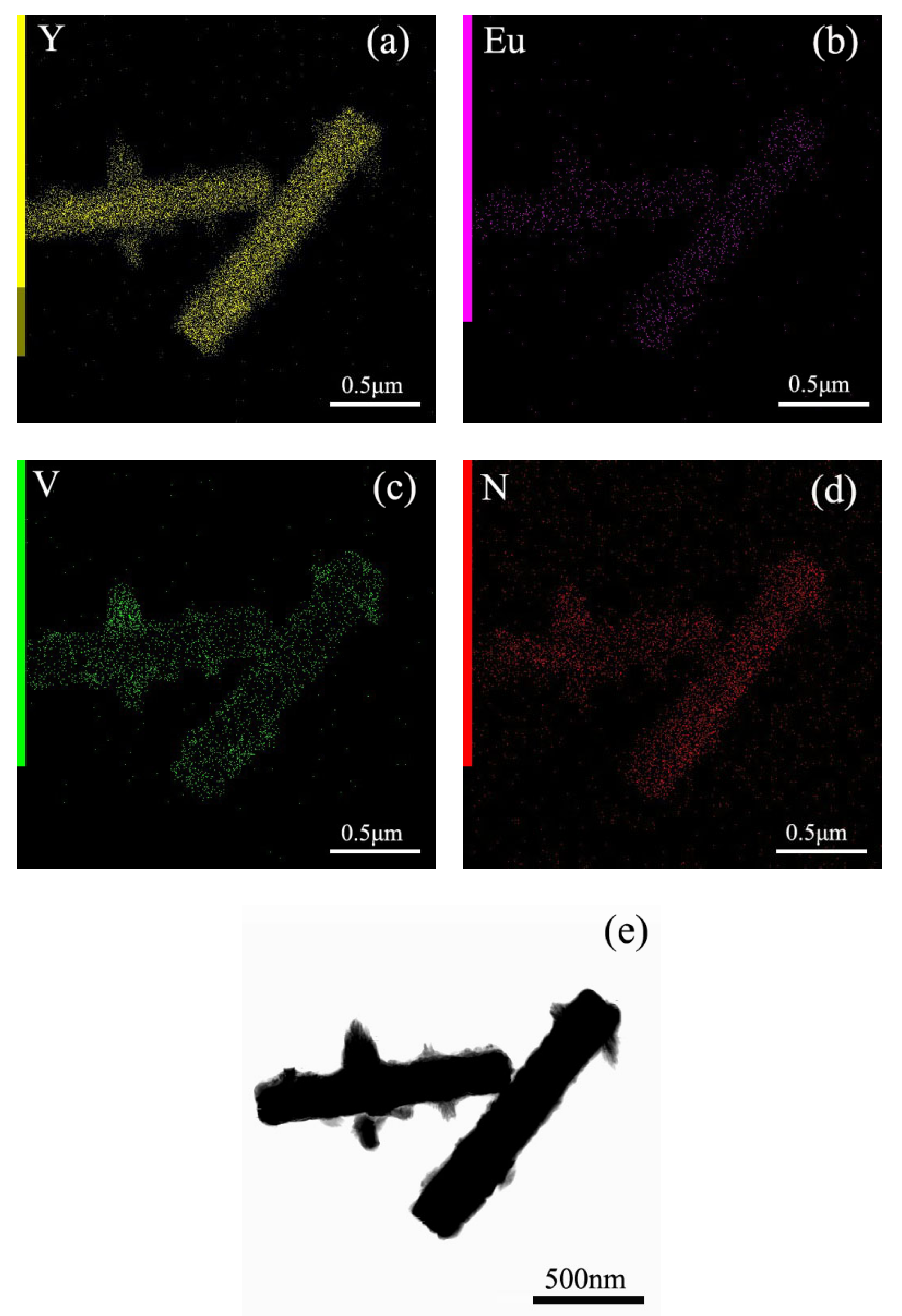
3−, V is distributed among the particles, except for the original component elements of Y, Eu, and N (
Figure 6 and
Figure 7). The TEM images of MC-
RV confirmed that there are nano-sized crystals on the surface of the prisms, and they grow up and tend to dendritic growth. Evidently, the materials needed for growth are obtained from the dissolution of the MC surface, similar to the surface corrosion of metal. The diameter of the prisms became slimmer at a higher
R value, further presenting direct evidence. Selected area electron diffraction (SAED) yields circular patterns, suggesting the MC-
RV consists of polycrystalline (
Figure 6). The calculated planar spacings of ~0.374 nm, ~0.301 nm, ~0.287 nm, and ~0.184 nm correspond well with the (140), (310), (320), and (500) planes of Y
4O(OH)
9NO
3, i.e., d(140) = 0.374026 nm, d(310) = 0.301376 nm, d(320) = 0.287141 nm, and d(500) = 0.183968 nm (JCPDS File no. 79-1352). Evidently, amorphous diffraction circular patterns were found in the SAED patterns, confirming the existence of amorphous particles on the surface of the prism. Collectively, the results from these analyses confirmed that the reaction with VO
3− contributed to the formation of amorphous particles containing VO
3− on the surface of the prism, which is similar to the surface corrosion of metal, called “surface eroding”. Therefore, it can be said that the surface modification of MC was successfully achieved by eroding the surface of MC through vanadate ions.
Figure 8 shows the PLE and PL spectra for MC. By monitoring the
5D
0→
7F
2 emission at 617 nm, a series of sharp lines in the PLE spectrum ranging from 300 nm to 500 nm can be ascribed to the transitions within the Eu
3+ 4f
6 electronic configuration. Different from other rare earth hydroxide precursors, MC exhibited O
2−-Eu
3+ charge transfer (CT) transitions at ~255 nm, as were commonly found for Eu
3+-activated Y
2O
3 [
7,
8,
9,
10]. Upon excitation at 395 nm (intra-4f
6 transition of Eu
3+), the PL spectrum displayed the typical
5D
0→
7F
J(
J = 0–4) transitions of Eu
3+. The relative intensities of the transitions to different
J levels depended on the symmetry of the Eu
3+ environment and can be described in terms of the Judd–Ofelt theory [
7]. The Judd–Ofelt parity law predicts that the magnetic dipole
5D
0→
7F
1 transition is permitted while the electric dipole
5D
0→
7F
2 transition
is forbidden, and the latter is allowed only on the condition that the Eu
3+ ions occupy the asymmetric site [
7,
8,
9]. The MC, having the composition of (Y
0.95Eu
0.05)
4O(OH)
9NO
3, has a monoclinic structure and 2 kinds of Eu
3+ ions, which are 7-coordinated Eu
3+ ions in
C2v non-centrosymmetric sites and 9-coordinated Eu
3+ ions in
D3h centrosymmetric sites [
3]. Since the molar ratio of
C2v occupancy to that of
D3h is 3, most Eu
3+ ions occupy the asymmetric site, and thus the
5D
0→
7F
2 transition at 617 nm is stronger than the
5D
0→
7F
1 transition at 595 nm [
3]. However, the MC did not output a strong red light, mainly due to the fact that the hydroxyls directly coordinated to the rare earth ions Eu
3+, which resulted in serious luminescence quenching [
24].
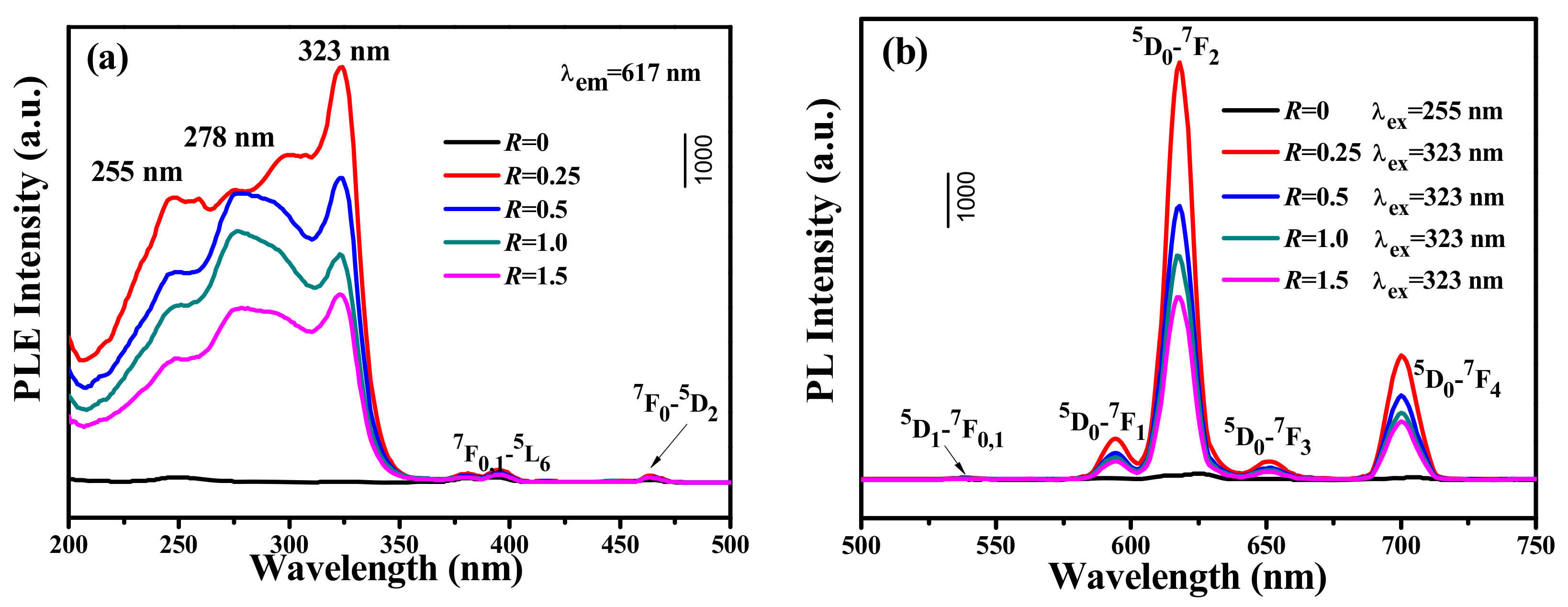
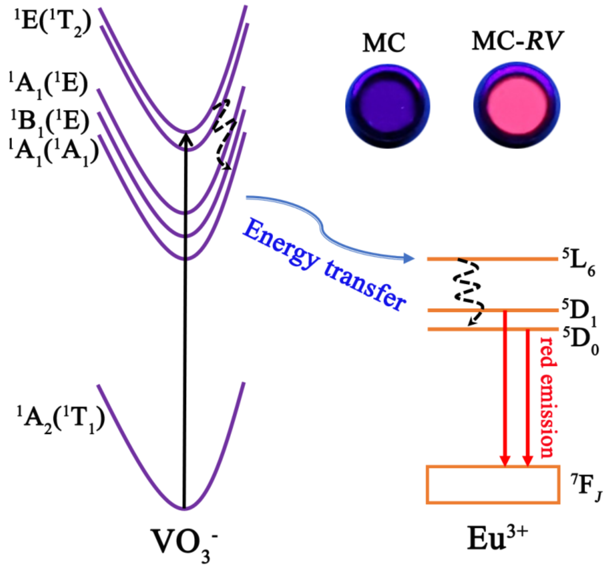
Figure 9 shows the PLE and PL spectra for MC-
RV, which was eroded by vanadate ions. The excitation spectrum consisted of a strong and broad absorption band ranging from 200 to 350 nm, which was assigned to the energy transfer from VO
3− to Eu
3+. The overlapped excitation was from 2 individual bands located at ~275 nm and ~323 nm, which corresponded to the transitions of
1A
2(
1T
1)→
1E (
1T
2) and
1A
2(
1T
2)→
1A
1 (
1E) of V
5+, respectively [
25,
26]. The band at ~255 nm was the contribution of the O
2−-Eu
3+ charge transfer [
7,
8,
9], while the very weak transitions of
7F
0,1→
5L
6 and
7F
0,1→
5D
2 at 395 nm and 463 nm for Eu
3+ were observed in the excitation spectra [
7,
8,
9]. Since the strongest excitation was located at ~323 nm, the excitation wavelength was chosen as 323 nm (
1A
2(
1T
2)→
1A
1 (
1E) transition of V
5+). Upon UV excitation at 323 nm, the PL spectra displayed strong emissions at 540 nm, 590 nm, 617 nm, 650 nm, and 702 nm, which were assigned to
5D
1→
7F
0,1,
5D
0→
7F
1,
5D
0→
7F
2,
5D
0→
7F
3, and
5D
0→
7F
4 transitions of Eu
3+, respectively. The emission at 617 nm attained the dominate role. This further confirmed the existence of VO
3−→Eu
3+ energy transfer. Interestingly, the emission intensity at 617 nm was greatly enhanced by increasing the
R value from 0 to 0.25, indicating that the VO
3−→Eu
3+ energy transfer contributed to the improved luminescence. The external/internal quantum efficiencies for
R = 0 were 6 ± 1%/11 ± 1%, and the external/internal quantum efficiencies for
R = 0.25 were 36 ± 1%/65 ± 1%, directly confirm the great enhancement of luminescence. However, a higher
R value induced a rougher particle surface, which contributed to the light scattering and quenching of the luminescence. Therefore, increasing the
R value further resulted in the luminescent decay. The external/internal quantum efficiencies for
R = 0.5,
R = 1.0, and
R = 1.5 were 25 ± 1%/49 ± 2%, 18 ± 2%/43 ± 1%, and 10 ± 1%/37 ± 2%, respectively. However, the emission intensity for MC-
RV is evidently stronger than that for MC on the whole, indicating that the erosion of the surface of rare earth microcrystals through vanadate ions can contribute to the considerable improvement to luminescence (
Figure 10).
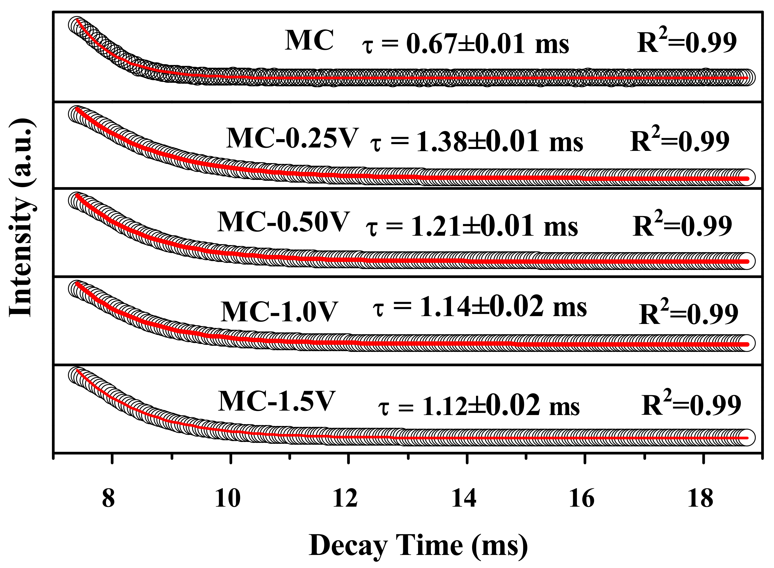
The decay kinetics of the
5D
0→
7F
2 transition at 617 nm for MC and MC-
RVre a investigated in
Figure 11. All the fluorescence decay curves can be fitted to single exponentials. The average lifetimes of the MC, MC-0.25V, MC-0.50V, MC-1.0V, and MC-1.5V samples determined in this work are ~0.67 ms, ~1.38 ms, ~1.21 ms, ~1.14 ms, and ~1.12 ms, respectively. Evidently, the lifetime for MC-
RV is longer than that for MC, since the energy transfer of VO
3− to Eu
3+ is more time consuming.


