Article

Photoluminescent and Photocatalytic Properties of Eu3+-Doped MgAl Oxide Coatings Formed by Plasma Electrolytic Oxidation of AZ31 Magnesium Alloy

by
Stevan Stojadinović
1,2,*,
Nenad Radić
3 and
Rastko Vasilić
1
1
Faculty of Physics, University of Belgrade, Studentski trg 12–16, 11000 Belgrade, Serbia
2
Faculty of Forestry, University of Belgrade, Kneza Višeslava 1, 11000 Belgrade, Serbia
3
IChTM-Department of Catalysis and Chemical Engineering, University of Belgrade, Njegoševa 12, 11000 Belgrade, Serbia
*
Author to whom correspondence should be addressed.
Coatings 2022, 12(12), 1830; https://doi.org/10.3390/coatings12121830
Submission received: 3 November 2022 / Revised: 24 November 2022 / Accepted: 25 November 2022 / Published: 26 November 2022

Abstract

:
The synthesis of Eu3+-doped MgAl oxide coatings containing MgO and MgAl2O4 was accomplished through plasma electrolytic oxidation of AZ31 magnesium alloy in aluminate electrolyte with the addition of Eu2O3 particles in various concentrations. Their morphological, structural, and above all, photoluminescent (PL) and photocatalytic activity (PA) were thoroughly investigated. PL emission spectra of MgAl oxide coatings feature characteristic emission peaks, which are ascribed to f–f transitions of Eu3+ ions from excited level 5D0 to lower levels 7FJ. The charge transfer state of Eu3+ or direct excitation of the Eu3+ ground state 7F0 into higher levels of the 4f-manifold are both attributed to the PL peaks that appear in the excitation PL spectra of the obtained coatings. The fact that the transition 5D07F2 (electrical dipole transition) in Eu3+-doped MgAl oxide coatings is considerably stronger than the transition 5D07F1 (magnetic dipole transition) indicates that Eu3+ ions occupied sites with non-inversion symmetry. Because of oxygen vacancy formation, the Eu3+-doped MgAl oxide coatings had a higher PA in the degradation of methyl orange than the pure MgAl oxide coating. The highest PA was found in Eu3+-doped MgAl oxide coating formed in an aluminate electrolyte with 4 g/L of Eu2O3 particles. The PA, morphology, and phase of Eu3+-doped MgAl oxide coatings did not change after several consecutive runs, indicating outstanding stability of these photocatalysts.

1. Introduction

Magnesium alloys are commonly used in the aircraft, automotive, military, medical, and electronic industries [1,2,3]. However, because of their high chemical reactivity, magnesium alloys are prone to corrosion, limiting their use in corrosive environments [4,5]. Surface modification is widely recognized as the most prevalent method for improving a magnesium alloy’s surface properties. A coating layer can enhance the corrosion resistance of the metallic substrate by acting as a barrier between the substrate and the environment [6,7,8]. Recently, many research groups have been investigating the plasma electrolytic oxidation (PEO) process as a surface-protective treatment for magnesium alloys [9,10,11,12,13,14].
PEO is a high-efficiency, low-priced, and environmentally friendly process for producing multicomponent oxide coatings with numerous functions on lightweight metals or metallic alloys [15,16,17]. The incidence of micro-discharging on the metal surface, which is complemented by gas evolution, indicates that the PEO process is connected with plasma creation [18,19,20]. Many processes such as light and heat emission and electrochemical and plasma-chemical reactions occur at the micro-discharge sites as a result of the elevated local temperature and pressure. These processes are responsible for altering the structure, content, and morphology of the oxide coatings that are produced.
Despite numerous articles investigating PEO of magnesium alloys [21,22,23,24], there is a lack of data describing the formation of PEO coatings on magnesium alloys with intended application as photocatalytic or as photoluminescent materials [25]. In this study, we showed that MgO and MgAl2O4 phases are present in coatings formed by PEO of AZ31 magnesium alloy in aluminate electrolyte. MgAl2O4 has been identified as a matrix component for the preparation of very efficient photoluminescent materials [26,27], whereas both MgO and MgAl2O4 have been recognized as efficient materials in the photocatalytic degradation of organic pollutants due to high concentration of surface vacancies and other defects [28,29]. MgO- and MgAl2O4-photoluminescent and photocatalytic active materials have previously been synthesized in powder form, limiting their practical applications, particularly in the field of photocatalysis, due to the high cost of photocatalyst separation and recovery from suspension. Additionally, the easy aggregation of photocatalyst particles renders them unsuitable for use in flow systems.
The main goal of presented research was to synthesize Eu3+-incorporated oxide coatings by PEO on AZ31 magnesium alloy in an aluminate electrolyte with the addition of Eu2O3 particles and to examine their photoluminescent and photocatalytic capabilities. Our recent investigation has showed that Eu3+-doped PEO-based coatings on different substrates (Ti, Zr, Nb, Hf, Gd, Y, Zn) can be formed in electrolyte containing Eu2O3 [30,31,32,33,34,35]. Among various trivalent rare earth ions, the Eu3+ ion is most promising among light-emitting materials, exhibiting intense PL red emission under UV excitation due to the 5D07F2 transition [36]. In addition, positive effects of Eu3+ ion doping on the photocatalytic capabilities of several materials have been described in the literature [37,38,39]. The only partially populated 4f orbital structure of Eu3+ ions is favorable for electron trapping, which boosts photocatalytic activity by decreasing charge carrier recombination.

2. Materials and Methods

In this research, an AZ31 magnesium alloy (96% Mg, 3% Al, 1% Zn, Alfa Aesar) plate was used as a substrate. The substrates were cut in rectangular pieces with dimensions of 25 mm × 10 mm × 0.81 mm and then ultrasonically cleaned with acetone, warm air stream dried, and covered with insulating resin, leaving only the 15 mm × 10 mm active surface exposed to the electrolyte. More details about the experimental setup can be found in [40]. PEO coatings were formed in an aluminate electrolyte (5 g/L NaAlO2) with Eu2O3 particles added in concentrations up to 8 g/L. PEO was done for 10 min at 150 mA/cm2 constant current density. The electrolyte was kept at a constant temperature of 20 ± 1 °C. Following the PEO procedure, the samples were rinsed with distilled water and air dried.
The structural and elemental analyses of the PEO coatings were carried out utilizing scanning electron microscope (SEM, JEOL 840A, Tokyo, Japan) with energy-dispersive X-ray spectroscopy (EDS, Oxford INCA, Abingdon, UK). A Rigaku Ultima IV diffractometer (XRD, Tokyo, Japan) equipped with a CuKα radiation source was used for phase identification of PEO coatings. The ultraviolet–visible diffusion reflectance spectra (DRS) were collected by a Shimadzu UV-3600 spectrophotometer (Tokyo, Japan) coupled with an integrating sphere. The spectrofluorometer (Horiba Jobin-Yvon Fluorolog FL3-22, Edison, NJ, USA) using a xenon lamp as radiation source was used to measure excitation and emission photoluminescence (PL) spectra at room temperature.
Under simulated solar radiation, photodegradation of methyl orange (MO) was investigated to probe the photocatalytic activity (PA) of formed coatings. In a 6.8 cm diameter open cylindrical thermostated pyrex glass reactor, a solution containing 8 mg/L of MO was kept in the dark for one hour to achieve absorption–desorption equilibrium. The concentration of MO remained nearly constant, indicating that MO adsorption was negligible. Following that, samples were exposed to the light of a 300 W incandescent lamp (Solimed BH Quarzlampen, Leipzig, Germany) placed 25 cm above the top surface of the solution. On the steel wire holder, the samples were placed 5 mm above the reactor bottom. PA was estimated by monitoring MO decomposition in selected time intervals after exposure to irradiation. A spectrophotometer (UV-Vis Thermo Electron Nicolet Evolution 500, Sittingbourne, England) was used to measure the MO concentration using the maximum MO absorption peak at 464 nm.
Free hydroxyl radicals (OH) have high potential rate to photodegrade MO and other organic pollutants. To identify possible creation of OH on irradiated PEO coatings, PL measurements were carried out using terephthalic acid, because it is observed that it reacts with OH, thus producing extremely fluorescent 2-hydroxyterephthalic acid with a PL maximum positioned at 425 nm [41]. The PEO coatings were placed in reactor with 10 mL of 5 × 10⁻⁴ mol/L terephthalic acid, which was diluted in aqueous solution of 2 × 10⁻³ mol/L NaOH. After the irradiation, PL spectra of the solution in which the reaction took place were collected utilizing the spectrofluorometer under the excitation wavelength of 315 nm.

3. Results and Discussion

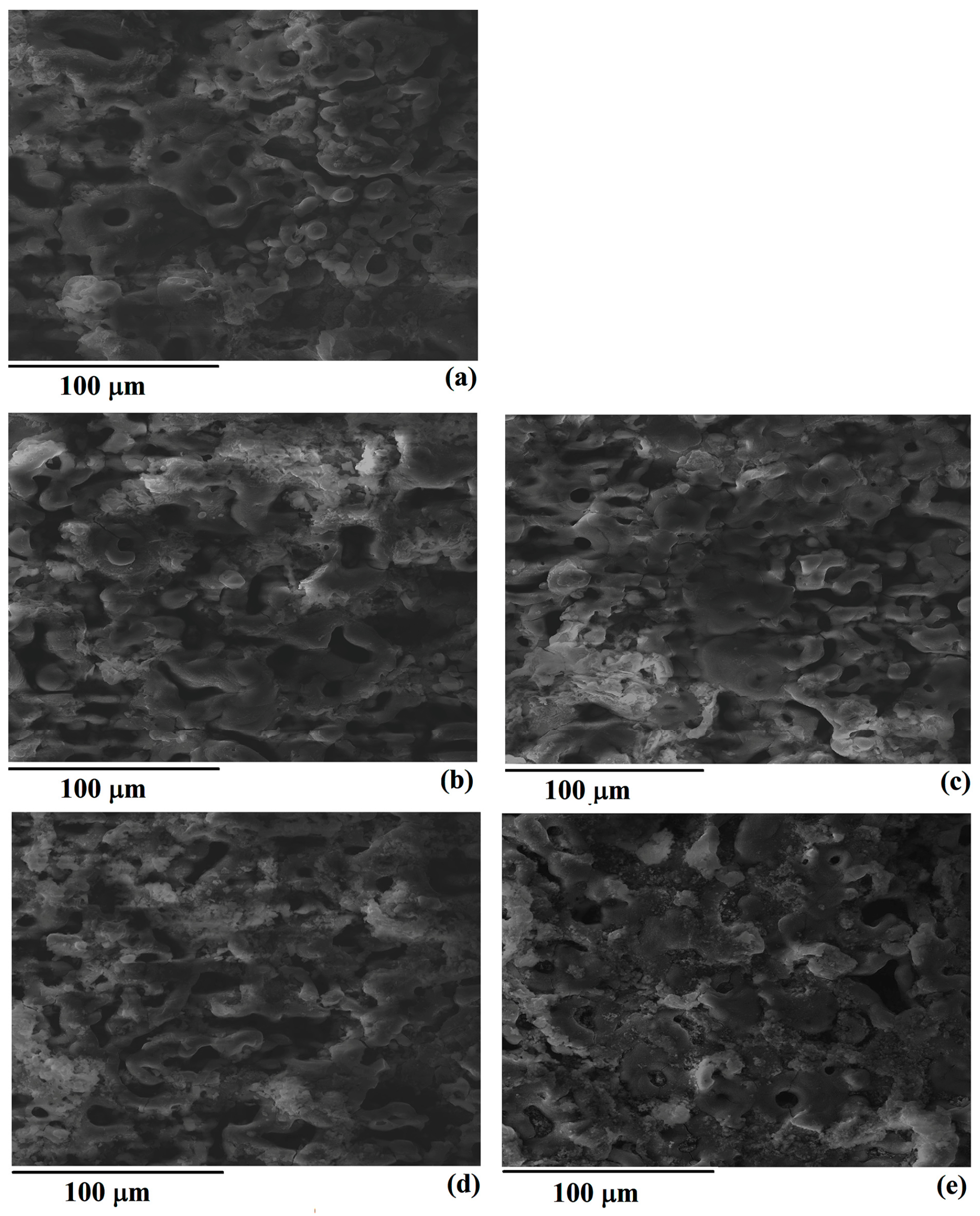
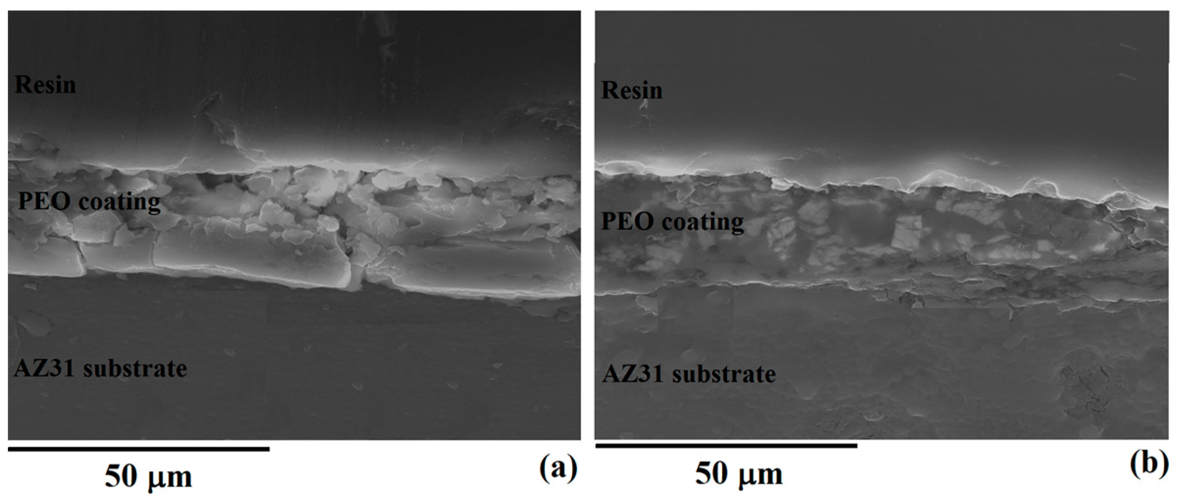
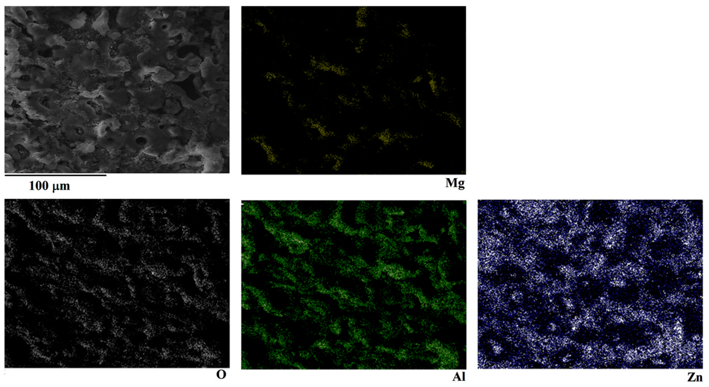
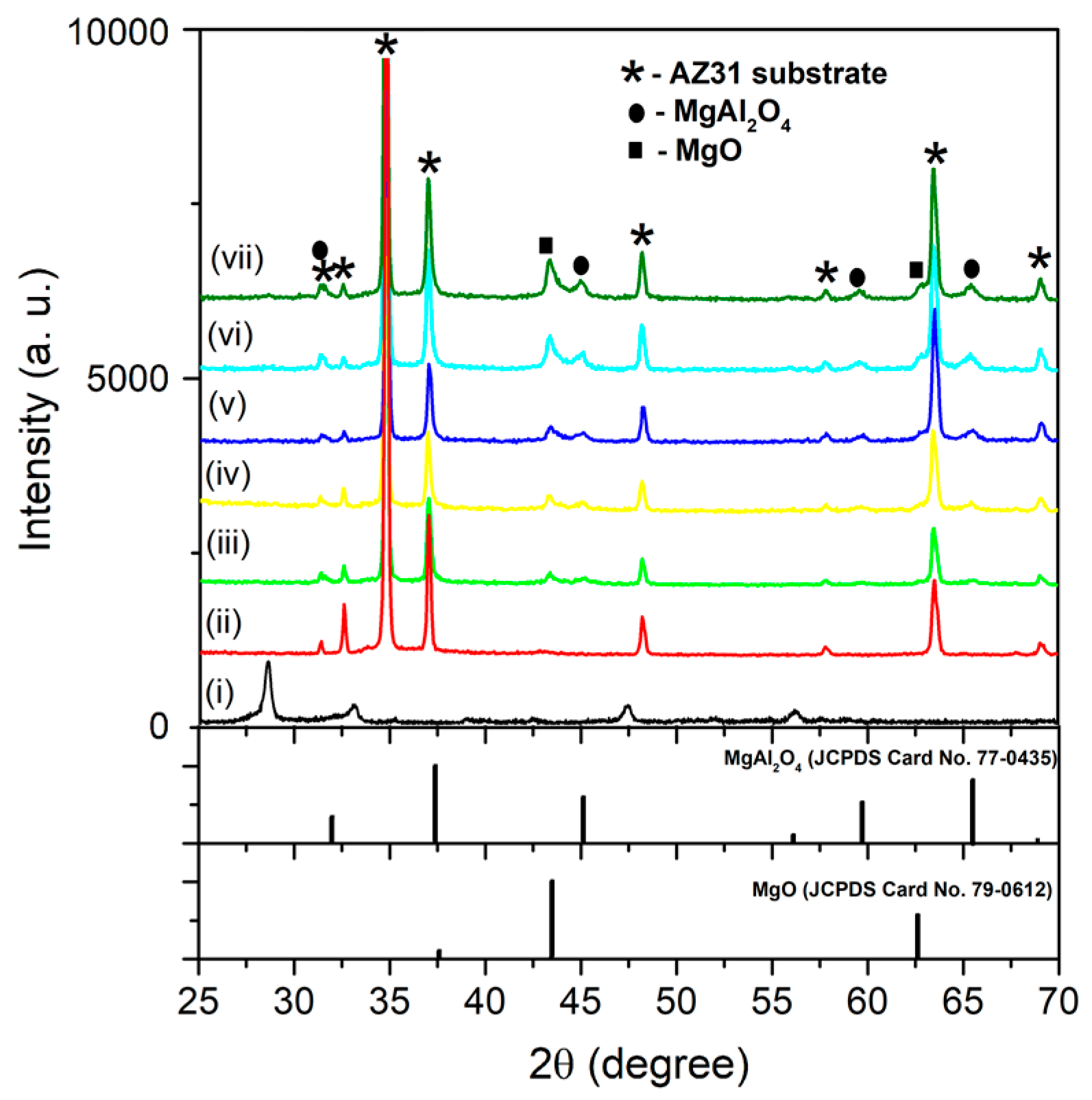
3.1. Morphology, Chemical, and Phase Composition of Eu-Doped MgAl Oxide Coatings

Figure 1 shows the top-surface morphology of coatings made in aluminate electrolyte with the addition of Eu2O3 particles. Obtained coatings are laced with a number of micro-discharging canals of changing diameter and areas formed by the freezing of molten materials between them. It can be also observed that the addition of Eu2O3 particles to aluminate electrolyte does not influence the surface morphology.
High temperatures which are inherent to PEO processing cause the melting of magnesium substrate, which enters the micro-discharging channels where it reacts with penetrated electrolyte. Products of these reactions, formed inside of micro-discharges, are ejected towards the oxide/electrolyte surface, where they promptly solidify upon contacting colder electrolyte. The repetition of these steps on a vast number of discrete locations where micro-discharging occurs results in thickening of the oxide coating. After 10 min of PEO, the formed coatings are roughly 22 μm thick and weakly dependent on the Eu2O3 particles’ concentration in the aluminate electrolyte (Figure 2).
Table 1 presents the elemental composition of the coatings in Figure 1. The dominant elements in the coatings are Mg, Al, O, and Eu. The observed amount of Eu in coatings surges as the concentration of Eu2O3 in electrolyte increases. EDS elemental mapping reveals that observed elements are rather uniformly distributed in obtained coatings (Figure 3).
The XRD patterns of pure Eu2O3 particles, AZ31 magnesium substrate, and formed coatings displayed in Figure 4. Observed diffraction patterns are characterized by XRD peaks that correspond to cubic phases of MgO (JCPDS card No. 79-0612) and MgAl2O4 (JCPDS card No. 77-0435). Diffraction peaks from the substrate are significant due to X-ray penetration through oxide coating and its reflection off the substrate. Despite the fact that elemental analysis confirmed the presence of Eu in the coatings, XRD analysis revealed no XRD maxima which can be attributed to Eu-containing crystalline phases.
The melting point of the Eu2O3 particles is around 2400 °C, which is considerably less than the estimated local temperature at the micro-discharging sites during PEO of AZ31 magnesium alloy [42]. Inside the micro-discharge channels, molten Eu2O3 particles can react with other components (originating either from substrate or from electrolyte) and participate in the formation of mixed-oxide coatings. The lack of diffraction peaks associated with Eu is most likely due to the uniform distribution of Eu throughout the surface coatings. It can also be observed that low Eu content in obtained coatings does not considerably change the crystalline structure of MgO and MgAl2O4.

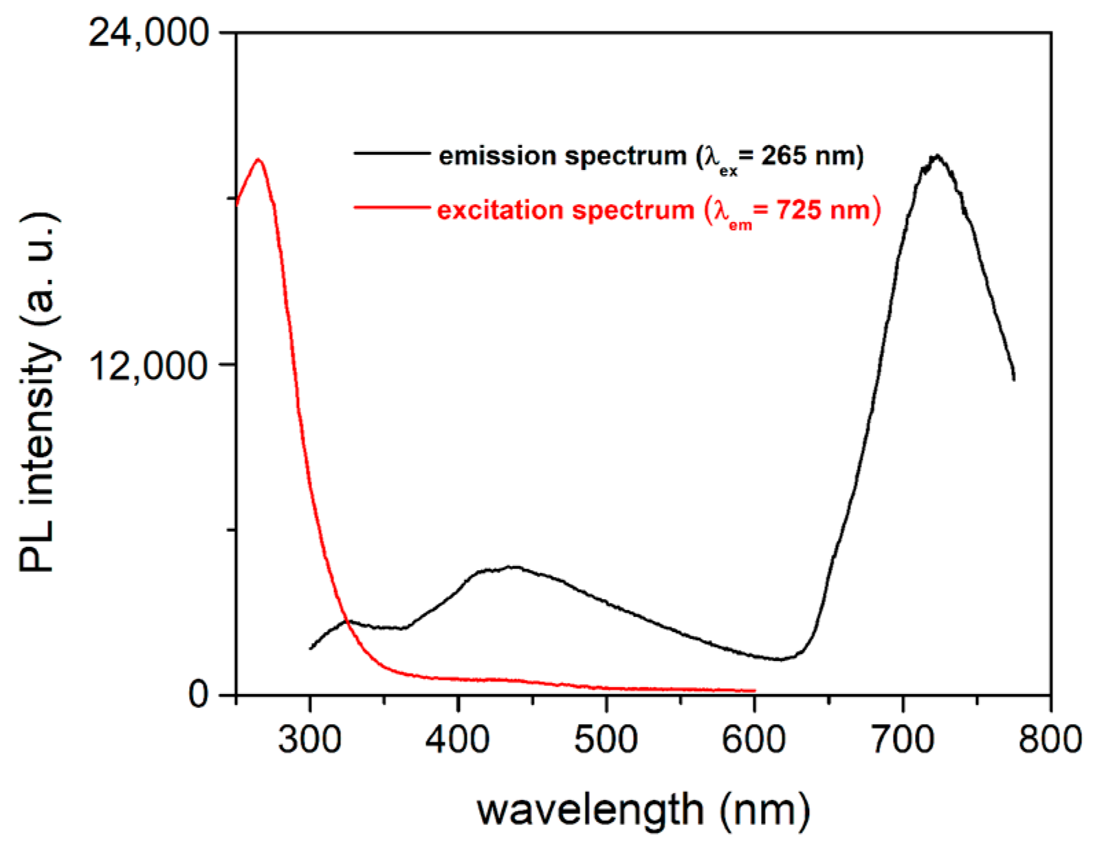
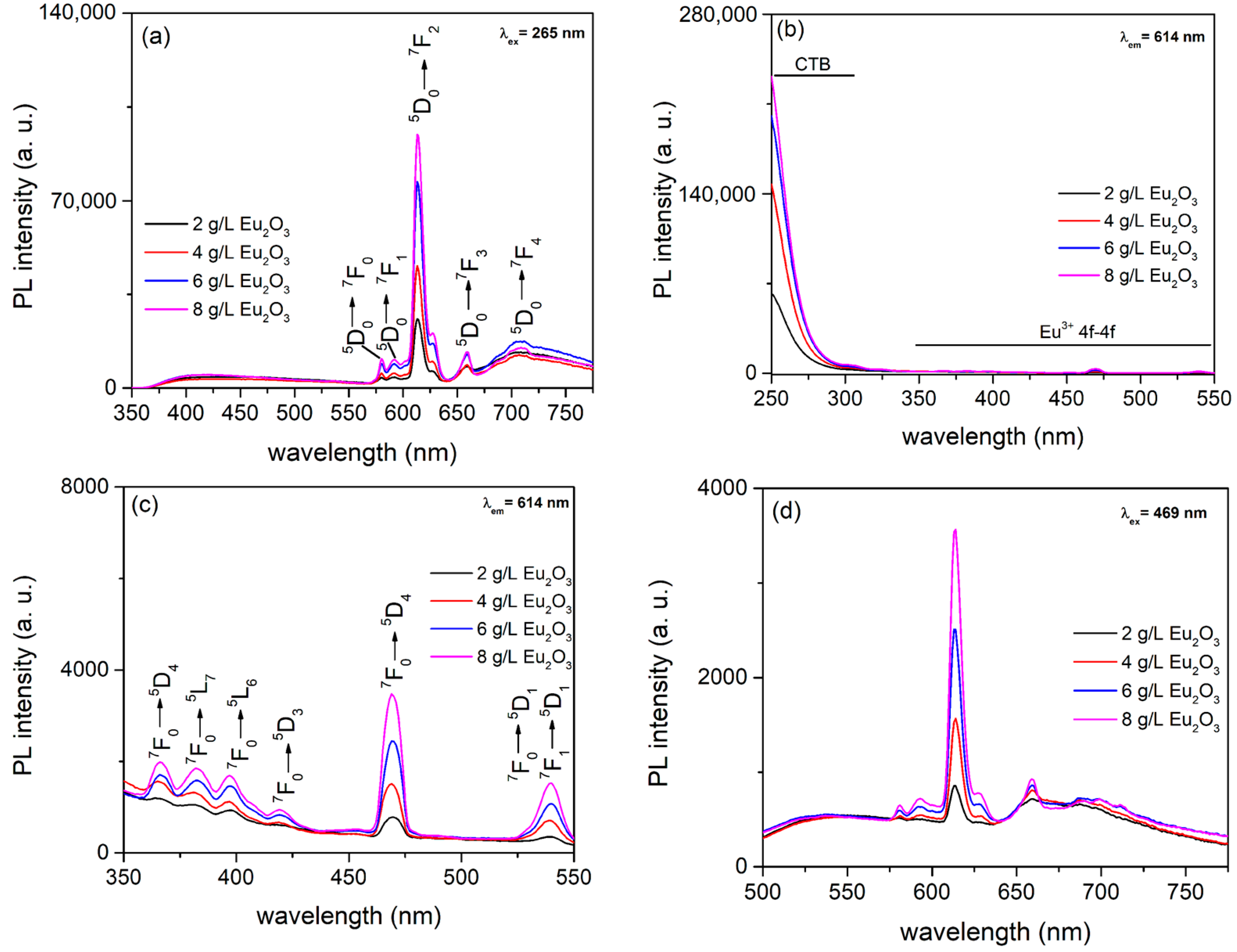
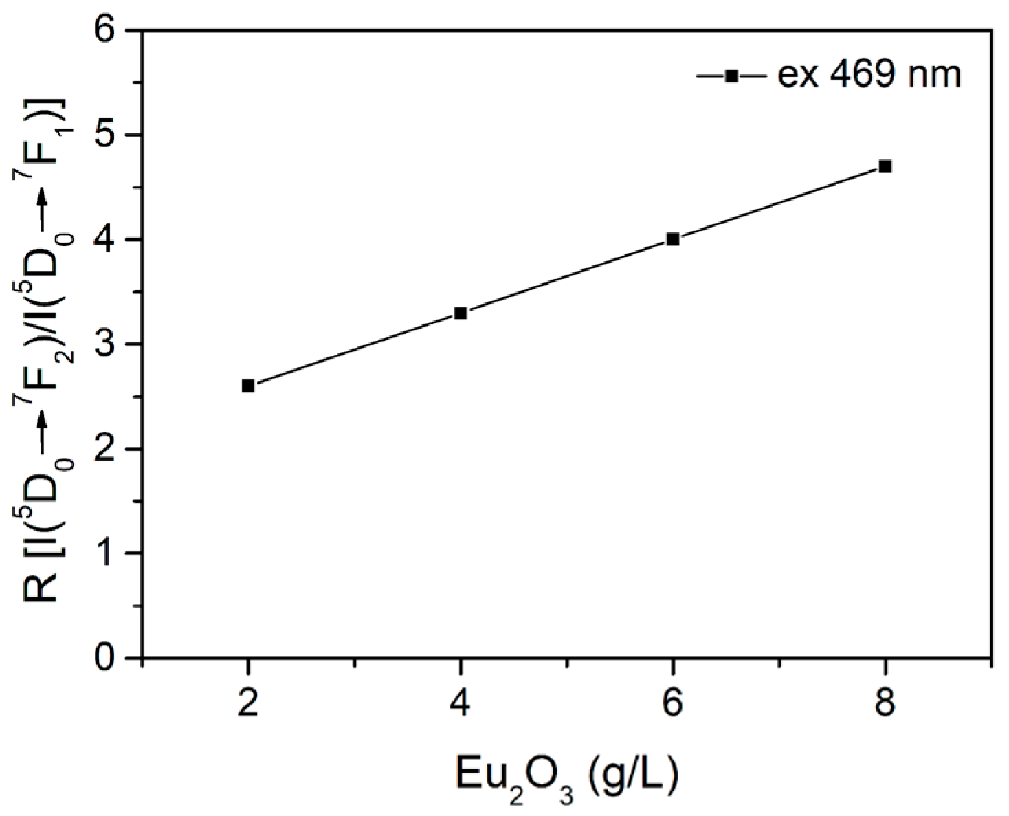
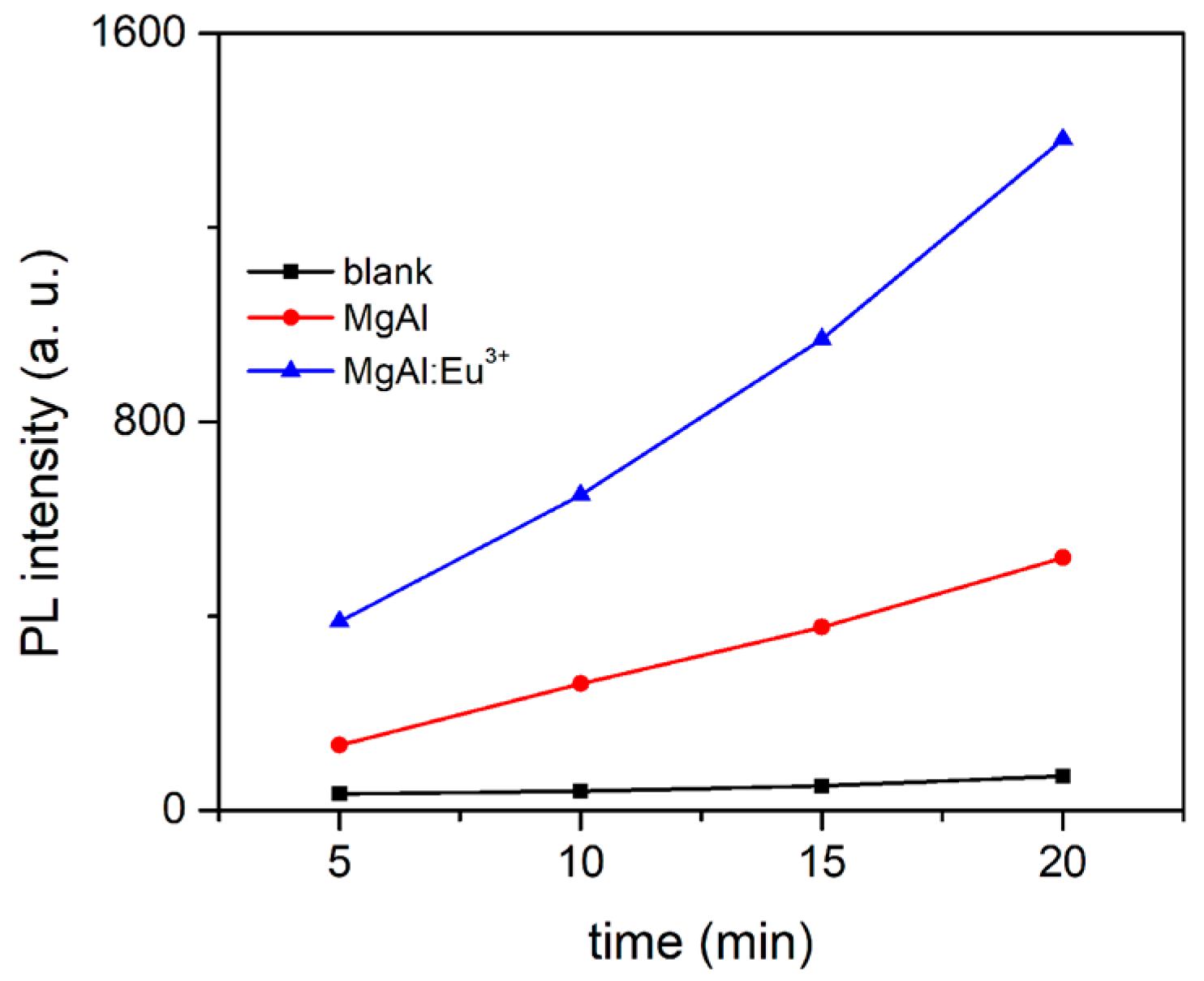
3.2. PL of Eu3+-Doped MgAl Oxide Coatings

The PL of MgAl oxide coatings with incorporated Eu is a combination of PL inherent to MgAl and Eu ions. PL excitation and emission spectra of MgAl oxide coating obtained in aluminate electrolyte are presented in Figure 5. The emission spectrum shows three dominant PL bands under ultraviolet excitation. The PL emission band centered at around 325 nm can be attributed to F+ centers in MgO [40], while the PL band from 360 nm to 600 nm can be attributed to structural defects in the coating, such as Mg vacancies and interstitials [43,44]. The band with a maximum near 725 nm can be related to F+ centers in MgAl2O4 [45].
The PL emission spectra of oxide coatings formed in aluminate electrolyte with varying concentration of Eu2O3 is presented in Figure 6a. Obviously, PL emission spectra show two discrete regions: the first one contains a typical broad PL band of MgAl oxide coatings, whereas the second one contains well-defined PL emission bands intrinsic to Eu3+ 4f–4f transitions from the excited 5D0 level to the lower 7FJ levels (J = 0, 1, 2, 3, and 4) superimposed on PL of MgAl oxide coatings [36]. The PL bands are positioned at about 580 nm (5D07F0), 592 nm (5D07F1), 614 nm (5D07F2), 659 nm (5D07F3), and 708 nm (5D07F4). The intensity of the PL emission of 4f–4f transitions of Eu3+ increases as the concentration of Eu3+ in MgAl oxide coatings rises, indicating that the concentration of Eu3+ which is necessary for concentration quenching has not yet been reached.
When PL excitation spectra are monitored at 614 nm (this wavelength corresponds to the most intense observed Eu3+ transition), an intense wide-ranging PL band in the region from 250 nm to 350 nm is observed (Figure 6b), which is followed by a series of narrow low-intensity bands (Figure 6c). Because the contribution of PL caused by optical transitions of MgAl oxide coatings is negligible, the broad PL band appears as a result of electron transfer between the fully occupied 2p orbital of O2− and the incompletely occupied 4f orbital of the Eu3+ [46]. Sharp bands, as shown in Figure 6c, mostly correspond to direct excitation of the Eu3+ ground state 7F0 into higher levels of the 4f-manifold [47]. The 7F05D2 transition at 469 nm is the most intense of these excitation transitions and is used to record the PL emission spectra (Figure 6d).
When PL emission spectra are monitored under 469 nm excitation, typical narrow bands corresponding to the intra-configurational 4f–4f transitions of Eu3+ are observed. The intensity of the observed narrow band increases when the concentration of Eu3+ in MgAl oxide coatings increases, but the peak positions remain constant. Another interesting observation is the very high intensity of hypersensitive electric dipole transition 5D07F2 at 614 nm compared to magnetic dipole transition 5D07F1 at 592 nm. The electric dipole transition 5D07F2 (ΔJ = ±2) is strongly dependent on changes in the environment around Eu3+ ions, while the magnetic dipole transition 5D07F1 (ΔJ = ±1) does not depend on the surrounding environment [36]. In other words, when Eu3+ ions are incorporated into non-inversion symmetry lattice sites, then 5D07F2 electric dipole transition becomes dominant. In contrast, if Eu3+ ions are positioned at the inversion symmetry sites then the 5D07F1 magnetic dipole transition becomes most prominent. In MgAl oxide coatings, the observed intensity of magnetic dipole transition is much lower than the corresponding intensity of electric dipole transition, suggesting that Eu3+ ions are positioned at non-inversion symmetry lattice sites.
The asymmetric ratio R between the transitions 5D07F2 and 5D07F1 provides information about the surroundings of incorporated Eu3+ ions. R values were calculated employing the equation [48]:
R = I ( 5 D 0     7 F 2 ) I ( 5 D 0     7 F 1 )
where I(5D07F2) and I(5D07F1) are the integrated intensities for the transitions 5D07F2 (600 nm to 635 nm) and 5D07F1 (585 nm to 600 nm), respectively. In MgAl oxide coatings, a high R value indicates an environment with a low degree of symmetry around the Eu3+ ions. Figure 7 clearly shows that an increase in Eu3+ concentration in the coatings results in higher R values, implying that increased concentration of Eu3+ in oxide coating results in lattice distortion and decreased symmetry around the Eu3+ ions. Because of differences in ionic radii between Eu3+ (95 pm), Mg2+ (66 pm), and Al3+ (51 pm), after replacing the Mg2+ or Al3+ ions, Eu3+ ions will create oxygen vacancies and cause the lattice distortion due to the size mismatch [49,50,51].

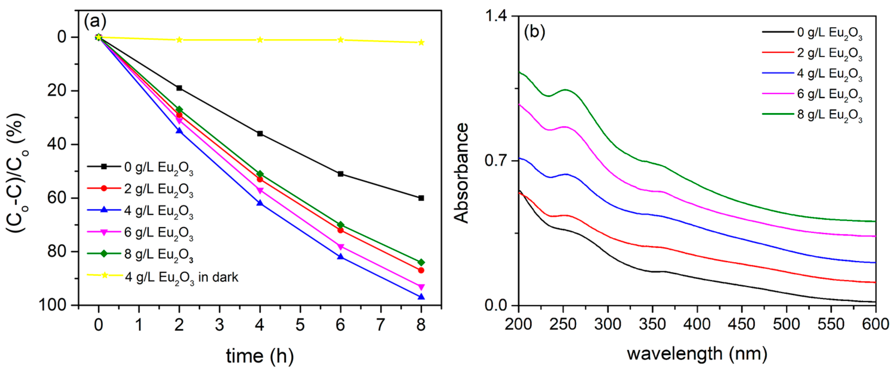
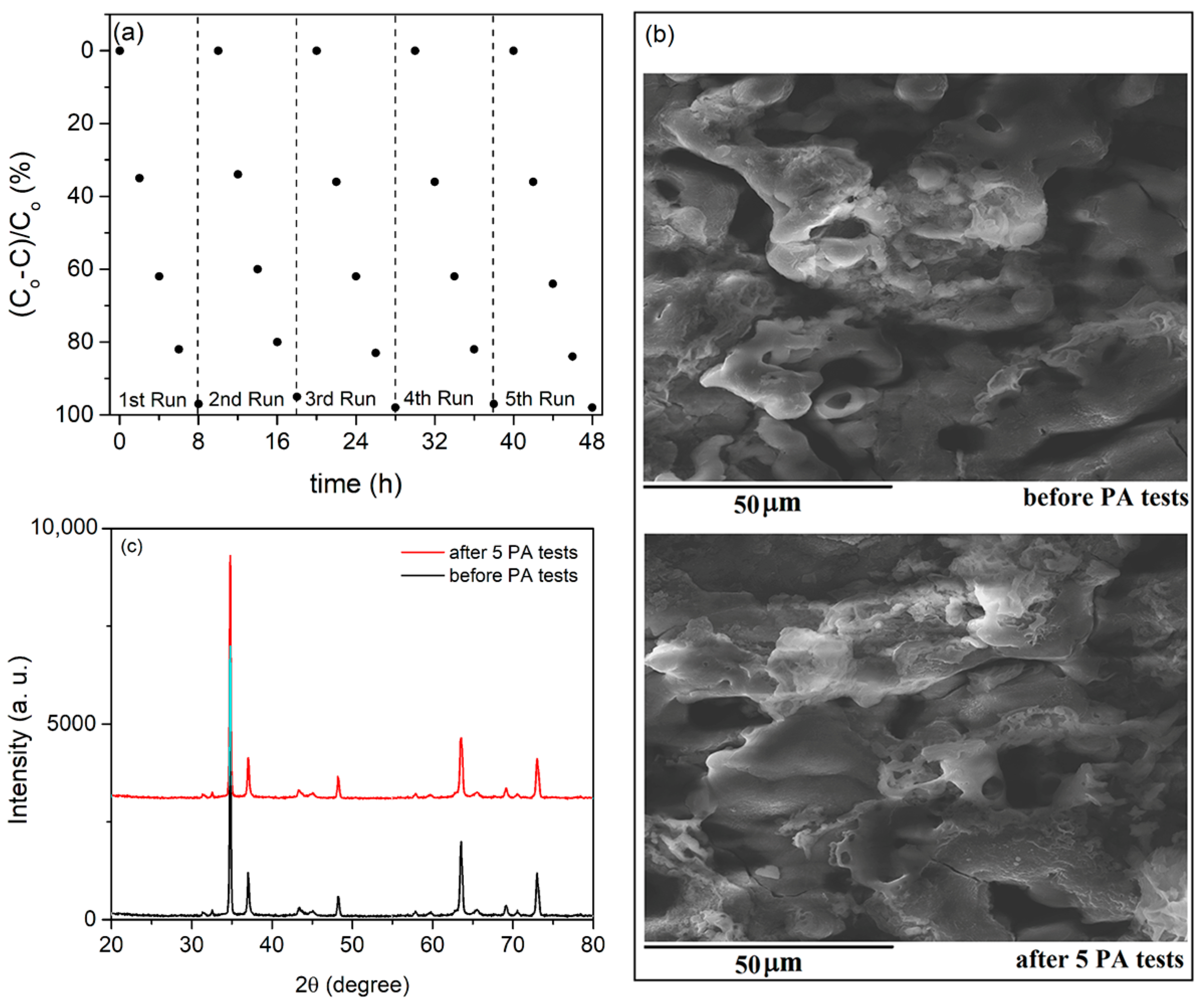
3.3. PA of Eu3+-Doped MgAl Oxide Coatings

PA and optical absorption properties of pristine and Eu3+ doped MgAl oxide coatings formed by PEO processing of AZ31 in aluminate electrolyte with varying concentration of Eu2O3 are presented in Figure 8. As a result of the large bandgap between MgO [43] and MgAl2O4 [52], the broad absorption band in the mid-UV region is observed for MgAl oxide coatings. Although the MgAl coating’s absorption band is detrimental for photocatalytic applications utilizing sunlight as the irradiation source, a high concentration of different types of oxygen vacancies and other defects generated in MgAl during the PEO processing results in good PA of MgAl coating. Because of the high concentration of surface vacancies, the PA of pure MgAl coating in MO degradation is considerably higher than that of commonly used TiO2 [30], MgO [25], ZrO2 [53], and Nb2O5 [54] coatings formed by PEO on titanium, AZ31 magnesium alloy, zirconium, and niobium substrates, respectively. Surface vacancies, which determine electron transfer between reactants and photocatalysts, were found to be closely related to active centers of heterogeneous photocatalytic reactions [55]. The PA of Eu3+-doped MgAl oxide coatings is significant in comparison with pristine MgAl oxide coating (Figure 8a). Because the incorporation of Eu3+ ions into MgAl oxide coatings has no effect on their optical absorption in comparison to pristine MgAl oxide coating (Figure 8b), increased PA can be related to the interaction of Eu3+ ions and MgAl oxide coatings. The incorporation of Eu3+ ions into MgAl oxide coatings during the PEO result in the formation of oxygen vacancy charge-trapping centers, which can lower the recombination rate of electrons and holes. As a result, Eu3+-doped MgAl oxide coatings have higher PA than pure MgAl oxide coatings.
It can be clearly seen that PA is dependent on the concentration of Eu2O3 particles which are added to aluminate electrolyte, i.e., Eu3+ in MgAl oxide coatings. The PA of the Eu3+-doped MgAl oxide coating formed in aluminate electrolyte with the addition of 4 g/L Eu2O3 is the highest, and it begins to decrease as the concentration of Eu2O3 particles increases. As a result, the most likely cause of PA decrease is that higher concentration of Eu3+ ions in MgAl oxide coatings increases the concentration of recombination centers for electron–hole pairs. Tests in the dark show that MO self-degradation is negligible.
Photo-generated electron–hole pairs formed on photocatalytic surfaces produce various active oxygen species such as the superoxide anion radical (O2−), hydrogen peroxide (H2O2), singlet oxygen (1O2), and the hydroxyl radical (OH) [56]. OH radicals are primarily responsible for organic pollutants’ degradation [57]. PL measurements performed in terephthalic acid on the surface of coatings formed in aluminate electrolyte with and without the addition of 4 g/L Eu2O3 particles are employed to test the formation of OH radicals. The quantity of produced OH radicals directly correlates with the intensity of PL [41]. Longer illumination times increase the PL intensity of both MgAl and Eu3+-doped MgAl coatings, but the latter exhibits higher PL intensity at 425 nm emission (Figure 9). This observation is not surprising, since Eu3+-doped MgAl coating shows higher PA.
One of the most important requirements for the photocatalyst’s applicability is its stability, which regulates the catalyst’s lifetime and operating costs. The PA after five consecutive runs using the most photocatalytically active formed coating is shown in Figure 10a. The sample was rinsed with water and dried between the runs. Figure 10a demonstrates that the PA did not decrease. Additionally, the morphology (Figure 10b) and phase structure (Figure 10c) did not change after five runs, proving the reliability and effectiveness of the Eu3+-doped MgAl oxide coatings formed by PEO as photocatalysts.

4. Conclusions

In summary, Eu3+-doped MgAl coatings were formed by plasma-electrolytic oxidation of AZ31 magnesium alloy in aluminate electrolyte which contains Eu2O3 particles. The coatings are crystallized and composed of MgO and MgAl2O4. DRS spectra of coatings exhibit a wide absorption band in the mid-UV region.
The emission PL spectra of Eu3+-doped MgAl coatings display narrow emission bands, which are attributed to Eu3+ ion 5D07FJ transitions. The excitation PL spectra of MgAl coatings doped with Eu3+ have a strong broad band which is related to the electron transfer transition from the 2p orbital of O2− ions to the 4f orbital of Eu3+ ions followed by weak, well-defined peaks corresponding to direct excitation of the Eu3+ ground state 7F0 into higher levels of the 4f-manifold. The electrical dipole transition 5D07F2 is significantly stronger than the magnetic dipole transition 5D07F1, indicating the presence of Eu3+ ions in an asymmetric environment.
The PA of MgAl coatings doped with Eu3+ is determined by the concentration of Eu2O3 particles in aluminate electrolyte, i.e., Eu3+ incorporated into MgAl coatings, with 4 g/L being the optimum Eu2O3 concentration in the aluminate electrolyte. Because the incorporation of Eu3+ particles into MgAl coatings has no significant effect on the absorption properties of formed coatings when compared to pure MgAl oxide coating, the increased PA of Eu3+-doped MgAl coatings is due to the formation of oxygen vacancy charge-trapping centers, which reduces the recombination rate of electrons and holes.

Author Contributions

Conceptualization, S.S.; methodology, S.S.; validation, S.S., investigation, S.S., N.R. and R.V.; writing—original draft preparation, S.S.; writing—review and editing, S.S. and R.V. All authors have read and agreed to the published version of the manuscript.

Funding

This research was funded by the Ministry of Education, Science, and Technological Development of the Republic of Serbia (Grants 451-03-9/2021-14/200162 and 451-03-9/2021-14/200026) and by the European Union Horizon 2020 Research and Innovation program under the Marie Sklodowska-Curie grant agreement No. 823942 (FUNCOAT).

Data Availability Statement

The data presented in this study are available on request from the corresponding author.

Conflicts of Interest

The authors declare no conflict of interest.

References

  1. Mordike, B.L.; Ebert, T. Magnesium: Properties—applications—potential. Mat. Sci. Eng. A 2001, 302, 37–45. [Google Scholar] [CrossRef]
  2. Ali, M.; Hussein, M.A.; Al-Aqeeli, N. Magnesium-based composites and alloys for medical applications: A review of mechanical and corrosion properties. J. Alloys Compd. 2019, 792, 1162–1190. [Google Scholar]
  3. Ravichandran, N.; Mungara, S.R. Manufacturing and deployment of magnesium alloy parts for light weighting applications. Mater. Today Proc. 2021, 47, 4838–4843. [Google Scholar]
  4. Liu, H.; Cao, F.; Song, G.-L.; Zheng, D.; Shi, Z.; Dargusch, M.S.; Atrens, A. Review of the atmospheric corrosion of magnesium alloys. J. Mater. Sci. Technol. 2019, 35, 2003–2016. [Google Scholar] [CrossRef]
  5. Esmaily, M.; Svensson, J.E.; Fajardo, S.; Birbilis, N.; Frankel, G.S.; Virtanen, S.; Arrabal, R.; Thomas, S.; Johansson, L.G. Fundamentals and advances in magnesium alloy corrosion. Prog. Mater. Sci. 2017, 89, 92–193. [Google Scholar]
  6. Daroonparvar, M.; Bakhsheshi-Rad, H.R.; Saberi, A.; Razzaghi, M.; Kasar, A.K.; Ramakrishna, S.; Menezes, P.L.; Misra, M.; Ismail, A.F.; Shariff, S.; et al. Surface modification of magnesium alloys using thermal and solid-state cold spray processes: Challenges and latest progresses. J. Magnes. Alloys 2022, 10, 2025–2061. [Google Scholar] [CrossRef]
  7. Liu, Y.; Zhang, Y.; Wang, Y.-L.; Tian, Y.-Q.; Chen, L.-S. Research progress on surface protective coatings of biomedical degradable magnesium alloys. J. Alloys Compd. 2021, 885, 161001. [Google Scholar] [CrossRef]
  8. Zhang, D.; Peng, F.; Liu, X. Protection of magnesium alloys: From physical barrier coating to\smart self-healing coating. J. Alloys Compd. 2021, 853, 157010. [Google Scholar] [CrossRef]
  9. Lin, Z.; Wang, T.; Yu, X.; Sun, X.; Yang, H. Functionalization treatment of micro-arc oxidation coatings on magnesium alloys: A review. J. Alloys Compd. 2021, 879, 160453. [Google Scholar] [CrossRef]
  10. Yao, W.; Wu, L.; Wang, J.; Jiang, B.; Zhang, D.; Serdechnova, M.; Shulha, T.; Blawert, C.; Zheludkevich, M.L.; Pan, F. Micro-arc oxidation of magnesium alloys: A review. J. Mater. Sci. Technol. 2022, 118, 158–180. [Google Scholar] [CrossRef]
  11. Farshid, S.; Kharaziha, M. Micro and nano-enabled approaches to improve the performance of plasma electrolytic oxidation coated magnesium alloys. J. Magnes. Alloys 2021, 9, 1487–1504. [Google Scholar] [CrossRef]
  12. Narayanan, T.S.N.S.; Park, I.S.; Lee, M.H. Strategies to improve the corrosion resistanceof microarc oxidation (MAO) coated magnesium alloys for degradable implants: Prospects and challenges. Prog. Mater. Sci. 2014, 60, 1–71. [Google Scholar] [CrossRef]
  13. Chen, Y.; Lu, X.; Lamaka, S.V.; Ju, P.; Blawert, C.; Zhang, T.; Wang, F.; Zheludkevich, M.L. Active protection of Mg alloy by composite PEO coating loaded with corrosion inhibitors. Appl. Surf. Sci. 2020, 504, 144462. [Google Scholar] [CrossRef]
  14. Fattah-alhosseini, A.; Chaharmahali, R.; Babaei, K. Effect of particles addition to solution of plasma electrolytic oxidation (PEO) on the properties of PEO coatings formed on magnesium and its alloys: A review. J. Magnes. Alloys 2020, 8, 799–818. [Google Scholar] [CrossRef]
  15. Yerokhin, A.L.; Nie, X.; Leyland, A.; Matthews, A.; Dowey, S.J. Plasma electrolysis for surface engineering. Surf. Coat. Technol. 1999, 122, 73–93. [Google Scholar] [CrossRef]
  16. Tsai, D.-S.; Chou, C.-C. Review of the soft sparking issues in plasma electrolytic oxidation. Metals 2018, 8, 105. [Google Scholar] [CrossRef] [Green Version]
  17. Kaseem, M.; Fatimah, S.; Nashrah, N.; Ko, Y.G. Recent progress in surface modification of metals coated by plasma electrolytic oxidation: Principle, structure, and performance. Prog. Mater. Sci. 2021, 117, 100735. [Google Scholar] [CrossRef]
  18. Stojadinović, S.; Vasilić, R.; Perić, M. Investigation of plasma electrolytic oxidation on valve metals by means of molecular spectroscopy—A review. RSC Adv. 2014, 4, 25759. [Google Scholar] [CrossRef]
  19. Snizhko, L.O.; Yerokhin, A.L.; Pilkington, A.; Gurevina, N.L.; Misnyankin, D.O.; Leyland, A.; Matthews, A. Anodic processes in plasma electrolytic oxidation of aluminium in alkaline solutions. Electrochim. Acta 2004, 49, 2085–2095. [Google Scholar] [CrossRef]
  20. Hussein, R.O.; Nie, X.; Northwood, D.O.; Yekokhin, A.; Matthews, A. Spectroscopic study of electrolytic plasma and discharging behaviour during the plasma electrolytic oxidation (PEO) process. J. Phys. D 2010, 43, 105203. [Google Scholar] [CrossRef]
  21. Buling, A.; Zerrer, J. Increasing the application fields of magnesium by ultraceramic: Corrosion and wear protection by plasma electrolytical oxidation (PEO) of Mg alloys. Surf. Coat. Technol. 2019, 369, 142–155. [Google Scholar] [CrossRef]
  22. Darband, G.B.; Aliofkhazraei, M.; Hamghalam, P.; Valizade, N. Plasma electrolytic oxidation of magnesium and its alloys: Mechanism, properties and applications. J. Magnes. Alloys 2017, 5, 74–132. [Google Scholar] [CrossRef]
  23. Mashtalyar, D.V.; Nadaraia, K.V.; Plekhova, N.G.; Imshinetskiy, I.M.; Piatkova, M.A.; Pleshkova, A.I.; Kislova, S.E.; Sinebryukho, S.L.; Gnedenkov, S.V. Antibacterial Ca/P-coatings formed on Mg alloy using plasma electrolytic oxidation and antibiotic impregnation. Mater. Lett. 2022, 317, 132099. [Google Scholar] [CrossRef]
  24. Rúa, J.M.; Zuleta, A.A.; Ramírez, J.; Fernández-Morales, P. Micro-arc oxidation coating on porous magnesium foam and its potential biomedical applications. Surf. Coat. Technol. 2019, 360, 213–221. [Google Scholar] [CrossRef]
  25. Stojadinović, S.; Tadić, N.; Radić, N.; Grbić, B.; Vasilić, R. MgO/ZnO coatings formed on magnesium alloy AZ31 by plasma electrolytic oxidation: Structural, photoluminescence and photocatalytic investigation. Surf. Coat. Technol. 2017, 310, 98–105. [Google Scholar] [CrossRef]
  26. Kumar, G.K.; Bhargav, P.B.; Aravinth, K.; Ahmed, N.; Balaji, C. Photoluminescence and electrochemical performance evaluation of Eu3+ doped MgAl2O4 phosphors for LED and energy storage applications. Ceram. Int. 2022, 48, 36038–36045. [Google Scholar] [CrossRef]
  27. Golyeva, E.V.; Kolesnikov, I.E.; Lähderanta, E.; Kurochkin, A.V.; Mikhailov, M.D. Effect of synthesis conditions on structural, morphological and luminescence properties of MgAl2O4:Eu3+ nanopowders. J. Lumin. 2018, 194, 387–393. [Google Scholar] [CrossRef]
  28. Cao, N.; Zhao, X.; Gao, M.; Li, Z.; Ding, X.; Li, C.; Liu, K.; Du, X.; Li, W.; Feng, J.; et al. Superior selective adsorption of MgO with abundant oxygen vacancies to removal and recycle reactive dyes. Sep. Purif. Technol. 2021, 275, 119236. [Google Scholar] [CrossRef]
  29. Ball, J.A.; Murphy, S.T.; Grimes, R.W.; Bacorisen, D.; Smith, R.; Uberuaga, B.P.; Sickafus, K.E. Defect processes in MgAl2O4 spinel. Solid State Sci. 2008, 10, 717–724. [Google Scholar] [CrossRef]
  30. Stojadinović, S.; Radić, N.; Grbić, B.; Maletić, S.; Stefanov, P.; Pačevski, A.; Vasilić, R. Structural, photoluminescent and photocatalytic properties of TiO2:Eu3+ coatings formed by plasma electrolytic oxidation. Appl. Surf. Sci. 2016, 370, 218–228. [Google Scholar] [CrossRef]
  31. Stojadinović, S.; Vasilić, R. Formation and photoluminescence of Eu3+ doped zirconia coatings formed by plasma electrolytic oxidation. J. Lumin. 2016, 176, 25–31. [Google Scholar] [CrossRef]
  32. Stojadinović, S.; Vasilić, R. Orange–red photoluminescence of Nb2O5:Eu3+, Sm3+ coatings formed by plasma electrolytic oxidation of niobium. J. Alloys Compd. 2016, 685, 881–889. [Google Scholar] [CrossRef]
  33. Stojadinović, S.; Tadić, N.; Ćirić, A.; Vasilić, R. Photoluminescence properties of Eu3+ doped HfO2 coatings formed by plasma electrolytic oxidation of hafnium. Opt. Mater. 2018, 77, 19–24. [Google Scholar] [CrossRef]
  34. Ćirić, A.; Stojadinović, S.; Dramićanin, M.D. Luminescence temperature sensing using thin-films of undoped Gd2O3 and doped with Ho3+, Eu3+ and Er3+ prepared by plasma electrolytic oxidation. Ceram. Int. 2020, 46, 23223–23231. [Google Scholar] [CrossRef]
  35. Stojadinović, S.; Ćirić, A. Photoluminescence of ZnO:Eu3+ and ZnO:Tb3+ coatings formed by plasma electrolytic oxidation of pure zinc substrate. J. Lumin. 2021, 235, 118022. [Google Scholar] [CrossRef]
  36. Binnemans, K. Interpretation of europium(III) spectra. Coord. Chem. Rev. 2015, 295, 1–45. [Google Scholar]
  37. Kumar, M.; Singh, G.; Chauhan, M.S. Europium (Eu3+)—doped ZnO nanostructures: Synthesis, characterization, and photocatalytic, chemical sensing and preliminary assessment of magnetic properties. Ceram. Int. 2021, 47, 17023–17033. [Google Scholar] [CrossRef]
  38. Huang, F.; Li, Z.; Yan, A.; Dong, H.; Li, Q.; Hu, M.; Xiong, X.; Gao, Q. Boosted visible-light photocatalytic activity and retarded carrier recombination of Eu3+-doped Nb3O7F nanomaterials prepared by hydrothermal method. Nanotechnology 2020, 31, 18520. [Google Scholar] [CrossRef] [PubMed]
  39. Borlaf, M.; Moreno, R.; Ortiz, A.L.; Colomer, M.T. Synthesis and photocatalytic activity of Eu3+-doped nanoparticulate TiO2 sols and thermal stability of the resulting xerogels. Mater. Chem. Phys. 2014, 144, 8–16. [Google Scholar] [CrossRef]
  40. Ćirić, A.; Stojadinović, S. Photoluminescence of ZrO2:Gd3+ and ZrO2:Dy3+ coatings formed by the plasma electrolytic oxidation. J. Alloys Compd. 2020, 832, 154907. [Google Scholar] [CrossRef]
  41. Ishibashi, K.; Fujishima, A.; Watanabe, T.; Hashimoto, K. Detection of active oxidative species in TiO2 photocatalysis using the fluorescence technique. Electrochem. Commun. 2000, 2, 207–210. [Google Scholar] [CrossRef]
  42. Stojadinović, S.; Vasilić, R.; Radić-Perić, J.; Perić, M. Characterization of plasma electrolytic oxidation of magnesium alloy AZ31 in alkaline solution containing fluoride. Surf. Coat. Technol. 2015, 273, 1–11. [Google Scholar] [CrossRef]
  43. Mageshwari, K.; Mali, S.S.; Sathyamoorthy, R.; Patil, P.S. Template-free synthesis of MgO nanoparticles for effective photocatalytic applications. Powder Technol. 2013, 249, 456–462. [Google Scholar] [CrossRef]
  44. Jintakosol, T.; Singjai, P. Effect of annealing treatment on luminescence property of MgO nanowires. Curr. Appl. Phys. 2009, 9, 1288–1292. [Google Scholar] [CrossRef]
  45. Pathak, N.; Ghosh, P.S.; Gupta, S.K.; Mukherjee, S.; Kadam, R.M.; Arya, A. An insight into the various defects-induced emission in MgAl2O4 and their tunability with phase behavior: Combined experimental and theoretical approach. J. Phys. Chem. C 2016, 120, 4016–4031. [Google Scholar] [CrossRef]
  46. Lu, Y.; Wang, J.; Shi, Z.; Shi, J.; Liu, Y.; He, L. Site occupation and fluorescence properties of MgAl2O4: Eu3+ phosphors. Mater. Sci. Semicond. Proc. 2022, 137, 106233. [Google Scholar] [CrossRef]
  47. Kolesnikov, I.E.; Golyeva, E.V.; Kurochkin, A.V.; Mikhailov, M.D. Structural and luminescence properties of MgAl2O4:Eu3+ nanopowders. J. Alloys Compd. 2016, 654, 32–38. [Google Scholar] [CrossRef]
  48. Tanner, P.A. Some misconceptions concerning the electronic spectra of tri-positive europium and cerium. Chem. Soc. Rev. 2013, 42, 5090–5101. [Google Scholar] [CrossRef]
  49. Guan, W.; Li, J.; Wang, X. Luminescence properties of Eu3+-doped MgAl2O4 nanopowders synthesized by micro-emulsion method. Phys. Status Solidi A 2014, 211, 1778–1781. [Google Scholar] [CrossRef]
  50. Reddy, G.K.; Krishna, R.H.; Reddy, A.J.; Monika, D.L.; Manjunath, C.; Prasanna Kumar, S.G.; Preetham, R.; Gopal, G.R. Facile self-propagating combustion synthesis of MgO:Eu3+ orangered nanophosphor and luminescence investigation by Judd-Ofelt intensity parameters. Optik 2018, 174, 234–243. [Google Scholar]
  51. Wiglusz, R.J.; Grzyb, T.; Lis, S.; Strek, W. Hydrothermal preparation and photoluminescent properties of MgAl2O4:Eu3+ spinel nanocrystals. J. Lumin. 2010, 130, 434–441. [Google Scholar] [CrossRef]
  52. Jagadeeshwaran, C.; Madhan, K.; Murugaraj, R. Size effect and order–disorder phase transition in MgAl2O4: Synthesized by co-precipitation method. J. Mater. Sci. Mater. Electron. 2018, 29, 18923–18934. [Google Scholar] [CrossRef]
  53. Stojadinović, S.; Vasilić, R.; Radić, N.; Grbić, B. Zirconia films formed by plasma electrolytic oxidation: Photoluminescent and photocatalytic properties. Opt. Mater. 2015, 40, 20–25. [Google Scholar] [CrossRef]
  54. Stojadinović, S.; Tadić, N.; Radić, N.; Stefanov, P.; Grbić, B.; Vasilić, R. Anodic luminescence, structural, photoluminescent, and photocatalytic properties of anodic oxide films grown on niobium in phosphoric acid. Appl. Surf. Sci. 2015, 355, 912–920. [Google Scholar] [CrossRef]
  55. Wang, B.; Liu, J.; Yao, S.; Liu, F.; Li, Y.; He, J.; Lin, Z.; Huang, F.; Liu, C.; Wang, M. Vacancy engineering in nanostructured semiconductors for enhancing photocatalysis. J. Mater. Chem. A 2021, 9, 17143. [Google Scholar] [CrossRef]
  56. Nosaka, Y.; Nosaka, A.Y. Generation and detection of reactive oxygen species in photocatalysis. Chem. Rev. 2017, 117, 11302–11336. [Google Scholar] [CrossRef] [PubMed]
  57. Al-Mamun, M.R.; Kader, S.; Islam, M.S.; Khan, M.Z.H. Photocatalytic activity improvement and application of UV-TiO2 photocatalysis in textile wastewater treatment: A review. J. Environ. Chem. Eng. 2019, 7, 103248. [Google Scholar] [CrossRef]
Figure 1. SEM micrographs of coatings obtained in aluminate electrolyte containing various additions of Eu2O3: (a) 0 g/L; (b) 2 g/L; (c) 4 g/L; (d) 6 g/L; (e) 8 g/L.
Coatings 12 01830 g001
Figure 2. Cross-sections of coatings obtained in: (a) aluminate electrolyte; (b) aluminate electrolyte with 8 g/L of Eu2O3.
Coatings 12 01830 g002
Figure 3. EDS maps of coating made in aluminate electrolyte with 8 g/L of Eu2O3.
Coatings 12 01830 g003
Figure 4. XRD patterns of: (i) Eu2O3 particles; (ii) AZ31 magnesium alloy; coatings formed in aluminate electrolyte with addition of: (iii) 0 g/L Eu2O3; (iv) 2 g/L Eu2O3; (v) 4 g/L Eu2O3; (vi) 6 g/L Eu2O3; (vii) 8 g/L Eu2O3.
Coatings 12 01830 g004
Figure 5. PL spectra of MgAl oxide coating formed in aluminate electrolyte.
Coatings 12 01830 g005
Figure 6. (a) PL emission spectra with 265 nm excitation; (b) PL excitation spectra monitored at 614 nm; (c) High-resolution PL excitation spectra monitored at 614 nm; (d) PL emission spectra with 469 nm excitation.
Coatings 12 01830 g006
Figure 7. The asymmetric ratio between 5D0 7F2 and 5D0 7F1.
Coatings 12 01830 g007
Figure 8. (a) PA and (b) DRS spectra of MgAl oxide coatings formed in aluminate electrolyte with varying concentration of Eu2O3 particles.
Coatings 12 01830 g008
Figure 9. PL emission intensity monitored at 425 nm of terephthalic acid and coatings formed in aluminate electrolyte (MgAl) and aluminate electrolyte + 4 g/L Eu2O3 (MgAl:Eu3+).
Coatings 12 01830 g009
Figure 10. (a) Recycling PA test; (b) SEM micrographs before and after cycling; (c) XRD patterns before and after cycling.
Coatings 12 01830 g010
Table 1. EDS analysis of coatings in Figure 1.
SampleEu2O3 (g/L)Atomic (%)
OMgAlEu
Figure 1a065.9815.7818.24
Figure 1b265.2215.7618.850.18
Figure 1c465.3516.0218.210.41
Figure 1d665.4315.6718.170.74
Figure 1e864.9616.4417.670.92
Publisher’s Note: MDPI stays neutral with regard to jurisdictional claims in published maps and institutional affiliations.

