
A Novel Protection Method for Carbonate Stone Artifacts with Gypsum Weathering Crusts
 ,
,
Abstract
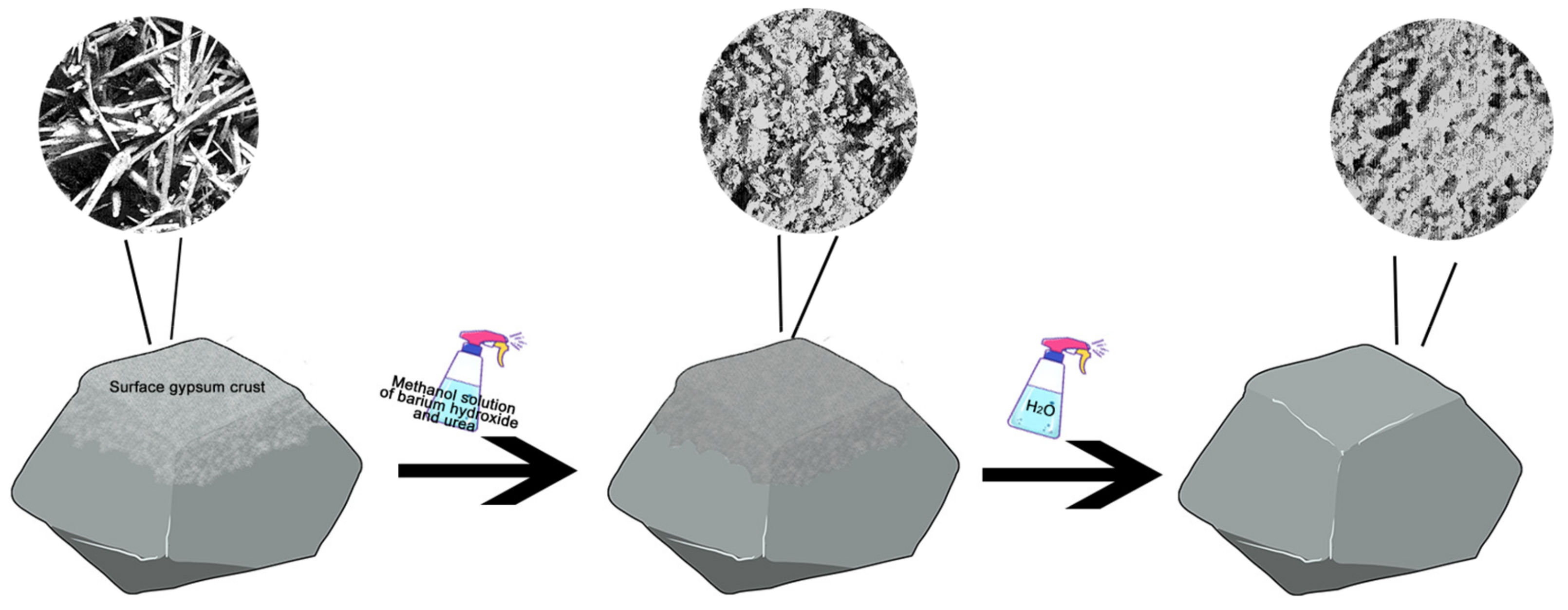
:1. Introduction
2. Materials and Methods
2.1. Sample Preparation
2.2. Characterization
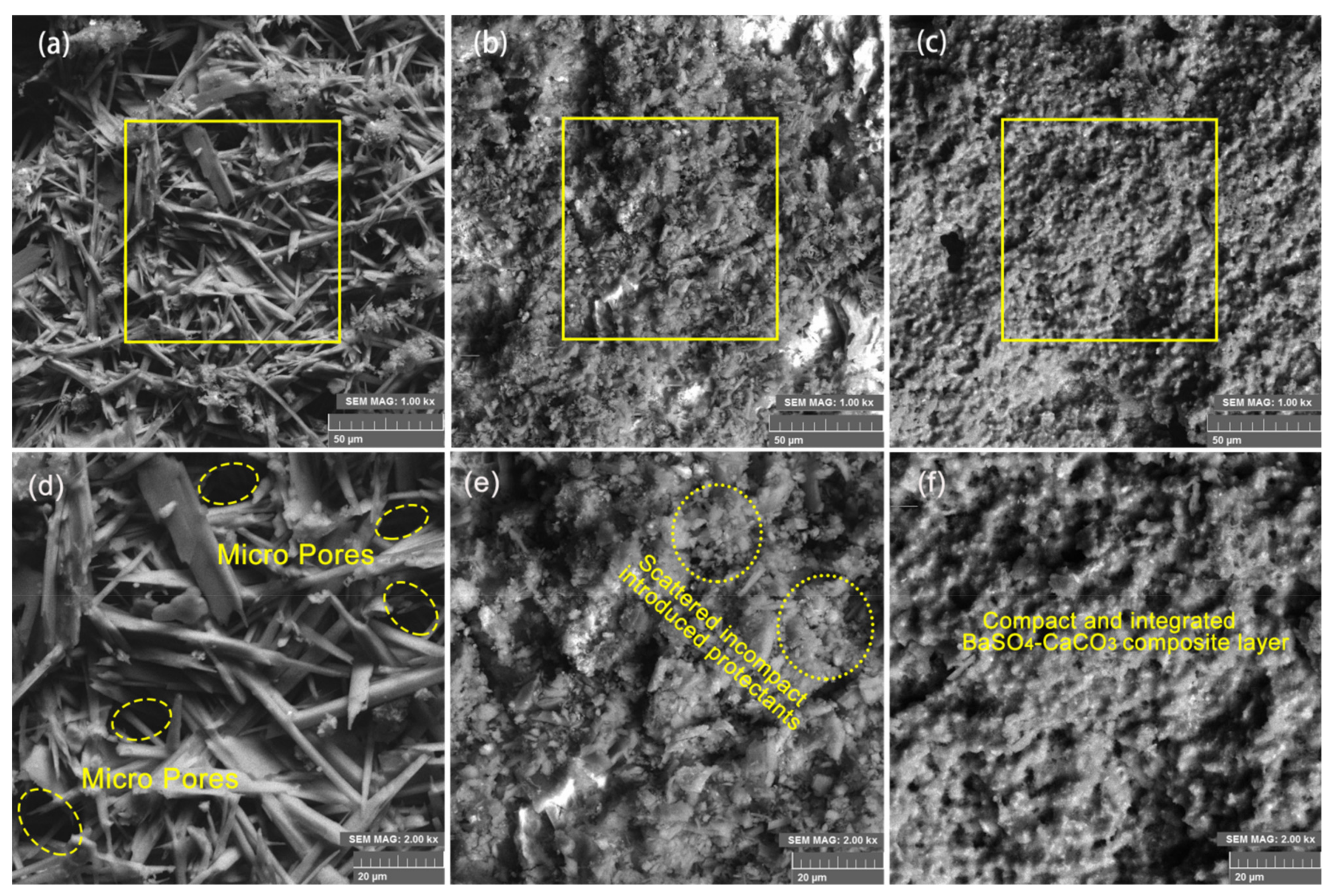
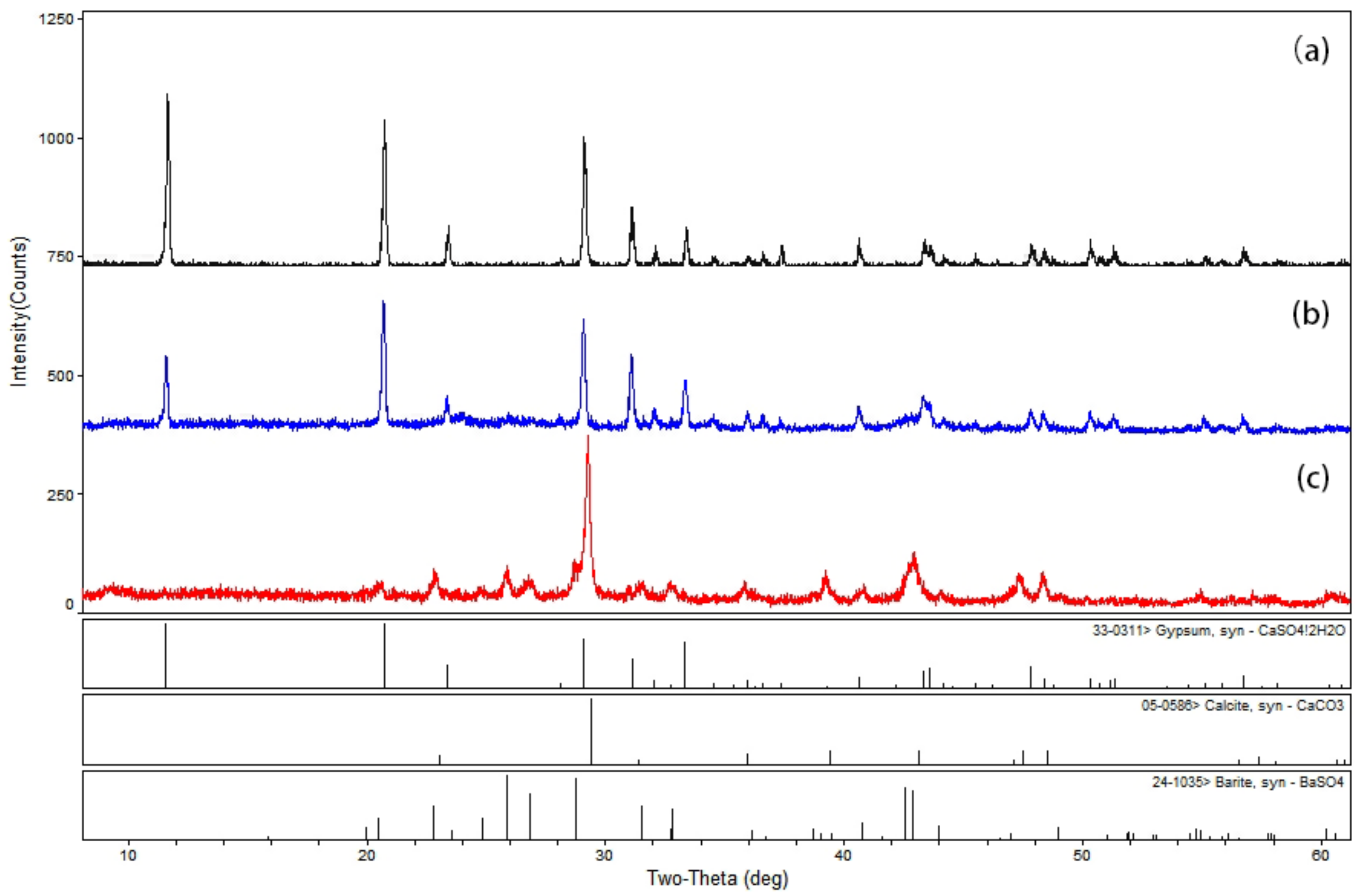
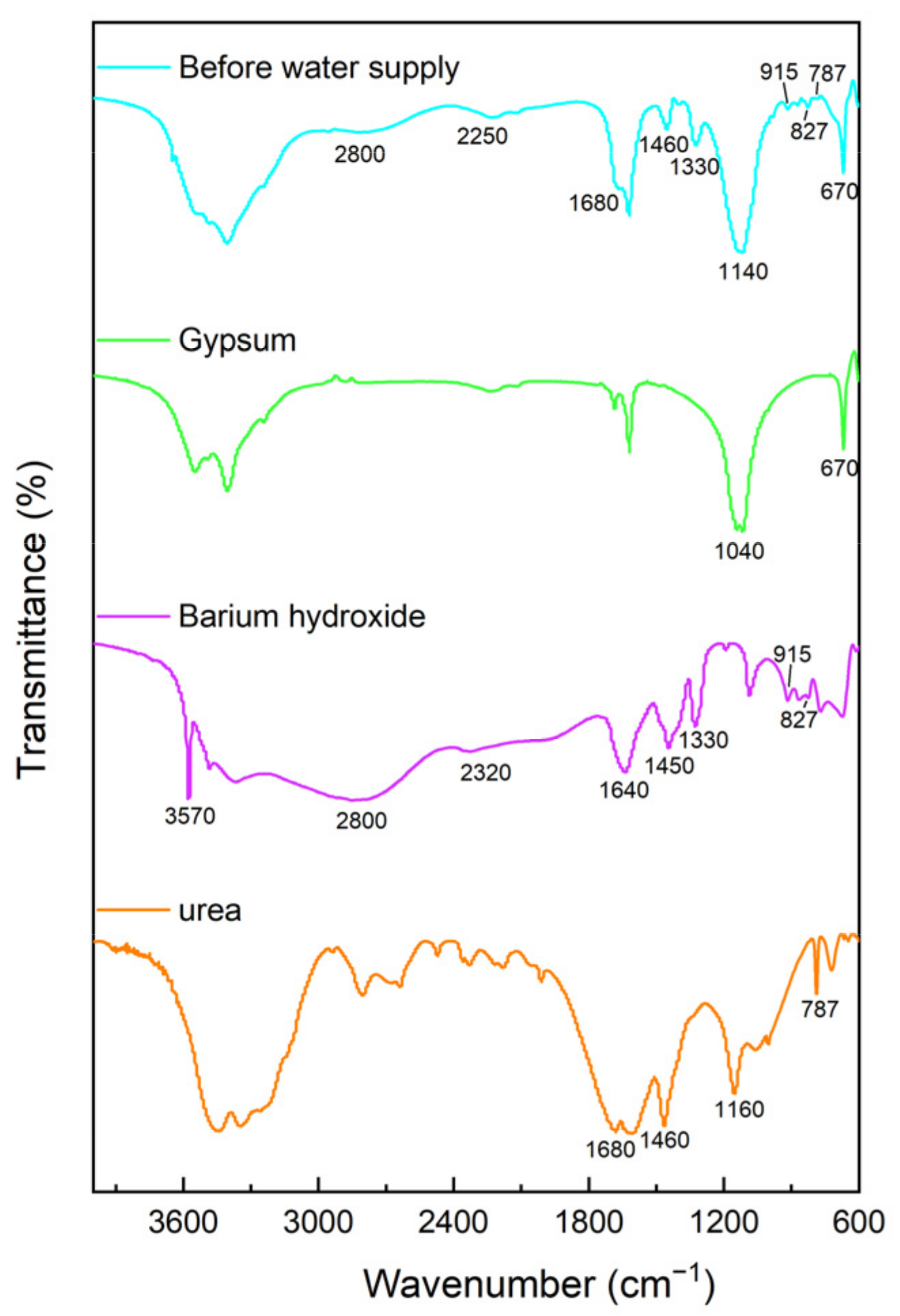
3. Results and Discussion
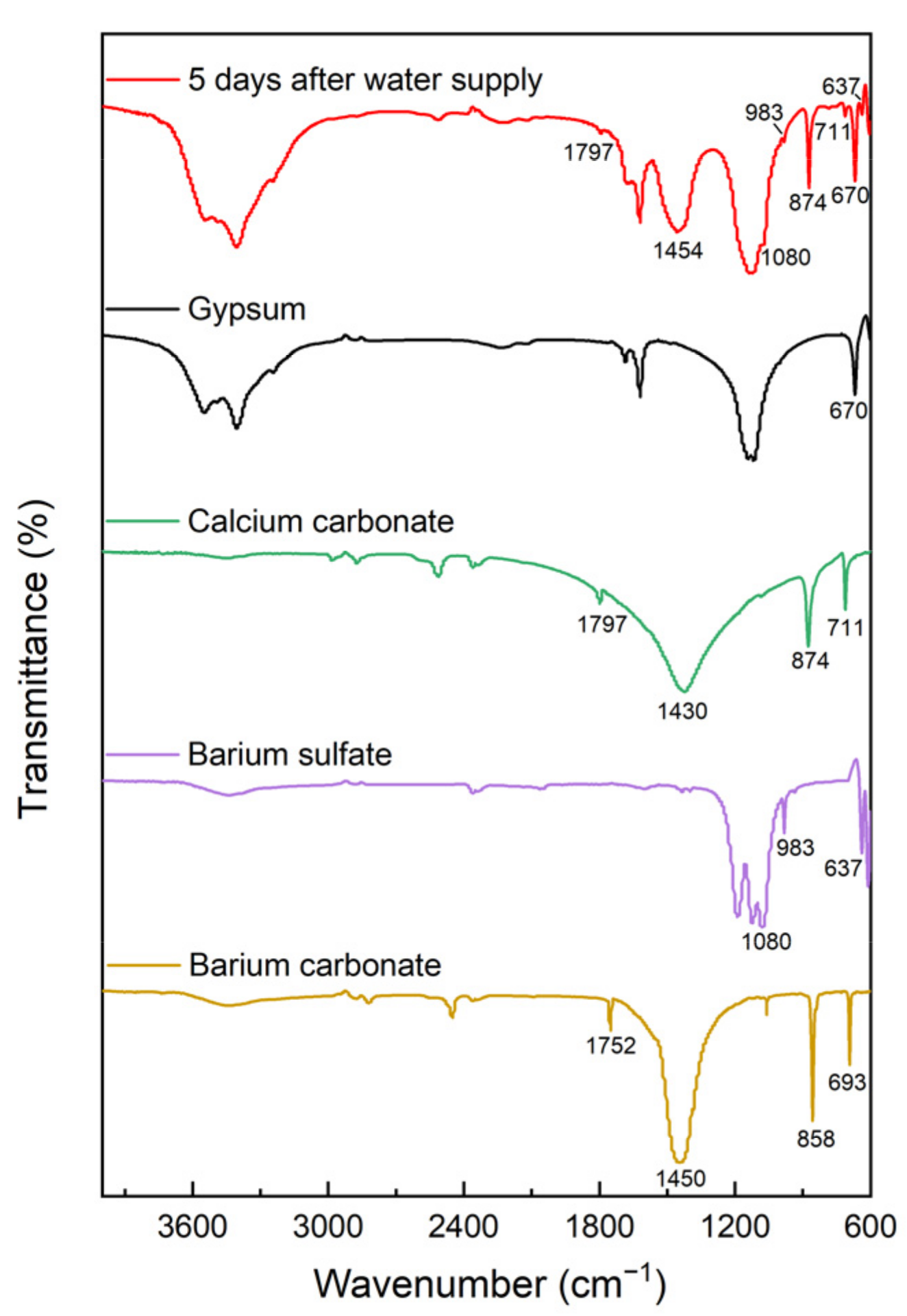
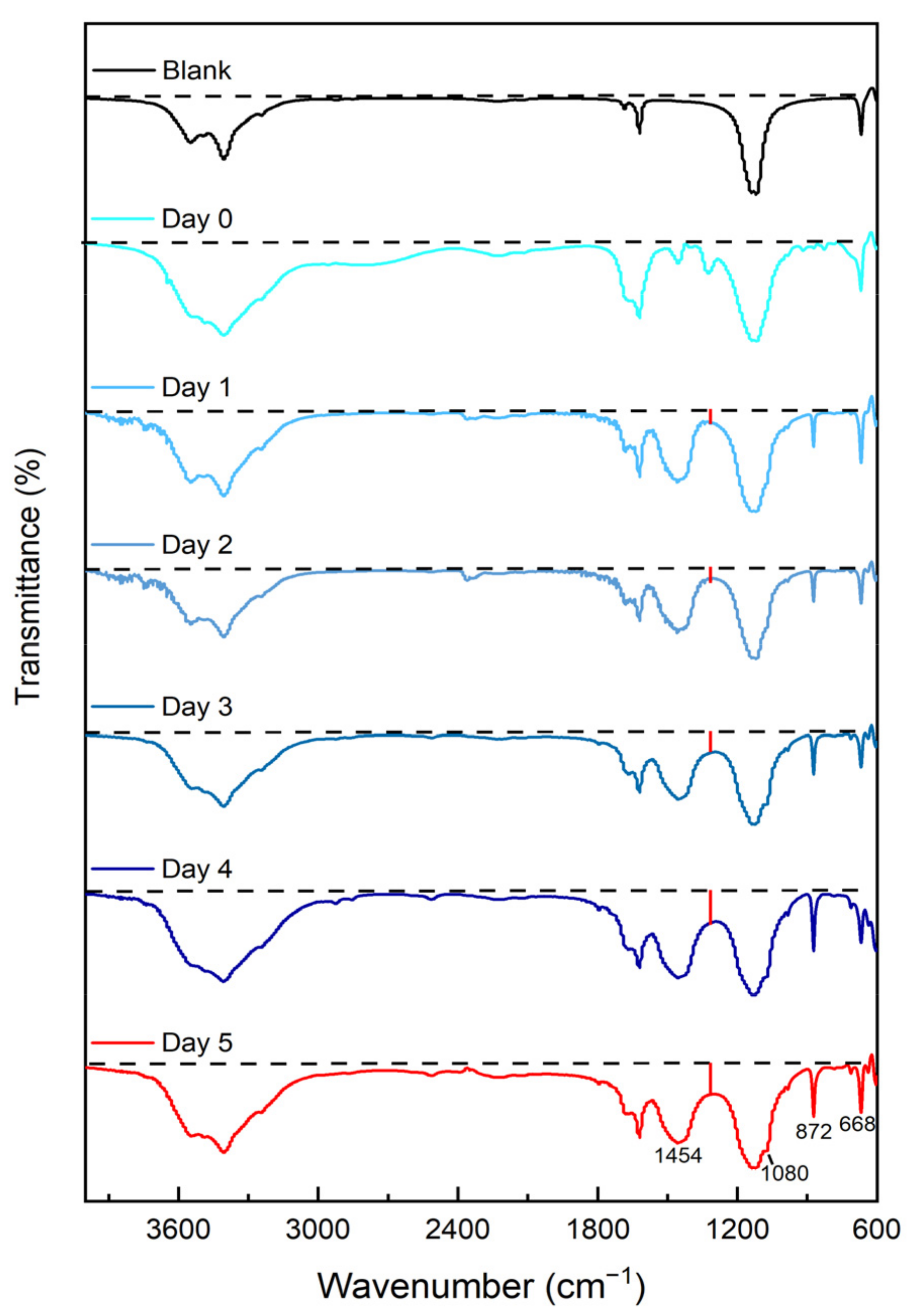
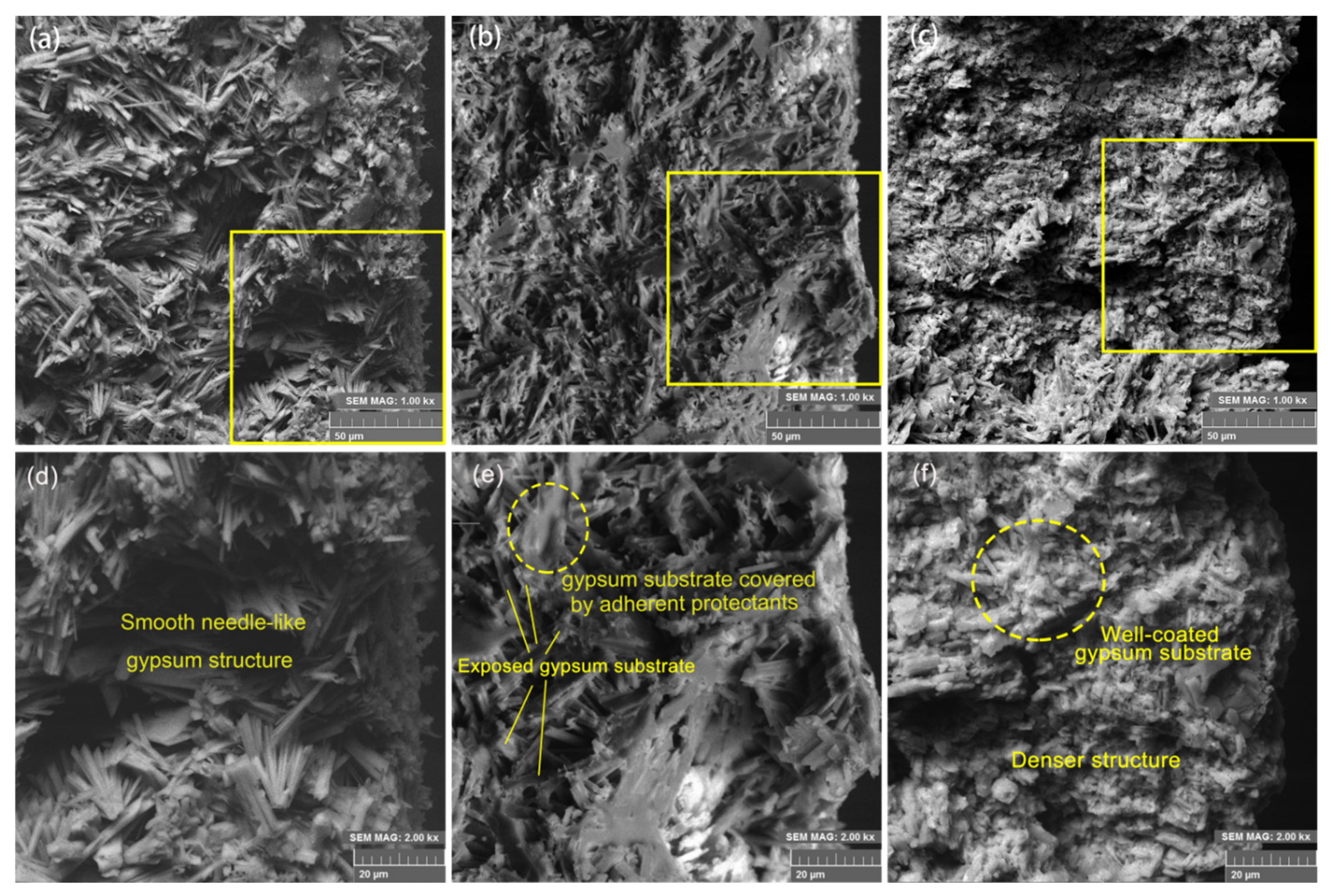
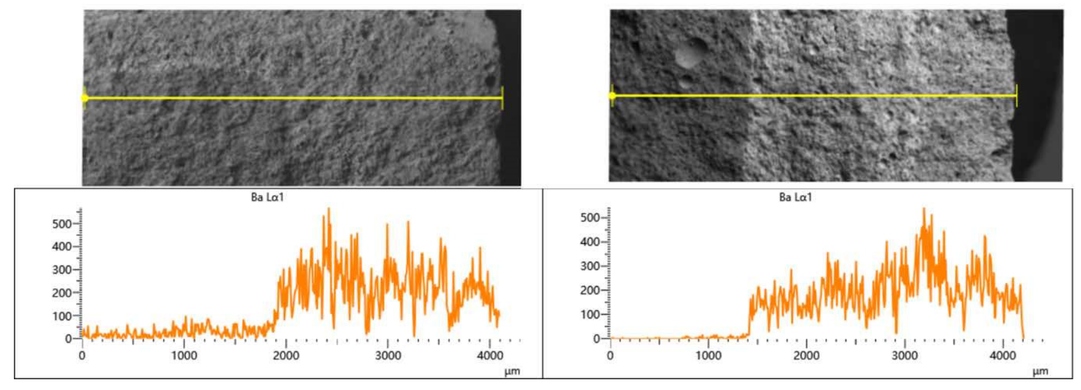
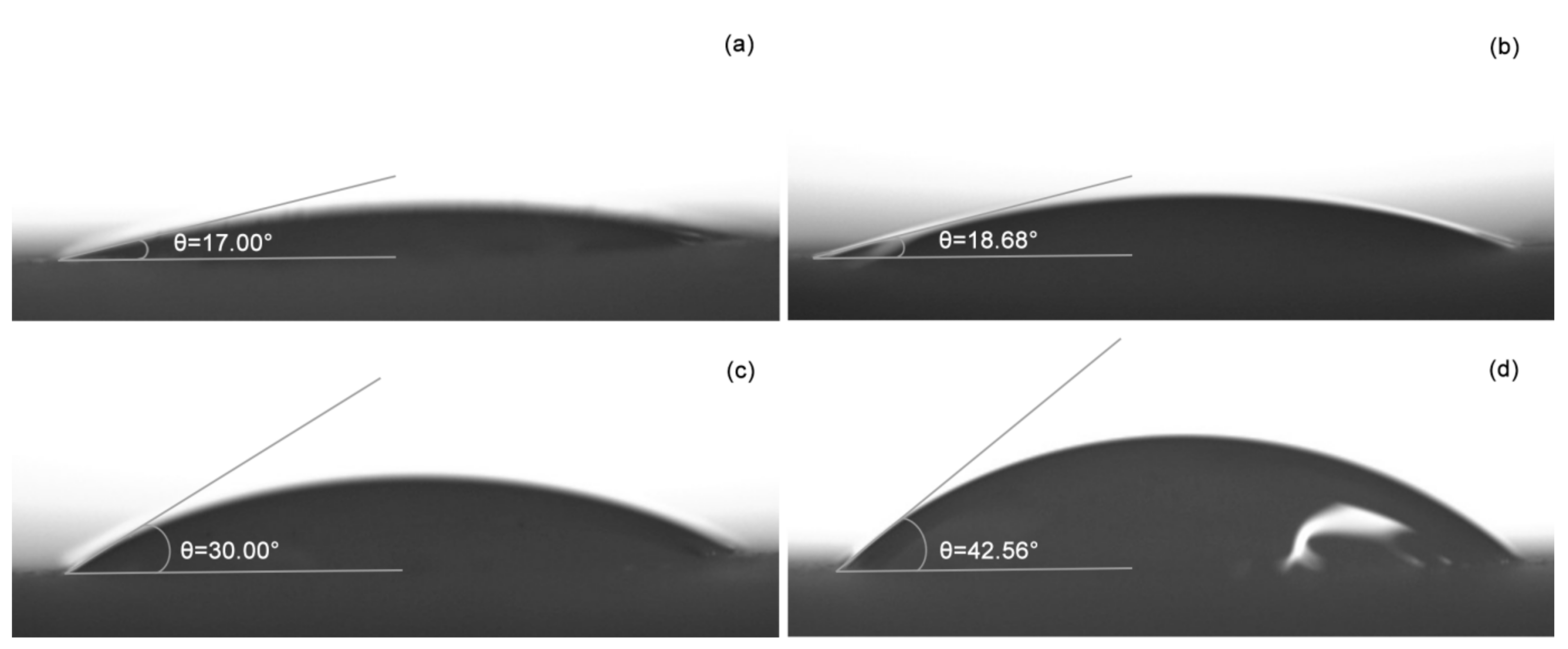
3.1. Morphological and Chemical Characterization
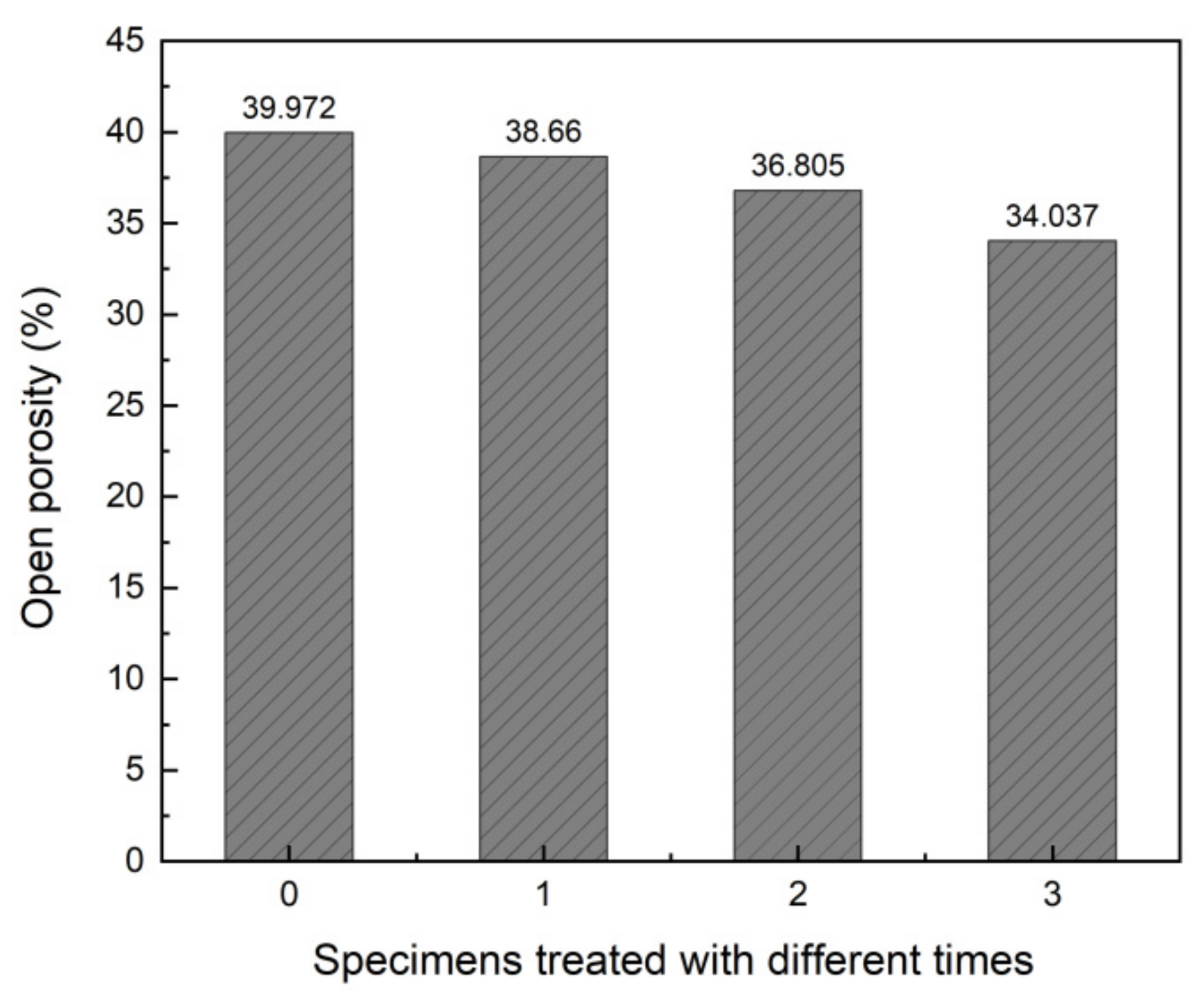
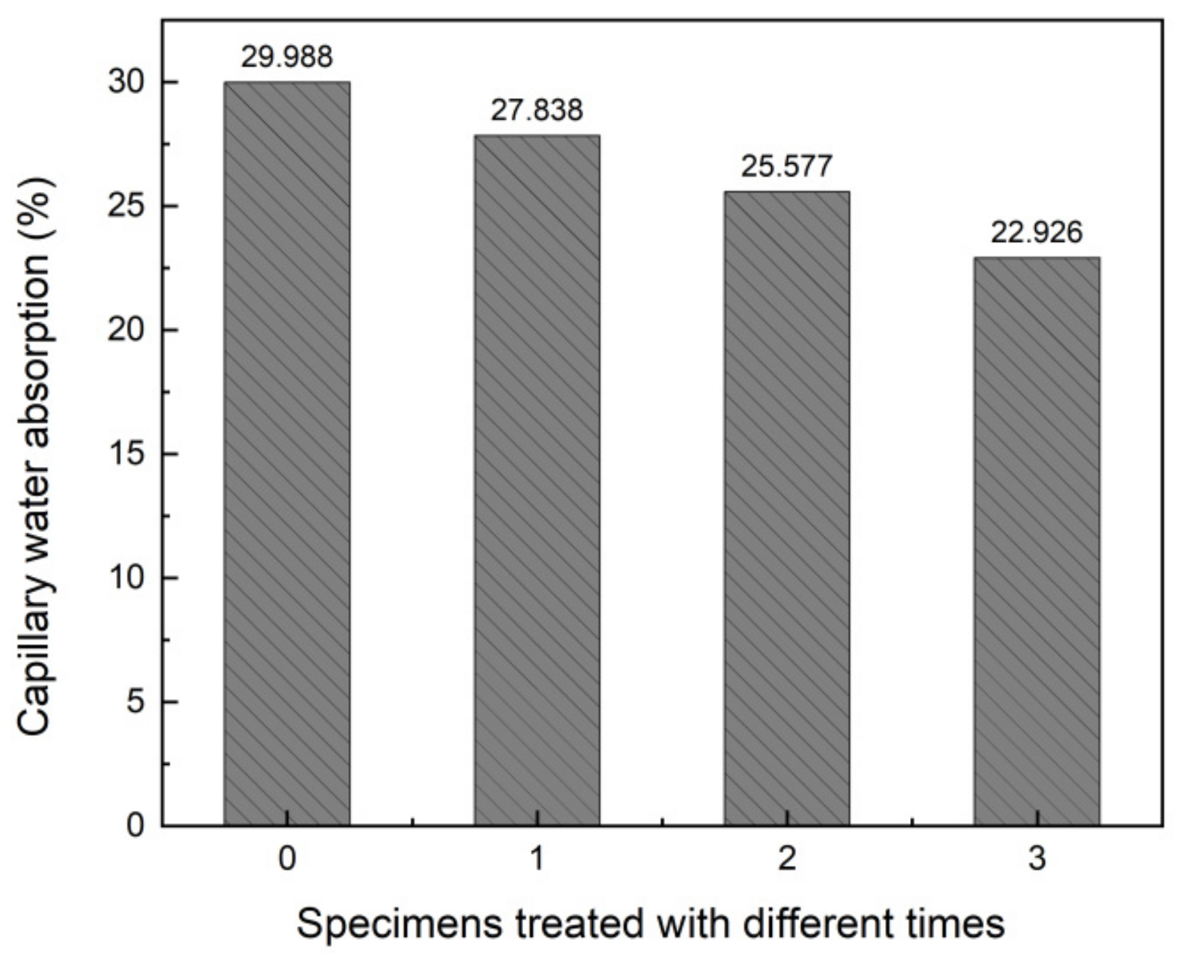
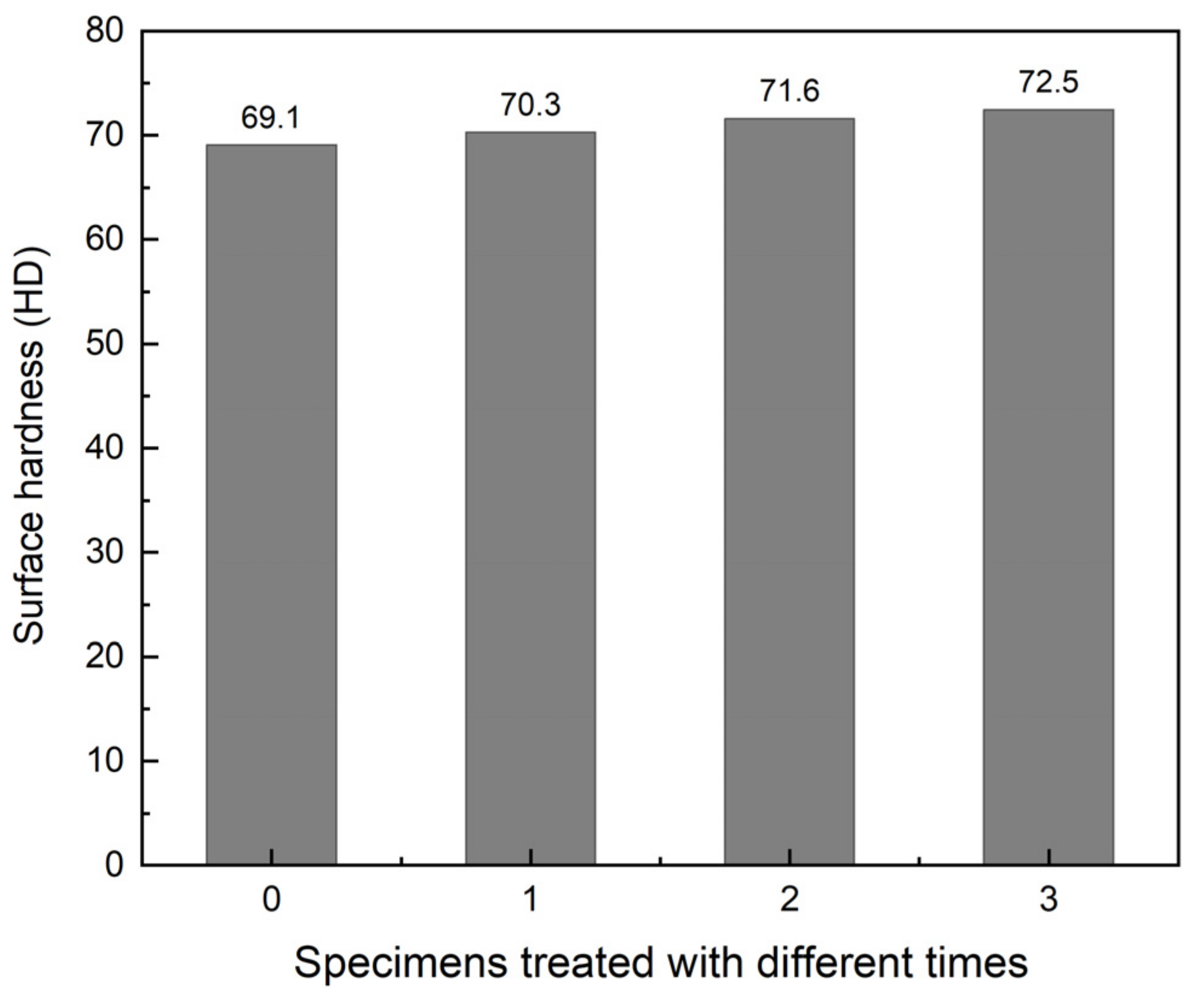
3.2. Physical Properties Characterization
4. Conclusions
Author Contributions
Funding
Institutional Review Board Statement
Informed Consent Statement
Data Availability Statement
Conflicts of Interest
References
- Bozdağ, A.; Ince, I.; Bozdağ, A.; Hatır, M.E.; Tosunlar, M.B.; Korkanç, M. An assessment of deterioration in cultural heritage: The unique case of Eflatunpınar Hittite Water Monument in Konya, Turkey. Bull. Eng. Geol. Environ. 2020, 79, 1185–1197. [Google Scholar] [CrossRef]
- Camuffo, D. Deterioration Processes of Historical Monuments. In Studies Environmental Science; Schneider, T., Ed.; Elsevier: Amsterdam, The Netherlands, 1986; Volume 30, pp. 189–221. [Google Scholar] [CrossRef]
- Charola, A.E.; Ware, R. Acid deposition and the deterioration of stone: A brief review of a broad topic. Geol. Soc. Lond. Spec. Publ. 2002, 205, 393–406. [Google Scholar] [CrossRef]
- Ausset, P.; Lefèvre, R.A.; Del Monte, M. Early mechanisms of development of sulphated black crusts on carbonate stone. In Proceedings of the 9th International Congress on Deterioration and Conservation of Stone, Venice, Italy, 19–24 June 2000; Fassina, V., Ed.; Elsevier Science B.V.: Amsterdam, The Netherlands, 2000; pp. 329–337. [Google Scholar] [CrossRef]
- Dean, J.A. Lange’s Handbook of Chemistry; Wei, J.F., Ed.; Science Press: Beijing, China, 2003. [Google Scholar]
- Kramar, S.; Urosevic, M.; Pristacz, H.; Mirtič, B. Assessment of limestone deterioration due to salt formation by micro-Raman spectroscopy: Application to architectural heritage. J. Raman Spectrosc. 2010, 41, 1441–1448. [Google Scholar] [CrossRef]
- Fronteau, G.; Schneider-Thomachot, C.; Chopin, E.; Barbin, V.; Mouze, D.; Pascal, A. Black-crust growth and interaction with underlying limestone microfacies. Geol. Soc. Lond. Speec. Publ. 2010, 333, 25–34. [Google Scholar] [CrossRef]
- Zanini, A.; Trafeli, V.; Bartoli, L. The laser as a tool for the cleaning of Cultural Heritage. IOP Conf. Ser. Mater. Sci. Eng. 2018, 364, 012078. [Google Scholar] [CrossRef]
- Gioventù, E.; Lorenzi, P. Bio-Removal of Black Crust from Marble Surface: Comparison with Traditional Methodologies and Application on a Sculpture from the Florence’s English Cemetery. Procedia Chem. 2013, 8, 123–129. [Google Scholar] [CrossRef] [Green Version]
- Pozo-Antonio, J.; Rivas, T.; López, A.; Fiorucci, M.; Ramil, A. Effectiveness of granite cleaning procedures in cultural heritage: A review. Sci. Total Environ. 2016, 571, 1017–1028. [Google Scholar] [CrossRef]
- Liu, Y.; Dong, T.; Zhang, K.; Yang, F.; Wang, L. Preliminary Study of the Targeted Cleaning of an Artificial Gypsum Layer on White Marble. Coatings 2021, 11, 37. [Google Scholar] [CrossRef]
- Zezza, F. The Monument Stone: An Eternal Link of Past Civilizations. In 10th International Symposium on the Conservation of Monuments in the Mediterranean Basin: Natural and Anthropogenic Hazards and Sustainable Preservation; Koui, M., Zezza, F., Kouis, D., Eds.; Springer International Publishing: Cham, Switzerland, 2018; pp. 17–28. [Google Scholar] [CrossRef]
- Artesani, A.; Di Turo, F.; Zucchelli, M.; Traviglia, A. Recent Advances in Protective Coatings for Cultural Heritage—An Overview. Coatings 2020, 10, 217. [Google Scholar] [CrossRef] [Green Version]
- Zhang, H.; Liu, Q.; Liu, T.; Zhang, B. The preservation damage of hydrophobic polymer coating materials in conservation of stone relics. Prog. Org. Coat. 2013, 76, 1127–1134. [Google Scholar] [CrossRef]
- Frigione, M.; Lettieri, M. Novel Attribute of Organic–Inorganic Hybrid Coatings for Protection and Preservation of Materials (Stone and Wood) Belonging to Cultural Heritage. Coatings 2018, 8, 319. [Google Scholar] [CrossRef] [Green Version]
- Li, W.; Lin, J.; Zhao, Y.; Pan, Z. The Adverse Effects of TiO2 Photocatalycity on Paraloid B72 Hybrid Stone Relics Protective Coating Aging Behaviors under UV Irradiation. Polymers 2021, 13, 262. [Google Scholar] [CrossRef] [PubMed]
- Hansen, E.F.; Doehne, E.; Fidler, J.M.; Larson, J.D.; Martin, B.R.; Matteini, M.; Rodriguez-Navarro, C.; Pardo, E.S.; Price, C.; De Tagle, A.; et al. A review of selected inorganic consolidants and protective treatments for porous calcareous materials. Stud. Conserv. 2003, 48, 13–25. [Google Scholar] [CrossRef]
- Drdácký, M.; Slížková, Z. Calcium hydroxide based consolidation of lime mortars and stone. In Proceedings of the International Symposium ‘Stone Consolidation in Cultural Heritage’, Lisbon, Portugal, 23–25 March 2022; pp. 299–308. Available online: https://www.researchgate.net/publication/313064616 (accessed on 31 October 2022).
- Hall, L.R.; Matero, F.G. Considerations on Complex Sequential Treatments of Gypsum Crusts: The Carrara Marble Capitals of the Philadelphia Merchants’ Exchange. J. Am. Inst. Conserv. 2011, 50, 123–148. [Google Scholar] [CrossRef]
- Sayre, E.V. Direct Deposition of Barium Sulfate from Homogeneous Solution within Porous Stone. Stud. Conserv. 1971, 16, 115–117. [Google Scholar] [CrossRef]
- Baglioni, P.; Chelazzi, D.; Giorgi, R. Nanotechnologies in the Conservation of Cultural Heritage: A Compendium of Materials and Techniques; Baglioni, P., Chelazzi, D., Giorgi, R., Eds.; Springer: Amsterdam, The Netherlands, 2015; pp. 15–59. [Google Scholar] [CrossRef]
- Magrini, D.; Bartolozzi, G.; Bracci, S.; Carlesi, S.; Cucci, C.; Picollo, M. Evaluation of the efficacy and durability of the “barium hydroxide method” after 40 years. Multi-analytical survey on the Crocifissione by Beato Angelico. J. Cult. Herit. 2020, 45, 362–369. [Google Scholar] [CrossRef]
- Toniolo, L.; Colombo, C.; Realini, M.; Peraio, A.; Positano, M. Evaluation of barium hydroxide treatment efficacy on a dolomitic marble. Ann. Chim. 2001, 91, 813–821. [Google Scholar]
- Salvadori, B.; Errico, V.; Mauro, M.; Melnik, E.; Dei, L. Evaluation of Gypsum and Calcium Oxalates in Deteriorated Mural Paintings by Quantitative FTIR Spectroscopy. Spectrosc. Lett. 2003, 36, 501–513. [Google Scholar] [CrossRef]
- Pastuović, Ž.; Fazinić, S.; Jakšić, M.; Krstić, D.; Mudronja, D. The use of the RBI nuclear microprobe in conservation process studies of a church portal. Nucl. Instrum. Methods Phys. Res. Sect. B Beam Interact. Mater. Atoms 2005, 231, 546–552. [Google Scholar] [CrossRef]
- Sassoni, E.; Graziani, G.; Franzoni, E.; Scherer, G.W. Conversion of calcium sulfate dihydrate into calcium phosphates as a route for conservation of gypsum stuccoes and sulfated marble. Constr. Build. Mater. 2018, 170, 290–301. [Google Scholar] [CrossRef]
- Chelazzi, D.; Poggi, G.; Jaidar, Y.; Toccafondi, N.; Giorgi, R.; Baglioni, P. Hydroxide nanoparticles for cultural heritage: Consolidation and protection of wall paintings and carbonate materials. J. Colloid Interface Sci. 2013, 392, 42–49. [Google Scholar] [CrossRef] [PubMed]
- Saoud, K.M.; Ibala, I.; El Ladki, D.; Ezzeldeen, O.; Saeed, S. Digital Heritage. Progress in Cultural Heritage: Documentation, Preservation, and Protection; Ioannides, M., Ed.; Springer International Publishing: Berlin/Heidelberg, Germany, 2014; pp. 342–352. [Google Scholar] [CrossRef]
- Lu, R.; Wang, L.; Liu, Y.; Yang, F.; Yang, L.; Wang, L.; Gao, X. Conservation of surface gypsification stone relics using methanol solution of barium hydroxide as a novel treating agent. Appl. Phys. A 2021, 128, 37. [Google Scholar] [CrossRef]
- Durán-Suárez, J.; García-Beltrán, A.; Rodríguez-Gordillo, J. Colorimetric cataloguing of stone materials (biocalcarenite) and evaluation of the chromatic effects of different restoring agents. Sci. Total Environ. 1995, 167, 171–180. [Google Scholar] [CrossRef]
- Camuffo, D.; Del Monte, M.; Sabbioni, C.; Vittori, O. Wetting, deterioration and visual features of stone surfaces in an urban area. Atmos. Environ. 1982, 16, 2253–2259. [Google Scholar] [CrossRef]
- Cabañas, M.V.; Rodríguez-Lorenzo, L.M.; Vallet-Regí, M. Setting Behavior and in Vitro Bioactivity of Hydroxyapatite/Calcium Sulfate Cements. Chem. Mater. 2002, 14, 3550–3555. [Google Scholar] [CrossRef]
- Favaro, M.; Tomasin, P.; Ossola, F.; Vigato, P.A. A novel approach to consolidation of historical limestone: The calcium alkoxides. Appl. Organomet. Chem. 2008, 22, 698–704. [Google Scholar] [CrossRef]
- Rodriguez-Navarro, C.; Suzuki, A.; Ruiz-Agudo, E. Alcohol dispersions of calcium hydroxide nanoparticles for stone conservation. Langmuir 2013, 29, 11457–11470. [Google Scholar] [CrossRef] [PubMed]
- Kroftová, K.; Škoda, D.; Kuřitka, I.; Kubát, J. Technology of preparation of barium and magnesium hydroxide nanodispersion and possibilities of their use in monument care. Contem. Mater. Technol. Civ. Eng. 2018, 2019, 21–23. [Google Scholar] [CrossRef]
- Starikova, Z.A.; Kessler, V.G.; Turova, N.Y.; Dantsker, I.A.; Bobyljov, A.P.; Mitiaev, A.S. Interaction of barium oxide and hydroxide with methanol: X-ray single crystal study of Ba(OH)2 methanol solvates. Polyhedron 2006, 25, 2401–2406. [Google Scholar] [CrossRef]
- Hussain, R.; Devi, R.R.; Maji, T.K. Controlled release of urea from chitosan microspheres prepared by emulsification and cross-linking method. Iran. Polym. J. 2012, 21, 473–479. [Google Scholar] [CrossRef]
- Chakrabarty, D.; Mahapatra, S. Aragonite crystals with unconventional morphologies. J. Mater. Chem. 1999, 9, 2953–2957. [Google Scholar] [CrossRef]
- Al Omari, M.M.H.; Rashid, I.S.; Qinna, N.A.; Jaber, A.M.; Badwan, A.A. Calcium Carbonate. In Profiles of Drug Substances, Excipients and Related Methodology; Elsevier: Amsterdam, The Netherlands, 2016; Volume 41, pp. 31–132. [Google Scholar]
- Zuena, M.; Zendri, E.; Costa, D.; Delgado-Rodrigues, J.; El Habra, N.; Tomasin, P. Calcium Ethoxide as Consolidant for Porous Limestones: Influence of the Solvent. Coatings 2019, 9, 83. [Google Scholar] [CrossRef] [Green Version]
- Ramaswamy, V.; Vimalathithan, R.M.; Ponnusamy, V. Synthesis and characterization of BaSO4 nano particles using micro emulsion technique. Adv. Appl. Sci. Res. 2010, 1, 197–204. [Google Scholar]
- Tomasin, P.; Mondin, G.; Zuena, M.; El Habra, N.; Nodari, L.; Moretto, L.M. Calcium alkoxides for stone consolidation: Investigating the carbonation process. Powder Technol. 2019, 344, 260–269. [Google Scholar] [CrossRef]
- Wilcox, L.V. Electrical Conductivity. J. AWWA 1950, 42, 775–776. [Google Scholar] [CrossRef]
- Sassoni, E.; Graziani, G.; Franzoni, E. An innovative phosphate-based consolidant for limestone. Part 1: Effectiveness and compatibility in comparison with ethyl silicate. Constr. Build. Mater. 2016, 102, 918–930. [Google Scholar] [CrossRef]
- Liu, Q.; Zhang, B.J. Assessment of Damage from Organic Protective Coating Treatments to Historic Stone Buildings and Sculptures. Appl. Mech. Mater. 2010, 44–47, 610–613. [Google Scholar] [CrossRef]
- García, O.; Malaga, K. Definition of the procedure to determine the suitability and durability of an anti-graffiti product for application on cultural heritage porous materials. J. Cult. Herit. 2012, 13, 77–82. [Google Scholar] [CrossRef]
- Tsakalof, A.; Manoudis, P.; Karapanagiotis, I.; Chryssoulakis, I.; Panayiotou, C. Assessment of synthetic polymeric coatings for the protection and preservation of stone monuments. J. Cult. Herit. 2007, 8, 69–72. [Google Scholar] [CrossRef]


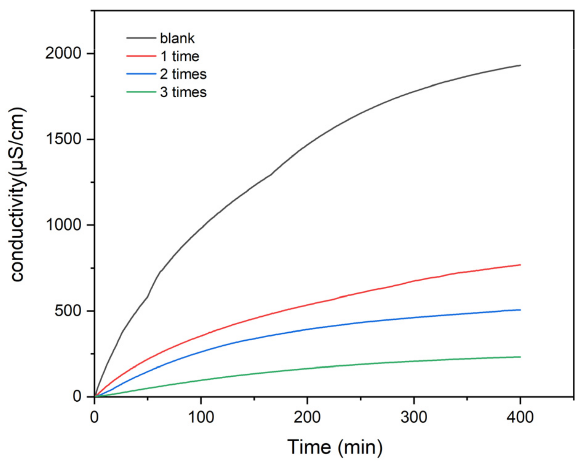
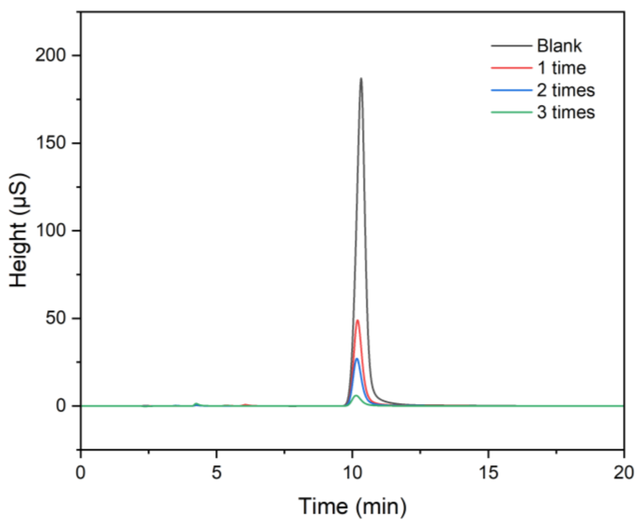
| Specimens with Different Treatment Numbers | Type | Area (S*min) | Height (S) | Amount (mg/L) |
|---|---|---|---|---|
| 0 (blank) | BMB | 75.083 | 186.735 | 65.413 |
| 1 | BMB | 19.226 | 48.921 | 16.783 |
| 2 | BMB | 10.940 | 27.031 | 9.569 |
| 3 | BMB | 2.575 | 5.929 | 2.287 |
| Specimens with Different Treatment Numbers | L* | a* | b* | ΔL* | Δa* | Δb* | ΔE* |
|---|---|---|---|---|---|---|---|
| 0 (blank) | 38.98 | 0.99 | 3.90 | - | - | - | - |
| 1 | 39.14 | 0.88 | 3.34 | 0.16 | −0.11 | −0.56 | 0.59 |
| 2 | 38.65 | 0.92 | 3.28 | −0.33 | −0.07 | −0.62 | 0.71 |
| 3 | 39.18 | 0.78 | 2.82 | 0.20 | −0.21 | −1.08 | 1.12 |
Publisher’s Note: MDPI stays neutral with regard to jurisdictional claims in published maps and institutional affiliations. |
© 2022 by the authors. Licensee MDPI, Basel, Switzerland. This article is an open access article distributed under the terms and conditions of the Creative Commons Attribution (CC BY) license (https://creativecommons.org/licenses/by/4.0/).
Share and Cite
Lu, R.; He, L.; Li, T.; Yang, F.; Liu, Y.; Zhang, K.; Chen, X. A Novel Protection Method for Carbonate Stone Artifacts with Gypsum Weathering Crusts. Coatings 2022, 12, 1793. https://doi.org/10.3390/coatings12111793
Lu R, He L, Li T, Yang F, Liu Y, Zhang K, Chen X. A Novel Protection Method for Carbonate Stone Artifacts with Gypsum Weathering Crusts. Coatings. 2022; 12(11):1793. https://doi.org/10.3390/coatings12111793
Chicago/Turabian StyleLu, Ruicong, Lu He, Ting Li, Fuwei Yang, Yan Liu, Kun Zhang, and Xinnan Chen. 2022. "A Novel Protection Method for Carbonate Stone Artifacts with Gypsum Weathering Crusts" Coatings 12, no. 11: 1793. https://doi.org/10.3390/coatings12111793