Article

Plasma–Solution Junction for the Formation of Carbon Material

1
Department of Chemical Systems Engineering, Graduate School of Engineering, Nagoya University, Furo-cho, Chikusa-ku, Nagoya 464-8603, Japan
2
Japan Science and Technology Corporation (JST), Strategic International Collaborative Research Program (SICORP), Furo-cho, Chikusa-ku, Nagoya 464-8603, Japan
3
Conjoint Research Laboratory in Nagoya University, Shinshu University, Furo-cho, Chikusa-ku, Nagoya 464-8603, Japan
4
Japan Science and Technology Corporation (JST), Open Innovation Platform with Enterprises, Research Institute and Academia (OPERA), Furo-cho, Chikusa-ku, Nagoya 464-8603, Japan
*
Author to whom correspondence should be addressed.
Coatings 2022, 12(11), 1607; https://doi.org/10.3390/coatings12111607
Submission received: 28 September 2022 / Revised: 17 October 2022 / Accepted: 20 October 2022 / Published: 22 October 2022
(This article belongs to the Special Issue Science and Engineering of Coating)

Abstract

:
The solution plasma process (SPP) can provide a low-temperature reaction field, leading to an effective synthesis of N-doped graphene with a high N content and well-structured planar structure. However, the interactions at the plasma–solution interface have not been well understood; therefore, it needs to be urgently explored to achieve the modulation of the SPP. Here, to address the knowledge gap, we experimentally determined the physical parameters of the spital distribution in the plasma phase, plasma–gas phase, and gas–liquid phase of the SPP by the Langmuir probe system with modification. Based on the assumption that plasma can act similarly to semiconductors with the Fermi level above the vacuum level, an energy band diagram of the plasma–solution junction could be proposed for the first time. It was observed that the Fermi level of the organic molecule could determine the magnitude of electron temperature in plasma, i.e., benzene produced the highest electron temperature, followed by phenol, toluene, and aniline. Finally, we found that the electron temperature at the interface could induce quenching, leading to the formation of multilayer large-size-domain carbon products. It provided significant evidence for achieving nonequilibrium plasma modulation of carbon nanomaterial synthesis.

Graphical Abstract

1. Introduction

The 2D carbon nanomaterials endowed with high conductivity, planar structure, large surface area, tunable electrical properties, transparency, and adjustable electron density have promoted them as promising electronic contact materials. They have been used in fuel cells, lithium-ion batteries, solar cells, sensors, superconductors, and so on [1,2,3,4,5,6,7]. The properties of carbon materials can be modulated by introducing heteroatoms to obtain a band gap. Nitrogen (N), which has an atomic size similar to carbon (C), is an excellent candidate and can provide a hole acceptor and electron donors in the carbon structure [8]. This demonstrates that N-doped graphene possesses outstanding semiconductor characteristics and significantly increases its potential for application in electronic devices [9,10]. In the last decade, heteroatom-doped graphene with different characteristics was obtained by using chemical vapor deposition (CVD) [11], a high-energy electrothermal process [12], and a template-based process [13]. However, it is difficult to improve the doping ratio while maintaining a planar structure for sufficient conductivity, as the bond between heteroatoms and carbon can break in the process of dehydrogenation at high temperatures [14,15]. Numerous reports have shown that the solution plasma process (SPP) is a successful route to obtain N-doped carbon with high N content at a low-temperature reaction field [16,17,18,19,20,21,22,23,24,25,26,27,28,29,30,31,32,33,34,35,36,37]. The SPP with a pin-to-pin electrode is a typical configuration and various precursors, e.g., cyclic or chain structure, of the raw molecules have been applied for the synthesis of different products [20]. The SPP can induce several unique reactions at the interfaces (e.g., plasma–liquid and gas–plasma), due to electric discharge, which causes a dielectric breakdown of the liquid similar to an “opening zipper”. This phenomenon results in the generation of strong shock waves, ultraviolet, various active radicals, and high-energy electrons [38,39,40]. In addition, the SPP has the advantages of fast chemical reactions with a simple experimental system, inexpensiveness, short-time operation, and easy recovery of products at the same time [18].
For the SPP, which is a nonequilibrium plasma process, in the interface where the plasma contacts with the liquid, the solution also acts as an electrode. There are secondary electron emissions from the molecules being ionized by collisions, i.e., γ effect [32]. These cations, produced by the collisional ionization of the molecules, play the role of elemental components in the subsequent polymerization and carbonization processes to form carbon products. It was previously proposed that the interaction between plasma and solution determines the plasma’s properties and the chemical reactions based on electron transfer [37]. In the field of nonequilibrium plasmas in solution, various reports have shown that plasma with different properties greatly influences the formation of carbon products, for example, low-discharge-current plasma could promote the substitution of C atoms with N atoms in the carbon framework without altering the π-bonded configuration [38,39,41,42]. However, there is a serious lack of existing work that can address the phenomena at the plasma–solution interfaces. Therefore, it is difficult to provide a clear explanation of the mechanism and kinetics.
In the field of classical semiconductor physics as well as electrical engineering, the existence of heterojunctions between different phases in the same system, such as the p–n junction, successfully explains the kinetic transfer processes of electrons and holes. It is noteworthy that electrons and ions in plasmas have common characteristics with those in semiconductors. For the nonequilibrium plasma, the electron temperature is much higher than the ion temperature; thus, the electrons are considered as free moving particles, while the velocities of the ions are indeed much lower than those of the electrons due to the significant difference in mass [43]. Arumugam et al. tried to propose the concept of energy band diagrams for the case of the plasma–metal junction [44]. Inspired by these studies, the presence of a plasma–solution junction in the SPP reaction field was investigated for the first time. The difference between solution and metal is in the bonding configuration. The metal has metal bonds, leading to electrons being free to move. The solution, on the other hand, contains covalent or ionic bonds, resulting in electrons bounded to specific orbitals within the molecule. Thus, the solution has a specific band gap. The interpretation of a junction between the plasma and solution is more complicated. Determining the Fermi energy level of the plasma is challenging. In this study, the determination of Fermi energy levels in different regions was effectively achieved by the probe method system with modification to suit the plasma in solution.
A Langmuir probe is a well-known method, which is generally used to measure low-density plasma parameters at the local location of the probe [42,44,45,46,47,48]. This method has the advantages of low cost, easy resolution, and relatively simple device, compared to other methods such as optical emission spectroscopy (OES), laser-induced fluorescence, laser interferometry, and laser scattering [44,46,49,50]. However, in the case of the SPP, the mean free path λ of electrons undergoes a certain degree of decrease at room temperature and atmospheric pressure, compared with low-pressure plasma ( λ = k B T 2 π d 2 P , where k B , T , d , and P are the Boltzmann constant, temperature, diameter of molecules, and pressure, respectively [51]). High-density plasma is generated around the probe, and the sheath layer on the surface becomes thicker [42,47]. Therefore, the relationship between the probe size and the Debye shielding is a key factor limiting the application of the probe method to this thermal nonequilibrium plasma diagnosis. Kaneko et al. made a great and pioneering attempt to apply the probe method of measurement in liquid plasma, by the designed electrostatic probe, and the potential information was explained [50]. This method differs from the Langmuir probe system because it has no variation in the externally applied bias voltage and, thus, it is not possible to obtain more information about the temperature of the SPP. In order to obtain accurate plasma characteristics using the Langmuir probe method, based on the assumption that the electrons in the plasma have a Maxwell distribution, we need to overcome the possible effects of the probe’s large electron current on the plasma [47,48]. Therefore, a more suitable theoretical model is needed to diagnose and calculate the Langmuir probe I–V signal.
In this study, a modified Langmuir probe method was proposed to measure physical parameters at different locations in the SPP with a pin-to-pin electrode structure system. The response characteristics of the probe were measured in four solutions of benzene, toluene, phenol, and aniline with different functional groups. The modified model was applied to calculate the plasma potential, electron temperature, and other parameters of the plasma in solution with a high collision rate and high particle density. The results were cross-validated using spatially resolved optical emission spectroscopy (SROES), and the concept of the plasma–solution junction was proposed to explain the physical mechanism of action in this study by assuming plasma as a new understanding of semiconductor materials. Furthermore, the characterizations of the obtained products were conducted well by using X-ray diffraction (XRD), Raman spectroscopy, X-ray photoelectron spectroscopy (XPS), and scanning electron microscopy (SEM) to confirm their properties and quality. The plasma–solution junction proposed in this study could provide direct evidence of the plasma–liquid interface. It showed the notable relationship between plasma and carbon products, and is also guidance information for physicists, chemists, and engineers involved in the application of the solution plasma field.

2. Experimental Procedures

2.1. Experimental Setup

Figure 1a shows the schematic diagram of the SPP setup, and the schematic illustration of the experimental process is shown in Figure S1, including solution plasma, filtration cleaning, separation drying, and characterization of carbon products. The pin-to-pin electrode system was constructed to carry out intensive investigation analysis by using a pair of tungsten electrodes (W, φ 1.0 mm, Nilaco Co., Tokyo, Japan) covered with ceramic tubes and placed 1 mm apart. Benzene (C6H6, >99.5%, Kanto Chemical Co., Inc., Tokyo, Japan), toluene (C6H5CH3, >99.5%, Kanto Chemical Co., Inc., Tokyo, Japan), aniline (C6H5NH2, >99.5%, Kanto Chemical Co., Inc., Tokyo, Japan), and phenol (C6H5OH, >99.5%, Kanto Chemical Co., Inc., Tokyo, Japan) were used sequentially as the solutions for the experiments. The experiment was carried out in a glass container with a volume of 200 mL, and the electrode was placed at the center of the glass reactor. The SPP was driven by a bipolar pulsed power supply (MPS-R06K01C-WP1-6CH, Kurita, Kyoto, Japan) under the conditions of a voltage of 1.6 kV, pulse width of 1.5 µs, and frequency of 11 kHz. The entire discharge process was carried out with a magnetic stirrer. After the SPP synthesis, the obtained black powder products by vacuum filtration were repeatedly washed to remove the residual organic components by using ethanol and dried at 80 °C for 8 h in an oven.

2.2. Langmuir Probe Measurement and SROES Measurement

The modified Langmuir probe measurement system consisted of two parts, i.e., the probe and the filter. The probe consisted of a tungsten wire (radius rp = 50 μm) covered with a 0.4 mm inner-diameter ceramic tube. An intravenous injection needle was used as a fixed holder between the ceramic tube and the tungsten to prevent the probe from contacting the ceramic surface and creating interference with charge injection. An exposed tip of 0.1 mm was left uncovered by the ceramic tube in the front section of the probe for the detection of plasma. The probe was measured by applying a DC bias voltage of −50 V–50 V, and the current flowing through the probe was recorded sequentially. For the same DC voltage value, collection and calculation of the average value for ten current data samples were performed to obtain accurate experimental results. The high-frequency noise from the power supply was filtered out by an RC low-pass filter. During the experiments, the response characteristics of the probe were measured at three locations, X1, X2, and X3, from the inside of the plasma to the interface of the solution, for each of the four solutions in turn. The voltage of the discharge part was measured by a high-voltage probe (P6015A, 1000×, 3.0 pF, 100 MΩ, Tektronix, Beaverton, OR, USA), and the discharge current and the Langmuir probe current were measured by current probes (PR30, Yokogawa, Tokyo, Japan). The voltage and current signals were finally displayed and recorded by a digital oscilloscope (DLM2024, 2.5 GS/s, 200 MHz, Yokogawa, Tokyo, Japan). To verify the accuracy of the experimental results, the generated species during the SPP discharge were revealed by using the enhanced SROES, as shown in Figure 1b. The SROES consisted of a pinhole with a size of 1 mm, three lenses (L1: 4 mm of diameter, 6 mm of focal length; L2: 4 mm of diameter, 12 mm of focal length; L3: 4 mm of diameter, 5 mm of focal length), optical fiber, and an optical spectrometer (USB2000+, Ocean Optics, Winter Park, FL, USA) with the wavelength range of 200–900 nm. This method could overcome the problem of unidirectional spectroscopic detection posed by conventional ways using fibers alone, and it collected light from the entire plasma region for analyzing the composition of the spectrum.

2.3. Product Characterization

Furthermore, we conducted the structural characterization of the carbon samples synthesized by benzene, toluene, aniline, and phenol using X-ray diffraction (XRD, Smartlab, Rigaku Co., Tokyo, Japan. Cu Kα X-rays source), a Raman spectra microscope (Raman, Leica DM 2500 M Ren (RL/TL), Renishaw Plc, England. 532.5 laser wavelength), X-ray photoelectron spectroscopy (XPS, ULVAC-PHI 5000 VERSAPROBE-II, Inc., Tokyo, Japan. Mg Kα X-ray source), and scanning electron microscopy (SEM, JSM-7610 F, JEOL Ltd., Tokyo, Japan, at the accelerating voltage of 10 kV).

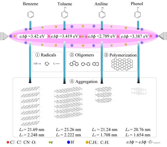
3. Results and Discussion

An important criterion for the Langmuir probe system to appropriately detect plasma in solution is to determine whether the presence of a metal probe causes serious perturbations to the discharge [47,52,53]. Therefore, the investigation of the electric field distribution before the occurrence of breakdown discharge is the theoretical basis for applying the Langmuir probe to discharges in organic solution. Here, the electric field of the discharge geometry with and without the Langmuir probe under the same excitation was analyzed using Ansys Maxwell software based on the finite element electric field simulation method, as shown in Figure 2a. In the electrode system without the probe structure, the region of a strong electric field over 2 × 10⁷ V/m was mainly concentrated at the tips of the two metal electrodes. The electric field distribution in the central region was stable at 1×10⁷ V/m. When a probe with ceramic protected was introduced, the charge accumulated on the surface of the ceramic material could potentially change the local potential distribution. Therefore, the spatial distribution of the electric field strength slight decreased. It is important to note that the electric field did not change sharply at the location where the probe was presented in the central region, as shown in Figure S2. This shows that the probe had a negligible effect on the development and evolution of the discharge process. After the plasma generation, the effect of Debye shielding could rapidly establish a localized region of a positively charged sheath layer surrounding the probe when such a solid material was introduced into the plasma system [47,54]. The plasma appeared electrically neutral in the scale of space larger than the Debye length. In other words, the radius of the probe should be smaller than the Debye length. Therefore, we calculated the Debye length of the SPP based on bubble theory according to λ D S P = ε 0 k B T e e 2 N e , where ε 0 , k B , N e , and T e are the vacuum dielectric constant, Boltzmann constant, plasma electron density, and plasma electron temperature, respectively [54,55]. By employing the parameter ranges from previous studies [25,26,56,57,58,59], the result showed that 69 λ D S P 154 µm. It could be seen that the probe with rp = 50 µm used in this work satisfied the theoretical requirement of detection accuracy. The above analysis provided the physical basis for the feasibility of the probe method in the SPP.
Figure 2b shows a typical discharge voltage and current waveform in benzene solution with 1.6 kV excitation conditions and the Langmuir probe voltage and current response waveform detected at position X3. We could observe the presence of only a noise component belonging to the positive plasma during probe detection, although the discharge current proved that the plasma was formed normally at this period. These similar phenomena were also reported in previous studies [49,55,56]. Probe current variation with the bias voltage could be detected normally in the negative discharge plasma. A comparison with the Langmuir probe signals from vacuum plasma, gas–phase plasma, and space physical plasma showed that the probe signals of the SPP had a consistent instantaneous variation characteristic [48,54,57]. This demonstrated that the proposed Langmuir probe approach in this research could effectively absorb electron and ion currents.
Based on the detected waveform, we further obtained the standard response characteristics of the Langmuir probe in the negative-polarity plasma corresponding to X1 (plasma phase), X2 (plasma–gas phase), and X3 (gas-liquid phase) of benzene, toluene, phenol, and aniline, as shown in Figure 2c–f. Furthermore, the primary differential dI/dV was also calculated. As the bias voltage gradually changed from −50 V to 50 V, the probe response went through the ion saturation current region (probe potential Vp < suspension potential ϕf (i.e., Ip = 0) < plasma potential ϕp), the transition region (suspension potential ϕf (i.e., Ip = 0) < probe potential Vp < plasma potential ϕp), and the electron saturation current region (plasma potential ϕp < probe potential Vp). As the mass of the electron is much smaller than the mass of the ion, the electron saturation current is much larger than the ion saturation current [58]. In addition, the currents collected by the probe from X1 to X3 decreased sequentially, which was the reason for the reduction in the Debye length due to the lower electron temperature and the higher electron density [59,60]. The current signal did not fluctuate up or down by more than ten percent, which was a highly sensitive response of the probe signal. The electric field of Debye shielding could be rapidly established over a much smaller area; thus, the capability of the probe to intercept electrons was reduced. The plasma potential ϕp was obtained from the minimum value of dI/dV as in Table 1. However, as mentioned earlier, in the SPP, there were a large number of collisional ionization processes inside the thick sheath, which rapidly built up on the probe surface. It significantly affected the effectiveness of the probe measurements [48]. Therefore, existing theories are difficult to be fully applicable to different experimental conditions, especially for a reasonable interpretation of the probe V–I curve. In other words, it was difficult to achieve accurate plasma parameter measurements based on traditional collision-free computational models. This is the main bottleneck that limits the application of Langmuir probes to collision-rich plasmas such as atmospheric pressure discharge or arc discharge. In this case, the researchers summarized several different criteria based on which to calculate the plasma parameters of the collision-rich probe signal [60,61]. In this work, combining the above factors, a modified computational model similar to Smy et al. as shown in the supporting information (Equation (1)–(5)) was applied to calculate the plasma characteristic parameters [48,62,63,64]. The effect of the excitation ionization process caused by the presence of the sheath layer was accommodated [48,62,63,64]. The obtained plasma parameters (Ne, Te) of different phases and different plasma are shown in Table 1.
According to the calculation results in Table 1, we plotted the mapping structure of the temperature spatial distribution of plasma in four solutions, as shown in Figure S3. It could be seen that there was an obvious interface stratification from the plasma to the solution. Such a temperature spatial feature had not been shown in similar studies in the past. The electron temperature was maximum in the plasma phase, and a steep gradient drop occurred in the plasma–gas phase. Radicals, including electrons, ions, and photons, were inactivated in a process similar to quenching. This characteristic guaranteed that the particles had enough energy to undergo carbonization and dehydrogenation processes. This could be the primary reason that the SPP enabled a wide range of applications for different kinds of allotropic carbon nanomaterials (e.g., amorphous carbon, graphite, graphene, and heteroatom-doped carbon). It was worth noting that owing to the clear grasp of electron energy distribution information, we could further realize the tuning of the products based on the temperature characteristics (e.g., higher electron temperature enhanced the reaction rate and sped up the polymerization of the carbon products process, resulting in reducing the sp3/sp2 ratio and enhancing the degree of crystallization of the products). The variation in the properties of the organics was brought by the introduction of different functional groups and the linkage to the benzene ring was an important reason for the difference in plasma electron temperature. Thus, we analyzed the variation in temperature with relative electronegativity (functional group element/C) at different interfaces of the plasma, as shown in Figure S4 in the plasma phase. The electron temperature gradually decreased with increasing electronegativity of the functional group elements, while in the plasma–gas phase and gas–liquid phase, the electron temperature appeared to have an abnormal increase in the solution plasma generated by the phenol.
The emission peaks obtained by SROES from four solutions, as shown in Figure 3a–d, exhibited Swan bands, representing the decomposition of the benzene ring structure into C2 radicals and Hα emission, indicating the abstracted hydrogen atoms [32,37]. The CN radical peaks also appeared, which could refer to the dissociation of the C6H5-NH2 bond of aniline [16]. The ratios between the characteristic peaks of the emitted species were almost the same, which implied that there was not a significant difference in the temperature of the plasma in the four dielectric solutions. Based on the continuum evident in the spectral lines in the measurement range of 340–800 nm (as shown by the red dashed line of the fitted blackbody function) and applying the following function, I ( λ ) = 8 π h c λ 5 ( 1 e hc / λ k B T b 1 ) , where I(λ), h, c, λ, and kB are the emission intensity, Planck’s constant, speed of light, wavelength, and Boltzmann constant [65], respectively, we further calculated the averaged blackbody temperature Tb. Tb should be the lower limit of the electron temperature by assuming the condition of local thermal equilibrium (LTE) based on Plank’s theory, as shown in Table 1.
Due to the strong and chaotic electric field of up to 107 V/m generated between the small gaps of the electrodes by the bipolar pulse voltage, further splitting or shifting of the emission spectrum occurred. This corresponds to the excited particles having a strong dipole moment, and thus the emission peak of the substance produced a significant Stark broadening [65,66,67]. The emission peak of ·Hα did not show any redshift or blueshift, which implied that there was no significant change in pressure during the electron avalanche development (α effect), as well as the secondary electron emission (γ effect) of the SPP. Thus, the van der Waals broadening with significant effects did not appear in the present study. Based on the above analysis, we fitted the ·Hα emission peak using the Lorentz function and calculated its full-width at half-maxima Δ λ F W H M , according to the equation λ F W H M = 8.33 × 10 3 ( N e s t a r k 10 20 ) 2 / 3 [68], and the plasma electron density was derived, as shown in Table 1. The obtained results of electron density were maintained in the order of 1024 m−3. In addition, the values of the electron density obtained in this study are consistent with the previous work, which could verify the accuracy of the probe method proposed in this study.
To further analyze the influence of plasma characteristics on the formation of the obtained carbon products, we characterized the structural features of the products. The XRD patterns are shown in Figure 4a. Two main characteristic broad peaks, corresponding to the (002) and (111) carbon planes, could be observed at around 23.5° and 43.5°, respectively [37]. The inter-planar spacing d and crystallite size (edge planes) Lc of the products were calculated by employing the Bragg equation (d = λ/2sinθ) and the Scherrer equation (Lc = 0.91 × λ/β/cosθ), where λ is the wavelength of X-rays, and θ and β are the radius values of the half diffraction angle and the FWHM of the peak, respectively [69]. The number of layers n was calculated by the equation n = Lc/d. According to Table 2, the results showed that the samples synthesized from aniline and phenol had a decreasing crystallite size and number of layers. The additional peaks (36° and 42°) were found from the tungsten-carbide (WC1−x), which could be possibly generated by the sputtering of the tungsten electrodes due to the γ effect, leading to the ion bombardment [37]. As shown in Table 2, for the calculation of IWC-36°/IC002, the content of W as impurities being turbid is mainly dependent on the electron temperature of the plasma phase.
Raman spectroscopic measurements were also employed to carry out the degree of detailed structural disorder from the samples, as shown in Figure 4b. The characteristic peaks were observed, including the D band (~1340 cm−1), G band (~1580 cm−1), and 2D band (~2710 cm−1) [17,18,37]. The pronounced G peaks are attributed to the E2g mode, which represents the stretching of carbon bonds from a pair of sp2 lattice carbon atoms. The D peak corresponds to the A1g mode, which represents a disbalance of symmetry from a disordered structure (defect, lattice distortion, bond length disordered, and impurity) in sp2-hybridized carbon [37]. In addition, the ratios of D and G peak intensity (ID/IG), which refer to the degree of graphitization, disorder, crystallinity, and crystallite size of the in-plane La (La= (2.4 × 10⁻¹⁰)λ4raman(ID/IG)−1), were also calculated [32], as shown in Table 2. The ID/IG increased with the presence of heteroatomic functional groups (-NH2, -OH) in the chemical structure of the organic solution. The product obtained from toluene exhibited the largest La (23.26 nm), followed by samples of benzene, aniline, and phenol, in this order. It was possible that the -CH3 conjugated to the benzene ring in toluene resulted in the in-plane sp2 structure of carbon products obtained by the SPP in toluene.
The elements and the bonding state presented in the synthesized products from the four precursors were performed by XPS measurement. The surveyed spectra as shown in Figure 4c were composed of a C 1 s peak (284.5 eV) and an O 1 s peak (532.5 eV). The N 1 s peak (399.5 eV) was shown specifically in the product of aniline due to its -NH2 functional group [18]. From the quantitative analysis of the elemental composition, the contents of C, O, and N are shown in Table S1, respectively. The presence of O in the products was due to the oxidation of the surface carbon layer during the preparation or drying process in an air environment [20,21,22]. N was found to be doped into the carbon product obtained from the SPP in aniline. The N content was around 3.53%. The content of pyrrolic N (399.7 eV) was found to be dominant, at 61.70%, according to the deconvolution of N peaks. Meanwhile, other N configurations were revealed as follows: pyridinic N (398.7 eV), 13.73%, and quaternary N (401.2 eV), 25.57%.
The morphology of carbon was further investigated by the SEM, as shown in Figure S5. It was found that all carbon products exhibited agglomerates of uniform nanoscale carbon particles with the three-dimensionally interconnected structure. The different precursors of the SPP also resulted in the change in the microporous morphology.
The obtained results in this work indicated that different precursors led to plasmas with different characteristics, which further led to the formation of carbon products with different properties. The explanation based on molecular orbital theory and the assumption that plasma is a special semiconductor material was proposed to explain this phenomenon that occurred in this work. It was proposed in previous studies that the characteristics of the electron–hole properties in semiconductor materials and electron–ion properties in plasma were similar [44]. Therefore, the highest energy level that could be filled by electrons in plasma was believed to be the Fermi level, i.e., the plasma potential that can be equated to the plasma Fermi level (eϕp = EF) [44]. As the Fermi level characterizes the consumed energy of the electron to be moved to the vacuum level, in the case of plasma, it needs to release a certain amount of energy to move electrons to the vacuum level. Therefore, the Fermi level of plasma should be above the vacuum level. When the plasma is in contact with the solution, the transfer of electrons should occur as the system tends to equilibrium, which is the reason that different solutions can cause the plasma to have different temperature characteristics and carbon product properties [44,68,69]. As shown in Figure 5a, the energy diagram was produced to indicate the charge transfer process in different solutions. In this diagram, the E0NHE = −4.44 eV was obtained by corresponding the hydrated protons per unit activity to the electrons in a vacuum near the surface of the solution [17]. It can unify the absolute scale (abs) in the field of describing metals or semiconductors with the electrochemical scale (relative scale: normal hydrogen electrode (NHE)) in the liquid redox process. The process of Fermi level alignment is also a process of electron energy transfer. The energy level diagram also shows the Fermi energy level of the metal probe (−4.55 eV), the plasma potential, i.e., its Fermi energy level (ϕp), and the HOMO (i.e., ionization potential) and LUMO (electron affinity) parameters of the four solution molecules [17,40,67,69]. In the contact zone of plasma–metal/solution, the electrons of the higher-energy states in the plasma could automatically flow to the lower energy levels in the metal or solution molecules. The departure of electrons in the plasma region possibly led to the formation of a positively charged region boundary, and the injection of electrons in the metal or solution region might result in the formation of a negatively charged region boundary. As a result, the accumulation of charges rapidly built up an internal electric field in the region of the plasma-metal/solution and blocked the infinite flow of electrons, leading to the escape of positive charges. Accordingly, this might cause the formation of the plasma–metal junction and the plasma–solution junction. In the junction region, the Fermi level proceeded in the opposite direction of the built-in electric field. The junction structure was consistent with the sheath structure in classical plasma theory, and this feature was the positive and negative double-layer structure, which was observed by SROES.
The reaction path of the formation of carbon products from liquid-phase molecules (benzene, toluene, phenol, and aniline) is shown in Figure 5b. Based on this plasma diagnosis of the SPP, the distribution of heavy reactive particles was found to be a key. As the result of OES (Figure 3), C2 and H radicals were the main component in the plasma phase of the four precursors. In addition, CN radicals were generated from aniline, and O radicals were generated from phenol [32]. C2 radicals were further converted into C22− or C24− forms after the adsorption of electrons enriched in the plasma. A O radical could be easily attracted by electrons and undergo the transformation process, O → O2 → O2−, due to the large electronegativity of O [50]. As O, C, and other composite particles have larger masses, they were maintained inside the plasma, and the lighter particles, such as H+ particles, could transport to the boundary layer between the plasma and the solution. In the boundary layer between the plasma and the solution, the solution molecules could act as liquid electrodes and provide a large number of secondary electrons for the maintenance of the discharge, resulting in the generation of positive ions such as C6H5+ [37]. At the same time, C2 radicals at the boundary were susceptible to hydrogenation processes to produce C2Hx+. The above analysis showed that there were negative charge layers, which could be dominated by C22−, C24−, CN, and O2, in the inner region of the plasma and positive charge layers, which could be dominated by C6H5+, C2Hx+, and H+, at the edges of the plasma, which constituted the electrical double layer inside the plasma. The electrical double layer promoted various types of radicals (CN, C6H5, C6, C6 + C6, etc.) and further went through the process of oligomerization, polymerization, and aggregation to assemble into a graphite framework. In addition, the significant formation of CN· ensured the generation of uniform heteroatom-doped carbon structures.
In Figure 5c, the ∆Te (between the plasma and liquid phases), Lc (edge crystallite size of carbon), and La (in-plane size of carbon) with the potential difference ∆ϕ between the plasma and the solution Fermi energy were used to illustrate the correlation of plasma and precursors. It was found that as the ∆ϕ increased, ∆Te increased. The larger potential difference of the plasma and solution Fermi energy level could induce more electrons to transfer to the solution molecules. More energy of the plasma was transferred to its surrounding medium. In other words, the larger potential difference could refer to the lower HOMO level of the solution molecule and, thus, the lower ionization potential. Under the condition of the same input energy, the higher temperature of the plasma was obtained. In the plasma phase, the phenol plasma had a distinct anomaly. The reason for this phenomenon might be the introduction of the typical electronegative element O. The electron energy needed an additional transfer to the cross-section of larger O2, O2, etc., particles compared to the solution plasma such as benzene discharge, and, thus, its electron temperature was lowest [50].
The Lc and La of carbon products also increased gradually with increasing ∆ϕ of phenol, aniline, toluene, and benzene, and were positively correlated with ∆Te. As a typical nonequilibrium plasma was generated in this study, a large temperature gradient difference was generated in the plasma surrounded by the solution, and substances such as ions, radicals, electrons, and photons were inactivated in this quenching-like process and rich chemical reactions were generated [32,37]. It was obvious that the ∆Te induced by potential difference ∆ϕ was the main factor for the effect of the carbon framework. The major factor in the formation of the above phenomenon could be the mobility of the active species in the SPP, which might be greatly promoted during a greater degree of quenching, and, thus, the number of nucleation sites formed greatly increased, increasing the layer number of the graphene structure. The carbon product formed by using aniline as a precursor in this study had the structural characteristics of few layers and small size. In addition, the lower electron temperature ensured the formation of a proper carbon framework while ensuring a sufficient N-doping ratio.

4. Conclusions

By using the modified Langmuir probe system and the calculation model for collision-rich sheath structures, we investigated the spatial characteristics in four plasmas in solution, including benzene, toluene, phenol, and aniline. Simultaneously, the plasma–solution junction was obtained by assuming that plasma in solution could act as a semiconductor material and the plasma potential could be considered as the Fermi level. The electron energy was mainly transferred to the molecules with lower Fermi levels at the interface between plasma and solution. When the molecule had a lower HOMO energy, its ionization could easily occur. By further analyzing the structural characteristics of carbon products, we found that the temperature difference at the interface between the plasma phase and the plasma solution was what determined the carbon structure, and the larger the temperature gradient difference, the easier it was to form the larger edge and in-plane crystallite size. To be specific, the product obtained from toluene exhibited the largest crystallite size of the in-plane basal plane; by using aniline as a precursor, with the lower electron temperature, the obtained product had the structural characteristics of few layers and small size, with a sufficient N-doping ratio. The obtained evidence could be used to explain the phenomena that occurred in the SPP under organic solutions and their relationship to the formation of carbon. Consequently, it may lead to further development of the reaction field by plasma in solution to produce novel materials.

Supplementary Materials

The following supporting information can be downloaded at: https://www.mdpi.com/article/10.3390/coatings12111607/s1, Figure S1: Schematic illustration of solution plasma process experiment, including solution plasma, filtration cleaning, separation drying, and characterization of carbon products; Figure S2: The electric field strength with or without the Langmuir probe system; Figure S3: Temperature structure characteristics of the negative-polar-discharge plasma in four solutions of (a) benzene, (b) toluene, (c) phenol, and (d) aniline mapping; Figure S4: Variation in electron temperature with relative electronegativity ((C = 2.55, N = 3.04, O = 3.44)/(C = 2.55)) at different interfaces of solution plasma. Figure S5: SEM image carbon products of (a) benzene, (b) toluene, (c) aniline, and (d) phenol. Table S1: Bulk and surface elemental composition of products from benzene, toluene, phenol, and aniline obtained from XPS measurement; nitrogen bonding composition of product from aniline.

Author Contributions

J.N. and N.S. conceived the idea; J.N. analyzed the data; X.W. conducted partial characterization; J.N. wrote the manuscript; N.S., C.C. and Y.S. gave advice and revised the manuscript. All authors have read and agreed to the published version of the manuscript.

Funding

This research was financially supported by the Open Innovation Platform with Enterprises, Research Institute and Academia (OPERA, Grant JPMJOP1843), and the Strategic International Collaborative Research Program (SICORP, Grant JPMJSC18H1) of the Japan Science and Technology Agency (JST).

Institutional Review Board Statement

Not applicable.

Informed Consent Statement

Not applicable.

Data Availability Statement

Data sharing in the article is not applicable.

Acknowledgments

J.N. thanks the China Scholarship Council for financial support through No. 201909110064.

Conflicts of Interest

The authors declare no conflict of interest.

References

  1. Sibul, R.; Kibena-Põldsepp, E.; Ratso, S.; Kook, M.; Sougrati, M.T.; Käärik, M.; Merisalu, M.; Aruväli, J.; Paiste, P.; Treshchalov, A.; et al. Iron- and nitrogen-doped graphene-based catalysts for fuel cell applications. Chem. ElectroChem. 2020, 7, 1739–1747. [Google Scholar]
  2. Han, M.; Lin, Z.; Ji, X.; Mu, Y.; Li, J.; Yu, J. Growth of flexible and porous surface layers of vertical graphene sheets for accommodating huge volume change of silicon in lithium-ion battery anodes. Mater. Today Energy 2020, 17, 100445. [Google Scholar] [CrossRef]
  3. Shaari, N.; Kamarudin, S.K.; Bahru, R. Carbon and graphene quantum dots in fuel cell application: An overview. Int. J. Energy Res. 2020, 45, 1396–1424. [Google Scholar] [CrossRef]
  4. Basu, K.; Selopal, G.S.; Mohammadnezad, M.; Akilimali, R.; Wang, Z.M.; Zhao, H.; Vetrone, F.; Rosei, F. Hybrid graphene/metal oxide anodes for efficient and stable dye sensitized solar cell. Electrochim. Acta 2020, 349, 136409. [Google Scholar] [CrossRef]
  5. Mo, R.; Tan, X.; Li, F.; Tao, R.; Xu, J.; Kong, D.; Wang, Z.; Xu, B.; Wang, X.; Wang, C.; et al. Tin-graphene tubes as anodes for lithium-ion batteries with high volumetric and gravimetric energy densities. Nat. Commun. 2020, 11, 1374. [Google Scholar] [CrossRef] [Green Version]
  6. Wu, Q.; Qiao, Y.; Guo, R.; Naveed, S.; Hirtz, T.; Li, X.; Fu, Y.; Wei, Y.; Deng, G.; Yang, Y.; et al. Triode-mimicking graphene pressure sensor with positive resistance variation for physiology and motion monitoring. ACS Nano 2020, 14, 10104–10114. [Google Scholar] [CrossRef]
  7. Park, J.M.; Cao, Y.; Watanabe, K.; Taniguchi, T.; Jarillo-Herrero, P. Tunable strongly coupled superconductivity in magic-angle twisted trilayer graphene. Nature 2021, 590, 249–255. [Google Scholar] [CrossRef]
  8. Guo, B.; Fang, L.; Zhang, B.; Gong, J.R. Graphene doping: A review. Insci. J. 2011, 1, 80–89. [Google Scholar] [CrossRef] [Green Version]
  9. Novodchuk, I.; Kayaharman, M.; Ibrahim, K.; Al-Tuairqi, S.; Irannejad, M.; Abdel-Rahman, E.; Sanderson, J.; Bajcsy, M.; Yavuz, M. B/n co-doped graphene oxide gel with extremely-high mobility and ion/ioff for large-area field effect transistors. Carbon 2020, 158, 624–630. [Google Scholar] [CrossRef]
  10. Ye, S.; Luo, F.; Xu, T.; Zhang, P.; Shi, H.; Qin, S.; Wu, J.; He, C.; Ouyang, X.; Zhang, Q.; et al. Boosting the alkaline hydrogen evolution of ru nanoclusters anchored on b/n–doped graphene by accelerating water dissociation. Nano Energy 2020, 68, 104301. [Google Scholar] [CrossRef]
  11. Zan, R.; Altuntepe, A. Nitrogen doping of graphene by cvd. J. Mol. Struct. 2020, 1199, 127026. [Google Scholar] [CrossRef]
  12. Li, D.; Duan, X.; Sun, H.; Kang, J.; Zhang, H.; Tade, M.O.; Wang, S. Facile synthesis of nitrogen-doped graphene via low-temperature pyrolysis: The effects of precursors and annealing ambience on metal-free catalytic oxidation. Carbon 2017, 115, 649–658. [Google Scholar] [CrossRef] [Green Version]
  13. Wang, Y.; Huang, J.; Chen, X.; Wang, L.; Ye, Z. Powder metallurgy template growth of 3d n-doped graphene foam as binder-free cathode for high-performance lithium/sulfur battery. Carbon 2018, 137, 368–378. [Google Scholar] [CrossRef]
  14. Peyghan, A.A.; Beheshtian, J. Application of hexa-peri-hexabenzocoronene nanographene and its b, n, and bn doped forms in na-ion batteries: A density functional theory study. Thin Solid Films 2020, 704, 137979. [Google Scholar] [CrossRef]
  15. Yonezawa, T.; Čempel, D.; Nguyen, M.T. Microwave-induced plasma-in-liquid process for nanoparticle production. Bull. Chem. Soc. Jpn. 2018, 91, 1781–1798. [Google Scholar] [CrossRef]
  16. Bratescu, M.A.; Kim, K.; Saito, N. Quantitative spectrochemical analysis of solution plasma in aromatic molecules. Plasma Process. Polym. 2019, 16, e1900012. [Google Scholar] [CrossRef]
  17. Chae, S.; Bratescu, M.A.; Saito, N. Synthesis of few-layer graphene by peeling graphite flakes via electron exchange in solution plasma. J. Phys. Chem. C 2017, 121, 23793–23802. [Google Scholar] [CrossRef]
  18. Chae, S.; Panomsuwan, G.; Bratescu, M.A.; Teshima, K.; Saito, N. P-type doping of graphene with cationic nitrogen. ACS Appl. Nano Mater. 2019, 2, 1350–1355. [Google Scholar] [CrossRef]
  19. Heo, Y.K.; Lee, S.H.; Bratescu, M.A.; Kim, S.M.; Lee, G.J.; Saito, N. Generation of non-equilibrium condition in solution plasma discharge using low-pass filter circuit. Plasma Process. Polym. 2016, 14, 1600163. [Google Scholar] [CrossRef]
  20. Hyun, K.; Saito, N. The solution plasma process for heteroatom-carbon nanosheets: The role of precursors. Sci. Rep. 2017, 7, 3825. [Google Scholar] [CrossRef] [Green Version]
  21. Hyun, K.; Ueno, T.; Panomsuwan, G.; Li, O.L.; Saito, N. Heterocarbon nanosheets incorporating iron phthalocyanine for oxygen reduction reaction in both alkaline and acidic media. Phys. Chem. Chem. Phys. 2016, 18, 10856–10863. [Google Scholar] [CrossRef] [PubMed]
  22. Hyun, K.; Ueno, T.; Saito, N. Synthesis of nitrogen-containing carbon by solution plasma in aniline with high-repetition frequency discharges. Jpn. J. Appl. Phys. 2016, 55, 01AE18. [Google Scholar] [CrossRef]
  23. Islam, M.Z.; Watthanaphanit, A.; Chae, S.; Saito, N. Li–air battery and orr activity of nanocarbons produced with good synthesis rate by solution plasma process. Mater. Adv. 2021, 2, 2636–2641. [Google Scholar] [CrossRef]
  24. Jedsukontorn, T.; Ueno, T.; Saito, N.; Hunsom, M. Facile preparation of defective black tio2 through the solution plasma process: Effect of parametric changes for plasma discharge on its structural and optical properties. J. Alloy Compd. 2017, 726, 567–577. [Google Scholar] [CrossRef]
  25. Kim, D.W.; Li, O.L.; Saito, N. Enhancement of orr catalytic activity by multiple heteroatom-doped carbon materials. Phys. Chem. Chem. Phys 2015, 17, 407–413. [Google Scholar] [CrossRef]
  26. Kim, D.-w.; Li, O.L.; Pootawang, P.; Saito, N. Solution plasma synthesis process of tungsten carbide on n-doped carbon nanocomposite with enhanced catalytic orr activity and durability. RSC Adv. 2014, 4, 16813–16819. [Google Scholar] [CrossRef]
  27. Kim, K.; Chokradjaroen, C.; Saito, N. Solution plasma: New synthesis method of n-doped carbon dots as ultra-sensitive fluorescence detector for 2,4,6-trinitrophenol. Nano Express 2020, 1, 020043. [Google Scholar] [CrossRef]
  28. Lee, H.; Bratescu, M.A.; Ueno, T.; Saito, N. Solution plasma exfoliation of graphene flakes from graphite electrodes. RSC Adv. 2014, 4, 51758–51765. [Google Scholar] [CrossRef]
  29. Lee, H.; Ueno, T.; Saito, N. The effect of electrode gap distance on the synthesis of carbon materials by using solution plasma process. JOM 2015, 67, 2550–2556. [Google Scholar] [CrossRef]
  30. Lee, S.; Heo, Y.; Bratescu, M.A.; Ueno, T.; Saito, N. Solution plasma synthesis of a boron-carbon-nitrogen catalyst with a controllable bond structure. Phys. Chem. Chem. Phys. 2017, 19, 15264–15272. [Google Scholar] [CrossRef]
  31. Lee, S.; Saito, N. Enhancement of nitrogen self-doped nanocarbons electrocatalyst via tune-up solution plasma synthesis. RSC Adv. 2018, 8, 35503–35511. [Google Scholar] [CrossRef] [PubMed]
  32. Morishita, T.; Ueno, T.; Panomsuwan, G.; Hieda, J.; Yoshida, A.; Bratescu, M.A.; Saito, N. Fastest formation routes of nanocarbons in solution plasma processes. Sci. Rep. 2016, 6, 36880. [Google Scholar] [CrossRef] [Green Version]
  33. Panomsuwan, G.; Saito, N.; Ishizaki, T. Simple one-step synthesis of fluorine-doped carbon nanoparticles as potential alternative metal-free electrocatalysts for oxygen reduction reaction. J. Mater. Chem. A 2015, 3, 9972–9981. [Google Scholar] [CrossRef]
  34. Panomsuwan, G.; Saito, N.; Ishizaki, T. Nitrogen-doped carbon nanoparticles derived from acrylonitrile plasma for electrochemical oxygen reduction. Phys. Chem. Chem. Phys. 2015, 17, 6227–6232. [Google Scholar] [CrossRef]
  35. Panomsuwan, G.; Saito, N.; Ishizaki, T. Fe–n-doped carbon-based composite as an efficient and durable electrocatalyst for the oxygen reduction reaction. RSC Adv. 2016, 6, 114553–114559. [Google Scholar] [CrossRef]
  36. Phan, P.Q.; Chae, S.; Pornaroontham, P.; Muta, Y.; Kim, K.; Wang, X.; Saito, N. In situ synthesis of copper nanoparticles encapsulated by nitrogen-doped graphene at room temperature via solution plasma. RSC Adv. 2020, 10, 36627–36635. [Google Scholar] [CrossRef]
  37. Niu, J.; Chokradjaroen, C.; Saito, N. Graphitic n-doped graphene via solution plasma with a single dielectric barrier. Carbon 2022, 199, 347–356. [Google Scholar] [CrossRef]
  38. Banno, M.; Akaike, K.; Yui, H. Time-resolved optical diagnostics of solution plasma formed with graphite electrodes. Jpn. J. Appl. Phys. 2018, 57, 0102B3. [Google Scholar] [CrossRef] [Green Version]
  39. Banno, M.; Kanno, K.; Someya, Y.; Yui, H. Nanosecond time-resolved microscopic spectroscopy for diagnostics of an atmospheric-pressure discharge plasma formed in aqueous solution. Jpn. J. Appl. Phys. 2015, 54, 066101. [Google Scholar] [CrossRef]
  40. Saito, N.; Bratescu, M.A.; Hashimi, K. Solution plasma: A new reaction field for nanomaterials synthesis. Jpn. J.Appl. Phys. 2018, 57, 0102A4. [Google Scholar] [CrossRef]
  41. Nascimento, J.C.; Aragão, E.C.B.B.; Fernandes, A.D.; Barbosa, F.T.F.; Costa, L.M.S.; Sousa, D.C.; Oliveira, C.; Abreu, G.J.P.; Grigorov, K.G.; Getsov, P.; et al. Optical measurements of an atmospheric pressure microplasma jet aiming surface treatment. Am. J. Condens. Matter Phys. 2014, 4, 8–18. [Google Scholar] [CrossRef]
  42. Wang, C.; An, H.H.; Xiong, J.; Fang, Z.H.; Wang, Y.W.; Zhang, Z.; Hua, N.; Sun, J.R.; Wang, W. A pinhole camera for ultrahigh-intensity laser plasma experiments. Rev. Sci. Instrum. 2017, 88, 113501. [Google Scholar] [CrossRef] [PubMed]
  43. Chokradjaroen, C.; Wang, X.; Niu, J.; Fan, T.; Saito, N. Fundamentals of solution plasma for advanced materials synthesis. Mater. Today Adv. 2022, 14, 100244. [Google Scholar] [CrossRef]
  44. Arumugam, S.; Perumal, M.; Anjana, K.P.; Satyanarayna, S.V.M.; Sinha, S.K. Plasma–metal junction. Phys. Plasmas 2020, 27, 023512. [Google Scholar] [CrossRef]
  45. Chen, C.; Fu, W.; Zhang, C.; Lu, D.; Han, M.; Yan, Y. Langmuir probe diagnostics with optical emission spectrometry (oes) for coaxial line microwave plasma. Appl. Sci. 2020, 10, 8117. [Google Scholar] [CrossRef]
  46. Hippler, R.; Cada, M.; Hubicka, Z. Time-resolved langmuir probe diagnostics of a bipolar high power impulse magnetron sputtering discharge. Appl. Phys. Lett. 2020, 116, 064101. [Google Scholar] [CrossRef]
  47. Smy, P.R. The use of langmuir probes in the study of high pressure plasmas. Adv. Phys. 1976, 25, 517–553. [Google Scholar] [CrossRef]
  48. Smy, P.R.; Noor, A.I. High-pressure langmuir probe in a weak flowing plasma or a plasma sheath. J. Appl. Phys. 1976, 47, 1327–1331. [Google Scholar] [CrossRef]
  49. Chen, Q.; Kaneko, T.; Hatakeyama, R. Characterization of pulse-driven gas-liquid interfacial discharge plasmas and application to synthesis of gold nanoparticle-DNA encapsulated carbon nanotubes. Curr. Appl. Phys. 2011, 11, S63–S66. [Google Scholar] [CrossRef]
  50. Chen, Q.; Kaneko, T.; Matsuda, N.; Hatakeyama, R. Potential structure of discharge plasma inside liquid directly measured by an electrostatic probe. Appl. Phys. Lett. 2013, 102, 244105. [Google Scholar] [CrossRef]
  51. Zhang, T.; Mariaux, G.; Vardelle, A.; Li, C.-J. Numerical simulation of plasma jet characteristics under very low-pressure plasma spray conditions. Coatings 2021, 11, 726. [Google Scholar] [CrossRef]
  52. Kiel, R.E. Continuum electrostatic probe theory for large sheaths on spheres and cylinders. J.Appl. Phys. 1969, 40, 3668–3673. [Google Scholar] [CrossRef]
  53. Liu, W.; Niu, J.; Zhao, S.; Chai, M. Study on atmospheric pressure glow discharge based on ac-dc coupled electric field. J. Appl. Phys. 2018, 123, 023303. [Google Scholar] [CrossRef] [Green Version]
  54. Qayyum, A.; Naseer, M.U.; Deeba, F.; Ahmad, S.; Shah, S.I.W.; Imran, M.; Hussain, S.; Zakaullah, M. Swept langmuir probe investigation of a time varying dc discharge. SN Appl. Sci. 2021, 3, 84. [Google Scholar] [CrossRef]
  55. Kaneko, T.; Chen, Q.; Harada, T.; Hatakeyama, R. Structural and reactive kinetics in gas–liquid interfacial plasmas. Plasma Sources Sci. Technol. 2011, 20, 034014. [Google Scholar] [CrossRef]
  56. Kaneko, T.; Harada, T.; Qiang, C.; Hatakeyama, R. Synthesis of nanoparticles conjugated with carbon nanotubes using gas-liquid interfacial plasmas. In Proceedings of the Tencon 2010–2010 IEEE Region 10 Conference, Fukuoka, Japan, 21–24 November 2010; pp. 149–153. [Google Scholar]
  57. Cui, W.; Liu, W.; Gao, Y. A “self-triggered” arc initiation applied in vacuum arc thrusters. Europhys. Lett. 2019, 125, 15001. [Google Scholar] [CrossRef]
  58. Cherrington, B.E. The use of electrostatic probes for plasma diagnostics--a review. Plasma Chem. Plasma Process. 1982, 2, 113–140. [Google Scholar] [CrossRef]
  59. Fanara, C.; Vilarinho, L. Electrical characterization of atmospheric pressure arc plasmas. Eur. Phys. J. D At. Mol. Opt. Phys. 2004, 28, 241–251. [Google Scholar] [CrossRef]
  60. Fanara, C. Sweeping electrostatic probes in atmospheric pressure arc plasmas-part i: General observations and characteristic curves. IEEE Trans. Plasma Sci. 2005, 33, 1072–1081. [Google Scholar] [CrossRef]
  61. Richardson, C.F.a.I.M. A langmuir multi-probe system for the characterization of atmospheric pressure arc plasmas. J. Phys. D Appl. Phys. 2001, 34, 2715–2725. [Google Scholar]
  62. Blair, L.; Xu, K.G. Langmuir probe diagnostics of an atmospheric-pressure microplasma. In Proceedings of the 46th AIAA Plasmadynamics and Lasers Conference, Dallas, TX, USA, 22–26 June 2015. [Google Scholar]
  63. Clements, R.M.; Smy, P.R. Ion current from a collision-dominated flowing plasma to a cylindrical electrode surrounded by a thin sheath. J. Appl. Phys. 1970, 41, 3745–3749. [Google Scholar] [CrossRef]
  64. Xu, K.G.; Doyle, S.J. Measurement of atmospheric pressure microplasma jet with langmuir probes. J. Vac. Sci. Technol. A Vac. Surf. Films 2016, 34, 051301. [Google Scholar] [CrossRef]
  65. Vanraes, P.; Nikiforov, A.; Leys, C. Electrical and spectroscopic characterization of underwater plasma discharge inside rising gas bubbles. J. Phys. D Appl. Phys. 2012, 45, 245206. [Google Scholar] [CrossRef]
  66. Fan, W.; Zhang, Q.; Wang, Y. Semiconductor-based nanocomposites for photocatalytic H2 production and CO2 conversion. Phys. Chem. Chem. Phys. 2013, 15, 2632–2649. [Google Scholar] [CrossRef] [PubMed]
  67. Michael, G.; Walter, E.L.W.; McKone, J.R.; Boettcher, S.W.; Mi, Q.; Santori, E.A.; Lewis, N.S. Solar water splitting cells. Chem. Rev. 2010, 110, 6446–6473. [Google Scholar]
  68. van der Horst, R.M.; Verreycken, T.; van Veldhuizen, E.M.; Bruggeman, P.J. Time-resolved optical emission spectroscopy of nanosecond pulsed discharges in atmospheric-pressure N2 and N2/H2O mixtures. J. Phys. D Appl. Phys. 2012, 45, 345201. [Google Scholar] [CrossRef] [Green Version]
  69. Coros, M.; Varodi, C.; Pogacean, F.; Gal, E.; Pruneanu, S.M. Nitrogen-doped graphene: The influence of doping level on the charge-transfer resistance and apparent heterogeneous electron transfer rate. Sensors 2020, 20, 1815. [Google Scholar] [CrossRef] [PubMed]
Figure 1. (a) Schematic illustration of the SPP experiment arrangement, Langmuir probe system, and schematic diagram of (b) SROES setup.
Coatings 12 01607 g001
Figure 2. (a) The schematic diagram of the distribution of electric field with or without the Langmuir probe system. (b) Typical discharge voltage, discharge current, Langmuir probe voltage, and current response characteristics (DC bias voltage from −50 V–50 V) at position X3 in benzene solution discharge under the excitation of 1.6 kV. (cf) The response characteristics IV curve and the first differential of the Langmuir probes of the negative SPP at different locations of X1 (plasma phase), X2 (plasma–gas phase), and X3 (gas–liquid phase) in benzene, toluene, phenol, and aniline.
Coatings 12 01607 g002
Figure 3. The SROES characterization of (a) benzene, (b) toluene, (c) phenol, and (d) aniline. (CN particles transit from B2Σ+ to X2Σ+ at 388 nm, Swan bands of C2 diatomic molecules transit from d3Πg to a3Πu at the range of 400–630 nm, and Balmer atomic hydrogen (Hα) is at 656 nm. The continuum evident in the spectral lines in the measurement range of 340–800 nm was fitted by the red dashed line).
Coatings 12 01607 g003
Figure 4. (a) XRD patterns; (b) Raman spectra; (c) XPS survey spectra of samples from benzene, toluene, phenol, and aniline; (d) high-resolution XPS scans of N 1 s peak for sample from aniline.
Coatings 12 01607 g004
Figure 5. (a) Energy diagram based on equating the plasma to a semiconductor containing the Fermi energy level of the metal probe (−4.55 eV), the plasma potential, i.e., its Fermi energy level (ϕp), and the HOMO (i.e., ionization potential) and LUMO (electron affinity) parameters of benzene, toluene, phenol, and aniline molecules. (b) Relationship of the potential difference ∆ϕ (the difference value of the plasma potential ϕp and EF-organic of each organic solution) with temperature gradient ∆Te of plasma phase and gas–liquid phase, edge crystallite size Lc from XRD, and in-plane crystallite size La from Raman. (c) Proposed schematic mechanism of growth and formation of carbon products from benzene, toluene, phenol, and aniline.
Coatings 12 01607 g005
Table 1. Electron density Ne, electron temperature Te, and plasma potential ϕp of different phases of benzene, toluene, phenol, and aniline from Langmuir probe system and plasma blackbody temperature Tb, and electron density Ne-stark of Stark broadening from SROES system.
Plasma Phase X1Plasma–Gas Phase X2Gas–Liquid Phase X3SROES
Ne
(a)
Te
(eV)
ϕp
(V)
Ne
(a)
Te
(eV)
ϕp
(V)
Ne
(a)
Te
(eV)
ϕp
(V)
Te
(eV)
Ne-stark
(a)
Benzene1.993.660.123.500.560.122.980.510.080.3337.64
Toluene2.813.380.295.020.350.333.240.300.020.3274.16
Phenol2.533.180.154.760.600.553.700.500.200.3354.80
Aniline2.503.250.223.660.320.222.660.260.210.3222.98
(a): ×1024 m−3.
Table 2. Structural parameters obtained from X-ray diffraction (XRD) and Raman characterization of all the samples synthesized by benzene, toluene, phenol, and aniline.
PrecursorXRD Raman
2θ/θd/(nm)Lc/(nm)nIWC-36°/IC002ID/IGI2D/IGLa/(nm)
Benzene23.660.3842.2485.8541.8500.890.3421.69
Toluene23.280.3902.2225.6970.8800.830.3923.26
Phenol24.000.3791.6544.3640.7430.930.3420.76
Aniline23.740.3831.7084.4601.0890.910.4821.24
Publisher’s Note: MDPI stays neutral with regard to jurisdictional claims in published maps and institutional affiliations.

