

Laser-Assisted Method for Cleaning and Analysis of Archaeological Metallic Coins


Abstract
:1. Introduction
2. Materials and Methods
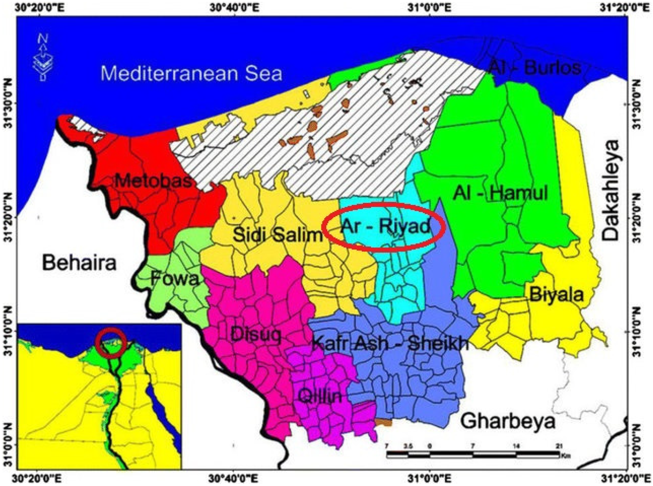
2.1. Description of the Studied Corroded Ancient Coins
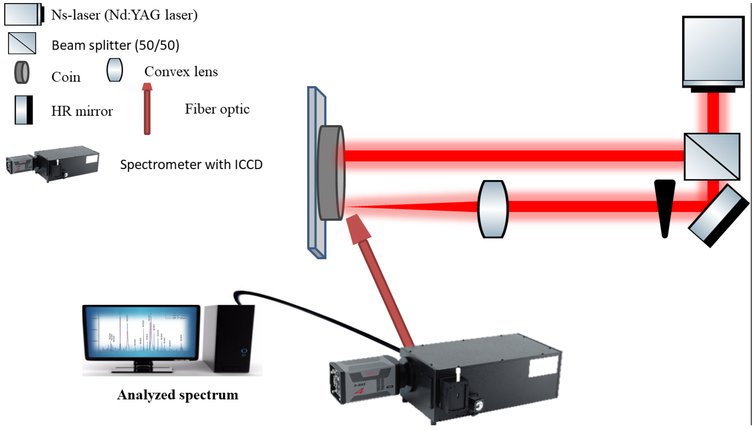
2.2. Experimental Set-Up for the Cleaning and Investigation of Corroded Coins

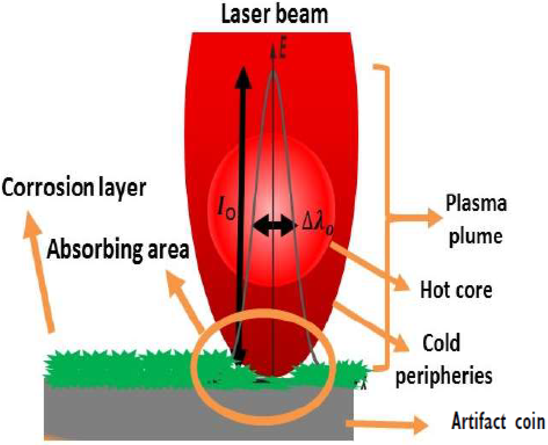
2.3. Pulsed Laser for Cleaning
2.4. Pulsed Laser Ablation for LIBS Analysis
3. Result and Discussion
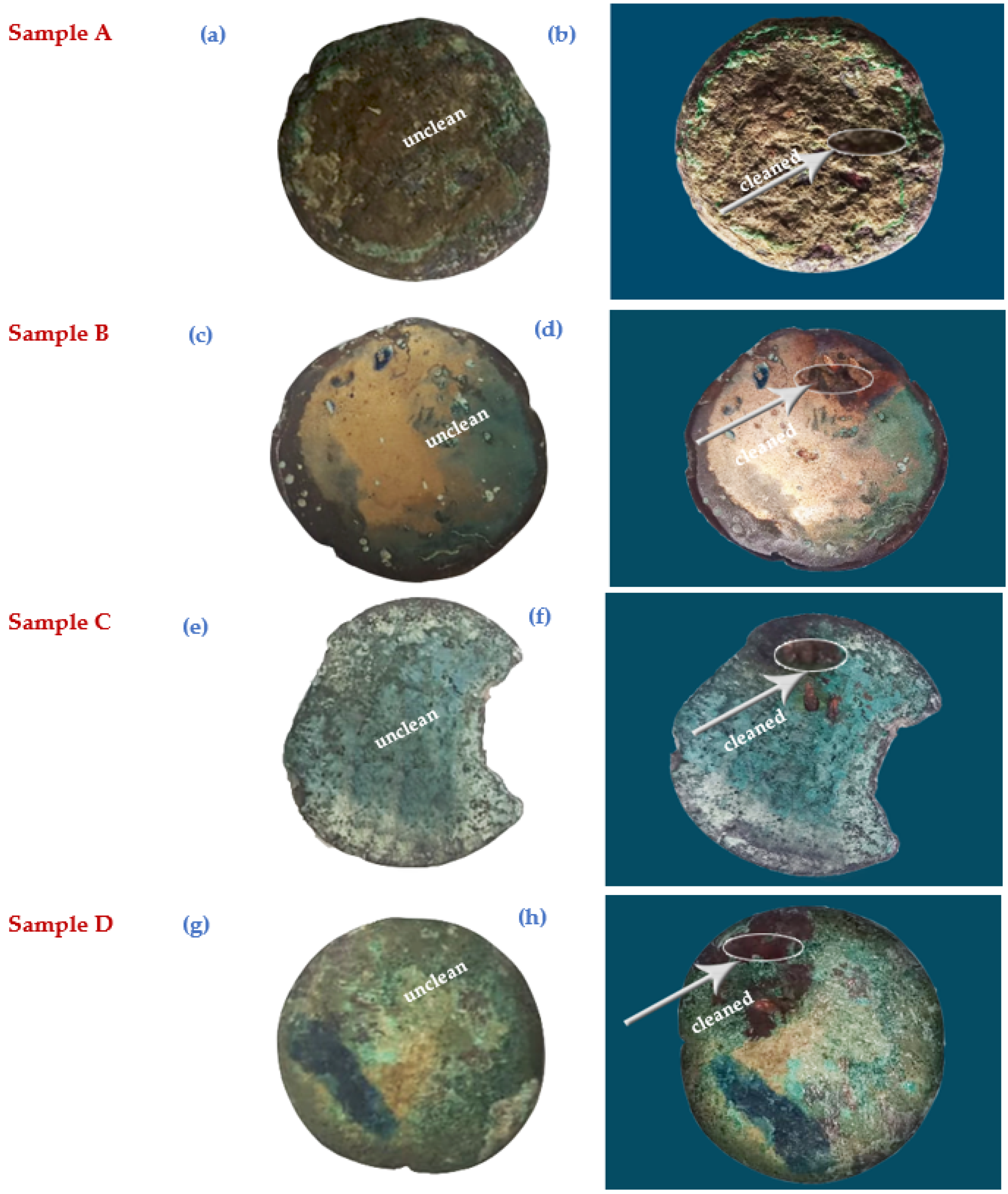
3.1. Visual Examination of Coin Samples
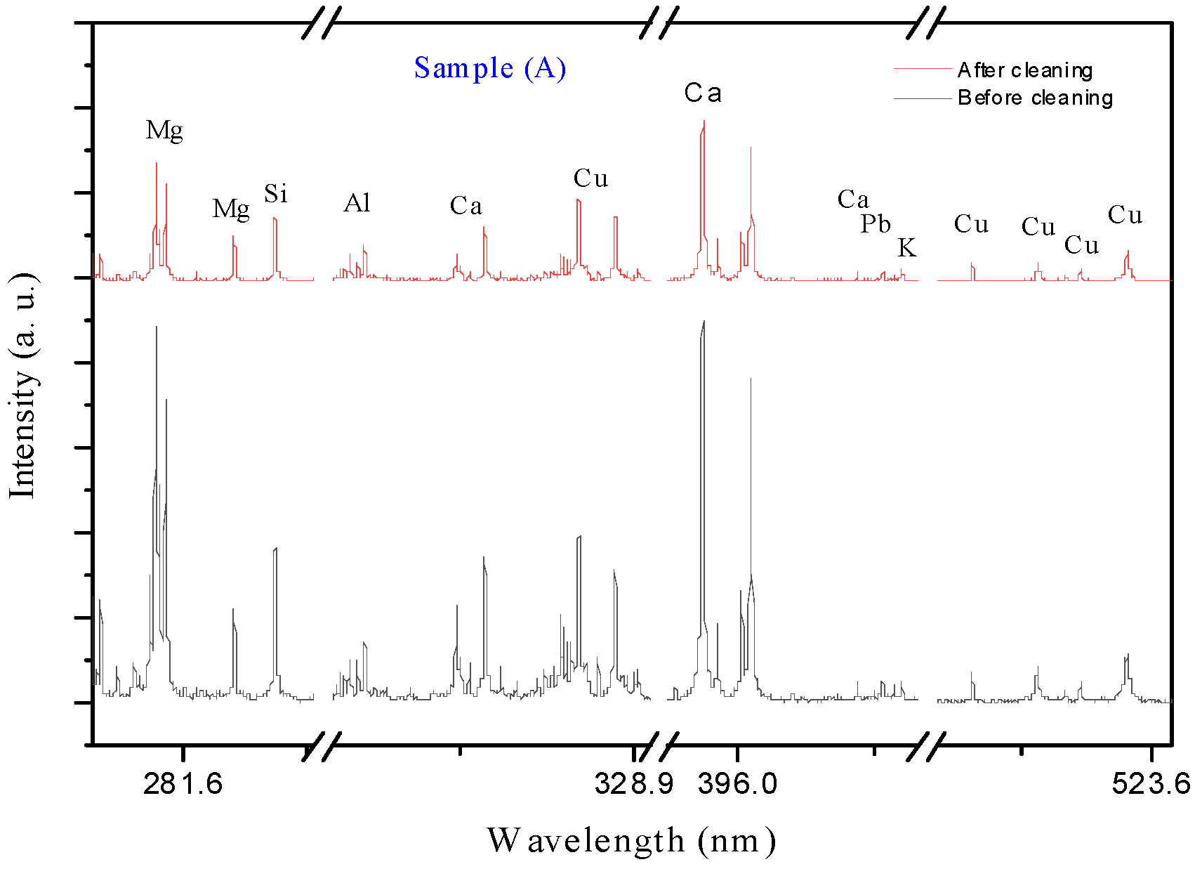
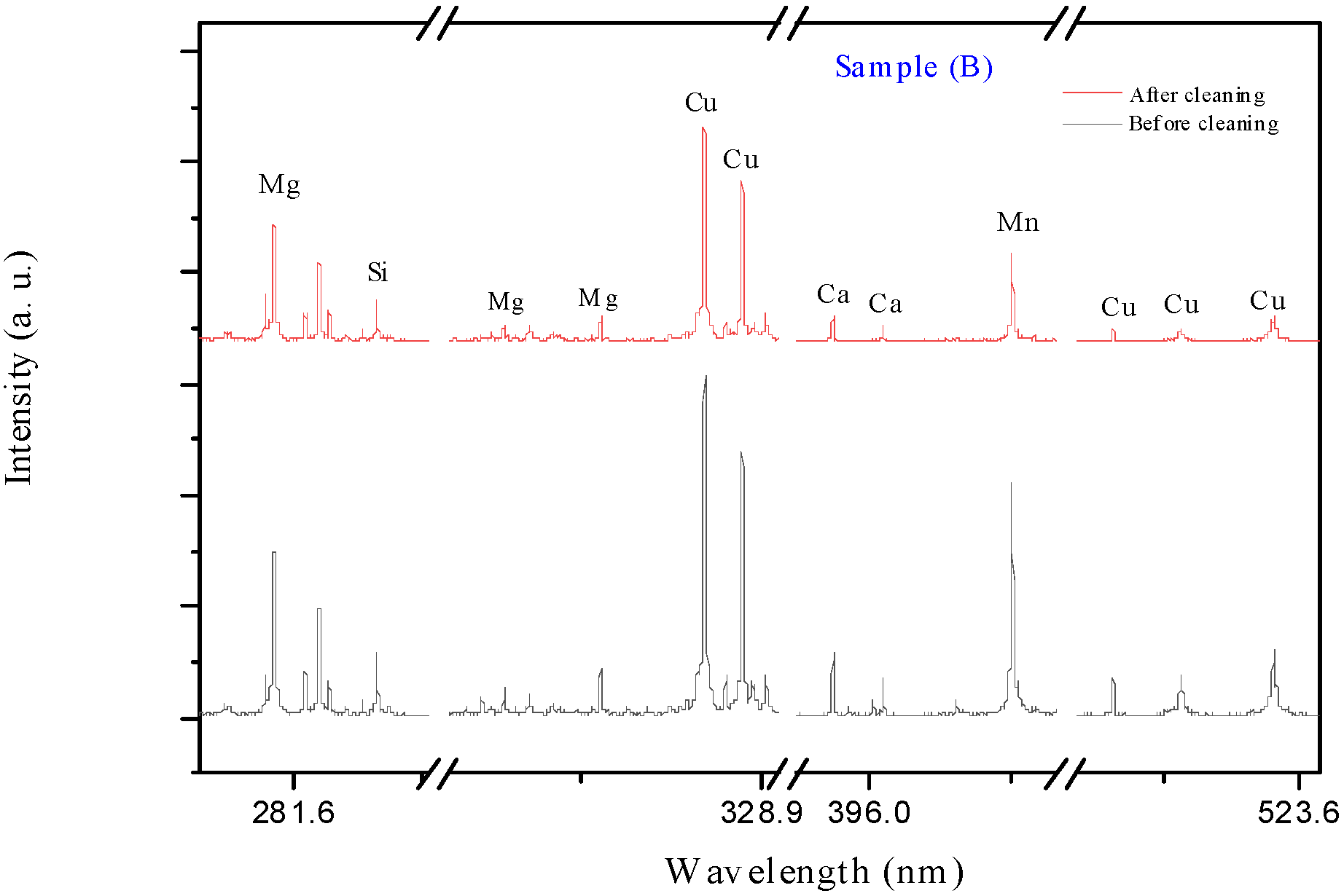
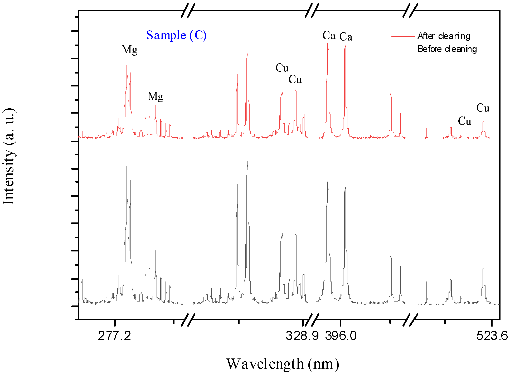
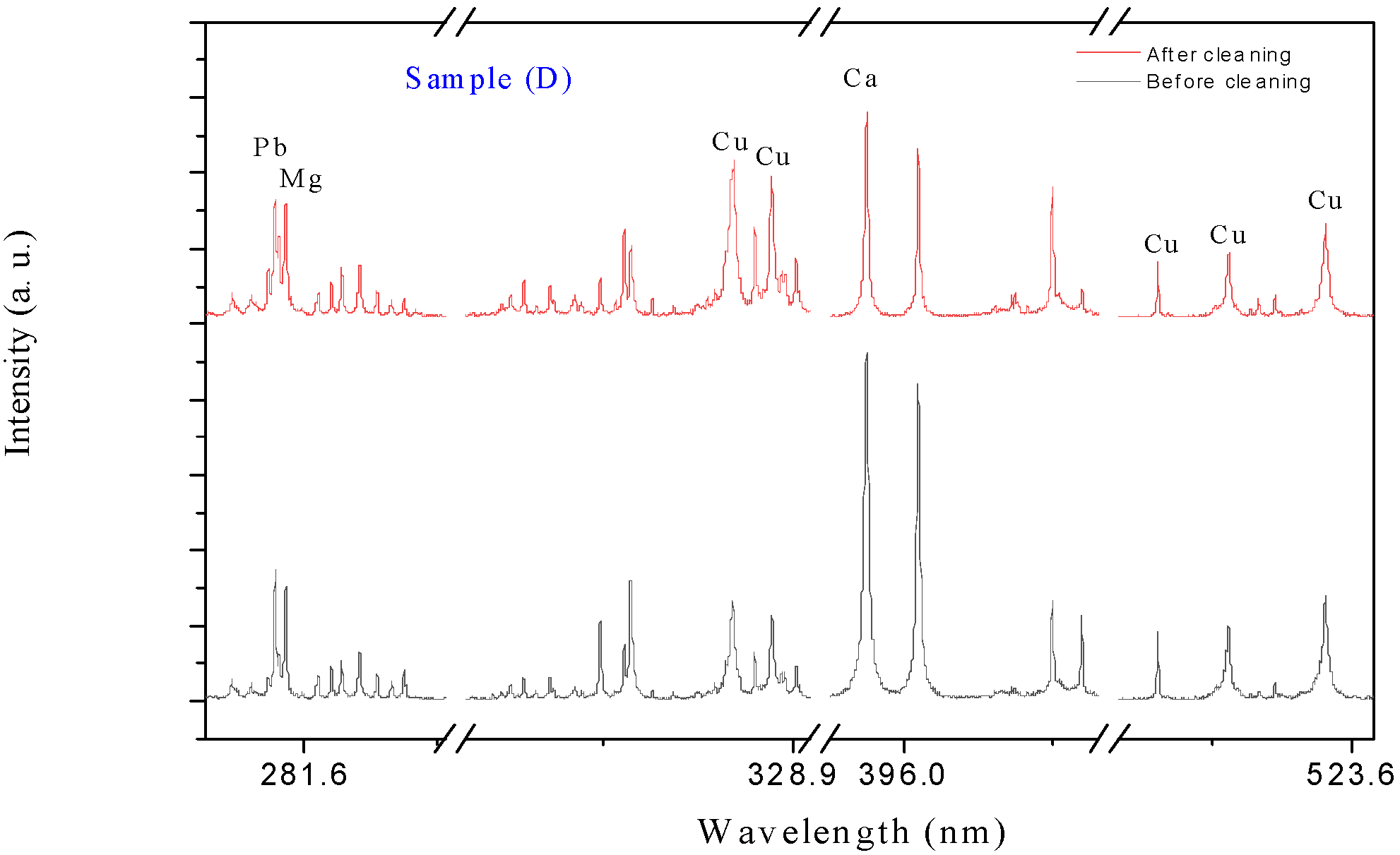
3.2. LIBS Analysis
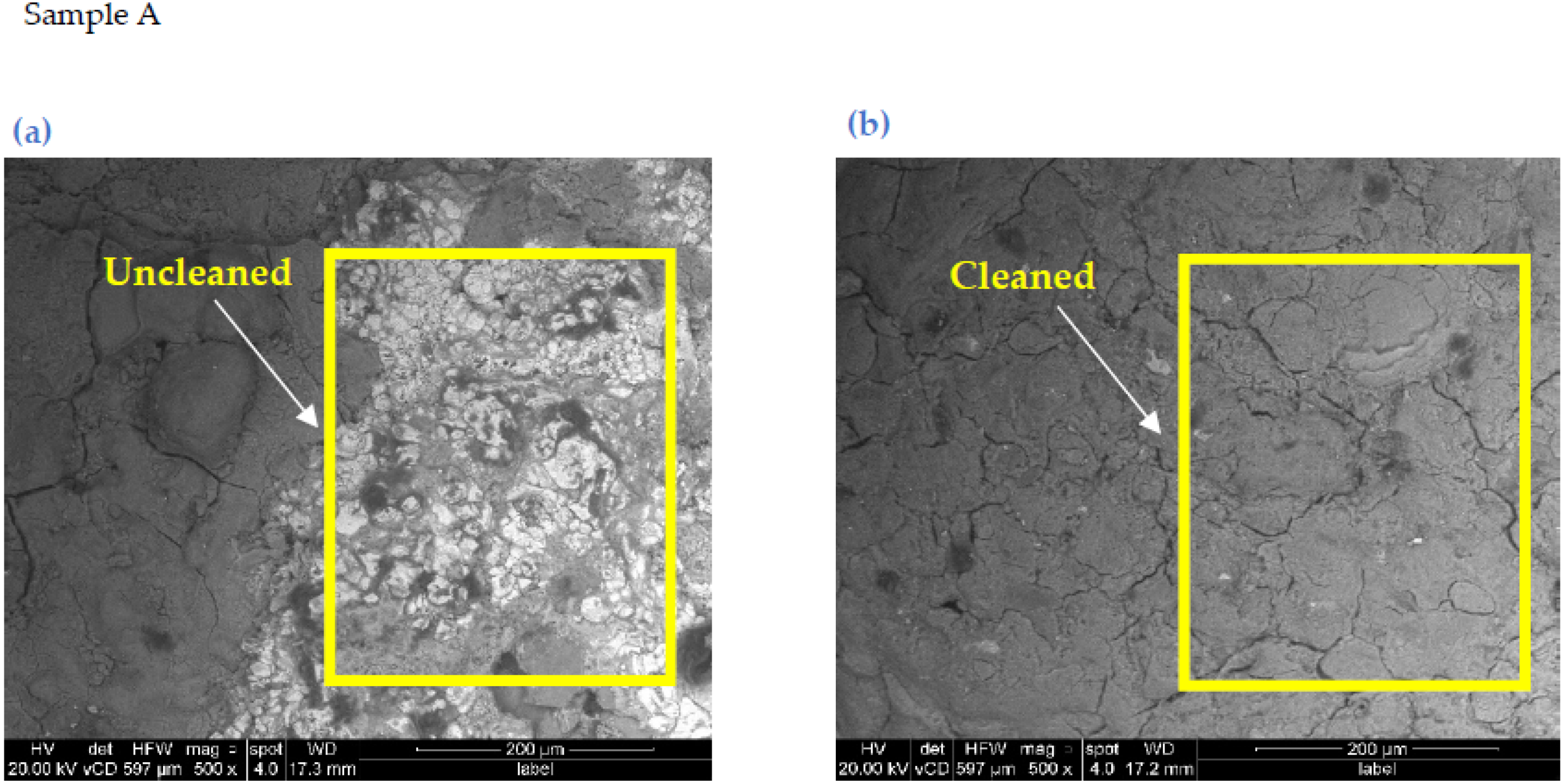
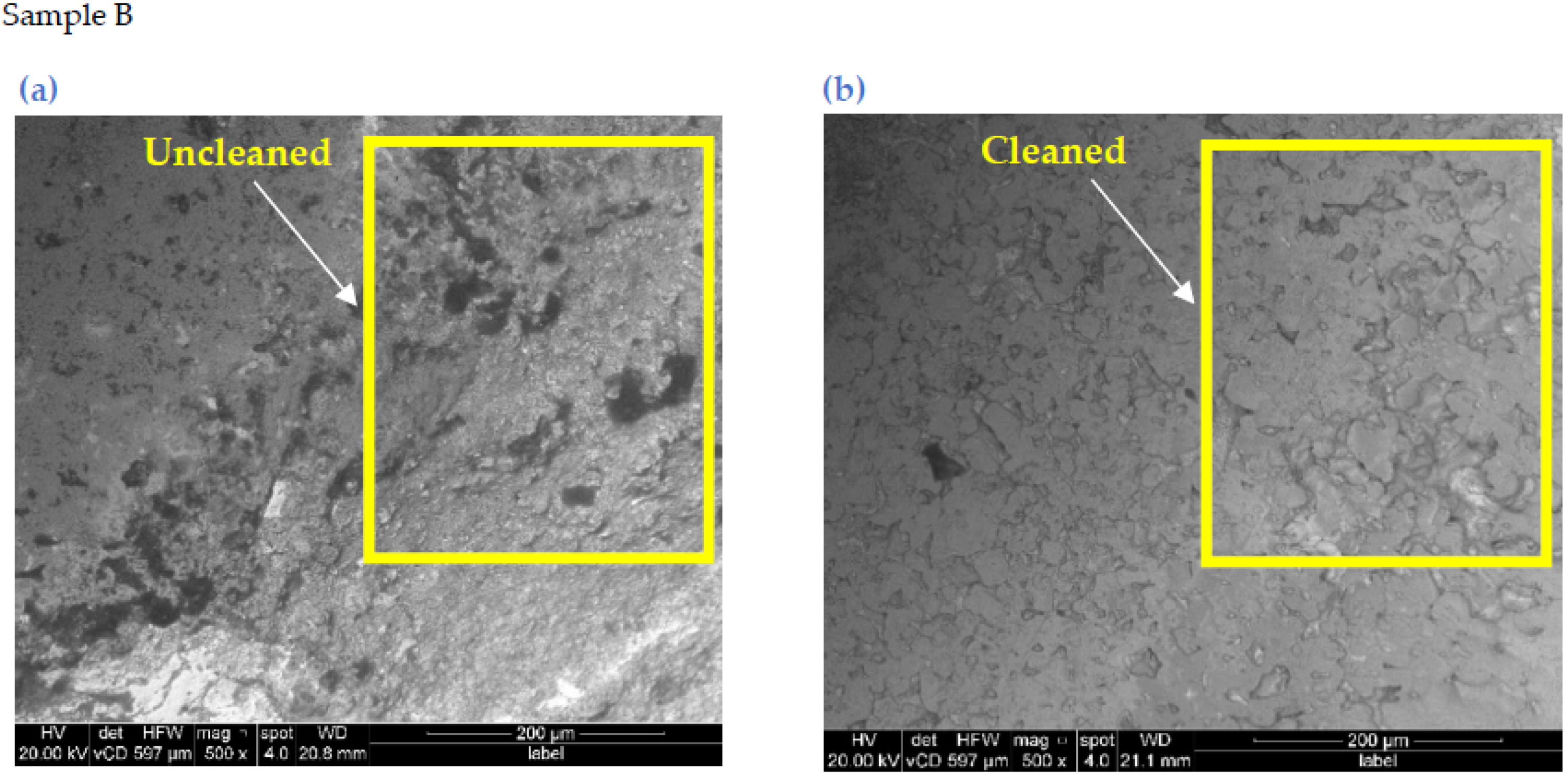
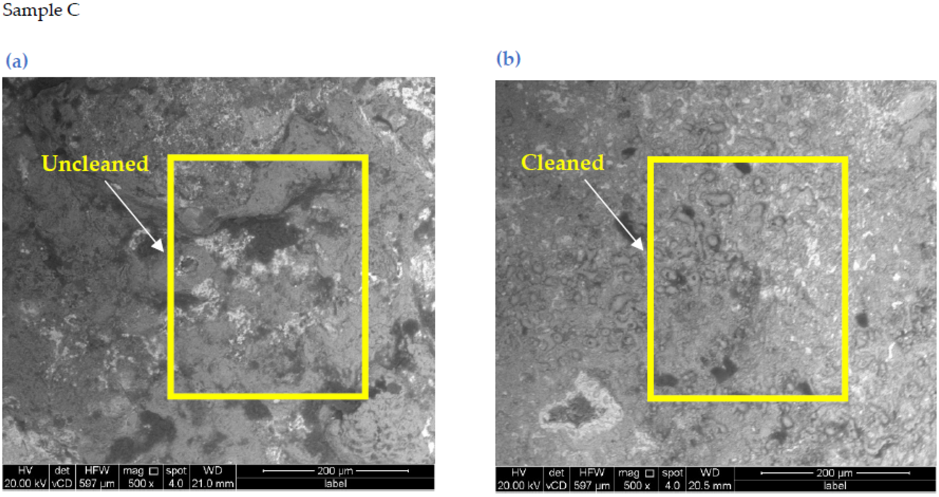
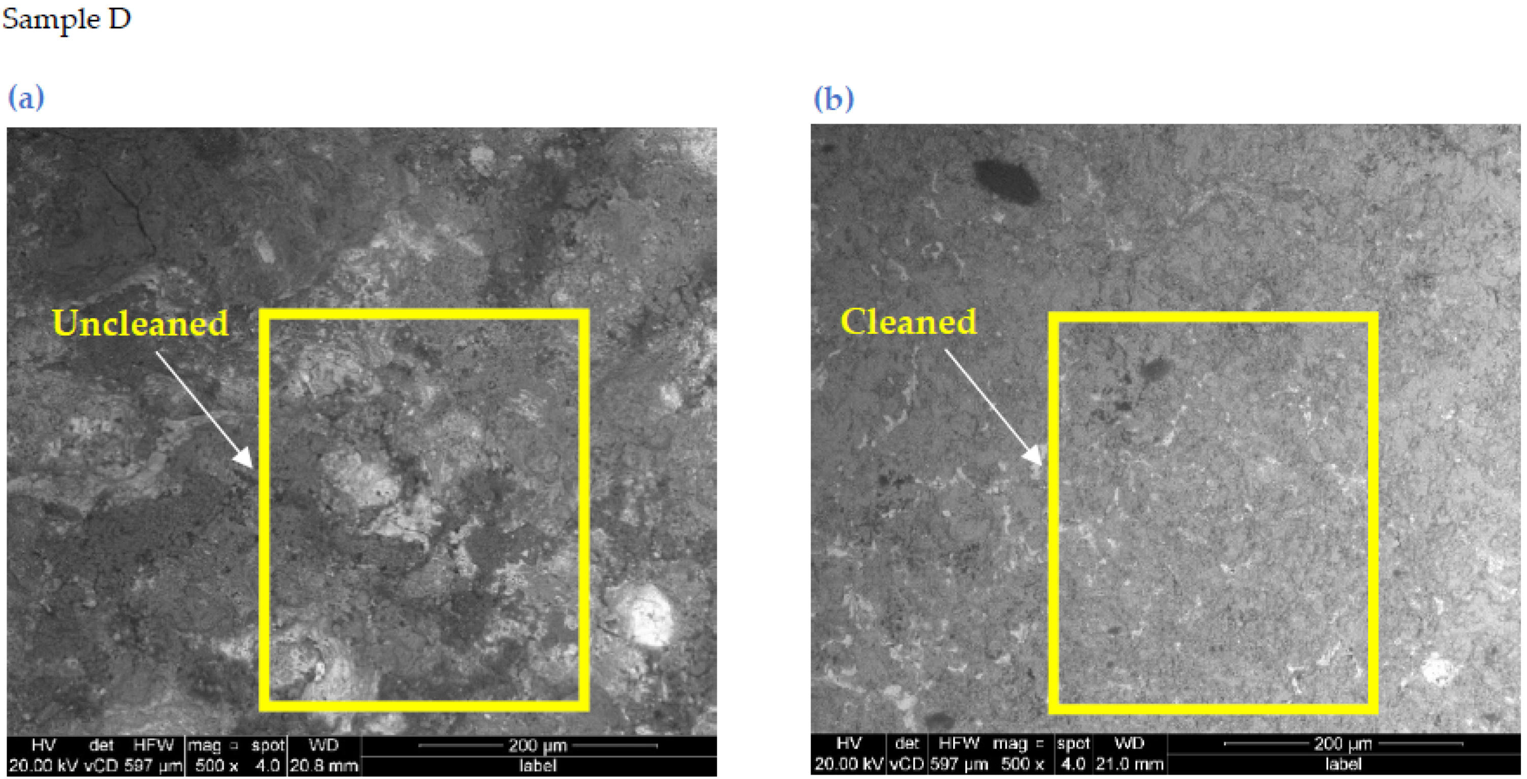
3.3. SEM Analysis
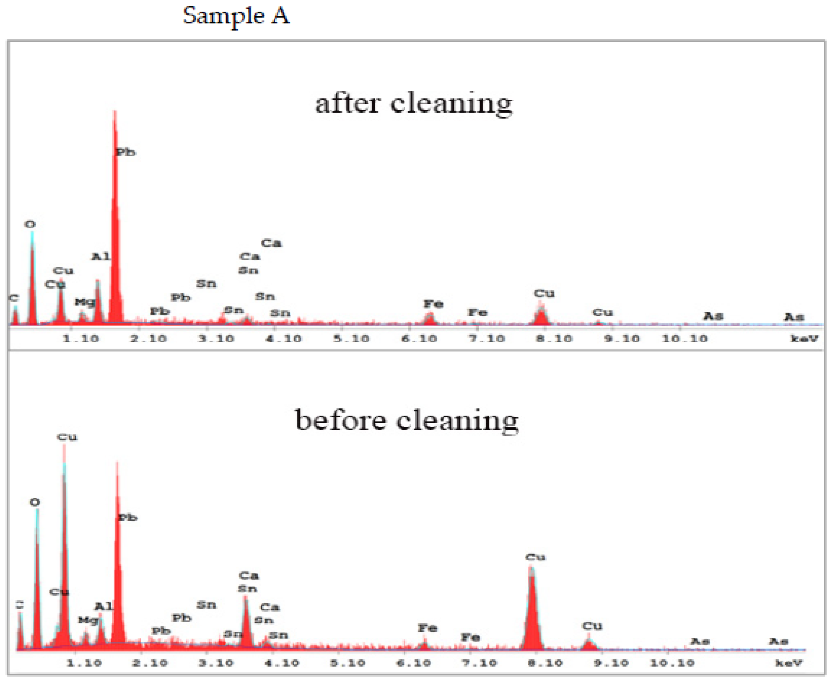
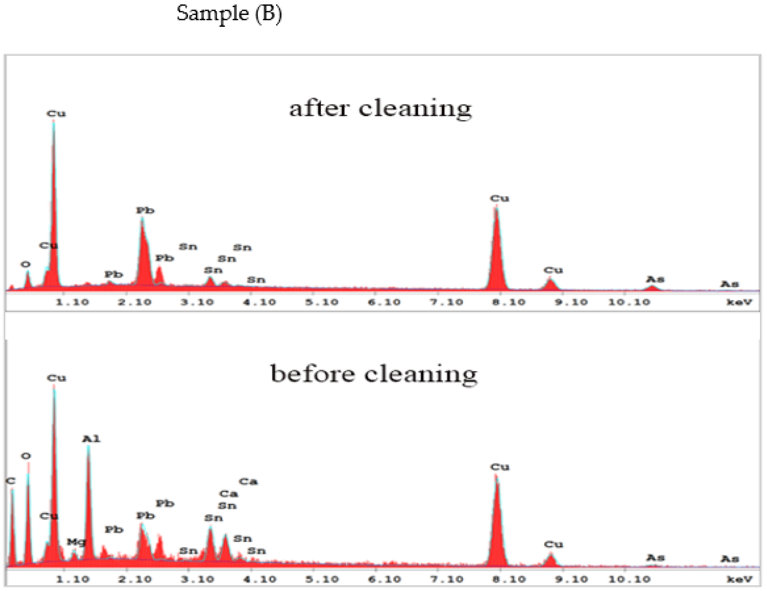
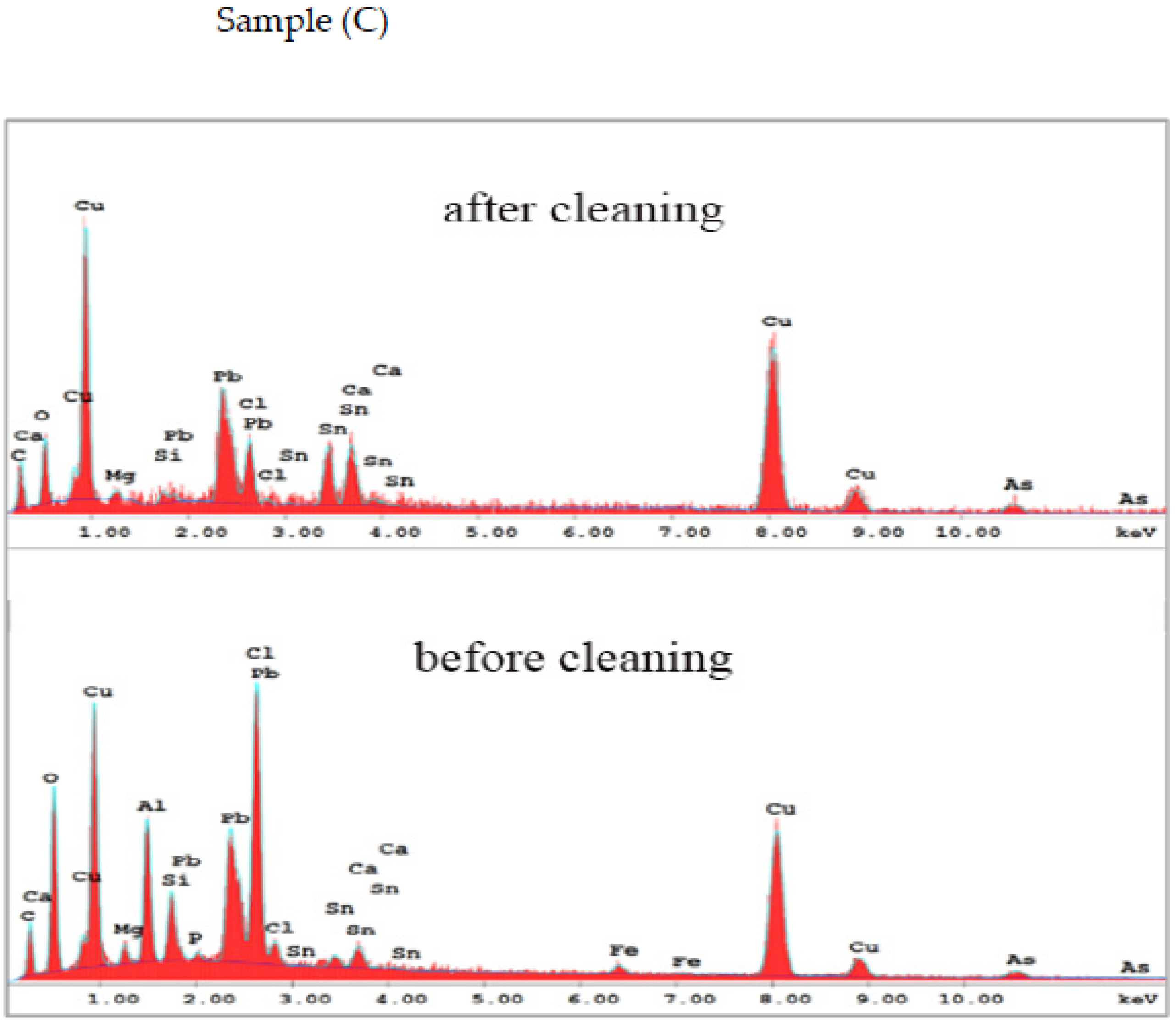
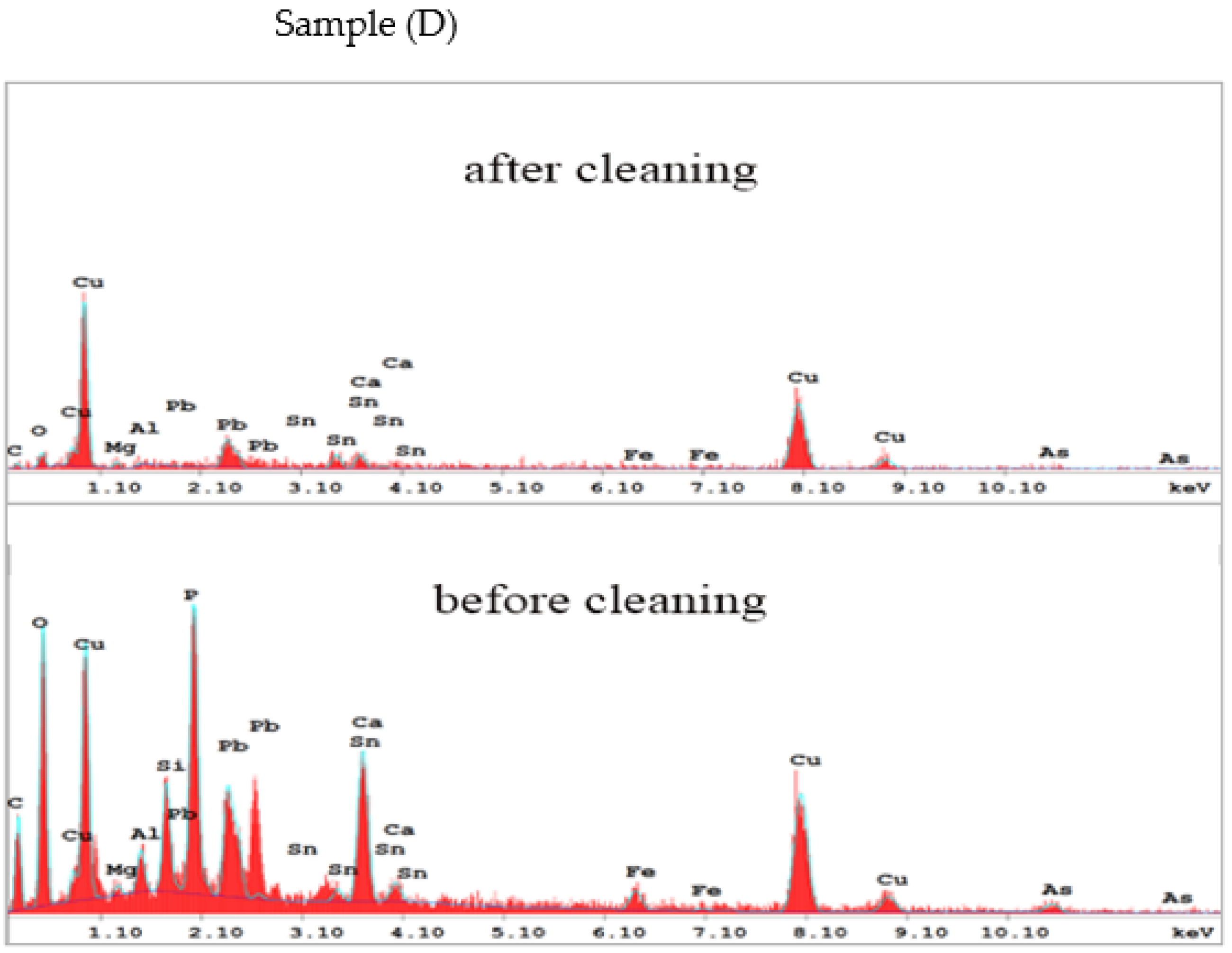
3.4. EDX Analysis
4. Conclusions
Author Contributions
Funding
Institutional Review Board Statement
Informed Consent Statement
Acknowledgments
Conflicts of Interest
References
- Awasthi, S.; Kumar, R.; Rai, G.K.; Rai, A.K. Study of archaeological coins of different dynasties using libs coupled with multivariate analysis. Opt. Lasers Eng. 2016, 79, 29–38. [Google Scholar] [CrossRef]
- Wilson, P. Landscape s of the Bashmur: Settlements and Monasteries in the Northern Egyptian Delta from 7th–9th Century. Mainz Hist. Cult. Sci. 2017, 36, 345. [Google Scholar]
- Hogarth, D.G. Three North Delta Nomes. J. Hell. Stud. 1904, 24, 1–19. [Google Scholar] [CrossRef] [Green Version]
- Elmorshedy, H.; Bergquist, R.; Abou El-Ela, N.E.; Eassa, S.M.; Elsakka, E.E.; Barakat, R. Can human schistosomiasis mansoni control be sustained in high-risk transmission foci in Egypt? Parasites Vectors 2015, 8, 372. [Google Scholar] [CrossRef] [Green Version]
- Sharma, G.R. The Excavations at Kausambi (1957-59): The Defences and Syenaciti of the Purusamedha; Department of Ancient History, Culture and Archaeology, University of Allahabad: Allahabad, India, 1960. [Google Scholar]
- Sharma, G.R. Excavations at Kausambi 1949–1950; MASI No. 74; Archaeological Survey of India: Delhi, India, 1969. [Google Scholar]
- Reale, R.; Plattner, S.H.; Guida, G.; Sammartino, M.P.; Visco, G. Ancient coins: Cluster analysis applied to find a correlation between corrosion process and burial soil characteristics. Chem. Central J. 2012, 6, S9. [Google Scholar] [CrossRef] [Green Version]
- Al-Zahrani, M.; Ghoniem, M. A Characterization of coins from the Nagran Hoard, Saui Arabia, prior to conservation. Int. J. Conserv. Sci. 2012, 3, 143–152. [Google Scholar]
- Li, M.; Ruan, F.; Li, R.; Zhou, J.; Zhang, T.; Tang, H.; Li, H. In situ simultaneous quantitative analysis multi-elements of archaeological ceramics via laser-induced breakdown spectroscopy combined with machine learning strategy. Microchem. J. 2022, 182, 107928. [Google Scholar] [CrossRef]
- Bertasa, M.; Korenberg, C. Successes and challenges in laser cleaning metal artefacts: A review. J. Cult. Herit. 2022, 53, 100. [Google Scholar] [CrossRef]
- Rimmer, D.T.M.; Watkinson, D.; Ganiaris, H. Guidelines for the Storage and Display of Archaeological Metalwork; English Heritage: London, UK, 2013. [Google Scholar]
- ICOM-Symposium. Conservation of Metal Statuary and Architectural Decoration in Open-Air Exposure, in, Paris; Symposium: Paris, France, 1986. [Google Scholar]
- Weil, P.D. Conservation of metal statuary and architectural decoration in open-air exposure: An overview of current status with suggestions regarding needs and future trends. In Conservation of Metal Statuary and Architectural Decoration in Open-Air Exposure= Conservation des Oeuvres d’art et Decorations, en Métal Exposées en Plein Air; Symposium: Paris, France, 1986; Volume 6–8. X. pp. 1–22. [Google Scholar]
- Bertholon, R. The location of the original surface: A review of the conservation literature. In Proceedings of the International Conference on Metals Conservation, Metal 2001, Santiago, Chile, 2–6 April 2001; pp. 167–179. [Google Scholar]
- Garbassi, F.; Mello, E. Surface spectroscopic studies on patinas of ancient metalobjects. Stud. Conserv. 1984, 29, 172–180. [Google Scholar]
- Scott, D.A. An examination of the patina and corrosion morphology of some Roman bronzes. J. Am. Inst. Conserv. 1994, 33, 1–23. [Google Scholar] [CrossRef]
- Siano, S.; Agresti, J.; Cacciari, I.; Ciofini, D.; Mascalchi, M.; Osticioli, I.; Mencaglia, A. Laser cleaning in conservation of stone, metal, and painted artifacts: State of the art and new insights on the use of the Nd: YAG lasers. Appl. Phys. A 2012, 106, 419–446. [Google Scholar] [CrossRef]
- Sanz, M.; Oujja, M.; Ascaso, C.; de los Ríos, A.; Pérez-Ortega, S.; Souza-Egipsy, V.; Wierzchos, J.; Speranza, M.; Cañamares, M.V.; Castillejo, M. Infrared and ultraviolet laser removal of crustose lichens on dolomite heritage stone. Appl. Surf. Sci. 2015, 346, 248–255. [Google Scholar] [CrossRef] [Green Version]
- Senesi, G.S.; Carrara, I.; Nicolodelli, G.; Milori, D.M.B.P.; De Pascale, O. Laser Cleaning and laser-induced breakdown spectroscopy applied in removing and characterizing black crusts from limestones of Castello Svevo; Bari, Italy: A case study. Microchem. J. 2016, 124, 296–305. [Google Scholar] [CrossRef]
- Zhao, H.; Qiao, Y.; Du, X.; Wang, S.; Zhang, Q.; Zang, Y.; Liu, X. Laser cleaning performance and mechanism in stripping of Polyacrylate resin paint. Appl. Phys. A 2020, 126, 360. [Google Scholar] [CrossRef]
- Wang, W.; Ayhan, B.; Kwan, C.; Qi, H.; Vance, S. A Novel and Effective Multivariate Method for Compositional Analysis using Laser Induced Breakdown Spectroscopy. Earth Environ. Sci. 2014, 17, 12208. [Google Scholar] [CrossRef] [Green Version]
- Li, X.; Guan, Y. Real-Time Monitoring of Laser Cleaning for Hot-Rolled Stainless Steel by Laser-Induced Breakdown Spectroscopy. Metals 2021, 11, 790. [Google Scholar] [CrossRef]
- Marimuthu, S.; Sezer, H.K.; Kamara, A.M. Applications of laser cleaning process in high value manufacturing industries. In Developments in Surface Contamination and Cleaning: Application of Cleaning Techniques; Kohli, R., Mittal, K.L., Eds.; Elsevier: Amsterdam, The Netherlands, 2019; Volume 11, pp. 251–288. [Google Scholar]
- Veiko, V.; Samohvalov, A.; Ageev, E. Laser cleaning of engraved rolls coupled with spectroscopic control. Opt. Laser Technol. 2013, 54, 170–175. [Google Scholar] [CrossRef]
- Hu, G.Q.; Song, Y.; Guan, Y.C. Tailoring metallic surface properties induced by laser surface processing for industrial applications. Nanotechnol. Precis. Eng. 2019, 2, 29–34. [Google Scholar] [CrossRef]
- Mateo, M.P.; Nicolas, G.; Piñon, V.; Ramil, A.; Yañez, A. Laser cleaning, an alternative method for removing oil-spill fuel residues. Appl. Surf. Sci. 2005, 247, 333–339. [Google Scholar] [CrossRef]
- Zhang, Z.Y.; Zhang, J.Y.; Wang, Y.B.; Zhao, S.S.; Lin, X.C.; Li, X.Y. Removal of paint layer by layer using a 20 kHz 140 ns quasi-continuous wave laser. Optics 2018, 174, 46–55. [Google Scholar] [CrossRef]
- Han, J.H.; Cui, X.D.; Wang, S.; Feng, G.Y.; Deng, G.L.; Hu, R.F. Laser effects based optimal laser parameter identifications for paint removal from metal substrate at 1064nm: A multi-pulse model. J. Mod. Opt. 2017, 64, 1947–1959. [Google Scholar] [CrossRef]
- Li, H.; Costil, S.; Liao, H.L.; Coddet, C. Surface preparation by using laser cleaning in thermal spray. J. Laser Appl. 2008, 20, 12–21. [Google Scholar] [CrossRef]
- Zhou, C.; Li, H.G.; Chen, G.Y.; Wang, G.; Shan, Z.Z. Effect of single pulsed picosecond and 100 nanosecond laser cleaning on surface morphology and welding quality of aluminium alloy. Opt. Laser Technol. 2020, 127, 1061197. [Google Scholar] [CrossRef]
- Zhang, G.X.; Hua, X.M.; Huang, Y.; Zhang, Y.L.; Li, F.; Shen, C.; Cheng, J. Investigation on mechanism of oxide removal and plasma behavior during laser cleaning on aluminum alloy. Appl. Surf. Sci. 2020, 506, 144666. [Google Scholar] [CrossRef]
- Lee, J.M.; Yu, J.E.; Koh, Y.S. Experimental study on the effect of wavelength in the laser cleaning of silver threads. J. Cult. Herit. 2003, 4, 157–161. [Google Scholar] [CrossRef]
- Lee, J.M. In-Process and Intelligent Monitoring Systems for Laser Cleaning Process. Ph.D. Thesis, The University of Liverpool, Liverpool, UK, 1999. [Google Scholar]
- Siano, S.; Salimbeni, R.; Pini, R.; Giusti, A.; Matteini, M. Laser cleaning methodology for the preservation of the Porta del Paradiso by Lorenzo Ghiberti. J. Cult. Herit. 2003, 4, 140–146. [Google Scholar] [CrossRef]
- Mostafa, A.M.; Hamed, S.A.M.; Afifi, H.; Mohamady, S. A comparative study on the color change of pigments due to the consolidation of conventional spectroscopic techniques and laser-induced breakdown spectroscopy. Appl. Phys. A 2019, 125, 559. [Google Scholar] [CrossRef]
- ElFaham, M.M.; Okil, M.; Mostafa, A.M. Effects of post-laser irradiation on the optical and structure properties of Al2O3 nanoparticles produced by laser ablation. J. Appl. Phys. 2020, 128, 153104. [Google Scholar] [CrossRef]
- ElFaham, M.M.; Okil, M.; Mostafa, A.M. Limit of detection and hardness evaluation of some steel alloys utilizing optical emission spectroscopic techniques. Opt. Laser Technol. 2018, 108, 634–641. [Google Scholar] [CrossRef]
- Singh, M.G.; Aradhana, J.; Rohit, K.; Kumar, A.; Rai, A.K. Analysis of deposited impurity material on the surface of the optical window of the Tokamak using LIBS. Phys. Scr. 2014, 89, 075601. [Google Scholar] [CrossRef]
- Fotakis, C.; Anglos, D.; Zafiropulos, V.; Georgiou, S.; Tornari, V. Lasers in the Preservation of Cultural Heritage Principles and Applications; Taylor & Francis: New York, NY, USA, 2007. [Google Scholar]
- Pathak, A.K.; Kumar, R.; Singh, V.K.; Agrawal, R.; Rai, S.; Rai, A.K. Assessment of LIBS for spectrochemical analysis: A review. Appl. Spectrosc. Rev. 2013, 47, 14–40. [Google Scholar] [CrossRef]
- Fortes, F.J.; Cabalín, L.M.; Laserna, J.J. The potential of laser-induced breakdown spectrometry for real time monitoring the laser cleaning of archaeometallurgical objects. Spectrochim. Acta Part B 2008, 63, 1191–1197. [Google Scholar] [CrossRef]
- Awasthi, S.; Kumar, R.; Pandey, R.; Rai, A. New Insights on Modern Age Coins by Calibration-Free Laser-Induced Breakdown Spectroscopy Method and Chemometric Approaches. J. Appl. Spectrosc. 2022, 89, 780–789. [Google Scholar] [CrossRef]
- Salem, Y.; Mohamed, E. The role of archaeometallurgical characterization of ancient coins in forgery detection. Nucl. Instrum. Methods Phys. Res. B 2019, 461, 247–255. [Google Scholar] [CrossRef]
- Fortes, F.J.; Cuñat, J.; Cabalín, L.M.; Laserna, J.J. In situ analytical assessment and chemical imaging of historical buildings using a man-portable laser system. Appl. Spectrosc. 2007, 61, 558–564. [Google Scholar] [CrossRef]
- Caridi, F.; Torrisi, L.; Cutroneo, M.; Barreca, F.; Gentile, C.; Serafino, T.; Castrizio, D. XPS and XRF depth patina profiles of ancient silver coins. Appl. Surf. Sci. 2013, 272, 82–87. [Google Scholar] [CrossRef]
- Navas, M.J.; Asuero, A.G.; Jiménez, A.M. A review of energy dispersive X-ray fluorescence (EDXRF) as an analytical tool in numismatic studies. Appl. Spectrosc. 2016, 70, 207–221. [Google Scholar] [CrossRef] [PubMed] [Green Version]
- Ager, F.J.; Moreno-Suarez, A.I.; Scrivano, S.; Ortega-Feliu, I.; Gomez-Tubio, B.; Respaldiza, M.A. Silver surface enrichment in ancient coins studied by micro-PIXE. Nucl. Instrum. Methods Phys. Res. B 2013, 306, 241–244. [Google Scholar] [CrossRef]
- Pieta, E.; Lekki, J.; Meléndez, J.M.; Paluszkiewicz, C.; Nowakowski, M.; Matosz, M.; Kwiatek, W. Surface characterization of medieval silver coins minted by the early Piasts: FTIR mapping and SEM/EDX studies. Surf. Interface Anal. 2018, 50, 78–86. [Google Scholar] [CrossRef]
- Gaudiuso, R.; Uhlir, K.; Griesser, M. Micro-invasive depth profile analysis by laser-induced breakdown spectroscopy (LIBS): The case of mercury layers on Sasanian coins. J. Anal. At. Spectrom. 2019, 34, 2261–2272. [Google Scholar] [CrossRef]
- Oudbashi, O.; Hasanpour, A.; Jahanpoor, A.; Rahjoo, Z. Microscopic and microanalytical study on Sasanian metal objects from Western Iran: A case study. STAR Sci. Technol. Archaeol. Res. 2017, 3, 194–205. [Google Scholar] [CrossRef] [Green Version]
- Mendoza-López, M.L.; Pérez-Bueno, J.J.; Rodríguez-García, M.E. Characterizations of silver alloys used in modern Mexican coins. Mater. Charact. 2009, 60, 1041–1048. [Google Scholar] [CrossRef]
- Inberg, A.; Ashkenazi, D.; Cohen, M.; Iddan, N.; Cvikel, D. Corrosion products and microstructure of copper alloy coins from the Byzantine-period Ma’agan Mikhael B shipwreck, Israel. Microchem. J. 2018, 143, 400–409. [Google Scholar] [CrossRef]
- Torrisi, L.; Cutroneo, M.; Torrisi, A. Mass Quadrupole Spectrometry Coupled to Laser Ablation for Cultural Heritage Applications. In Handbook of Cultural Heritage Analysis; Springer International Publishing: Cham, Switzerland, 2022; pp. 445–464. [Google Scholar]
- Ioanid, E.G.; Ioanid, A.; Rusu, D.E.; Doroftei, F. Surface investigation of some medieval silver coins cleaned in high-frequency cold plasma. J. Cult. Herit. 2011, 12, 220–226. [Google Scholar] [CrossRef]


| Sample | Composition (wt. %) | |||||||||||||
|---|---|---|---|---|---|---|---|---|---|---|---|---|---|---|
| Cu | Ca | Mg | Pb | C | O | Al | Sn | Fe | As | Cl | P | Si | ||
| Coin A | Before cleaning | 72 | 4.7 | 1.2 | 0.54 | 6.29 | 9.91 | 2.1 | 0.7 | 2.55 | 0.05 | _ | _ | _ |
| After cleaning | 45.3 | 2.2 | 2.1 | 2.29 | 8.86 | 18.1 | 7.8 | 2.7 | 10.6 | 0.01 | _ | _ | _ | |
| Coin B | Before cleaning | 77.2 | 0.6 | 0.4 | 17.2 | 0.08 | 1.46 | 0 | 3 | _ | 0.02 | _ | _ | _ |
| After cleaning | 61.4 | 1.1 | 0.7 | 6.29 | 10.5 | 5.91 | 6.7 | 7.5 | _ | 0.01 | _ | _ | _ | |
| Coin C | Before cleaning | 67.6 | 2 | 0.4 | 12.1 | 4.21 | 2.67 | 0.2 | 8 | 0.13 | 0.02 | 2.2 | 0.1 | 0.4 |
| After cleaning | 55.2 | 0.8 | 0.7 | 12.8 | 5.28 | 6.78 | 4.4 | 1.5 | 1.31 | 0.02 | 9.3 | 0.3 | 1.8 | |
| Coin D | Before cleaning | 79.7 | 1.4 | 0.8 | 8.8 | 1.27 | 1.5 | 0.3 | 5.1 | 0.66 | 0.04 | _ | 0.2 | 0.3 |
| After cleaning | 46.4 | 6.8 | 0.3 | 11 | 7.78 | 11 | 1.3 | 1.5 | 3.09 | 0.04 | _ | 8 | 2.8 | |
Publisher’s Note: MDPI stays neutral with regard to jurisdictional claims in published maps and institutional affiliations. |
© 2022 by the authors. Licensee MDPI, Basel, Switzerland. This article is an open access article distributed under the terms and conditions of the Creative Commons Attribution (CC BY) license (https://creativecommons.org/licenses/by/4.0/).
Share and Cite
Rezk, R.A.; Abdel Ghany, N.A.; Mostafa, A.M. Laser-Assisted Method for Cleaning and Analysis of Archaeological Metallic Coins. Coatings 2022, 12, 1548. https://doi.org/10.3390/coatings12101548
Rezk RA, Abdel Ghany NA, Mostafa AM. Laser-Assisted Method for Cleaning and Analysis of Archaeological Metallic Coins. Coatings. 2022; 12(10):1548. https://doi.org/10.3390/coatings12101548
Chicago/Turabian StyleRezk, Reham A., Nabil Ahmed Abdel Ghany, and Ayman M. Mostafa. 2022. "Laser-Assisted Method for Cleaning and Analysis of Archaeological Metallic Coins" Coatings 12, no. 10: 1548. https://doi.org/10.3390/coatings12101548