Article

Effects of Autologous Bone Marrow Mesenchymal Stem Cells and Platelet-Rich Plasma on Bone Regeneration and Osseointegration of a Hydroxyapatite-Coated Titanium Implant

1
IRCCS Istituto Ortopedico Rizzoli, Complex Structure of Surgical Sciences and Technologies, Via di Barbiano 1/10, 40136 Bologna, Italy
2
IRCCS Istituto Ortopedico Rizzoli, Complex Structure of Reconstructive Orthopedic Surgery Innovative Techniques, Via G.C. Pupilli 1, 40136 Bologna, Italy
*
Author to whom correspondence should be addressed.
Coatings 2021, 11(7), 840; https://doi.org/10.3390/coatings11070840
Submission received: 18 June 2021 / Revised: 8 July 2021 / Accepted: 10 July 2021 / Published: 12 July 2021
(This article belongs to the Special Issue Bioactive Coatings on Medical Implants)

Abstract

:
Bone regeneration remains one of the major clinical needs in orthopedics, and advanced and alternative strategies involving bone substitutes, cells, and growth factors (GFs) are mandatory. The purpose of this study was to evaluate whether the association of autologous bone marrow mesenchymal stem cells (BMSC), isolated by ‘one-step surgical procedure’, and activated platelet rich plasma (PRP) improves osseointegration and bone formation of a hydroxyapatite-coated titanium (Ti-HA) implant, already in clinical use, in a rabbit cancellous defect. The GFs present in plasma, in inactivated and activated PRP were also tested. At 2 weeks, histology and histomorphometry highlighted increased bone-to-implant contact (BIC) in Ti-HA combined with BMSC and PRP in comparison to Ti-HA alone and Ti-HA + PRP. The combined effect of BMSC and PRP peaked at 4 weeks where the BIC value was higher than all other treatments. At both experimental times, newly formed bone (Trabecular Bone Volume, BV/TV) in all tested treatments showed increased values in comparison to Ti-HA alone. At 4 weeks Ti-HA + PRP + BMSC showed the highest BV/TV and the highest osteoblasts number; additionally, a higher osteoid surface and bone formation rate were found in Ti-HA + BMSC + PRP than in all other treatments. Finally, the analyses of GFs revealed higher values in the activated PRP in comparison to plasma and to non-activated PRP. The study suggests that the combination of autologous activated PRP, as a carrier for BMSCs, is a promising regenerative strategy for bone formation, osseointegration, and mineralization of bone implants.

1. Introduction

1.1. Titanium Implants

Titanium (Ti) and its alloys have been widely used in the clinic as bone implants due to their biocompatibility and mechanical properties [1]. However, despite the remarkable developments of these materials, the bio-inertness of the Ti surfaces is still a problem for the promotion of bone healing and for the osseointegration between the host bone and the implant [2]. Biological osseointegration is a long and complex process that involves several processes implicated in bone healing around implants [3]. This last in turn involves cellular and extracellular biological events that take place at the bone-implant interface [3]. This cascade of biological events is controlled by growth factors and differentiation factors released by the hematopoietic cells activated at the bone–implant interface [4]. Alteration of these biological processes leads to late bone healing and poor osseointegration at the bone–implant interface which can affect the implant stability and increase the risk of aseptic loosening [3]. This is particularly true for aging populations, and in presence of co-morbidities and low bone mass at the implantation site that affects the biological regenerative response [4]. To date, to avoid these problems Ti surfaces are often modified thus to achieve a more robust and earlier osseointegration between the implant and surrounding bone [5]. Over the years, the research and analysis on biomaterials have led to the knowledge that functionalizing Ti implants with different coatings can be a successful strategy in determining the achievement of the osteointegration process. Among coating materials, the plasma spraying of hydroxyapatite (HA) has been one of the most used and consolidated coatings, also with clinical and commercial success [5,6,7,8]. However, as no specific strategy can effectively satisfy all the clinical needs for bone healing, the idea of supplying different critical elements for tissue regeneration, such as osteoconductive matrix, osteogenic cells, and growth factors, has been suggested by numerous researchers and clinicians.

1.2. Bone Marrow Mesenchymal Stem Cells and Platelet Rich Plasma

Bone marrow mesenchymal stem cells (BMSCs) and growth factors (GFs), derived from platelet rich plasma (PRP), have been widely used to improve the bone healing and osseointegration process [9,10,11,12,13,14,15,16]. The regenerative bone capability of BMSCs has been extensively reported in vivo with promising results [17,18,19]. Concerning their use, currently, three different approaches can be employed to engineer the bone biomaterials/grafts [20]. In the first approach, BMSCs are cultured for 2–3 weeks in a cell therapy unit to isolate and augment the MSC fraction, and then several million of these cells are injected into a bone defect alone or seeded onto a graft/biomaterial before implantation [21]. In the second strategy after the bone marrow harvesting, the BMSCs are isolated and expanded for several weeks and then seeded on a graft/biomaterial, where they are cultured for a further week and finally transplanted into bone defects [22]. The third approach consists of aspirating bone marrow, followed by centrifugation to concentrate MSCs and implant them immediately into the bone defect with or without a graft/biomaterial, with no need for cell expansion (one-step procedure) [15,16,23]. Although these multiple stem cell-based approaches are all effective, the most useful strategy for clinical practice is the one-step derivation of MSCs [24]. An additional strategy to improve bone regeneration is the use of activated platelet-derived products. Activated platelets secrete numerous GFs, such as platelet-derived growth factor (PDGFaa, PDGFbb, PDGFab), transforming growth factors (TGFs) β1 and β2, epidermal growth factor (EGF), insulin-like growth factor (IGF), vascular endothelial growth factor (VEGF), bone morphogenetic protein (BMP) and many other bioactive molecules expressed during the different tissue healing phases [25]. Platelets mainly participate in the early phases of regeneration and through their degranulation, they release numerous growth factors that initiate and preserve the healing process [26]. The use of PRP is an inexpensive strategy to obtain numerous platelets and therefore numerous growth factors, many of which have a direct role in bone healing [27,28,29]. Activated PDGF attaches to transmembrane receptors on osteoblasts, osteoclasts, and macrophages stimulating angiogenesis, bone remodeling, and phagocytosis of damaged tissue during usual wound and fracture healing [30,31]. TGFβ regulates proliferation, differentiation, chemotaxis, and adhesion of progenitor cells in the wound bed [25,32,33]. Concerning VEGF, it acts synergistically with osteogenic proteins, such as bone morphogenetic protein (BMP)-4 and BMP-2, by enhancing cell recruitment, prolonging cell survival, increasing angiogenesis, and enhancing bone mineralization [34,35,36,37]. Despite the presence of conflicting reports about the effect of PRP on bone healing and regeneration mainly due to differences in the ‘preparation protocol’ (blood source and volume for extraction, instruments, frequency of rotation, time of centrifugation, final platelet concentrations, processes of activation, other cell types inclusion/exclusion, dose, and intervals of dose, etc), it appears that most preclinical and clinical research supports a positive role in bone regeneration [38,39]. In addition, the presence of osteogenic growth factors within PRP have led to the hypothesis that it can act synergistically with BMSCs to accelerate osteogenesis [40,41,42]. However, few studies have analyzed the effects of their association and most of them employed manipulated cells (e.g., in vitro culture expansion, osteogenic differentiation) whereas the typical clinical applications use cells with no manipulation [42,43,44,45,46,47,48,49]. Thus, PRP with BMSCs is not a proven therapy, the lack of PRP standardization and the use of manipulated BMSCs introduce a great variability preventing specific conclusions.
The purpose of this study was to evaluate whether the association of autologous BMSCs (isolated by ‘one-step surgical procedure’) and activated PRP improves the pattern of early osseointegration and bone formation of a hydroxyapatite-coated titanium (Ti-HA) implant, an implant already of clinical use, in an in vivo animal model.

2. Material and Methods

2.1. In Vivo Study

The research protocol was approved by the Ethical Committees of IRCCS Istituto Ortopedico Rizzoli and by the public authorities as provided by the current law at the time of the study (Law by Decree 116/92). Sixteen New Zealand rabbits (Harlan Laboratories SRL, Udine, Italy), with a body weight of 2.7 ± 0.3 kg, were housed at a controlled temperature, 22 ± 1 °C, and relative humidity of 55% ± 5% in single boxes and fed a standard diet (Mucedola, Milano, Italy) with filtered tap water ad libitum. At the time of surgery, animals were randomly divided into two groups, group 1 (2 weeks) and group 2 (4 weeks), and submitted to the same surgical procedure. General anesthesia was induced by intramuscular injection of 44 mg/kg ketamine (Imalgene 1000, Merial Italia S.p.A, Assago-Milan, Italy) and 3 mg/kg xylazine (Rompun, Bayer SpA, Milano, Italy), under assisted ventilation with O2/air (1/0.4 L/min) mixture and 2.5% isofluorane (Forane, Abbot SpA, Latina, Italy). Before surgery, the animals were submitted to the withdrawal of blood (7.0 mL) from the auricular artery for PRP preparation and of bone marrow (about 6.0–7.0 mL) from the iliac crest for the preparation of BMSC as described below. After this, bilateral confined cancellous defects 4 mm in diameter and 10 mm in depth were drilled in the distal femurs and Ti-HA alone, Ti-HA in combination with PRP and BMSCs, Ti-HA in combination with BMSCs, and Ti-HA in combination with PRP were randomly implanted in the left or right-side distal femurs. When using PRP, 0.9 mL of PRP was mixed with 0.1 mL autologous thrombin/calcium chloride solution to permit the gel formation. Ti-HA was tightly positioned inside the defects. When using the combination of BMSCs and PRP, BMSCs were manually mixed in PRP, and then thrombin was added. Antibiotic therapy (Cefazolin, 100 mg/kg, Pfizer Italia Srl, Latina, Italy) was administered preoperatively and immediately after surgery. Postoperative analgesia was performed using metamizole chloride, 50 mg/kg (Farmolisina Ceva Vetem, S.p.A, Agrate Brianza, Milan, Italy).
To monitor the bone ingrowth around implants dynamically the animals received intramuscular (i.m.) injections of oxytetracycline (30 mg/kg, Pfizer Italiana, Latina, Italy) before euthanasia at 4 weeks. Oxytetracycline was administered consecutively for two days, suspended for ten days, and again administered for another two days. After 2 and 4 weeks, animals were euthanized with an intravenous (i.v.) administration of Tanax (Hoechst, Frankfurt am Main, Germany) under general anesthesia.

2.2. Bone Marrow Stromal Cells (BMSCs)

A volume of 6.0–7.0 mL of bone marrow was collected in sterile vials and an isopycnic centrifugation was carried out to concentrate stem cells. An equivalent volume of phosphate buffered saline (PBS) was added to the bone marrow and the mix was layered over undiluted Ficoll-Paque (Sigma-Aldrich, Milano, Italy) and centrifuged. At the end of this process, the band of light-density cells was separated, washed, counted (4 ± 2 × 10⁶ cells/mL), and resuspended in 200 mL of PBS for implantation.

2.3. Platelet-Rich Plasma (PRP) Gel

The blood from the auricular artery (7.0 mL) was withdrawn into siliconized tubes containing 3.8% sodium citrate (blood/citrate ratio: 9/1) to obtain PRP by centrifugation. After a dilution of 1/100 with ammonium oxalate, the platelets number was evaluated on whole blood and PRP using a hemocytometer. The percentage yield was calculated as number of platelets in PRP on number of platelets in the blood. The mean ± standard error of platelet number in blood was 359.6 ± 16.2 × 10³/mL (range 125.0–592.5 × 10³/mL); the mean number of platelets in PRP after centrifugation was 730.5 ± 33.6 × 10³/mL (range: 235.0–1255.0 × 103/mL), with a mean percentage yield of 210.5 ± 8.4% (range 119.3%–321.3%). After the count of platelets, the PRP was eliminated and centrifuged. Platelet was re-suspended in platelet poor plasma (PPP) to achieve a platelet concentrate equal to 1 × 10⁶/mL plasma [10]. To activate the platelets, autologous thrombin was reconstituted with CaCl2 0.025 M to a concentration of 1000 U/mL, sterilized by filtration, and added to the platelet suspension (1/9 parts of thrombin/platelet concentrate). It was stirred and gel formed in some seconds. To activate PRP, thrombin was added promptly prior to use.

2.4. Growth Factor Measurements

Basal plasma aliquots, inactivated PRP, and CaCl2 activated PRP were examined in each rabbit to quantify the GFs content by enzyme-linked immunosorbent assay (ELISA) (Quantitative-Immunoassay, R&D Systems, Minneapolis, MN, USA): transforming growth factor-β1 (TGF-β1), platelet-derived growth factor-AB (PDGF-AB), vascular-endothelial growth factor (VEGF), and interleukin 1β (IL-1β) following manufacturer’s instructions.

2.5. Histology and Histomorphometry

Distal femurs were fixed in 4% buffer paraformaldehyde, washed with distilled water, and dehydrated with ethyl alcohol solutions at increasing concentrations. After that the samples were infiltrated and finally embedded in methyl-methacrylate, (Merck, Hohenbrunn, Germany). After polymerization, the femur blocks were sectioned along a plane perpendicular to the implant height using a Leica 1600 diamond saw microtome (Leica SpA, Milano, Italy), obtaining at least 15 sections of 40 ± 10 μm in thickness from each sample. These sections were evaluated at a magnification from 1.25× to 20× and the most central (n = 9) of them was selected (Olympus BX-51, Kyoto, Japan). Selected sections were thinned with a grinding system using abrasive papers of different granulation (Struers, Milano, Italy), up to a thickness of 20 ± 5 μm (EXAKT Grinding Systems, Norderstedt, Germany). After this, 6 sections were stained, 3 with Toluidine Blue, Acid Fucsin, and Fast Green for osseointegration evaluation, and 3 with Stevenel’s Blue and Picro-Fuchsin for the evaluation of bone turnover, the other 3 sections were left unstained for dynamic histomorphometry evaluation.
For histomorphometric evaluations, 4 Regions of Interest (ROI) were selected around the implants in a coronal area of 500 µm in thickness at 10× magnification for static morphometry and at 20× for dynamic ones (Leica Imaging System, Milano, Italy). The following static and dynamic histomorphometric parameters were measured independently by two investigators: Bone-to-Implant Contact (BIC, %), Trabecular Bone Volume (BV/TV, %), Osteoid Volume (Os.V/BV, %), Osteoid Surface (Os.S/BS, %), Osteoid Thickness (Os.Th, µm), Osteoblast number (N.Ob/BS, n/mm2), Mineral Apposition Rate (MAR, μm/day), and Bone Formation Rate (BFR/BS, μm2/μm3/day).

2.6. Statistical Analyses

SPSS v.12.1 software (SPSS Inc., Chicago, IL, USA) was used to perform the statistical analysis. Data were reported as mean ± SD at a significance level of p < 0.05. After checking normal distribution and homogeneity of variance, a one-way ANOVA was performed for comparison between groups. Finally, Scheffé’s post hoc multiple comparison tests were used to detect significant differences between groups.

3. Results

3.1. Growth Factor Measurements
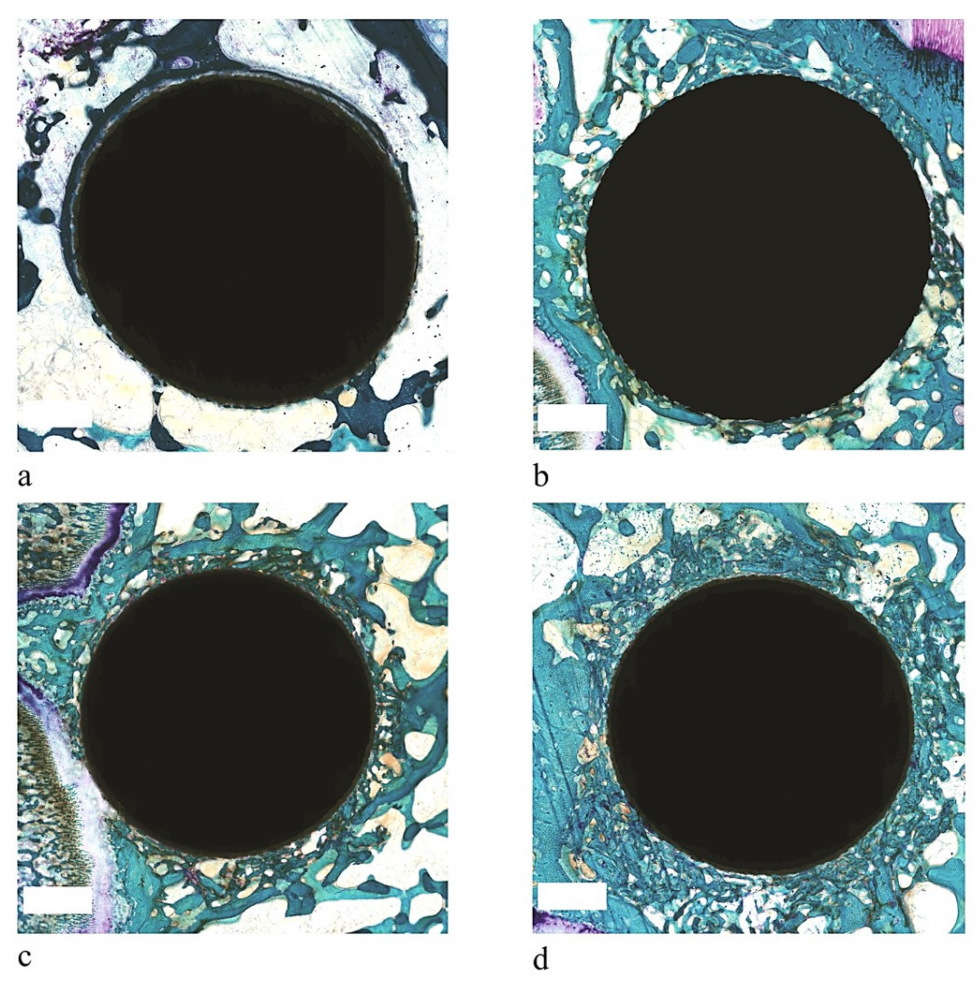
As shown in Table 1 TGF-β1, PDGF-AB, VEGF, and IL-1β levels were higher in the activated PRP (p < 0.0005) in comparison with plasma and non-activated PRP values.

3.2. Histology

At 2 weeks, thin and dense trabeculae reached the implant surfaces of Ti-HA, Ti-HA + BMSC, Ti-HA + PRP, Ti-HA + BMSC + PRP implants (Figure 1). As shown in Figure 1a areas of poor bone-to-implant contact were observed on the Ti-HA implant. Conversely, Ti-HA + PRP (Figure 1b), Ti-HA + BMSC (Figure 1c), Ti-HA + PRP + BMSC (Figure 1d) exhibited a well-integrated and well-defined continuous layer of new bone around the implants with no intervening fibrous tissue. However, in Ti-HA + PRP + BMSC, the bone tissue response was more marked, not only in comparison to Ti-HA alone but also in comparison to Ti-HA + PRP and Ti-HA + BMSC (Figure 1). Thin bone trabeculae around the Ti-HA + PRP + BMSC implant and more mature trabecular tissue at the periphery of the implant were seen (Figure 1d).
Ti-HA + PRP + BMSC implant showed higher cellular activity with numerous osteoblasts and a large amount of osteoid tissue along the newly formed trabeculae (Figure 2a–c).
At 4 weeks, bone trabeculae at the interface of all implants had grown in length and diameter and contained well mineralized woven bone (Figure 3a–d). Bone osseointegration and remodeling were more evident with a higher amount of bone integrated with the implants without the interposition of fibrous tissue (Figure 3a–d). However, as at 2 weeks in the Ti-HA + PRP + BMSC implant, the bone tissue response was more evident in comparison to all the other implants and it was almost completely covered with mineralized bone and new bone formation increased (Figure 3d).

3.3. Histomorphometry

As shown in Table 2, at 2 weeks, BIC was significantly higher in Ti-HA + PRP + BMSC than in Ti-HA and Ti-HA + PRP (p < 0.05). BV/TV showed significantly higher values in Ti-HA + PRP (p < 0.05), Ti-HA + BMSC (p < 0.005) and in Ti-HA + PRP + BMSC (p < 0.0005) in comparison to Ti-HA alone. The same trend was observed for Os.V/BV where significant differences were detected between Ti-HA + PRP, Ti-HA + BMSC and Ti-HA + PRP + BMSC in comparison to Ti-HA alone (p < 0.05). Os.S/BS was significantly higher in Ti-HA + PRP + BMSC when compared to all the other implants (p < 0.005). Concerning Os.S/BS, it was significantly higher in Ti-HA + BMSC when compared to Ti-HA alone (p < 0.05). Significant changes in Os.Th occurred in Ti-HA + PRP + BMSC when compared to Ti-HA alone (p < 0.05). Finally, in Ti-HA + PRP, Ti-HA + BMSC and Ti-HA + PRP + BMSC the N.Ob/BS showed a significant increase when compared to Ti-HA alone. The N.Ob/BS changed significantly also in Ti-HA + PRP + BMSC in comparison to Ti-HA + BMSC.
As shown in Table 3, at 4 weeks, Ti-HA associated with PRP and BMSC showed the highest BIC value among all tested implants (p < 0.0005). All treatments showed significantly higher BV/TV compared with the Ti-HA alone. Moreover, Ti-HA + PRP + BMSC also highlighted significantly higher BV/TV compared with Ti-HA + PRP and Ti-HA + BMSC (p < 0.005). Supplementary evaluations of Os.V/BV and Os.S/BS showed significant differences between Ti-HA + PRP, Ti-HA + BMSC, Ti-HA + PRP + BMSC and Ti-HA alone. Additionally, Ti-HA + PRP + BMSC implants showed significant higher (p < 0.005) Os.Th than TI-HA alone. Data concerning N.Ob/BS revealed significantly higher values for Ti-HA + PRP, Ti-HA + BMSC and Ti-HA + PRP + BMSC when compared with Ti-HA alone (p < 0.0005). N.Ob/BS revealed also significantly higher values in Ti-HA + PRP + BMSC when compared to Ti-HA + PRP (p < 0.0005) and Ti-HA + BMSC (p < 0.005).
Finally, MAR values showed significant higher values in all implants compared to Ti-HA alone (p < 0.0005). Moreover, Ti-HA + PRP + BMSC revealed significantly higher values when compared to Ti-HA + PRP (p < 0.05). BFR/BS values demonstrated significantly higher values in Ti-HA + PRP + BMSC in comparison to all the other implants (p < 0.0005). Finally, BFR/BS also highlighted significantly higher values between Ti-HA + PRP, Ti-HA + BMSC and Ti-HA alone (p < 0.005).

4. Discussion

Bone tissue regeneration and osseointegration remain difficult challenges in orthopedic surgery especially for the elderly population and in presence of co-morbidities and prosthesis revision surgeries. In these cases, a reduction in the number of stem cells, a reduction in the migration, proliferation, or differentiation capacity of these cells, and an altered vascularization, as well as altered bone tissue properties lead to a lower regenerative capacity [50,51,52,53,54,55,56]. To date, several strategies have been developed based on the dogma that the efficiency and efficacy of any successful bone implants commonly can be ascribed to one or more of three properties: osteogenic cells, osteoconduction, and osteoinduction [57]. In this context, regenerative medicine appeared as one of the most promising approaches. MSCs are a well-known system for bone tissue regeneration, and, for the clinical practice, a more advantageous approach would be the so-called ‘one-step surgical procedure’ [58]. Although only a limited number of clinical case series have been performed, this approach has the potential to overcome specific limitations of ‘cell-based two-step procedures’, representing a promising option for bone tissue regeneration and osseointegration [58]. However, bone regeneration depends not only on the seed cells but also on the extracellular cytokines. Therefore, the application of platelet products, such as PRP, capable of releasing and storing cytokines appears to be an attractive approach in tissue regeneration. PRP contains cell adhesion molecules and chemotactic properties that support the recruitment of MSCs and fibroblasts to the repair site [59]. In the present study, we evaluated whether the combined use of BMSCs and PRP is able to improve bone regeneration and osseointegration of a clinical used HA-coated Ti implant. We have chosen this implant since it has shown excellent clinical results in terms of osseointegration and bone healing and thus allows us to better highlight the effect of the tested biological adjuvants, i.e., BMSCs and PRP, even in the presence of a surface with a high osseointegrative potential. In addition, to assess the potential of an implant to effectively form bone, it is critical to understand the primary biological response short investigation times (2 and 4 weeks after implantation) have been selected in this study. These short evaluation times have been chosen because in longer times the effective osseointegration of Ti covered by HA would not have made it possible to analyze the effects linked to the presence of PRP and BMSCs in the speeding up of the biological bone healing process.
The study demonstrated that PRP and BMSCs applied to an HA-coated Ti implant were effective in promoting bone regeneration and osseointegration. However, when used in association, the PRP and BMSCs have synergistic effects that result in better bone repair and osseointegration as compared to PRP or MSCs alone. An accelerated bone healing at the bone–implant interface was observed in the combined treatments, i.e., Ti-HA + PRP, Ti-HA + BMSC, and Ti-HA + BMSC + PRP, at both 2 and 4 weeks after materials implantation. However, at 4 weeks Ti-HA + PRP + BMSC implants showed the highest volume of regenerated bone also when compared to Ti-HA + PRP and Ti-HA + BMSC. Similar results were also found for osseointegration. At 2 weeks, morphometric data highlighted higher osseointegration in Ti-HA associated with both biological stimulators in comparison to Ti-HA alone and Ti-HA + PRP. The synergic effect of PRP and BMSCs was maximized at 4 weeks when the highest osseointegration values were found in Ti-HA + BMSC + PRP implants. These data were further confirmed by the bone turnover parameters. As for newly formed bone, the osteoid volume and surface were higher in the combined treatments than in Ti-HA alone, providing more viable tissue at both experimental times. Likewise, osteoid thickness revealed higher values in Ti-HA + PRP + BMSC than in Ti-HA alone at each experimental time. The increases in osteoid parameters in Ti-HA combined with PRP and BMSC are an indicator of improved mineralization processes, thus highlighting osteoblast activation, also confirmed by a significant increase in the number of osteoblasts at both experimental times. In addition, at 4 weeks the percentage of mineralizing bone surface, i.e., mineral apposition rate (MAR), showed the highest value in Ti-HA + BMSC + PRP. In this study, the combined use of autologous BMSCs and activated PRP had a stimulating effect on bone formation, mineralization, and osseointegration. This is potentially due to the fact that when PRP was activated the platelet degranulation led to an increased release of GFs, as detected by the higher values of GFs in the activated PRP in comparison with plasma and inactivated PRP detected in our study. Moreover, as further confirmed in our study by the higher IL-1β values in activated PRP, platelets are a source of IL-1β that is implicated to differing levels in stimulating the chemotaxis, proliferation, and maturation of the cells, regulating inflammatory molecules and attracting leukocytes. These results confirmed that the GFs within the PRP effect and stimulate BMSCs [60,61]. Some studies showed that the efficacy of MSCs therapies is frequently restricted due to two factors, i.e., the decreased survival capacity of the transplanted cells due to the harsh acidic, hypoxic, and avascular microenvironment at the lesion site, and the absence of control over their differentiation into the damaged tissues [62,63]. Thus, since GFs such as IGF, TGF-β, and VEGF present in platelets can stimulate BMSCs proliferation, reduce apoptosis and chromosomal instability, delay the appearance of senescent features, and enhance bone mineralization, the use of PRP in association with BMSCs may help to overcome the limit linked to the efficacy of MSCs therapies [25,32,33,34,35,36,37]. Additionally, MSCs cultured with PRP were also able to maintain their immune-privileged by modifying cytokine secretion of immune cells (T lymphocytes, B cells, and natural killer cells) to an anti-inflammatory profile and hindered amplification of adaptive immune response [64,65,66]. Finally, besides the stimulating action on MSCs, the GFs released by PRP could also have a direct effect on the cells residing at the defect site [64].
Although this study was limited by a small sample size, our findings support the use of a combination of autologous BMSCs, isolated by one-step procedure, with autologous activated PRP on the biomechanically stable bone implant, already of clinical use, as an easy and physiological way of application for cells and GFs for the improvement of bone regeneration and osseointegration, satisfying the requirements of the ‘ideal’ bone implant, i.e., progenitor cells for osteogenesis, material for osteoconduction, and growth factors for osteoinduction. Noticeably, following the recent advances in material technologies with novel smart or intelligent materials, magnetoelectric material, biomimetic 3D and 4D printing materials, nanomaterials, and new and advanced implants coatings, the efficacy and effect of these biological enhancers will have to be evaluated also with these materials, thus, to be able to work in the multi- and interdisciplinary biomaterials arena, extending from their design to novel uses.
In conclusion, the results of this study could be easily translated into the clinical scenario particularly for patients at risk for osteolysis and aseptic loosening: a surface already used in the clinical setting was tested, PRP is already used clinically for several pathologies and the use of a ‘one-step surgical procedure’ to harvest BMSCs was followed to overcome lifelong and costly in vitro culture expansion, thus preventing the cell phenotype transformation and overwhelming the strict rules on cell manipulation that require Good Manufacturing Practices facility for MSCs clinical applications.

Author Contributions

Conceptualization, F.S., D.D., and M.F.; Data curation, N.D.P., M.S., G.D.S., and C.S.; Formal analysis, G.G.; Investigation, F.S., M.S., and L.M.; Methodology, F.S., N.D.P., M.S., G.G., L.M., G.D.S., and C.S.; Supervision, M.F.; Validation, M.F.; Writing—original draft, F.S.; Writing—review & editing, N.D.P., M.S., G.G., L.M., G.D.S., C.S., D.D., and M.F. All authors have read and agreed to the published version of the manuscript.

Funding

This work was supported by Grants from IRCCS-Istituto Ortopedico Rizzoli (Ricerca Corrente).

Institutional Review Board Statement

The study was conducted according to current law at the time of the study (Law by Decree 116/92) and approved by the Ethical Committees of IRCCS Istituto Ortopedico Rizzoli and by the public authorities.

Informed Consent Statement

Not applicable.

Data Availability Statement

Data sharing not applicable.

Conflicts of Interest

The authors declare that there are no conflict of interest.

References

  1. Kirmanidou, Y.; Sidira, M.; Drosou, M.-E.; Bennani, V.; Bakopoulou, A.; Tsouknidas, A.; Michailidis, N.; Michalakis, K. New Ti-Alloys and Surface Modifications to Improve the Mechanical Properties and the Biological Response to Orthopedic and Dental Implants: A Review. BioMed Res. Int. 2016, 2016, 1–21. [Google Scholar] [CrossRef] [Green Version]
  2. Liu, X.; Chu, P.K.; Ding, C. Surface modification of titanium, titanium alloys, and related materials for biomedical applications. Mater. Sci. Eng. R Rep. 2004, 47, 49–121. [Google Scholar] [CrossRef] [Green Version]
  3. Hartmann, E.S.; Köhler, M.I.; Huber, F.; Redeker, J.I.; Schmitt, B.; Schmitt-Sody, M.; Summer, B.; Fottner, A.; Jansson, V.; Mayer-Wagner, S. Factors regulating bone remodeling processes in aseptic implant loosening. J. Orthop. Res. 2017, 35, 248–257. [Google Scholar] [CrossRef] [Green Version]
  4. Overmann, A.L.; Aparicio, C.; Richards, J.T.; Mutreja, I.; Fischer, N.G.; Wade, S.M.; Potter, B.K.; Davis, T.A.; Bechtold, J.E.; Forsberg, J.A.; et al. Orthopaedic osseointegration: Implantology and future directions. J. Orthop. Res. 2019, 38, 1445–1454. [Google Scholar] [CrossRef]
  5. Ahn, T.K.; Lee, D.H.; Kim, T.; Jang, G.C.; Choi, S.; Oh, J.B.; Ye, G.; Lee, S. Modification of Titanium Implant and Titanium Dioxide for Bone Tissue Engineering. In Novel Biomaterials for Regenerative Medicine; Chun, H.J., Park, K., Kim, C.-H., Khang, G., Eds.; Springer: Singapore, 2018; Volume 1077, pp. 355–368. [Google Scholar]
  6. Heimann, R.B. Osseoconductive and Corrosion-Inhibiting Plasma-Sprayed Calcium Phosphate Coatings for Metallic Medical Implants. Metals 2017, 7, 468. [Google Scholar] [CrossRef] [Green Version]
  7. Lombardi, A.V.; Berend, K.R.; Mallory, T.H. Hydroxyapatite-coated Titanium Porous Plasma Spray Tapered Stem: Experience at 15 to 18 Years. Clin. Orthop. Relat. Res. 2006, 453, 81–85. [Google Scholar] [CrossRef] [PubMed]
  8. Fu, T.; Li, H.; Sun, J.; Li, G.; Li, W.; Zhang, H. Facile hydrothermal synthesis of TiO2–CaP nano-films on Ti6Al4V alloy. Trans. Nonferrous Met. Soc. China 2015, 25, 1122–1127. [Google Scholar] [CrossRef]
  9. Fini, M.; Filardo, G.; Tschon, M.; Pischedda, M.; Pacorini, A.; Kon, E.; Giardino, R. Regenerative medicine for the treatment of musculoskeletal overuse injuries in competition horses. Int. Orthop. 2011, 35, 1569–1576. [Google Scholar] [CrossRef] [Green Version]
  10. Weibrich, G.; Hansen, T.; Kleis, W.; Buch, R.; Hitzler, W.E. Effect of platelet concentration in platelet-rich plasma on peri-implant bone regeneration. Bone 2004, 34, 665–671. [Google Scholar] [CrossRef] [PubMed]
  11. Jensen, T.B.; Rahbek, O.; Overgaard, S.; Søballe, K. Platelet rich plasma and fresh frozen bone allograft as enhancement of implant fixation an experimental study in dogs. J. Orthop. Res. 2004, 22, 653–658. [Google Scholar] [CrossRef] [PubMed]
  12. Roldán, J.; Jepsen, S.; Miller, J.; Freitag, S.; Rueger, D.C.; Açil, Y.; Terheyden, H. Bone formation in the presence of platelet-rich plasma vs. bone morphogenetic protein-7. Bone 2004, 34, 80–90. [Google Scholar] [CrossRef] [PubMed]
  13. Mazor, Z.; Peleg, M.; Garg, A.K.; Luboshitz, J. Platelet-Rich Plasma for Bone Graft Enhancement in Sinus Floor Augmentation With Simultaneous Implant Placement: Patient Series Study. Implant. Dent. 2004, 13, 65–72. [Google Scholar] [CrossRef] [PubMed]
  14. Fontana, S.; Olmedo, D.G.; Linares, J.A.; Guglielmotti, M.B.; Crosa, M.E. Effect of Platelet-Rich Plasma on the Peri-implant Bone Response::An Experimental Study. Implant Dent. 2004, 13, 73–78. [Google Scholar] [CrossRef]
  15. Yin, D.; Wang, Z.; Gao, Q.; Sundaresan, R.; Parrish, C.; Yang, Q.; Krebsbach, P.H.; Lichtler, A.C.; Rowe, D.W.; Hock, J.; et al. Determination of the Fate and Contribution of Ex Vivo Expanded Human Bone Marrow Stem and Progenitor Cells for Bone Formation by 2.3ColGFP. Mol. Ther. 2009, 17, 1967–1978. [Google Scholar] [CrossRef] [PubMed]
  16. Jäger, M.; Hernigou, P.; Zilkens, C.; Herten, M.; Li, X.; Fischer, J.; Krauspe, R. Cell therapy in bone healing disorders. Orthop. Rev. 2010, 2, e20. [Google Scholar] [CrossRef] [Green Version]
  17. Livingston, T.L.; Gordon, S.; Archambault, M.; Kadiyala, S.; McIntosh, K.; Smith, A.; Peter, S.J. Mesenchymal stem cells combined with biphasic calcium phosphate ceramics promote bone regeneration. J. Mater. Sci. Mater. Electron. 2003, 14, 211–218. [Google Scholar] [CrossRef] [PubMed]
  18. Mankani, M.H.; Kuznetsov, S.A.; Robey, P.G. Formation of hematopoietic territories and bone by transplanted human bone marrow stromal cells requires a critical cell density. Exp. Hematol. 2007, 35, 995–1004. [Google Scholar] [CrossRef]
  19. Fang, D.; Seo, B.-M.; Liu, Y.; Sonoyama, W.; Yamaza, T.; Zhang, C.; Wang, S.; Shi, S. Transplantation of Mesenchymal Stem Cells Is an Optimal Approach for Plastic Surgery. Stem Cells 2007, 25, 1021–1028. [Google Scholar] [CrossRef]
  20. Stanovici, J.; Le Nail, L.-R.; Brennan, M.Á.; Vidal, L.; Trichet, V.; Rosset, P.; Layrolle, P. Bone regeneration strategies with bone marrow stromal cells in orthopaedic surgery. Curr. Res. Transl. Med. 2016, 64, 83–90. [Google Scholar] [CrossRef]
  21. Brennan, M.Á.; Renaud, A.; Amiaud, J.; Rojewski, M.T.; Schrezenmeier, H.; Heymann, D.; Trichet, V.; Layrolle, P. Pre-clinical studies of bone regeneration with human bone marrow stromal cells and biphasic calcium phosphate. Stem Cell Res. Ther. 2014, 5, 114. [Google Scholar] [CrossRef] [Green Version]
  22. Dong, Y.; Chen, X.; Hong, Y. Tissue-Engineered Bone Formation In Vivo for Artificial Laminae of the Vertebral Arch Using β-Tricalcium Phosphate Bioceramics Seeded With Mesenchymal Stem Cells. Spine 2013, 38, E1300–E1306. [Google Scholar] [CrossRef]
  23. Hernigou, P.; Poignard, A.; Beaujean, F.; Rouard, H. Percutaneous Autologous Bone-Marrow Grafting for NonunionsInfluence of the Number and Concentration of Progenitor Cells. J. Bone Jt. Surg. Am. Vol. 2005, 87, 1430–1437. [Google Scholar] [CrossRef]
  24. Schottel, P.C.; Warner, S.J. Role of Bone Marrow Aspirate in Orthopedic Trauma. Orthop. Clin. N. Am. 2017, 48, 311–321. [Google Scholar] [CrossRef]
  25. Lieberman, J.R.; Daluiski, A.; Einhorn, T.A. The role of growth factors in the repair of bone. J. Bone Jt. Surg. Am. Vol. 2002, 84, 1032–1044. [Google Scholar] [CrossRef]
  26. Tschon, M.; Fini, M.; Giardino, R.; Filardo, G.; Dallari, D.; Torricelli, P.; Martini, L.; Giavaresi, G.; Kon, E.; Maltarello, M.C.; et al. Lights and shadows concerning platelet products for musculoskeletal regeneration. Front. Biosci. 2011, E3, 96–107. [Google Scholar] [CrossRef] [Green Version]
  27. Cole, B.J.; Seroyer, S.T.; Filardo, G.; Bajaj, S.; Fortier, L.A. Platelet-Rich Plasma: Where Are We Now and Where Are We Going? Sports Health A Multidiscip. Approach 2010, 2, 203–210. [Google Scholar] [CrossRef]
  28. Salamanna, F.; Veronesi, F.; Maglio, M.; Della Bella, E.; Sartori, M.; Fini, M. New and Emerging Strategies in Platelet-Rich Plasma Application in Musculoskeletal Regenerative Procedures: General Overview on Still Open Questions and Outlook. BioMed Res. Int. 2015, 2015, 1–24. [Google Scholar] [CrossRef] [PubMed]
  29. Veronesi, F.; Fini, M.; Sartori, M.; Parrilli, A.; Martini, L.; Tschon, M. Pulsed electromagnetic fields and platelet rich plasma alone and combined for the treatment of wear-mediated periprosthetic osteolysis: An in vivo study. Acta Biomater. 2018, 77, 106–115. [Google Scholar] [CrossRef]
  30. Heldin, C.-H.; Westermark, B. PDGF-like growth factors in autocrine stimulation of growth. J. Cell. Physiol. 1987, 133, 31–34. [Google Scholar] [CrossRef] [PubMed]
  31. Andrew, J.G.; Hoyland, J.A.; Freemont, A.J.; Marsh, D.R. Platelet-derived growth factor expression in normally healing human fractures. EMC Cardiol. Angéiologie 1995, 16, 455–460. [Google Scholar] [CrossRef]
  32. Crane, J.L.; Cao, X. Bone marrow mesenchymal stem cells and TGF-β signaling in bone remodeling. J. Clin. Investig. 2014, 124, 466–472. [Google Scholar] [CrossRef] [Green Version]
  33. Barry, F.; Boynton, R.E.; Liu, B.; Murphy, J.M. Chondrogenic Differentiation of Mesenchymal Stem Cells from Bone Marrow: Differentiation-Dependent Gene Expression of Matrix Components. Exp. Cell Res. 2001, 268, 189–200. [Google Scholar] [CrossRef]
  34. Gerber, H.-P.; Vu, T.H.; Ryan, A.M.; Kowalski, J.; Werb, Z.; Ferrara, N. VEGF couples hypertrophic cartilage remodeling, ossification and angiogenesis during endochondral bone formation. Nat. Med. 1999, 5, 623–628. [Google Scholar] [CrossRef] [PubMed]
  35. Maes, C.; Stockmans, I.; Moermans, K.; Van Looveren, R.; Smets, N.; Carmeliet, P.; Bouillon, R.; Carmeliet, G. Soluble VEGF isoforms are essential for establishingepiphyseal vascularization and regulating chondrocyte development and survival. J. Clin. Investig. 2004, 113, 188–199. [Google Scholar] [CrossRef] [PubMed]
  36. Peng, H.; Wright, V.; Usas, A.; Gearhart, B.; Shen, H.-C.; Cummins, J.; Huard, J. Synergistic enhancement of bone formation and healing by stem cell–expressed VEGF and bone morphogenetic protein-4. J. Clin. Investig. 2002, 110, 751–759. [Google Scholar] [CrossRef] [PubMed]
  37. Peng, H.; Usas, A.; Olshanski, A.; Ho, A.M.; Gearhart, B.; Cooper, G.M.; Huard, J. VEGF Improves, Whereas sFlt1 Inhibits, BMP2-Induced Bone Formation and Bone Healing Through Modulation of Angiogenesis. J. Bone Miner. Res. 2005, 20, 2017–2027. [Google Scholar] [CrossRef] [PubMed]
  38. Oryan, A.; Alidadi, S.; Moshiri, A. Platelet-rich plasma for bone healing and regeneration. Expert Opin. Biol. Ther. 2015, 16, 213–232. [Google Scholar] [CrossRef]
  39. Eskan, M.A.; Greenwell, H.; Hill, M.; Morton, D.; Vidal, R.; Shumway, B.; Girouard, M.-E. Platelet-Rich Plasma–Assisted Guided Bone Regeneration for Ridge Augmentation: A Randomized, Controlled Clinical Trial. J. Periodontol. 2014, 85, 661–668. [Google Scholar] [CrossRef] [PubMed]
  40. Liebig, B.E.; Kisiday, J.D.; Bahney, C.S.; Ehrhart, N.P.; Goodrich, L.R. The platelet-rich plasma and mesenchymal stem cell milieu: A review of therapeutic effects on bone healing. J. Orthop. Res. 2020, 38, 2539–2550. [Google Scholar] [CrossRef]
  41. Kasten, P.; Vogel, J.; Geiger, F.; Niemeyer, P.; Luginbühl, R.; Szalay, K. The effect of platelet-rich plasma on healing in critical-size long-bone defects. Biomaterials 2008, 29, 3983–3992. [Google Scholar] [CrossRef]
  42. Yamada, Y.; Ueda, M.; Naiki, T.; Takahashi, M.; Hata, K.-I.; Nagasaka, T. Autogenous Injectable Bone for Regeneration with Mesenchymal Stem Cells and Platelet-Rich Plasma: Tissue-Engineered Bone Regeneration. Tissue Eng. 2004, 10, 955–964. [Google Scholar] [CrossRef]
  43. Qi, Y.; Niu, L.; Zhao, T.; Shi, Z.; Di, T.; Feng, G.; Li, J.; Huang, Z. Combining mesenchymal stem cell sheets with platelet-rich plasma gel/calcium phosphate particles: A novel strategy to promote bone regeneration. Stem Cell Res. Ther. 2015, 6, 1–16. [Google Scholar] [CrossRef] [Green Version]
  44. Yu, T.; Pan, H.; Hu, Y.; Tao, H.; Wang, K.; Zhang, C. Autologous platelet-rich plasma induces bone formation of tissue-engineered bone with bone marrow mesenchymal stem cells on beta-tricalcium phosphate ceramics. J. Orthop. Surg. Res. 2017, 12, 178. [Google Scholar] [CrossRef] [Green Version]
  45. Kitoh, H.; Kitakoji, T.; Tsuchiya, H.; Katoh, M.; Ishiguro, N. Transplantation of culture expanded bone marrow cells and platelet rich plasma in distraction osteogenesis of the long bones. Bone 2007, 40, 522–528. [Google Scholar] [CrossRef] [PubMed]
  46. Filho Cerruti, H.; Kerkis, I.; Kerkis, A.; Tatsui, N.H.; da Costa Neves, A.; Bueno, D.F.; Da Silva, M.C.P. Allogenous Bone Grafts Improved by Bone Marrow Stem Cells and Platelet Growth Factors: Clinical Case Reports. Artif. Organs 2007, 31, 268–273. [Google Scholar] [CrossRef] [PubMed]
  47. Shimizu, S.; Tsuchiya, S.; Hirakawa, A.; Kato, K.; Ando, M.; Mizuno, M.; Osugi, M.; Okabe, K.; Katagiri, W.; Hibi, H. Design of a Randomized Controlled Clinical Study of tissue-engineered osteogenic materials using bone marrow-derived mesenchymal cells for Maxillomandibular bone defects in Japan: The TEOM study protocol. BMC Oral Health 2019, 19, 1–8. [Google Scholar] [CrossRef] [Green Version]
  48. Tobita, M.; Uysal, C.A.; Guo, X.; Hyakusoku, H.; Mizuno, H. Periodontal tissue regeneration by combined implantation of adipose tissue-derived stem cells and platelet-rich plasma in a canine model. Cytotherapy 2013, 15, 1517–1526. [Google Scholar] [CrossRef]
  49. Yamada, Y.; Nakamura, S.; Ueda, M.; Ito, K. Osteotome technique with injectable tissue-engineered bone and simultaneous implant placement by cell therapy. Clin. Oral Implant. Res. 2011, 24, 468–474. [Google Scholar] [CrossRef] [PubMed]
  50. Kasper, G.; Mao, L.; Geißler, S.; Draycheva, A.; Trippens, J.; Kühnisch, J.; Tschirschmann, M.; Kaspar, K.; Perka, C.; Duda, G.N.; et al. Insights into Mesenchymal Stem Cell Aging: Involvement of Antioxidant Defense and Actin Cytoskeleton. Stem Cells 2009, 27, 1288–1297. [Google Scholar] [CrossRef] [PubMed]
  51. Geissler, S.; Textor, M.; Kühnisch, J.; Könnig, D.; Klein, O.; Ode, A.; Pfitzner, T.; Adjaye, J.; Kasper, G.; Duda, G.N. Functional Comparison of Chronological and In Vitro Aging: Differential Role of the Cytoskeleton and Mitochondria in Mesenchymal Stromal Cells. PLoS ONE 2012, 7, e52700. [Google Scholar] [CrossRef] [Green Version]
  52. Yukata, K.; Xie, C.; Li, T.-F.; Takahata, M.; Hoak, D.; Kondabolu, S.; Zhang, X.; Awad, H.A.; Schwarz, E.M.; Beck, C.A.; et al. Aging periosteal progenitor cells have reduced regenerative responsiveness to bone injury and to the anabolic actions of PTH 1-34 treatment. Bone 2014, 62, 79–89. [Google Scholar] [CrossRef] [Green Version]
  53. D’Ippolito, G.; Schiller, P.C.; Ricordi, C.; Roos, B.A.; Howard, G.A. Age-Related Osteogenic Potential of Mesenchymal Stromal Stem Cells from Human Vertebral Bone Marrow. J. Bone Miner. Res. 1999, 14, 1115–1122. [Google Scholar] [CrossRef]
  54. Lu, C.; Hansen, E.; Sapozhnikova, A.; Hu, D.; Miclau, T.; Marcucio, R.S. Effect of age on vascularization during fracture repair. J. Orthop. Res. 2008, 26, 1384–1389. [Google Scholar] [CrossRef] [Green Version]
  55. Checa, S.; Hesse, B.; Roschger, P.; Aido, M.; Duda, G.N.; Raum, K.; Willie, B.M. Skeletal maturation substantially affects elastic tissue properties in the endosteal and periosteal regions of loaded mice tibiae. Acta Biomater. 2015, 21, 154–164. [Google Scholar] [CrossRef]
  56. Aido, M.; Kerschnitzki, M.; Hoerth, R.; Checa, S.; Spevak, L.; Boskey, A.L.; Fratzl, P.; Duda, G.N.; Wagermaier, W.; Willie, B.M. Effect of in vivo loading on bone composition varies with animal age. Exp. Gerontol. 2015, 63, 48–58. [Google Scholar] [CrossRef] [PubMed] [Green Version]
  57. Bauer, T.W.; Muschler, G.F. Bone graft materials. An overview of the basic science. Clin. Orthop. Relat. Res. 2000, 371, 10–27. [Google Scholar] [CrossRef]
  58. Veronesi, F.; Giavaresi, G.; Tschon, M.; Borsari, V.; Aldini, N.N.; Fini, M. Clinical Use of Bone Marrow, Bone Marrow Concentrate, and Expanded Bone Marrow Mesenchymal Stem Cells in Cartilage Disease. Stem Cells Dev. 2013, 22, 181–192. [Google Scholar] [CrossRef]
  59. Gobbi, A.; Fishman, M. Platelet-rich Plasma and Bone Marrow–derived Mesenchymal Stem Cells in Sports Medicine. Sports Med. Arthrosc. Rev. 2016, 24, 69–73. [Google Scholar] [CrossRef] [PubMed] [Green Version]
  60. Vogel, J.P.; Szalay, K.; Geiger, F.; Kramer, M.; Richter, W.; Kasten, P. Platelet-rich plasma improves expansion of human mesenchymal stem cells and retains differentiation capacity andin vivobone formation in calcium phosphate ceramics. Platelets 2006, 17, 462–469. [Google Scholar] [CrossRef] [PubMed]
  61. Yun, J.-H.; Han, S.-H.; Choi, S.-H.; Lee, M.-H.; Lee, S.-J.; Song, S.U.; Oh, N. Effects of bone marrow-derived mesenchymal stem cells and platelet-rich plasma on bone regeneration for osseointegration of dental implants: Preliminary study in canine three-wall intrabony defects. J. Biomed. Mater. Res. Part B Appl. Biomater. 2014, 102, 1021–1030. [Google Scholar] [CrossRef]
  62. Rodrigues, M.; Griffith, L.G.; Wells, A. Growth factor regulation of proliferation and survival of multipotential stromal cells. Stem Cell Res. Ther. 2010, 1, 1–12. [Google Scholar] [CrossRef] [Green Version]
  63. Hua, P.; Liu, J.-Y.; Tao, J.; Yang, S.-R. Application and Progress of Combined Mesenchymal Stem Cell Transplantation in the Treatment of Ischemic Cardiomyopathy. BioMed Res. Int. 2015, 2015, 1–6. [Google Scholar] [CrossRef] [PubMed] [Green Version]
  64. Wiafe, B.; Metcalfe, P.D.; Adesida, A.B. Stem Cell Therapy: Current Applications and Potential for Urology. Curr. Urol. Rep. 2015, 16, 77. [Google Scholar] [CrossRef] [PubMed]
  65. Rubio-Azpeitia, E.; Andia, I. Partnership between platelet-rich plasma and mesenchymal stem cells: In vitro experi-ence. Muscles Ligaments Tendons J. 2014, 4, 52–62. [Google Scholar] [CrossRef] [PubMed]
  66. Lana, J.F.S.D.; da Fonseca, L.F.; Macedo, R.D.R.; Mosaner, T.; Murrell, W.; Kumar, A.; Purita, J.; de Andrade, M.A.P. Platelet-rich plasma vs bone marrow aspirate concentrate: An overview of mechanisms of action and orthobiologic synergistic effects. World J. Stem Cells 2021, 13, 155–167. [Google Scholar] [CrossRef] [PubMed]
Figure 1. Histological images at 2 weeks: (a) Ti-HA; (b) Ti-HA+ BMSC; (c) Ti-HA + PRP; (d) Ti-HA + BMSC + PRP. Toluidine Blue, Acid Fucsin, Fast Green staining; magnification 2.5×.
Coatings 11 00840 g001
Figure 2. Histological sections of a Ti-HA + BMSC + PRP implant after 2 weeks. Stain: Stevenel’s blue and Van Gieson’s picro-fuchsin. Magnification 20×. Calcified bone stains a bright pink. Osteoid stain bright green; osteoblasts stain blue. (a) Osteoid-like tissue layer (dashed arrow); (b) Extensive new bone formation as osteoblast-like cells; (c) Cuboidal osteoblast-like cells (continuous arrow).
Coatings 11 00840 g002
Figure 3. Histological images showing bone osteointegration 4 weeks after surgery. (a) Ti-HA; (b) Ti-HA + BMSC; (c) Ti-HA + PRP; (d) Ti-HA + BMSC + PRP. Toluidine Blu, Acid Fuchsin, and Fast Green staining; magnification 2.5×.
Coatings 11 00840 g003
Table 1. Quantitative ELISA analysis of TGF-β1, PDGF-AB, VEGF, and IL-1β content in plasma, inactivated PRP (iPRP), and in CaCl2 activated PRP (aPRP) (mean ± SD).
GFsPlasmaiPRPaPRP
TGF-β1 (ng/mL)52 ± 13175 ± 28 a401 ± 174 b,c
PDGF-AB (ng/mL)8 ± 128 ± 6116 ± 34 b,c
VEGF (ng/mL)230 ± 23382 ± 75 d817 ± 74 b,c
IL-1β (mg/mL)0.8 ± 0.21.3 ± 0.56.0 ± 1.0 b,c
Scheffé post hoc multiple comparison test: a, iPRP versus plasma (p < 0.05); b, aPRP versus plasma (p < 0.0005); c, aPRP versus iPRP (p < 0.0005); d, iPRP versus plasma (p < 0.0005).
Table 2. Histomorphometric data 2 weeks after surgery (median ± SD).
ParametersImplants
Ti-HATi-HA + PRPTi-HA + BMSCTi-HA + BMSC + PRP
BIC (%)53.7 ± 2.551.2 ± 7.061.6 ± 1.471.5 ± 2.0 *
BV/TV (%)51.6 ± 0.264.1 ± 3.7 *67.8 ± 1.3 **79.3 ± 3.3 ***
Os.V/BV (%)0.4 ± 0.11.0 ± 0.1 *1.1 ± 0.4 *2.5 ± 0.3 *
Os.S/BS (%)1.4 ± 0.72.8 ± 0.65.4 ± 1.0 *21.9 ± 4.7 **
Os.Th (μm)6.8 ± 2.814.2 ± 1.113.5 ± 3.517.2 ± 1.3 *
N.Ob/BS (n/mm2)14.6 ± 6.040.7 ± 2.4 *32.1 ± 1.7 *59.0 ± 7.8 *, **
Scheffé’s post hoc multiple comparison tests: BIC: Ti-HA + BMSC + PRP versus Ti-HA, Ti-HA + PRP (* p < 0.05); BV/TV: Ti-HA + PRP (* p < 0.05), Ti-HA + BMSC (** p < 0.005), Ti-HA + BMSC + PRP (*** p < 0.0005) versus Ti-HA; Os.V/BV: Ti-HA + PRP, Ti-HA + BMSC, Ti-HA + BMSC + PRP versus Ti-HA (* p < 0.05). Os.S/BS: Ti-HA + BMSC + PRP versus Ti-HA + PRP, Ti-HA + BMSC, Ti-HA (** p < 0.005); Ti-HA + BMSC versus Ti-HA (* p < 0.05). Os.Th: Ti-HA + BMSC + PRP versus Ti-HA (* p < 0.05). N.Ob/BS: Ti-HA + PRP (* p < 0.05), Ti-HA + BMSC (* p < 0.05), Ti-HA + BMSC + PRP (** p < 0.005) versus Ti-HA; Ti-HA + BMSC + PRP versus Ti-HA + BMSC (* p < 0.05).
Table 3. Histomorphometric data 4 weeks after surgery (median ± SD).
ParametersImplants
Ti-HATi-HA + PRPTi-HA + BMSCTi-HA + BMSC + PRP
BIC (%)55.6 ± 2.163.3 ± 5.365. 6 ± 4.281.7 ± 2.5 ***
BV/TV (%)52.9 ± 0.668.8 ± 0.4 **71.5 ± 2.0 **85.3 ± 4.4 ***, **
Os.V/BV (%)0.1 ± 0.00.6 ± 0.0 ***0.5 ± 0.1 **0.6 ± 0.1 ***
Os.S/BS (%)0.9 ± 0.15.2 ± 0.3 *6.6 ± 1.8 *5.7 ± 1.6 **
Os.Th (μm)2.9 ± 4.49.9 ± 0.29.2 ± 1.011.8 ± 1.5 **
N.Ob/BS (n/mm2)6.3 ± 2.936.9 ± 0.128.9 ± 0.650.7 ± 4.9 ***, **
MAR (μm/day)1.4 ± 0.32.4 ± 0.2 ***2.5 ± 0.1 ***3.0 ± 0.4 ***, *
BFR/BS (μm2/μm3/day)0.6 ± 0.11.6 ± 0.4 **1.7 ± 0.1 **3.3 ± 0.6 ***
Scheffé’s post hoc multiple comparison tests: BIC: Ti-HA + BMSC + PRP versus Ti-HA + PRP, Ti-HA + BMSC and Ti-HA (*** p < 0.0005). BV/TV: Ti-HA + PRP (** p < 0.005), Ti-HA + BMSC (** p < 0.005), Ti-HA + BMSC + PRP (*** p < 0.0005) versus Ti-HA; Ti-HA + BMSC+ PRP versus Ti-HA + PRP and Ti-HA + BMSC (** p < 0.005). Os.V/BV: Ti-HA + PRP (*** p < 0.0005), Ti-HA + BMSC (** p < 0.005), Ti-HA + BMSC + PRP (*** p < 0.0005) versus Ti-HA; Os.S/BS: Ti-HA + PRP(* p < 0.05), Ti-HA + BMSC (* p < 0.05), Ti-HA + BMSC + PRP (** p < 0.005) versus Ti-HA; Os.Th: Ti-HA + BMSC + PRP versus Ti-HA (** p < 0.005); N.Ob/BS: Ti-HA + PRP, Ti-HA + BMSC, Ti-HA + BMSC + PRP versus Ti-HA (*** p < 0.0005); Ti-HA + BMSC + PRP versus Ti-HA + PRP (*** p < 0.0005), Ti-HA + BMSC (** p < 0.005). MAR: Ti-HA + PRP, Ti-HA + BMSC, Ti-HA + BMSC + PRP versus Ti-HA (*** p < 0.0005); Ti-HA + BMSC + PRP versus Ti-HA + PRP (* p < 0.05); BFR/BS: Ti-HA + PRP, Ti-HA + BMSC versus Ti-HA (** p < 0.005); Ti-HA + BMSC + PRP (*** p < 0.0005) versus all tested materials.
Publisher’s Note: MDPI stays neutral with regard to jurisdictional claims in published maps and institutional affiliations.

