
Press-On Force Effect on the Efficiency of Composite Restorations Final Polishing—Preliminary In Vitro Study

 , ,
, , 
Abstract
:1. Introduction
- The increased press-on force affects the composite roughness.
- The pressure force of 2N is the most optimal for polishing.
- The composite materials with different filler diameters require a different pressure force.
2. Materials and Methods
2.1. Materials Used in the Study
2.2. Specimen Preparation
2.3. Finishing Procedures
2.4. Roughness Evaluation
- Ra—arithmetic mean deviation of the profile from the mean line, measured along with the measuring or elementary section
- Rq—mean square deviation of the profile from the mean line along the measurement or elementary section
- Rz—roughness height from the mean line along the measuring or elementary section
- Rt—total profile height.
2.5. Statistical Analysis
3. Results
4. Discussion
5. Conclusions
- The higher-pressure force of the individual polishing discs may generate greater roughness of the composite material at each polishing stage.
- According to our findings, a force of 1 N seems to be much more favorable for polishing.
- A smaller difference in filler diameters in composite material can promote improved polishing.
Author Contributions
Funding
Institutional Review Board Statement
Informed Consent Statement
Data Availability Statement
Acknowledgments
Conflicts of Interest
References
- Goldberg, M. In Vitro and in Vivo Studies on the Toxicity of Dental Resin Components: A Review. Clin. Oral Investig. 2008, 12, 1–8. [Google Scholar] [CrossRef] [PubMed]
- Rogalewicz, R.; Voelkel, A.; Kownacki, I. Application of HS-SPME in the Determination of Potentially Toxic Organic Compounds Emitted from Resin-Based Dental Materials. J. Environ. Monit. 2006, 8, 377–383. [Google Scholar] [CrossRef]
- Gupta, S.K.; Saxena, P.; Pant, V.A.; Pant, A.B. Release and Toxicity of Dental Resin Composite. Toxicol. Int. 2012, 19, 225–234. [Google Scholar] [CrossRef] [Green Version]
- Schmalz, G.; Cieplik, F. Biofilms on Restorative Materials. Monogr. Oral Sci. 2021, 29, 155–194. [Google Scholar] [CrossRef]
- Kurt, A.; Cilingir, A.; Bilmenoglu, C.; Topcuoglu, N.; Kulekci, G. Effect of Different Polishing Techniques for Composite Resin Materials on Surface Properties and Bacterial Biofilm Formation. J. Dent. 2019, 90, 103199. [Google Scholar] [CrossRef]
- Aykent, F.; Yondem, I.; Ozyesil, A.G.; Gunal, S.K.; Avunduk, M.C.; Ozkan, S. Effect of Different Finishing Techniques for Restorative Materials on Surface Roughness and Bacterial Adhesion. J. Prosthet. Dent. 2010, 103, 221–227. [Google Scholar] [CrossRef]
- Yadav, R.D.; Raisingani, D.; Jindal, D.; Mathur, R. A Comparative Analysis of Different Finishing and Polishing Devices on Nanofilled, Microfilled, and Hybrid Composite: A Scanning Electron Microscopy and Profilometric Study. Int. J. Clin. Pediatr. Dent. 2016, 9, 201–208. [Google Scholar] [CrossRef]
- Madhyastha, P.S.; Hegde, S.; Srikant, N.; Kotian, R.; Iyer, S.S. Effect of Finishing/Polishing Techniques and Time on Surface Roughness of Esthetic Restorative Materials. Dent. Res. J. 2017, 14, 326–330. [Google Scholar] [CrossRef]
- Radlanski, R.J. A New Carbide Finishing Bur for Bracket Debonding. J. Orofac. Orthop. 2001, 62, 296–304. [Google Scholar] [CrossRef]
- Nedeljkovic, I.; De Munck, J.; Vanloy, A.; Declerck, D.; Lambrechts, P.; Peumans, M.; Teughels, W.; Van Meerbeek, B.; Van Landuyt, K.L. Secondary Caries: Prevalence, Characteristics, and Approach. Clin. Oral Investig. 2020, 24, 683–691. [Google Scholar] [CrossRef] [PubMed]
- Alhareky, M.; Tavares, M. Amalgam vs. Composite Restoration, Survival, and Secondary Caries. J. Evid. Based Dent. Pract. 2016, 16, 107–109. [Google Scholar] [CrossRef]
- Schwendicke, F.; Lamont, T.; Innes, N. Removing or Controlling? How Caries Management Impacts on the Lifetime of Teeth. Monogr. Oral Sci. 2018, 27, 32–41. [Google Scholar] [CrossRef]
- Endo, T.; Finger, W.J.; Kanehira, M.; Utterodt, A.; Komatsu, M. Surface Texture and Roughness of Polished Nanofill and Nanohybrid Resin Composites. Dent. Mater. J. 2010, 29, 213–223. [Google Scholar] [CrossRef] [PubMed] [Green Version]
- Erdemir, U.; Sancakli, H.S.; Yildiz, E. The Effect of One-Step and Multi-Step Polishing Systems on the Surface Roughness and Microhardness of Novel Resin Composites. Eur. J. Dent. 2012, 6, 198–205. [Google Scholar] [CrossRef] [PubMed] [Green Version]
- Pala, K.; Tekçe, N.; Tuncer, S.; Serim, M.E.; Demirci, M. Evaluation of the Surface Hardness, Roughness, Gloss and Color of Composites after Different Finishing/Polishing Treatments and Thermocycling Using a Multitechnique Approach. Dent. Mater. J. 2016, 35, 278–289. [Google Scholar] [CrossRef] [Green Version]
- Sapra, V.; Taneja, S.; Kumar, M. Surface Geometry of Various Nanofiller Composites Using Different Polishing Systems: A Comparative Study. J. Conserv. Dent. 2013, 16, 559–563. [Google Scholar] [CrossRef] [PubMed] [Green Version]
- Pettini, F.; Corsalini, M.; Savino, M.G.; Stefanachi, G.; Venere, D.D.; Pappalettere, C.; Monno, G.; Boccaccio, A. Roughness Analysis on Composite Materials (Microfilled, Nanofilled and Silorane) After Different Finishing and Polishing Procedures. Open Dent. J. 2015, 9, 357–367. [Google Scholar] [CrossRef] [Green Version]
- Heintze, S.D.; Reinhardt, M.; Müller, F.; Peschke, A. Press-on Force during Polishing of Resin Composite Restorations. Dent. Mater. 2019, 35, 937–944. [Google Scholar] [CrossRef]
- Heintze, S.D.; Forjanic, M.; Rousson, V. Surface Roughness and Gloss of Dental Materials as a Function of Force and Polishing Time in Vitro. Dent. Mater. 2006, 22, 146–165. [Google Scholar] [CrossRef]
- Kocaagaoglu, H.; Aslan, T.; Gürbulak, A.; Albayrak, H.; Taşdemir, Z.; Gumus, H. Efficacy of Polishing Kits on the Surface Roughness and Color Stability of Different Composite Resins. Niger. J. Clin. Pract. 2017, 20, 557–565. [Google Scholar] [CrossRef] [Green Version]
- St-Pierre, L.; Martel, C.; Crépeau, H.; Vargas, M.A. Influence of Polishing Systems on Surface Roughness of Composite Resins: Polishability of Composite Resins. Oper. Dent. 2019, 44, E122–E132. [Google Scholar] [CrossRef] [PubMed]
- Barbosa, S.H.; Zanata, R.L.; Navarro, M.F.D.L.; Nunes, O.B. Effect of Different Finishing and Polishing Techniques on the Surface Roughness of Microfilled, Hybrid and Packable Composite Resins. Braz. Dent. J. 2005, 16, 39–44. [Google Scholar] [CrossRef] [PubMed]
- Bansal, K.; Gupta, S.; Nikhil, V.; Jaiswal, S.; Jain, A.; Aggarwal, N. Effect of Different Finishing and Polishing Systems on the Surface Roughness of Resin Composite and Enamel: An In Vitro Profilometric and Scanning Electron Microscopy Study. Int. J. Appl. Basic Med. Res. 2019, 9, 154–158. [Google Scholar] [CrossRef]
- Ehrmann, E.; Medioni, E.; Brulat-Bouchard, N. Finishing and Polishing Effects of Multiblade Burs on the Surface Texture of 5 Resin Composites: Microhardness and Roughness Testing. Restor. Dent. Endod. 2019, 44, e1. [Google Scholar] [CrossRef]
- Senawongse, P.; Pongprueksa, P. Surface Roughness of Nanofill and Nanohybrid Resin Composites after Polishing and Brushing. J. Esthet. Restor. Dent. 2007, 19, 265–273; discussion 274–275. [Google Scholar] [CrossRef]
- Da Silva, T.M.; Salvia, A.C.R.D.; Carvalho, R.F.; de Pagani, C.; da Rocha, D.M.; da Silva, E.G. Polishing for Glass Ceramics: Which Protocol? J. Prosthodont. Res. 2014, 58, 160–170. [Google Scholar] [CrossRef] [PubMed]
- Al Shayeb, K.N.; Turner, W.; Gillam, D.G. Accuracy and Reproducibility of Probe Forces during Simulated Periodontal Pocket Depth Measurements. Saudi Dent. J. 2014, 26, 50–55. [Google Scholar] [CrossRef] [Green Version]
- Kemaloglu, H.; Karacolak, G.; Turkun, L.S. Can Reduced-Step Polishers Be as Effective as Multiple-Step Polishers in Enhancing Surface Smoothness? J. Esthet. Restor. Dent. 2017, 29, 31–40. [Google Scholar] [CrossRef] [Green Version]
- Wilder, A.D.; Swift, E.J.; May, K.N.; Thompson, J.Y.; McDougal, R.A. Effect of Finishing Technique on the Microleakage and Surface Texture of Resin-Modified Glass Ionomer Restorative Materials. J. Dent. 2000, 28, 367–373. [Google Scholar] [CrossRef]
- De Oliveira, G.U.; Mondelli, R.F.L.; Charantola Rodrigues, M.; Franco, E.B.; Ishikiriama, S.K.; Wang, L. Impact of Filler Size and Distribution on Roughness and Wear of Composite Resin after Simulated Toothbrushing. J. Appl. Oral Sci. 2012, 20, 510–516. [Google Scholar] [CrossRef]
- Marghalani, H.Y. Effect of Filler Particles on Surface Roughness of Experimental Composite Series. J. Appl. Oral Sci. 2010, 18, 59–67. [Google Scholar] [CrossRef] [PubMed] [Green Version]


| Material (Manufacturer) | Type | Matrix | Average Particle Size | Filer Type | Filer Loading Vol % | Shade |
|---|---|---|---|---|---|---|
| Boston (Arcona, Niemce, Poland) | microhybrid composite | organic Bis-GMA, UDMA, Bis-Ema, TEGDMA | 0.7–2 µm | Glass filler, Ba-Al-Si, Ba-Al-B-Si, silica | 78 | A2 |
| Charisma Classic (Kulzer GmbH, Hanau, Germany) | microhybrid composite | Bis-GMA | 0.005–10 µm | Glass filler, Ba-Al-F, feldspar | 61 | A2 |
| Composite Resin | Force | Only Light-Cured | 40 µm Bur | 80 µm Sof-Lex | 40 µm Sof-Lex | 20 µm + 10 µm Sof-Lex |
|---|---|---|---|---|---|---|
| Boston | 1N |  |  |  |  |  |
| 2N |  |  |  |  | ||
| Charisma | 1N |  |  |  |  |  |
| 2N |  |  |  |  |
| Composite Resin | Boston | Charisma | |||||||
|---|---|---|---|---|---|---|---|---|---|
| Force | 1 N | 2 N | 1 N | 2 N | |||||
| Mean | SD | Mean | SD | Mean | SD | Mean | SD | ||
| Ra [µm] | only light-cured | 0.644 | 0.365 | 0.644 | 0.365 | 0.484 | 0.072 | 0.484 | 0.072 |
| 40 µm bur | 0.939 | 0.184 | 1.097 | 0.218 | 0.960 | 0.274 | 1.185 | 0.620 | |
| 80 µm Sof-lex | 0.925 | 0.151 | 2.725 | 2.384 | 1.141 | 0.332 | 1.033 | 0.238 | |
| 40 µm Sof-lex | 1.668 | 0.909 | 2.110 | 1.348 | 1.611 | 1.545 | 1.843 | 0.755 | |
| 20 µm + 10 µm Sof-lex | 1.017 | 0.429 | 2.670 | 1.178 | 1.154 | 0.793 | 1.233 | 0.447 | |
| Rq [µm] | only light-cured | 0.863 | 0.514 | 0.863 | 0.514 | 0.677 | 0.097 | 0.677 | 0.097 |
| 40 µm bur | 1.230 | 0.209 | 1.570 | 0.483 | 1.390 | 0.311 | 1.680 | 0.741 | |
| 80 µm Sof-lex | 1.175 | 0.199 | 3.300 | 2.732 | 1.477 | 0.404 | 1.483 | 0.448 | |
| 40 µm Sof-lex | 2.080 | 1.059 | 2.610 | 1.566 | 1.963 | 1.803 | 2.483 | 0.690 | |
| 20 µm + 10 µm Sof-lex | 1.252 | 0.436 | 3.323 | 1.409 | 1.464 | 1.001 | 1.650 | 0.750 | |
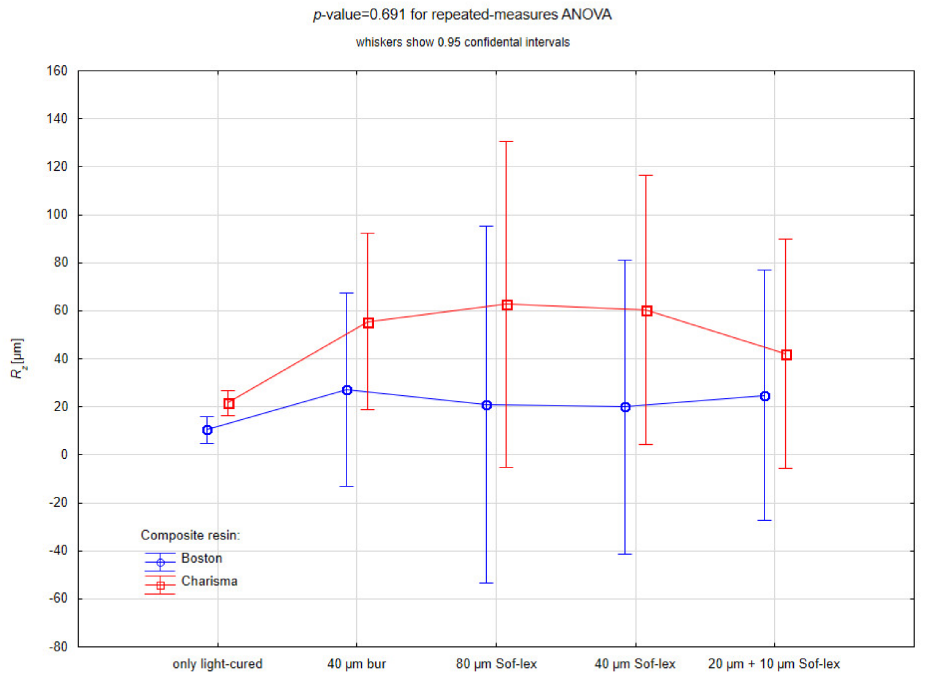
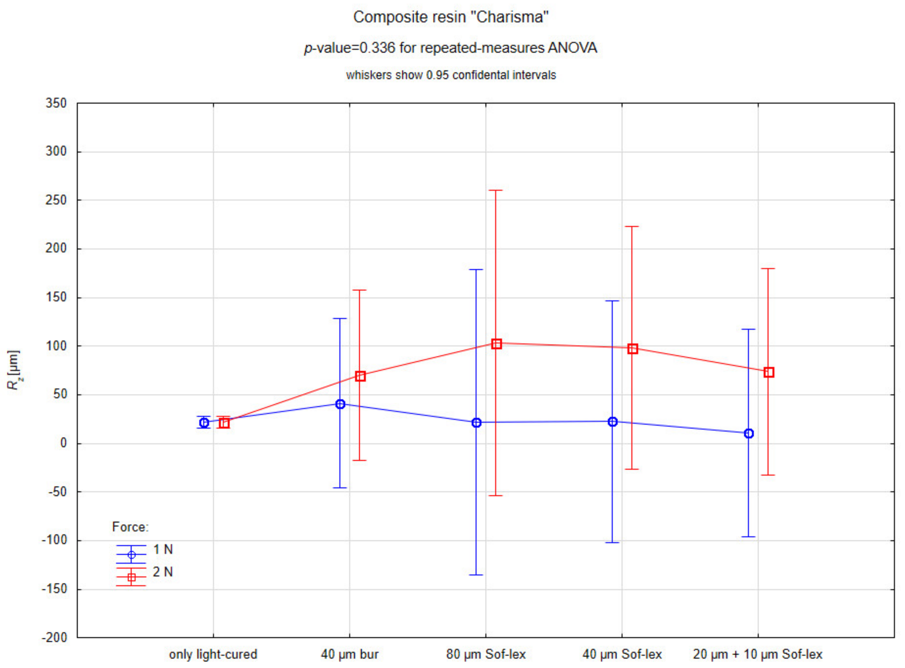
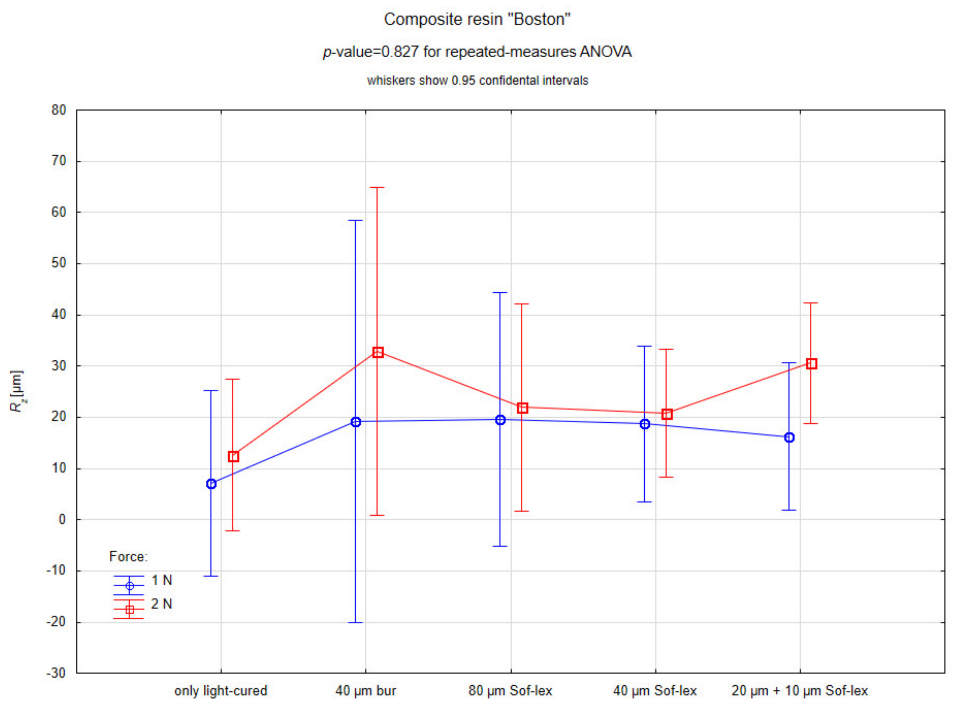
| Rz [µm] | only light-cured | 12.580 | 9.685 | 12.580 | 9.685 | 21.680 | 4.009 | 21.680 | 4.009 |
| 40 µm bur | 20.773 | 7.919 | 32.820 | 20.034 | 41.263 | 10.386 | 69.923 | 76.428 | |
| 80 µm Sof-lex | 20.430 | 7.691 | 21.950 | 11.198 | 21.897 | 14.282 | 103.507 | 137.832 | |
| 40 µm Sof-lex | 20.553 | 3.232 | 20.843 | 8.289 | 22.403 | 10.532 | 98.427 | 109.443 | |
| 20 µm + 10 µm Sof-lex | 16.200 | 1.428 | 30.643 | 7.789 | 10.610 | 6.955 | 73.667 | 93.510 | |
| Rt [µm] | only light-cured | 26.570 | 26.901 | 26.570 | 26.901 | 40.377 | 1.470 | 40.377 | 1.470 |
| 40 µm bur | 40.230 | 19.952 | 57.300 | 22.914 | 78.297 | 24.722 | 99.717 | 89.397 | |
| 80 µm Sof-lex | 51.430 | 23.827 | 41.357 | 23.331 | 33.383 | 21.447 | 153.943 | 199.767 | |
| 40 µm Sof-lex | 35.847 | 9.657 | 31.990 | 21.071 | 39.050 | 14.506 | 132.667 | 126.391 | |
| 20 µm + 10 µm Sof-lex | 39.640 | 4.384 | 49.997 | 16.016 | 20.043 | 10.612 | 94.657 | 110.706 | |
| All Samples | Boston | Charisma | |
|---|---|---|---|
| Ra | 0.575 | 0.318 | 0.985 |
| Rq | 0.541 | 0.307 | 0.966 |
| Rz | 0.431 | 0.827 | 0.336 |
| Rt | 0.529 | 0.882 | 0.296 |
Publisher’s Note: MDPI stays neutral with regard to jurisdictional claims in published maps and institutional affiliations. |
© 2021 by the authors. Licensee MDPI, Basel, Switzerland. This article is an open access article distributed under the terms and conditions of the Creative Commons Attribution (CC BY) license (https://creativecommons.org/licenses/by/4.0/).
Share and Cite
Lehmann, A.; Nijakowski, K.; Potempa, N.; Sieradzki, P.; Król, M.; Czyż, O.; Radziszewska, A.; Surdacka, A. Press-On Force Effect on the Efficiency of Composite Restorations Final Polishing—Preliminary In Vitro Study. Coatings 2021, 11, 705. https://doi.org/10.3390/coatings11060705
Lehmann A, Nijakowski K, Potempa N, Sieradzki P, Król M, Czyż O, Radziszewska A, Surdacka A. Press-On Force Effect on the Efficiency of Composite Restorations Final Polishing—Preliminary In Vitro Study. Coatings. 2021; 11(6):705. https://doi.org/10.3390/coatings11060705
Chicago/Turabian StyleLehmann, Anna, Kacper Nijakowski, Natalia Potempa, Paweł Sieradzki, Mateusz Król, Olaf Czyż, Agnieszka Radziszewska, and Anna Surdacka. 2021. "Press-On Force Effect on the Efficiency of Composite Restorations Final Polishing—Preliminary In Vitro Study" Coatings 11, no. 6: 705. https://doi.org/10.3390/coatings11060705