
Bacterial Adhesion on Prosthetic and Orthotic Material Surfaces

 ,
,
Abstract
:
1. Introduction
2. Materials and Methods
2.1. Materials for the Prosthesis and Orthosis
2.2. Bacteria
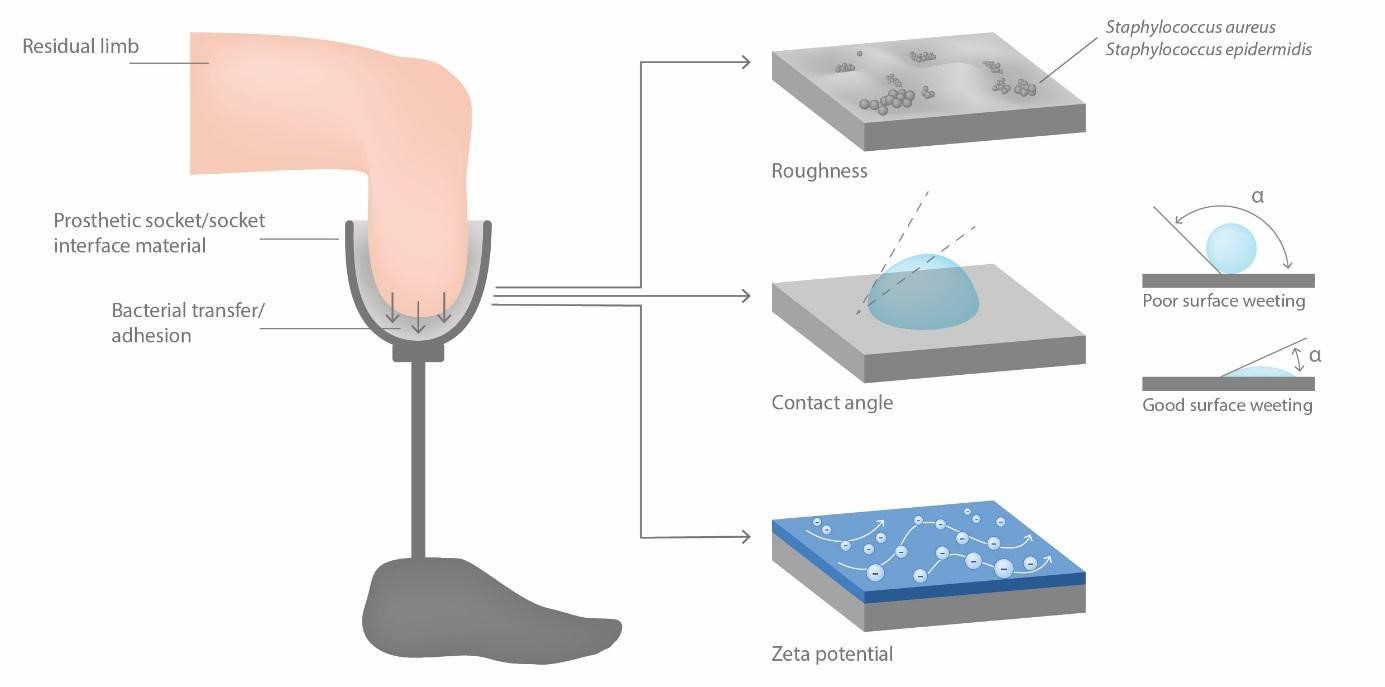
2.3. Roughness
2.4. Contact Angle
2.5. Streaming Potential
2.6. Monitoring the Adhesion Extent
3. Results
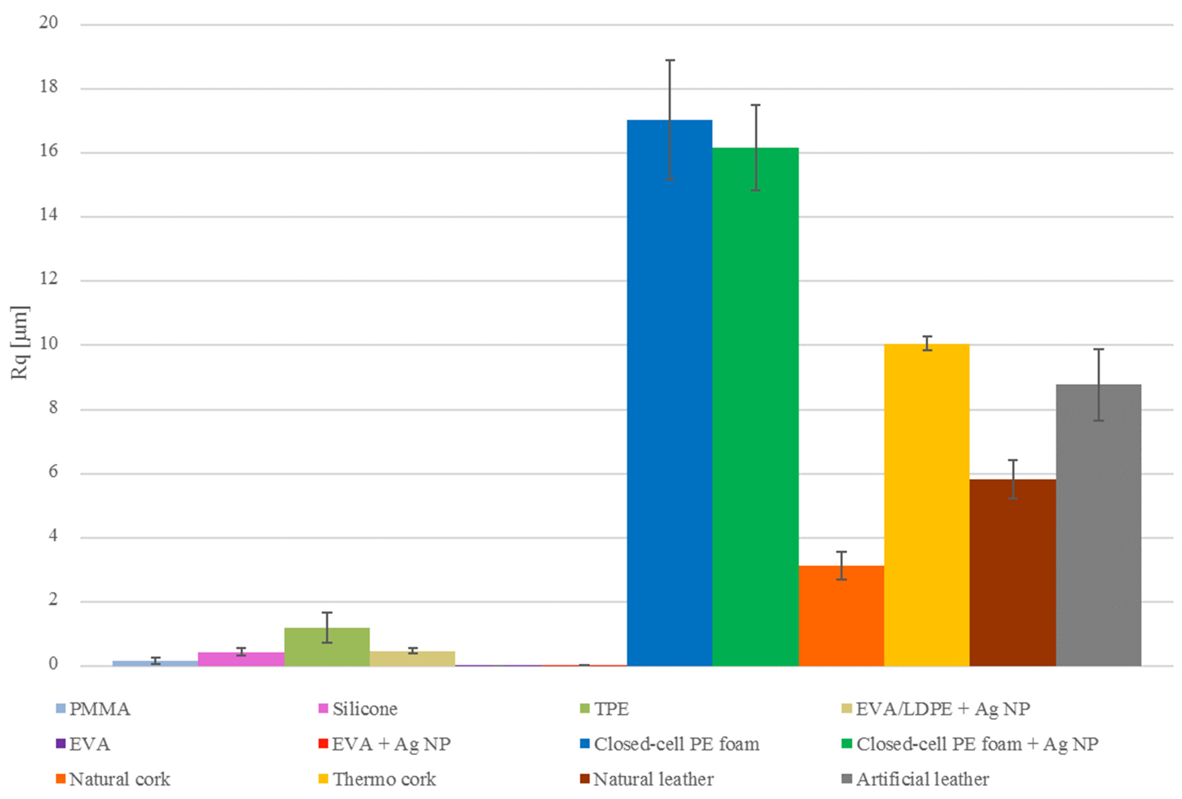
3.1. Roughness
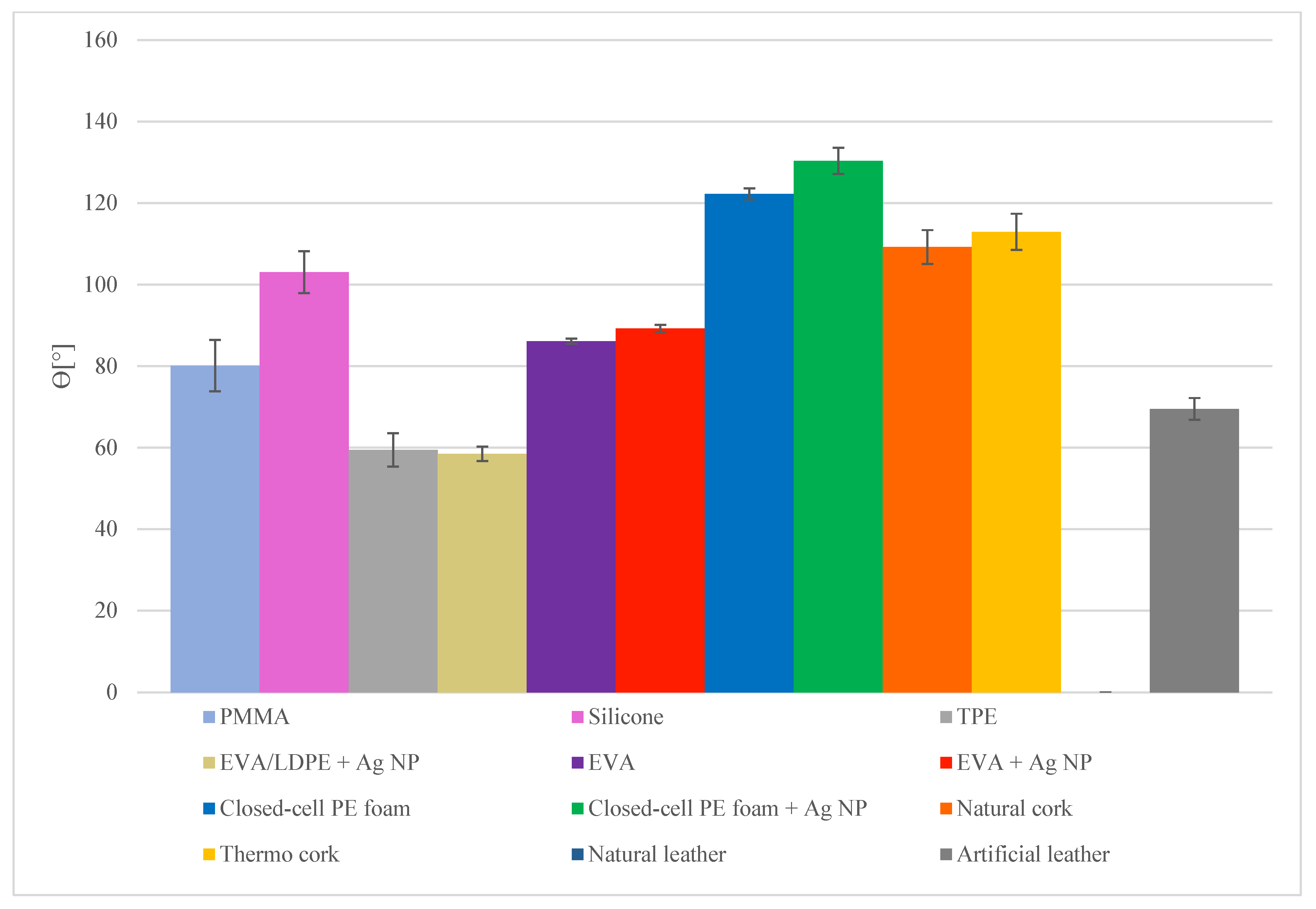
3.2. Contact Angle
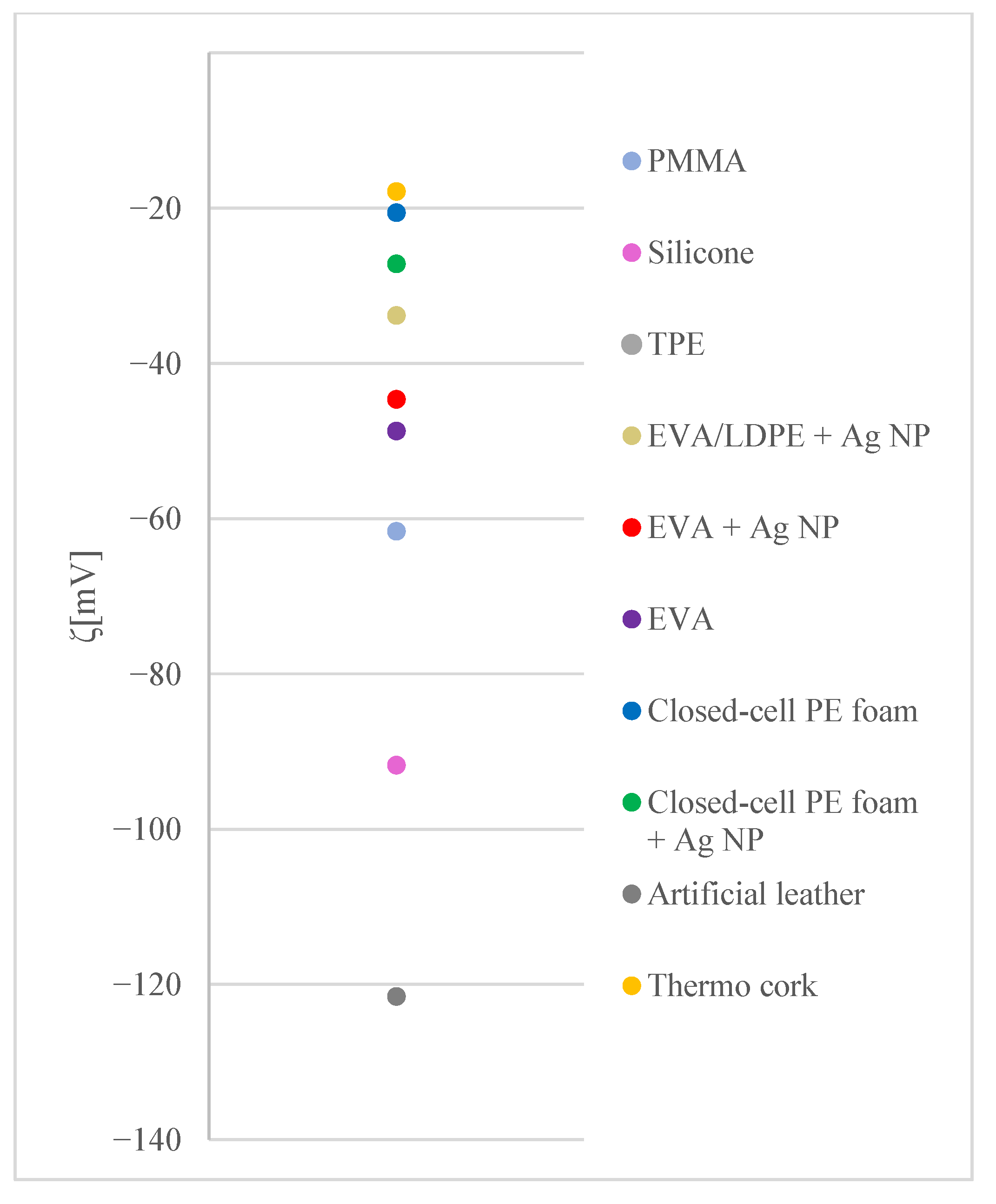
3.3. Zeta Potential
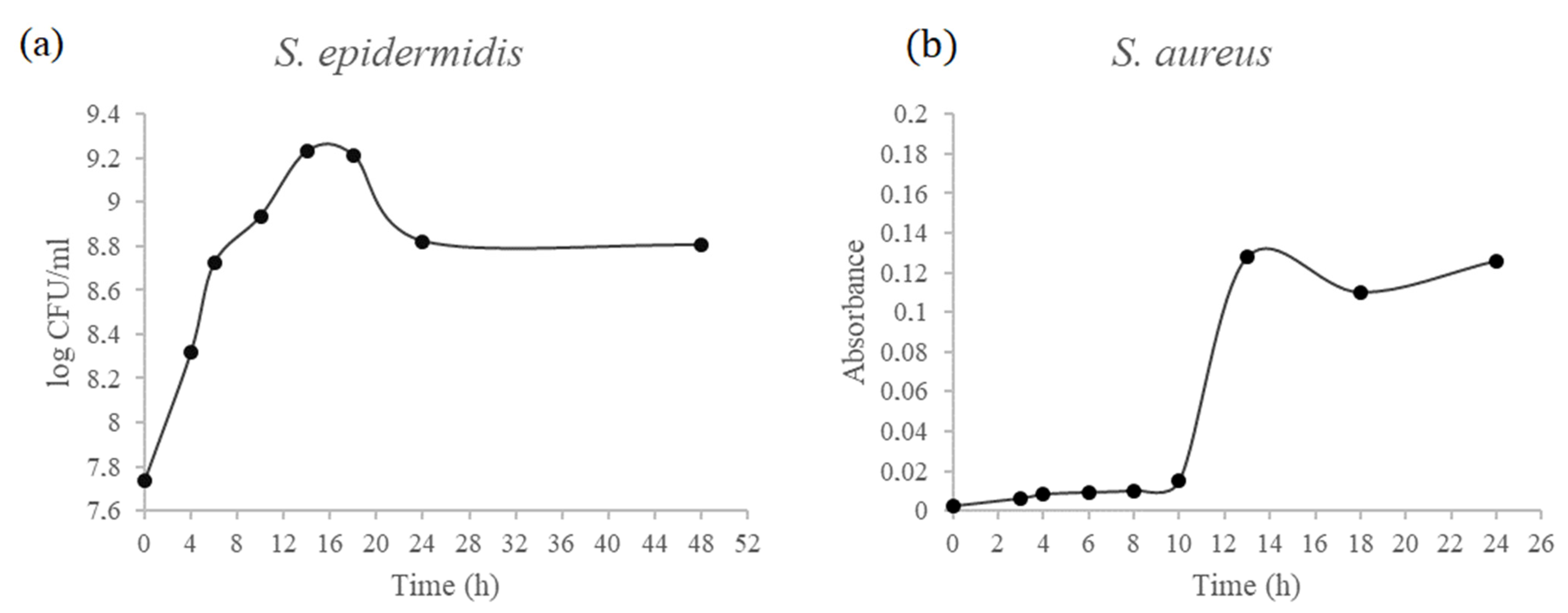
3.4. Growth Curves
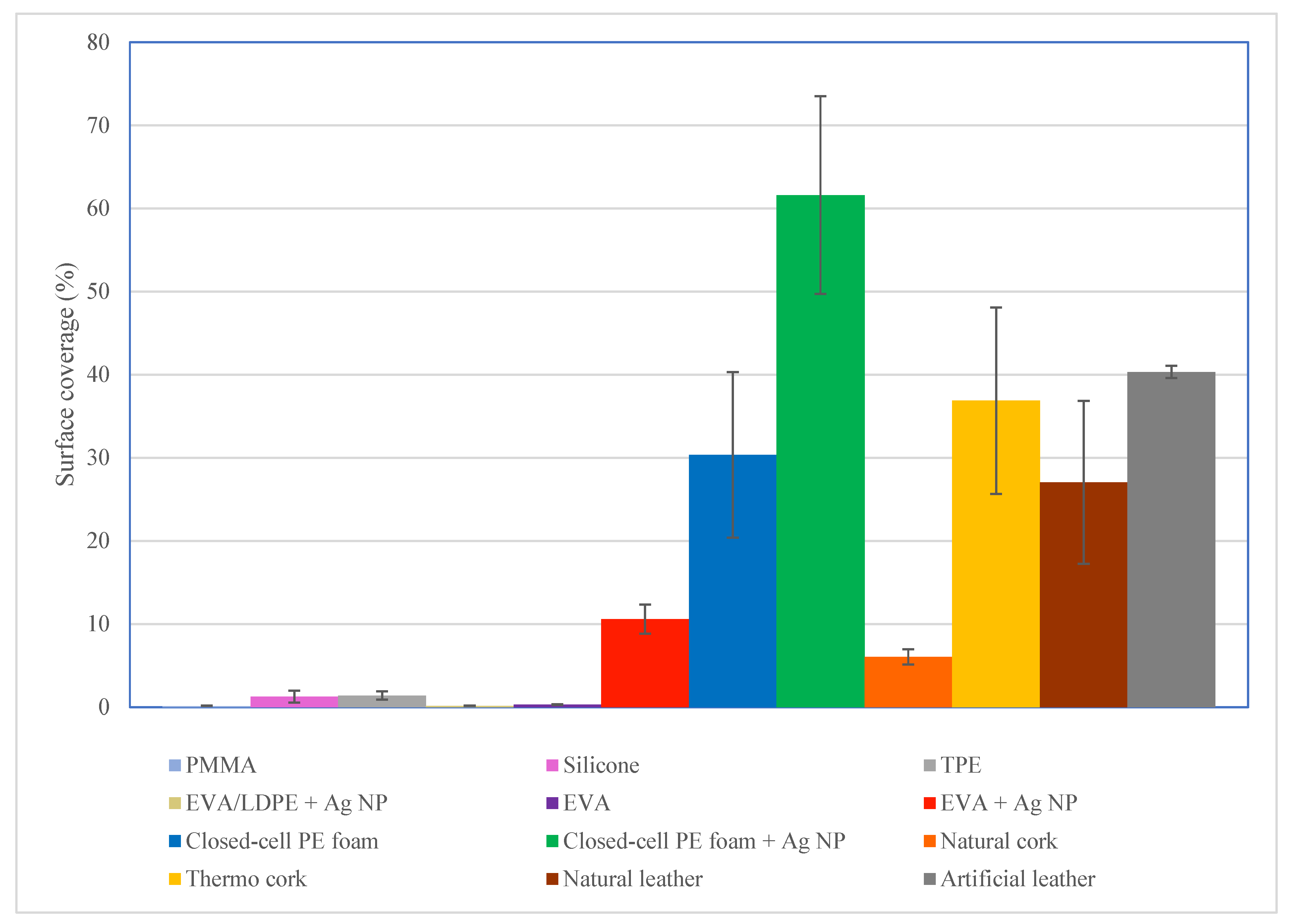
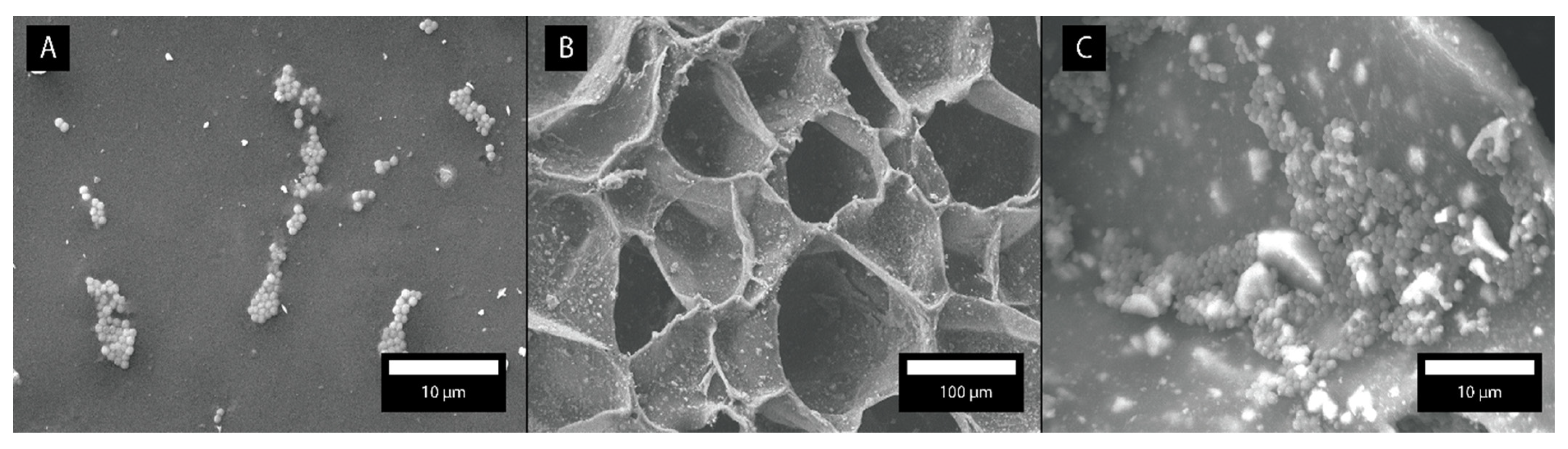
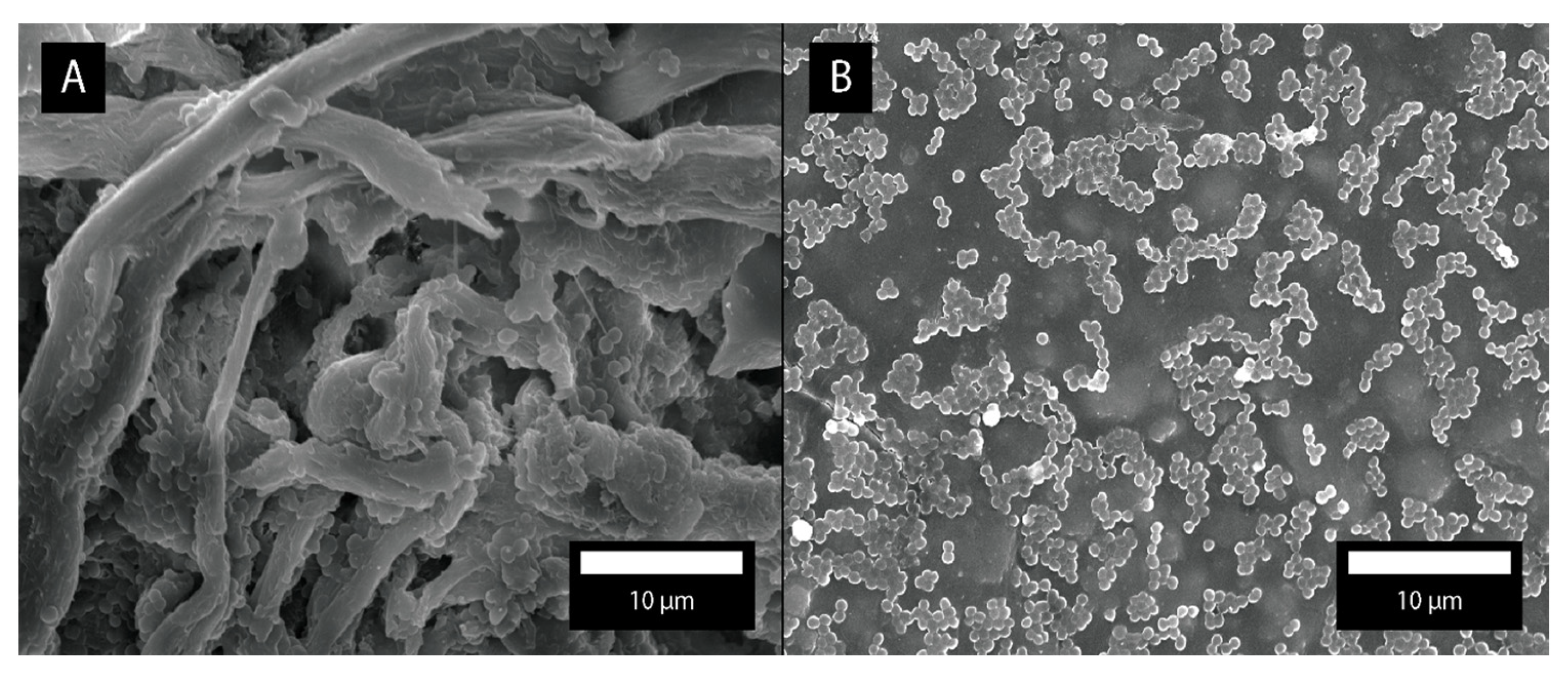
3.5. Bacterial Adhesion Rate
4. Discussion
5. Conclusions
Author Contributions
Funding
Institutional Review Board Statement
Informed Consent Statement
Data Availability Statement
Acknowledgments
Conflicts of Interest
References
- Kogler, G.F. Materials and technology. In Orthotics and Prosthetics in Rehabilitation, 3rd ed.; Lusardi, M., Jorge, M., Nielsen, C., Eds.; Saunders: St. Louis, MO, USA, 2012; pp. 143–160. [Google Scholar]
- Grice, E.A.; Serge, J.A. The skin microbiome. Nat. Rev. Microbiol. 2011, 9, 244–253. [Google Scholar] [CrossRef]
- Coates, R.; Moran, J.; Horsburg, M.J. Staphylococci: Colonisers and pathogens of human skin. Future Microbiol. 2014, 9, 75–91. [Google Scholar] [CrossRef] [PubMed]
- Dalton, H.M.; March, P.E. Molecular genetics of bacterial attachment and biofouling. Curr. Opin. Biotechnol. 1998, 9, 252–255. [Google Scholar] [CrossRef]
- An, Y.H.; Friedman, R.J. Concise review of mechanisms of bacterial adhesion to biomaterial surfaces. J. Biomed. Mater. Res. 1997, 43, 338–348. [Google Scholar] [CrossRef]
- Isaacson, R.E. Pilus Adhesins. In Bacterial Adhesion: Mechanisms and Physiological Significance; Savage, D.C., Madilyn, F., Eds.; Plenum Press: Coventry, UK, 1985; pp. 307–336. [Google Scholar]
- Fletcher, M.; Floodgate, G.D. An electron-microscopic demonstration of as acidic polysaccharide involved in the adhesion of a marine bacterium to solid surfaces. J. Gen. Microbiol. 1973, 74, 325–334. [Google Scholar] [CrossRef] [Green Version]
- Sousa, C.; Teixeira, P.; Oliveria, R. Influence of surface properties on the adhesion of Staphylococcus epidermidis to acrylic and silicone. Int. J. Biomater. 2009, 2009, 1–9. [Google Scholar] [CrossRef] [PubMed] [Green Version]
- Hogt, A.H.; Dankert, J.; de Vries, J.A.; Feijen, J. Adhesion of coagulase-negative staphylococci to biomaterials. J. Gen. Microbiol. 1983, 129, 1959–1968. [Google Scholar] [CrossRef] [Green Version]
- Rimondini, L.; Fare, S.; Brambilla, E.; Felloni, A.; Consonni, C.; Brossa, F.; Carrassi, A. The effect of surface roughness on early in vivo plaque colonization on titanium. J. Periodontol. 1997, 68, 556–562. [Google Scholar] [CrossRef] [PubMed]
- Whitehouse, D.J. Handbook of Surface Metrology; Institute of Physics Publishing: Philadelphia, PA, USA, 1994. [Google Scholar]
- Carvalho, I.; Henriques, M.; Carvalho, S. New strategies to fight bacterial adhesion. In Microbial Pathogens and Strategies for Combating Them: Science, Technology and Education; Méndez-Vilas, A., Ed.; Formatex Research Center: Badajoz, Spain, 2013; Volume 1, pp. 170–178. [Google Scholar]
- Staats, T.B.; Lundt, J. The UCLA total surface bearing suction below-knee prosthesis. Clin. Prosthet. Orthot. 1987, 11, 118–130. [Google Scholar]
- van Loosdrecht, M.C.M.; Lyklema, J.; Norde, W.; Schraa, G.; Zehnder, A.J. Electrophoretic mobility and hydrophobicity as a measure to predict the initial steps of bacterial adhesion. Appl. Environ. Microbiol. 1987, 53, 1898–1901. [Google Scholar] [CrossRef] [Green Version]
- van der Wall, A.; Norde, W.; Zehnder, A.J.B.; Lyklema, J. Determination of the total charge in the cell walls of gram-positive bacteria. Colloid Surf. B 1997, 9, 81–100. [Google Scholar] [CrossRef]
- Zhao, Y.Q.; Sun, Y.; Zhang, Y.; Ding, X.; Zhao, N.; Yu, B.; Zhao, H.; Duan, S.; Xu, F. Well-defined gold nanorod/polymer hybrid coating with inherent antifouling and photothermal bactericidal properties for treating an infected hernia. ACS Nano 2020, 14, 2265–2275. [Google Scholar] [CrossRef] [PubMed]
- Irawan, A.P.; Soemardi, T.P.; Widjajalaksmi, K.; Reksoprodjo, A.H.S. Tensile and flexural strength of ramie fiber reinforced epoxy composites for socket prosthesis application. Int. J. Mech. Mater. Eng. 2010, 6, 46–50. [Google Scholar]
- Shwartz, M. Encyclopedia of Materials, Parts, and Finishes, 2nd ed.; CRC Press LLC: Boca Raton, FL, USA, 2002. [Google Scholar]
- Knapp, D. Transtibial prosthetics. In Orthotics and Prosthetics in Rehabilitation, 3rd ed.; Lusardi, M., Jorge, M., Nielsen, C., Eds.; Saunders: St. Louis, MO, USA, 2012; pp. 628–629. [Google Scholar]
- Lovegreen, W.; Murphy, D.P.; Smith, W.K. Lower limb amputation and gait. In Braddom’s Physical Medicine and Rehabilitation, 5th ed.; Cifu, D., Ed.; Elsevier: Philadelphia, PA, USA, 2016; pp. 191–223. [Google Scholar]
- Bertels, T.; Kettewig, T. Breathable liner for transradial prostheses. In MEC ’11: Raising the Standard, Proceedings of the 2011 MyoElectric Controls Symposium, Fredericton, NB, Canada, 14–19 August 2011; University of New Brunswick: Fredericton, NB, Canada, 2011. [Google Scholar]
- Bottomley, J.M. Footwear: Foundation for lower extremity orthoses. In Orthotics and Prosthetics in Rehabilitation, 3rd ed.; Lusardi, M., Jorge, M., Nielsen, C., Eds.; Saunders: St. Louis, MO, USA, 2012; pp. 161–180. [Google Scholar]
- Gerba, C.P.; Pepper, I.L.; Newby, D.T. Microbial transport in the subsurface. In Environmental Microbiology, 3rd ed.; Pepper, I.L., Gerba, C.P., Gentry, T.J., Eds.; Academic Press: San Diego, CA, USA, 2015; pp. 321–325. [Google Scholar]
- Harris, J.M.; Martin, L.F. An in vitro study of the properties influencing Staphylococcus epidermidis adhesion to prosthetic vascular graft materials. Ann. Surg. 1987, 206, 612–620. [Google Scholar] [CrossRef] [PubMed]
- Zore, A.; Bezek, K.; Jevšnik, M.; Abram, A. Bacterial adhesion rate on food grade ceramics and Teflon as kitchen worktop surfaces. Int. J. Food Microbiol. 2020, 332, 1–5. [Google Scholar] [CrossRef]
- Bohinc, K.; Dražić, G.; Fink, R.; Oder, M.; Jevšnik, M.; Nipič, D.; Godič-Torkar, K.; Raspor, P. Available surface dictates microbial adhesion capacity. Int. J. Adhes. Adhes. 2014, 50, 265–272. [Google Scholar] [CrossRef]
- Todar, K. Todar’s Online Textbook of Bacteriology; Universty of Wisconsin, Department of Bacteriology: Madison, WI, USA, 2005. [Google Scholar]
- Wang, J.; Li, J.; Guo, G.; Wang, Q.; Tang, J.; Zhao, Y.; Qin, H.; Wahafu, T.; Shen, H.; Liu, X.; et al. Silver-nanoparticles-modified biomaterial surface resistant to staphylococcus: New insight into the antimicrobial action of silver. Sci. Rep. 2016, 6, 1–16. [Google Scholar] [CrossRef] [Green Version]
- Lin, W.; Klein, J. Control of surface forces through hydrated boundary layers. Curr. Opin. Colloid Interface Sci. 2019, 44, 94–106. [Google Scholar] [CrossRef]
- Armentano, I.; Arciola, C.R.; Fortunati, E.; Ferrari, D.; Mattioli, S.; Amoroso, C.F.; Rizzo, J.; Kenny, J.M.; Imbriani, M.; Visai, L. The interaction of bacteria with engineered nanostructured polymeric materials: A Review. Sci. World J. 2014, 2014, 410423. [Google Scholar] [CrossRef]
- Murray, P.R.; Rosenthal, K.S.; Pfaller, M.A. Medical Microbiology, 7th ed.; Elsevier Saunders: Philadelphia, PA, USA, 2013. [Google Scholar]
- Sandle, T. Bacterial adhesion: An introduction. J. Valid Technol. 2013, 19, 1–10. [Google Scholar]
- Song, F.; Koo, H.; Ren, D. Effects of material properties on bacterial adhesion and biofilm formation. J. Dent. Res. 2015, 94, 1027–1034. [Google Scholar] [CrossRef] [PubMed]
- Kovačević, D.; Pratnekar, R.; Godič-Torkar, K.; Salopek, J.; Dražić, G.; Abram, A.; Bohinc, K. Influence of polyelectrolyte multilayer properties on bacterial adhesion capacity. Polymers 2016, 8, 345. [Google Scholar] [CrossRef] [PubMed]
- Janovak, L.; Deak, A.; Tallosy, S.P.; Sebok, D.; Csapo, E.; Bohinc, K.; Abram, A.; Palinko, I.; Dekany, I. Hydroxyapatite-enhanced structural, photocatalytic and antibacterial properties of photoreactive TiO2/HAp/polyacrylate hybrid thin films. Surf. Coat. Technol. 2017, 326, 316–326. [Google Scholar] [CrossRef]
- Lin, W.; Zhang, J.; Wang, Z.; Chen, S. Development of robust biocompatible silicone with high resistance to protein adsorption and bacterial adhesion. Acta Biomater. 2011, 7, 2053–2059. [Google Scholar] [CrossRef] [PubMed]
- Vaterrodt, A.; Thallinger, B.; Daumann, K.; Koch, D.; Guebitz, G.M.; Ulbricht, M. Antifouling and Antibacterial Multifunctional Polyzwitterion/Enzyme Coating on Silicone Catheter Material Prepared by Electrostatic Layer-by-Layer Assembly. Langmuir 2016, 32, 1347–1359. [Google Scholar] [CrossRef] [PubMed]


| Material | Product Name | Manufacturer/Supplier |
|---|---|---|
| Ethylene-vinyl acetate (EVA) | ThermoLyn soft | Ottobock |
| Ethylene-vinyl acetate (EVA) with Ag NP | Anitbacterial ThermoLyn | Ottobock |
| Closed-cell polyethylene (PE) | Pedilin | Ottobock |
| Closed-cell polyethylene (PE) with Ag NP | Pedilin SilverShield | Ottobock |
| Natural cork | Flexo-Kork | Ortho-Reha Neuhof |
| Thermo-cork | Thermo-Kork | Ortho-Reha Neuhof |
| Natural leather | Walkleder-Hälse | Ortho-Reha Neuhof |
| Artificial leather | Kunstleder mit Gewebe | Ortho-Reha Neuhof |
| Silicone | Iceross Seal-In® X TF | Ossur |
| Thermoplastic elastomer (TPE) gel | Alpha Hybrid® Liner | WillowWood |
| Ethylene-vinyl acetate/low-density polyethylene (EVA/LDPE) with Ag NP | ThermoLyn EVA/LDPE SilverShield | Ottobock |
| Poly(methyl methacrylate) (PMMA) | Lamineirheartz C | Ortho-Rhea Neuhof |
Publisher’s Note: MDPI stays neutral with regard to jurisdictional claims in published maps and institutional affiliations. |
© 2021 by the authors. Licensee MDPI, Basel, Switzerland. This article is an open access article distributed under the terms and conditions of the Creative Commons Attribution (CC BY) license (https://creativecommons.org/licenses/by/4.0/).
Share and Cite
Abram, A.; Zore, A.; Lipovž, U.; Košak, A.; Gavras, M.; Boltežar, Ž.; Bohinc, K. Bacterial Adhesion on Prosthetic and Orthotic Material Surfaces. Coatings 2021, 11, 1469. https://doi.org/10.3390/coatings11121469
Abram A, Zore A, Lipovž U, Košak A, Gavras M, Boltežar Ž, Bohinc K. Bacterial Adhesion on Prosthetic and Orthotic Material Surfaces. Coatings. 2021; 11(12):1469. https://doi.org/10.3390/coatings11121469
Chicago/Turabian StyleAbram, Anže, Anamarija Zore, Urban Lipovž, Anita Košak, Maja Gavras, Žan Boltežar, and Klemen Bohinc. 2021. "Bacterial Adhesion on Prosthetic and Orthotic Material Surfaces" Coatings 11, no. 12: 1469. https://doi.org/10.3390/coatings11121469