Review

Antimicrobial Peptides-Coated Stainless Steel for Fighting Biofilms Formation for Food and Medical Fields: Review of Literature

1
UMET—Unité Matériaux et Transformations, University of Lille, CNRS, INRAE, Centrale Lille, UMR 8207, F-59000 Lille, France
2
Laboratoire d’analyses Chimiques et Microbiologiques, Faculté de Santé Publique, Université Libanaise, Saida 1600, Lebanon
*
Author to whom correspondence should be addressed.
Coatings 2021, 11(10), 1216; https://doi.org/10.3390/coatings11101216
Submission received: 18 August 2021 / Revised: 30 September 2021 / Accepted: 1 October 2021 / Published: 4 October 2021
(This article belongs to the Topic Multiple Application for Novel and Advanced Materials)

Abstract

:
Emerging technology regarding antimicrobial coatings contributes to fighting the challenge of pathogenic bacterial biofilms in medical and agri-food environments. Stainless steel is a material widely used in those fields since it has satisfying mechanical properties, but it, unfortunately, lacks the required bio-functionality, rendering it vulnerable to bacterial adhesion and biofilm formation. Therefore, this review aims to present the coatings developed by employing biocides grafted on stainless steel. It also highlights antimicrobial peptides (AMPs)used to coat stainless steel, particularly nisin, which is commonly accepted as a safe alternative to prevent pathogenic biofilm development.

Graphical Abstract

1. Introduction

In industrial and medical environments, bacteria adhere to accessible surfaces and can grow and develop into a dense biofilm. Biofilm-associated infections in medical devices and food equipment represent a serious public health burden and negatively impact the proper functioning of the instrument, resulting in huge economical loss.
The biofilm is constructed of a complex consortium of microorganisms encased in an extracellular polymeric matrix. Bacterial invasion of a substrate is a multi-stage phenomenon that includes biological and physico-chemical factors. Biofilm growth can be explained as a five-step process. The first step in biofilm formation is the adsorption of a so-called conditional layer that consists mainly of complex exopolymeric substances, as these substances are already present in the aqueous environment emitted by the microorganisms. Secondly, if the conditions are favorable, a transformation from reversible to irreversible adhesion occurs and extracellular polymers (EPS) are secreted by the bacteria; then early development of the biofilm structure starts and the formed micro-colonies develop into a mature biofilm. Finally, dispersion of the biofilm cells occurs in the neighboring environment [1]. Many efforts have been made to control biofilm formation in the food industry and hospitals, especially by actively cleaning and disinfecting surfaces. Yet, this antimicrobial treatment can be compromised due to disinfectants failing to penetrate the biofilm matrix that is attached to the surface, or bacterial resistance [2,3].
This paper focuses on the problematic pathogenic biofilm formation in the food and medical industries. Several materials can be used in these fields, such as Teflon, PET, titanium, or stainless steel. Each material has its own specificity, and the bacterial adhesion rate can be different on each material, depending on its nature and surface properties. For example, the adhesion rate of staphylococcal species on Teflon and stainless steel was compared and the results showed that the majority of strains had a moderate adhesion rate (< 20%) on stainless steel while a higher adhesion rate was observed on the Teflon surface [4]. This paper will focus on stainless steel because it is widely used in medical and food applications [5,6]. This is mainly due to its relatively low cost, ease of fabrication, good corrosion resistance, mechanical characteristics, and good biocompatibility. It is estimated that nearly 60% of surgical implants [5] and about 85% of surgical instruments used in the United States are made of stainless steel [7].
Different surface treatment strategies for the prevention of bacterial adhesion and biofilm formation have been developed. Coatings incorporating antimicrobials have been used under different conditions and their effectiveness depends on the nature of the surface and the surrounding environment [8]. The state of the art of the main classes of biocides grafted on stainless steel, including silver nanoparticles (AgNPs) [9], essential oils (EOs) [10], light-activated antimicrobials [11], cationic molecules [12], antibiotics [13], enzymes [14], and antimicrobial peptides (AMPs) [15], will be presented. Among these methods, surface treatment using AMPs is a very promising way to reduce bacterial contamination by killing adherent microorganisms. Special attention will be given to AMPs-based coatings for their interesting properties such as low toxicity and safety [16]. Generally, AMPs are significantly more effective than classical biocides. They present advantages over conventional antibiotics, with a large spectrum of antibacterial, antifungal, and antiviral properties [17,18]. They are also powerful with fast germ-killing capacity and low bactericidal concentration. They are even efficient on conventional antibiotic-resistant species and also have synergistic effects with classical antibiotics to neutralize endotoxins [17,19]. Moreover, these AMPs are safe, have fewer or even zero toxic side effects, and do not easily induce bacterial drug resistance compared to conventional biocides [20]. Special focus is paid to nisin, a widely used bacteriocin and a healthy option to fight biofilm formation [21]. This bacteriocin is approved by the Joint Food and Agriculture Organization/World Health Organization (FAO/WHO) as a safe food additive and is currently largely applied in biomedical fields [22]. The advantages and disadvantages of each applied biocide will also be given.

2. The Employment of Stainless Steel

In food manufacturing and hospitals, cleaning and disinfection are paramount. The material and equipment selected upstream will affect the future care and disinfection methods. In those fields, metals specifically can provide exceptional strength properties. However, strength is not the only aspect to consider. The most adequate material needs to be as inert and non-corrosive as possible.
Stainless steel is characterized by an addition of chromium of at least 10.5% of the total composition. Chromium is very reactive to oxygen and immediately forms a strong barrier on its outer surface. This barrier is highly resistant and protects internal structures from additional corrosion [23]. Indeed, this stainless-steel alloy is one of the most largely used materials in biomedical and food fields. This metal is selected for its mechanical and chemical stability, biocompatibility, good corrosion resistance, low price, and non-toxicity. It is mostly employed in medical sectors for orthopedic implants and prostheses, cardiovascular valves and stents, and also for 3D printing of custom-made implants [24]. Moreover, in agri-food industries, stainless steel is selected since it does not affect the food’s color or taste without contaminating it. It also provides amazing performance for maintaining food safety by being effortlessly and efficiently washed and sterilized [25].
It is of importance to note that the major issue for researchers is to provide antibacterial and antiadhesive properties to implantable stainless steel. Moreover, it is difficult to elaborate coatings with specific mechanical properties and an adequate antimicrobial/antiadhesive effect at the same time. That is why scientists have tried to modify stainless-steel surfaces via multiple strategies and elaborate coatings with the employment of several antimicrobial molecules.

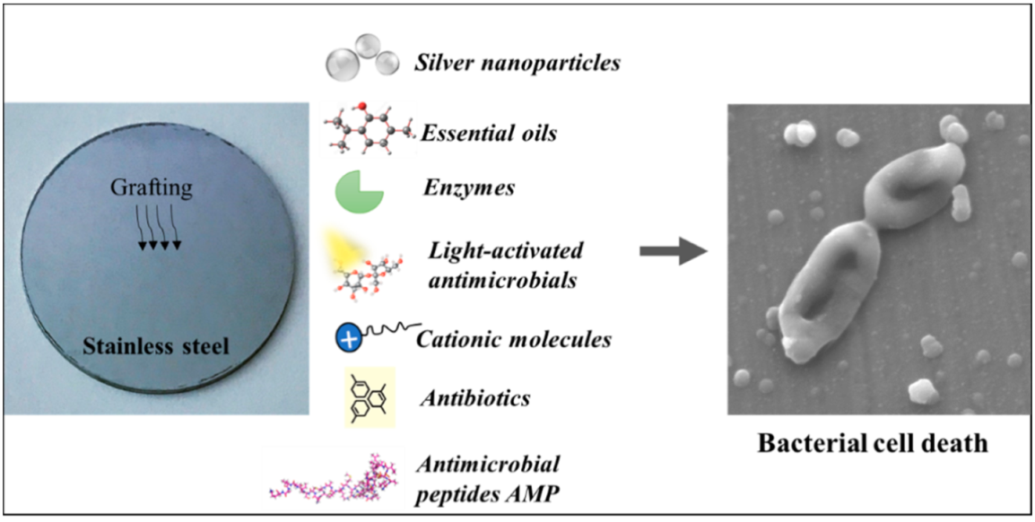
3. Antimicrobial Coatings on Stainless Steel Incorporating Biocides

3.1. Silver Nanoparticles Coated on Stainless Steel

Since ancient times, the most frequently used antibacterial heavy metal is silver. This metal species fights bacteria by disrupting enzymatic activities, disabling the membrane function, and damaging the DNA and oxidative stress [26]. Moreover, silver was demonstrated to exhibit an excellent bacteriostatic and bactericidal effect towards several bacterial species [27]. Different configurations of silver were used to develop the films. Ions, metallic nanoparticles, and silver halide nanoparticles were integrated into Layer-by-Layer (LbL) coatings on stainless steel and afterward released to kill bacteria. AgNPs have been extensively found to possess effective antimicrobial properties related to its oligodynamic action and multiple modes of its biocidal action [28]. Silver-based nanoparticle mixed with the cationic polymer poly(3,4-dihydroxy-L-phenylalanine)-co-poly(2-(methacryloxy)ethyl trimethylammonium chloride) (DOPA) permitted the adhesion enhancement of the LbL coating to a stainless-steel surface. The mixture formed an aqueous suspension with stable AgO and AgCl nanoparticles, which was gathered with the polyanion poly(styrene sulfonate) (PSS) on a stainless-steel surface. Micelles were formed by the interaction between the positively charged DOPA and the negatively charged PSS. This LbL coating imparted efficient antibacterial activity related to the silver ions released from the film, against Escherichia coli strains [29,30]. Moreover, antimicrobial coatings were elaborated by electrodeposition of Ag on stainless steel from an AgNO3 aqueous solution. These films were designed for a fracture repair implant and were harmless to human osteoblasts, preventing in vivo bacterial infection. The coatings were challenged with Pseudomonas aeruginosa, and 13-fold bacterial reduction was observed after 24 h [31]. Otherwise, Cowan et al. [9] evaluated the antibacterial efficacy of stainless-steel coupons coated with a zeolite matrix containing silver and zinc (AgION). These coatings showed antimicrobial behavior against Gram-positive strains like Listeria monocytogenes and Staphylococcus aureus and Gram-negative strains like Pseudomonas aeruginosa and Escherichia coli. In another study, antibacterial coatings based on chitosan and bioglass particles were developed on stainless steel using electrophoretic deposition (EPD). These coatings showed efficient antibacterial activity against Staphylococcus aureus [32]. Moreover, stainless-steel coatings based on AgNP thin layers, elaborated by the reduction of Tollen’s reagent, were developed using formaldehyde radiofrequency plasma functionalization. These coatings’ antimicrobial efficacy was tested and resulted in a 5-log reduction in Listeria monocytogenes populations after 5 h of exposure [33].
Several studies concerning AgNPs toxicity reported that a safe range can be established for the use of AgNPs in designing antimicrobial coatings [28]. However, despite silver’s effective antibacterial activity, the toxic effect of AgNPs against the mammalian cells limited their use [34]. Moreover, Sung et al. [35] outlined that constant exposure to AgNPs beyond 90 days caused inflammatory lesions that considerably weakened the lung function of rats.

3.2. Essential Oils Coated on Stainless Steel

Essential oils (EO) are natural compounds that are produced by plants with an antimicrobial effect for their protection against Gram-negative and Gram-positive bacteria, fungi, viruses, and yeasts [36].
EOs have a variety of structures, and their mechanism of action against microorganisms is not clearly understood. They can be bacteriostatic at a low concentration but bactericidal at higher concentrations [37]. They are known to include hydrophilic and hydrophobic interactions with cell membranes of targeted microorganisms. These interactions cause the increased membrane permeability, leading to the loss of the cell’s vital components such as ions and molecules and ATP production, and finally cell death [38].
The application of essential oils in the development of antimicrobial coatings on stainless steel is aimed precisely at edible particularity. A study demonstrated the effective antimicrobial activity of isoeugenol-coated stainless steel against Staphylococcus aureus, Listeria monocytogenes, and Pseudomonas fluorescens. Indeed, the surface eliminated all viable cells from the coated stainless-steel surface [10]. Moreover, another research study evaluated the bactericidal power of Mentha piperita essential oil-coated stainless steel. The coating highlighted anti-adhesive behavior against Escherichia coli [39].
However, the native odor of EOs might be problematic to their applications to industrials since a consequent concentration of EOs is needed to achieve their antimicrobial efficiency. Furthermore, the presence of EO on equipment used to process food could adversely impact its sensory perception [40]. EOs are effective after migration from the coating. Indeed, EO-coated material will lose its activity with time [41]. Moreover, EO activity can be considerably reduced when exposed to high pH levels and high and low temperatures. It is also diminished in the presence of lipids, proteins, oxygen, and polysaccharides [42]. Furthermore, their non-stability, volatility, and immiscibility in water may cause problems for their applications.

3.3. Light-Activated Antimicrobials Coated on Stainless Steel

Antimicrobial coatings can also be developed using light-activated antimicrobials that are photo-activated compounds. These species might be organic and aromatic like porphyrin, chlorophyll derivatives, or inorganic oxides like titanium dioxide (TiO2). The antimicrobial activity becomes efficient when those compounds are exposed to light of a specific wavelength. The reaction produces reactive oxygen species that interact with the bacterial protective membranes and DNA and results in bacterial death [43,44]. These species are regarded as bacteriostatic rather than bactericidal [45]. Regarding a stainless-steel-coated substrate with those photo-activated compounds, a study involved grafting a thin film of TiO2. This modified stainless steel showed efficient antibacterial activity against bacillus pumilus [11]. Moreover, the frequently touched surfaces play an important role in the spread of bacterial infections in hospitals. In research, nanostructured TiO2 coatings on stainless-steel surfaces were elaborated. They were made via chlorine chemistry and elaborated to kill bacteria under visible light, via enhanced photocatalytic activity. These coatings showed a greater than 3-log reduction in viable Escherichia coli after 4 h of visible light exposure [46]. However, some of these antimicrobial coatings can only be activated by UV light or by very-high-intensity light sources, which may be dangerous to human health [47].

3.4. Cationic Molecules Coated on Stainless Steel

Cationic molecules have the ability to impart bactericidal activity while released from an LbL film or when immobilized on the surface. They can be bacteriostatic when employed at a lower rate than the minimum inhibitory concentration (MIC) and bactericidal when used at a higher concentration than the MIC [48]. Bacteria will be killed once in contact with these positively charged molecules. The antimicrobial properties of the cationic functional groups stabilized on a surface persist more than the released ones [30]. In research, a type of coating using a polycation has been generated to impart antifouling and antimicrobial properties on stainless-steel surfaces. Chitosan, a versatile hydrophilic polysaccharide, was used for its large antimicrobial spectrum. The stainless-steel surface was grafted with chitosan via polymer brushes based on poly (2-hydroxyethyl methacrylate) (PHEMA). The coating was developed using the LbL process. Firstly, as an initiator, a layer of barnacle cement (BC) holds the alkyl bromide, and an atom that transfers radical polymerization (ATRP) of 2-hydroxyethyl methacrylate was fixed to the surface. The hydroxyl groups of PHEMA were then converted to carboxyl groups to link to chitosan. The coated surface reduced bacterial adhesion and presented an antibacterial efficacy against Escherichia coli [12].
The main positively charged structure used for its bactericide power is quaternary ammonium compounds (QACs) [49]. In a study, QACs were coated on silanized stainless-steel substrates via the alkylation of immobilized ethylene diamine using cold plasma techniques. The coated films showed bactericidal efficacy against Staphylococcus aureus and Klebsiella pneumonia [50].
In order to fight biofilm formation by preventing bacterial adhesion, highly hydrophobic coatings were elaborated by LbL assembly technology using fluorinated polyelectrolytes. Stainless steel was coated with 1% Nafion, a sulfonated tetrafluoroethylene-based fluoropolymer-copolymer characterized with ionic properties. This ionomer has a large number of sulfonate groups providing a negative surface charge. These coatings significantly reduced Escherichia coli adhesion thanks to electrostatic repulsion between the negatively charged bacterial cells and film surface [51].
However, the extensive employment of these biocides in the food and healthcare sectors constitutes a risk to patients and consumer’s health. Moreover, the extended use of cationic antimicrobials in these fields caused the development of bacterial resistance [52].

3.5. Antibiotics Coated on Stainless Steel

In multiple studies concerning fighting bacterial contamination, antibiotics were incorporated in coatings and released from films to exert bactericidal effects. Those therapeutics were used especially in favor of multifunctional biomedical coatings, as they can be bacteriostatic or bactericidal according to the drug type [53]. However, bacteria may develop resistance to antibiotics over time. Furthermore, antibiotic-grafted surfaces are not suitable for food industry employment [13,54,55]. However, in medical fields, antibiotic resistance develops naturally via mutation, and new bacterial resistance mechanisms are growing and disseminating worldwide, essentially owing to the overuse and/or misuse of antibiotics, as well as inconvenient infection prevention [56]. Antibiotic-resistant bacteria are regarded as an arising global disease and constitute a major public health problem [57]. Analyses conducted by the European Centre for Disease Prevention and Control showed that antimicrobial resistance remains an important threat to public health in Europe [58]. This is why researchers need to find another solution to fight against pathogens.

3.6. Enzymes Coated on Stainless Steel

Antimicrobial enzymes are ubiquitous in nature, participating meaningfully in the defense mechanisms of organisms against bacterial infection and fungi. They are wall-degrading enzymes that result in cell wall breakage and cell death [59]. They can be bacteriostatic and bactericidal depending on the concentration employed [60]. Antimicrobial enzymes are also employed for bactericidal film development. For example, stainless steel, pretreated with poly(ethylene imine) (PEI), was grafted with lysozyme and/or poly(ethylene glycol). It showed antimicrobial activity against Listeria ivanovii and Micrococcus luteus [14]. Moreover, the efficiency of serine protease trypsin in preventing biofilm formation was reported [61]. The trypsin-grafted bioactive coating developed on stainless steel showed efficient antibacterial activity against Staphylococcus epidermidis biofilms [61]. Despite their effectiveness, the economic cost of enzyme extraction and purification before use is very expensive [62].

4. Generalities on Antimicrobial Peptides

Multiple studies have aimed to develop stainless steel coated with biocides such as antibiotics, heavy metals, and other new synthesized antimicrobial compounds. In fact, the bacterial resistance and complexity of the biocide validation process and clinical phase analyses compromise their application in the food industry and medical fields. In this context, there is a pressing need to find alternative strategies for preventing microbial contamination on surfaces. AMPs were highlighted as promising candidates for antimicrobial surface elaboration due to their biocompatibility, low toxicity, and effectiveness [16]. AMPs are amino acid sequences that constitute a key component of the innate immune system of many types of species, such as animals, plants, insects, and microorganisms. The AMPs’ defense role grants them protection from invading microorganisms. Indeed, those protein-like antibacterial agents are obtained by extraction from living organisms [63]. They have an effective antimicrobial effect against a broad spectrum of microorganisms including antibiotic-resistant planktonic bacteria and biofilms [15]. Those agents are mostly cationic amphipathic peptides that connect to specific constituents of the negatively charged bacterial envelope via electrostatic interaction. Their commonly low molecular weight makes them capable of being entrapped within the cell wall. This reaction results in disruption, destabilization, and depolarization the bacterial plasma membrane conducting bacterial deadly permeabilization, after the leakage of the essential biomolecules [19,52]. In order to produce biocidal surfaces, AMPs can be linked covalently via their amine, hydroxyls, carboxylic acid, and thiol groups, to materials currently used in medical and food fields. There are several types of surfaces used for the antimicrobial coating procedure, such as stainless steel [64], titanium [65], polystyrene [66], glass [67], magnetic nanoparticles [68], and silicon [69]. More particularly, bacteriocins are AMPs that are ribosomally synthesized by bacteria of a certain type of species, as a bacterial defense system. Bacteriocin target cells are bacteria closely related to their producer strain, which stay unharmed thanks to their specific immunity proteins [70]. Those proteinaceous toxins are either bactericidal with or without cell lysis, or bacteriostatic inhibiting the growth of bacteria [71]. In many food industries, bacteriocins are employed to avoid food spoilage and bacterial contamination [72]. Easily degraded by proteolytic enzymes of the mammalian gastrointestinal tract, bacteriocins are believed to be safe [73]. Indeed, they have a high prevalence in nature and are established without an associated public health risk. Lactic acid bacteria (LAB), generating bacteriocins, and bacteriocins are used as natural preservatives in food industries [74]. Another efficient alternative for the medical and food sectors is the immobilization of bacteriocins on surfaces, which improves antimicrobial activity and stability of the equipment [75]. The most popular and most studied bacteriocin is nisin, a valuable compound for food and healthcare fields [76].

5. Stainless-Steel Coatings Incorporating Antimicrobial Peptides

In a previous study, covalent grafting of Antimicrobial Peptides (AMPs) such as magainin I and nisin was reported on a stainless-steel surface pre-coated with a chitosan polymeric layer. The results showed that the modified stainless steel decreased the Listeria ivanovii adhesion and underlined an efficient antibiofilm activity [6]. Another work using the LbL technique showed that nisin can be incorporated into a cross-linked coating with durable and strong antimicrobial power against Bacillus subtilis. The properties of such coatings were optimized using successive immersions of the substrate. Firstly, a solution of polycationic copolymer was strongly anchored to the surface, and secondly, successive dipping of the surface was carried out in a solution a poly(methacrylamide) bearing oxidized poly(3,4-dihydroxyphenylalanine) moieties and then in a solution of a polymer bearing primary amines [77]. Moreover, a study investigated the covalent fixation of the AMPs nisin, tritrpticin (Trp11), and 4K-C16, a tetrapeptide of lysine conjugated with a C16 aliphatic acid. The immobilization reaction was performed via a reaction between the amine groups on the peptides and the surface epoxy groups obtained by an organic–polymeric interlayer deposited by RF-glow discharge plasma on stainless steel. The modified stainless steel showed effective bactericidal power against Escherichia coli and Bacillus subtilis [78]. In another study, aiming to develop an easily cleaned stainless-steel surface, C. Vreuls et al. [79] elaborated multilayer coatings that included peptides with antibacterial properties such as nisin, Trp11, 4K-C16, and anti-adhesive properties like heparin and mucin. The antimicrobial behavior of the films was tested against Gram-positive and Gram-negative strains. Moreover, the coated surfaces with antiadhesive molecules showed a 95% effective reduction of S. epidermidis in comparison with uncoated stainless steel. The multilayered coatings obtained by combining both types of compounds, antibacterial and antiadhesive, demonstrated better cleanability. Indeed, the antiadhesive effect inhibited the formation of a dead bacterial layer that could have decreased the antibacterial efficacy by reducing the film’s exposure to cells. Furthermore, Cao et al. [80] carried out research where polished stainless steel 304 was treated with dopamine as a coupling agent, permitting strong binding ability with two types of synthetic peptides. The peptide-modified stainless-steel surfaces showed antibacterial capacity and anti-biofilm power against Staphylococcus aureus, correlating with topography characteristics. A group of researchers developed a two-step covalent bonding method, for the fixation of 1-Ethyl-3-(3-dimethylaminopropyl)carbodiimide/N-Hydroxysuccinimide (EDC/NHS)-functionalized nisin onto stainless steel, already coated with a stable polymerized aminosilica interlayer by an organosilicon-based cold plasma coatings system. It highlighted that the modified stainless steel acquired an effective antibacterial property that was demonstrated against Bacillus subtilis 168. Indeed, almost four log10 reductions of Gram-positive cells were reached in comparison with bare stainless steel [81]. The same researchers inverted a wet chemical strategy by functionalizing a stainless-steel surface with vinyltrimethoxysilane, maleic anhydride, and EDC/NHS. Nisin was then linked to the surface via an amide bond formed between its NH2 functional group and the EDC/NHS functionalized surface. The surfaces showed effective antimicrobial power. Indeed, each of these immobilization approaches would result in a different orientation of nisin that would affect the antibacterial efficacy [82,83].
Friedlander et al. [84] carried out a study aiming to find a solution for biofilm contamination on surfaces in dairy industries. Peptide-coated stainless steel was developed with an antiadhesive effect towards Gram-negative Pseudomonas aeruginosa and Gram-positive Bacillus licheniformis, preventing biofilm formation. In addition, the elaborated surfaces did not impact the technological properties of dairy products. Another approach was developed for stainless-steel 304L and titanium Ti-6Al-4V surfaces using an oxide coating deposited using nanosecond pulsed laser technology. Different surface morphologies were observed, namely a mudflat cracked surface on titanium but no cracks on the steel. The two surfaces were nisin-infused and showed an active anti-microbial performance against Listeria monocytogenes. Moreover, the nisin fixed on those surfaces reacted differently to the release tests. Indeed, it was slowly liberated from the non-cracked SS surface and showed no liberation from the Ti-6Al-4V surface due to its immobilization in the micro-cracks created on the surface [85].
Recently, Cao et al. reported an operative pathway to manage marine biofouling on the surface of SS that relies on the interactions between a derived AMP (M2-DA) and the surface of SS. M2-DA was formulated by conjugating magainin Ⅱ, a cationic amphipathic antimicrobial peptide, and dopamine, which was then used to modify the surface of SS. AFM, XPS, and contact angle measurements showed that M2-DA was successfully linked to the substrate surface, while the morphology and wettability significantly changed after the surface modification. The thickness and robustness of M2-DA on the coating were also determined. Vibrio natriegens and Citrobacter farmeri adhesion test results demonstrated that M2-DA-treated surfaces exhibit strong antibacterial behavior, and the bacterial adhesion rate decreased by 99.79% and 99.33%, respectively [86].
Another valuable consideration is to characterize the adhesion and durability of each type of surface coating prior to its use, regardless of the biocide used. Several storage and sterilization procedures were tested for the preservation of the integrity and efficiency of coated materials. Saini et al. (2016) showed that cellulose nanofibers treated using non-covalently bonded Nisin exhibited very low bactericidal activity against strong and resistant bacteria such as S. aureus after washing [87]. This means that non-covalent linkage of AMPs to the surface weakens the antibacterial efficiency and the service life of the coating; such findings corroborate those of Arakha et al. [88]. On the same wavelength, research showed that if the AMP employed is linked covalently to the surface, the coating durability will be affected positively [77]. For example, Espejo et al. (2019), Cao et al. (2020), and Qi et al. (2011) adopted covalent bonding of Nisin on the surface material. The elaborated coatings were demonstrated to be effective and have a good service life [85,86,89]. Dutz et al. (2017) demonstrate that the storage period of protein-coated materials might be extended by using optimized storing conditions (e.g., lower temperature, oxygen exclusion, reduced humidity) and that damaging effects of the coated materials could be suppressed using UV radiation for sterilization. Such studies suggest that the storage conditions and post-treatment of coated materials must be adapted and optimized for long-term use applications [90].

6. Nisin Qualification

Nisin is an AMP that has been applied in about 50 countries over the past 40 years for its bactericidal properties [91]. It was approved by the United States FDA (Food and drug administration) as “generally recognized as safe”, or GRAS, for its application as a safe additive and antimicrobial agent limiting pathogenic contamination during the manufacturing chain in food industries [92]. Nisin was also confirmed by the EU as a safe preservative and added to the list of food additives under the European number E 234 [93].
Nisin applications have been extended to biomedical fields since it can prevent the growth of antibiotic-resistant bacterial strains [22]. There are six variants of nisin, whereby nisin A is the originally isolated form of nisin, which is a cyclic polypeptide that consists of 34 amino acids, and the other five natural variants, Z, F, Q, U, and U2, differ by up to 10 amino acids in comparison with nisin A [94]. Nisin is a lantibiotic that belongs to class I bacteriocins [95]. It is a ribosomally elaborated and post-translationally modified lantibiotic, produced by Lactococcus species for the formation of nisin A, Z, F, and Q and Streptococcus species for the formation of nisin U and U2 [22,94].
In fact, the post-translational shift is responsible for providing lantibiotics, including nisin, with their biological activity [96]. Nisin is a positively charged polypeptide with a 3500 Da molar mass presenting 34 amino acids allocated in hydrophilic residues at the COOH-terminus and hydrophobic residues at the NH2-terminus [97,98].
It is distinguished by intramolecular rings established by the unusual thioether-bridged amino acids called lanthionine, by 3-methyllanthionine, and it also holds other uncommon dehydrated amino acids such as dehydrobutyrine (Dhb) and dehydroalanine (Dha) [99].
Moreover, according to De Vuyst and Vandamme [100], the rare amino acids present in its structure might be the reason for its thermo-stability and solubility in an acidic solution and its particular activity against bacteria. Nisin stability is directly linked to its solubility since nisin solutions are boilable in diluted hydrochloric acid at pH 2.5 or less without losing its activity. However, while the stability of nisin remains constant after autoclaving at an acidic pH close to 2, it gradually loses its activity as it increases to a basic pH.

7. Nisin Antimicrobial Properties and Mechanism of Action

Bacteriocins exert their antimicrobial activity mainly through interactions with the cell wall of its targeted microorganism [101]. Nisin bactericidal powers are efficient at pico to nanomolar concentrations relying on various mechanisms of action [102]. It interacts with the cell membrane precursor lipid II, implicating the inhibition of bacterial cell wall biosynthesis and pore formation in the membrane [103]. Nisin’s antibacterial mode of action firstly involves the binding of its cationic COOH-terminus with the anionic lipids of the bacterial cell wall via electrostatic interactions [104]. After nisin binds to the membrane, its amphiphilic character allows its hydrophobic NH2-terminus to slide, parallel to the membrane surface, into the lipidic membrane via hydrophobic interactions [105,106]. The increased concentration of anionic lipids is crucial for an effective and deeper insertion of the peptide into the lipidic part of the bacterial cell wall. Indeed, several studies indicated that the ring structures and NH2-terminus of nisin are relevant for its interaction with Lipid II. The different susceptibilities of bacterial strains to nisin and their minimum inhibitory concentration (MIC) ranges could be due to the differences in lipid II amounts between several strains [104,107,108]. Thereafter, a trans-membrane pore is created by nisin–lipid II complexes resulting in cytoplasmic membrane disruption and rapid efflux of essential cellular components like amino acids, ATP, and ions, leading to cellular death [93,106].

8. Nisin Antimicrobial Spectrum and Applications

Nisin is a well-studied compound of the lantibiotic family, and it has high antimicrobial activity against a broad range of Gram-positive bacteria while almost none against Gram-negative strains. Nisin is bioactive against many foodborne pathogens such as Staphylococcus aureus, Listeria monocytogenes, Clostridium botulinum, and Bacillus cereus [105]. Moreover, several investigations have revealed that nisin can inhibit the growth of antibiotic-resistant bacterial strains, such as Clostridium difficile, Streptococcus pneumoniae, methicillin-resistant Staphylococcus aureus, and Enterococci [22]. However, some studies showed that nisin, combined with other antimicrobials or bioengineered, has developed bactericidal potency against both Gram-positive and Gram-negative species [99,109]. Indeed, there is an important interest in employing nisin with another molecule to extend its target spectrum. A study showed that nisin exposed to chelating compounds or freezing or sub-lethal heat developed activity against Gram-negative bacteria [110]. The chelator EDTA damaged the outer cell wall, exposing the Gram-negative bacteria to bacteriocins [111]. Otherwise, bacterial strains can develop resistance to nisin by producing a nisin-destroying enzyme, nisinase. Furthermore, physiological changes can occur in the bacterial membrane composition [112,113]. That is why it is important to optimize the use of bacteriocins rather than exaggerate its use, in order to avoid bacterial adaptation [106].
The International Unit (IU) of nisin activity has been fixed as the activity encased in 1 µg of this International Reference Preparation, which is equivalent to nisaplin, the unique commercial nisin. Nisaplin is elaborated with activity standardized at the same level of 1 × 10⁶ IU / g. Thus, 1 g of nisaplin has an activity of 106 IU, when 1 g of pure nisin contains 40 × 106 IU. Bioactivity of 40 IU is consequently equal to 1 µg of pure nisin [100]. Nisin has found applications in cosmetics products as a natural antibacterial agent. A study showed that nisin, when combined with other preservatives usually causing allergies to consumers, enabled the use of smaller quantities of those chemicals [114]. It has also been applied in veterinary and biomedicine fields for certain infections treatments [115]. Moreover, in food sectors, nisin was used as a preservative and for extending products’ shelf life [110,113]. Moreover, for advanced technologies, it was employed in antimicrobial food packaging development [106]. Bacteriocins were largely employed in the food and medical sectors for antimicrobial coatings preparation. Several studies were carried out to set up innovative nisin-coated surfaces.

9. Conclusions

Antibacterial-treated stainless-steel surfaces are of everlasting relevance, but the requirements for such coatings are more challenging in biomedical and food applications. Preventing the formation of bacterial biofilms is a goal that researchers have attempted to achieve. From an overall perspective, it is difficult to predict which type of coatings and which molecules are most suitable for employment. This is determined by the surrounding environment, the conditions, and whether their applicability is intended to be short-term or long-term. It is also important to characterize the adhesion and service life of each coating type before its employment, regardless of the biocide used. Such tests can give an idea of the coated surface application, for disposable or long-term employment. AgNPs, EOs, light-activated antimicrobials, antibiotics, cationic molecules, and enzymes have been grafted on SS surfaces in several studies to be tested against pathogenic bacteria. However, the application and use of AMPs are of increasing importance. Indeed, AMPs are a diverse class of natural compounds that are generated as the first line of defense by all multicellular living entities. These peptides can display a broad range of activity to immediately destroy bacteria, yeasts, fungi, and viruses. Grafting these natural molecules onto stainless steel is a promising approach. However, it is important to try multi-strategies to control bacterial contamination and eradicate microbiological risk in the food and medical sectors.

Author Contributions

Conceptualization, M.H.; methodology, M.H.; writing—original draft preparation, M.H.; writing—review and editing, C.J., H.A. and N.-E.C.; supervision, C.J. All authors have read and agreed to the published version of the manuscript.

Funding

This research received no external funding.

Institutional Review Board Statement

Not applicable.

Informed Consent Statement

Not applicable.

Data Availability Statement

Not applicable.

Conflicts of Interest

The authors declare no conflict of interest.

References

  1. Costerton, J.W.; Cheng, K.J.; Geesey, G.G.; Ladd, T.I.; Nickel, J.C.; Dasgupta, M.; Marrie, T.J. Bacterial Biofilms in Nature and Disease. Annu. Rev. Microbiol. 1987, 41, 435–464. [Google Scholar] [CrossRef] [PubMed]
  2. Simões, M.; Pereira, M.O.; Machado, I.; Simões, L.C.; Vieira, M.J. Comparative antibacterial potential of selected aldehyde-based biocides and surfactants against planktonic Pseudomonas fluorescens. J. Ind. Microbiol. Biotechnol. 2006, 33, 741–749. [Google Scholar] [CrossRef] [Green Version]
  3. Carrascosa, C.; Raheem, D.; Ramos, F.; Saraiva, A.; Raposo, A. Microbial Biofilms in the Food Industry—A Comprehensive Review. Int. J. Environ. Res. Public Health 2021, 18, 2014. [Google Scholar] [CrossRef]
  4. Marino, M.; Frigo, F.; Bartolomeoli, I.; Maifreni, M. Safety-Related Properties of Staphylococci Isolated from Food and Food Environments: Safety-Related Properties of Staphylococci. J. Appl. Microbiol. 2011, 110, 550–561. [Google Scholar] [CrossRef] [PubMed]
  5. Beaupré, G.S.; Csongradi, J.J. Refracture Risk After Plate Removal in the Forearm. J. Orthop. Trauma 1996, 10, 87–92. [Google Scholar] [CrossRef] [PubMed]
  6. Héquet, A.; Humblot, V.; Berjeaud, J.-M.; Pradier, C.-M. Optimized Grafting of Antimicrobial Peptides on Stainless Steel Surface and Biofilm Resistance Tests. Colloids Surf. B Biointerfaces 2011, 84, 301–309. [Google Scholar] [CrossRef]
  7. Boyd, A.H.; Hylwa, S.A. Nickel Release from Surgical Instruments and Operating Room Equipment. Dermatol. Online J. 2018, 24. [Google Scholar] [CrossRef]
  8. Adlhart, C.; Verran, J.; Azevedo, N.F.; Olmez, H.; Keinänen-Toivola, M.M.; Gouveia, I.; Melo, L.F.; Crijns, F. Surface Modifications for Antimicrobial Effects in the Healthcare Setting: A Critical Overview. J. Hosp. Infect. 2018, 99, 239–249. [Google Scholar] [CrossRef] [Green Version]
  9. Cowan, M.M.; Abshire, K.Z.; Houk, S.L.; Evans, S.M. Antimicrobial Efficacy of a Silver-Zeolite Matrix Coating on Stainless Steel. J. Ind. Microbiol. Biotechnol. 2003, 30, 102–106. [Google Scholar] [CrossRef]
  10. Nielsen, C.K.; Subbiahdoss, G.; Zeng, G.; Salmi, Z.; Kjems, J.; Mygind, T.; Snabe, T.; Meyer, R.L. Antibacterial Isoeugenol Coating on Stainless Steel and Polyethylene Surfaces Prevents Biofilm Growth. J. Appl. Microbiol. 2018, 124, 179–187. [Google Scholar] [CrossRef] [PubMed] [Green Version]
  11. Yu, J.C.; Ho, W.; Lin, J.; Yip, H.; Wong, P.K. Photocatalytic Activity, Antibacterial Effect, and Photoinduced Hydrophilicity of TiO 2 Films Coated on a Stainless Steel Substrate. Environ. Sci. Technol. 2003, 37, 2296–2301. [Google Scholar] [CrossRef] [PubMed]
  12. Yang, W.J.; Cai, T.; Neoh, K.-G.; Kang, E.-T.; Dickinson, G.H.; Teo, S.L.-M.; Rittschof, D. Biomimetic Anchors for Antifouling and Antibacterial Polymer Brushes on Stainless Steel. Langmuir 2011, 27, 7065–7076. [Google Scholar] [CrossRef] [PubMed]
  13. Bastarrachea, L.J.; Denis-Rohr, A.; Goddard, J.M. Antimicrobial Food Equipment Coatings: Applications and Challenges. Annu. Rev. Food Sci. Technol. 2015, 6, 97–118. [Google Scholar] [CrossRef]
  14. Caro, A.; Humblot, V.; Méthivier, C.; Minier, M.; Salmain, M.; Pradier, C.-M. Grafting of Lysozyme and/or Poly (Ethylene Glycol) to Prevent Biofilm Growth on Stainless Steel Surfaces. J. Phys. Chem. B 2009, 113, 2101–2109. [Google Scholar] [CrossRef]
  15. Riool, M.; de Breij, A.; Drijfhout, J.W.; Nibbering, P.H.; Zaat, S.A.J. Antimicrobial Peptides in Biomedical Device Manufacturing. Front. Chem. 2017, 5, 63. [Google Scholar] [CrossRef]
  16. Andrade, C.A. Chemical Immobilization of Antimicrobial Peptides on Biomaterial Surfaces. Front. Biosci. 2016, 8, 129–142. [Google Scholar] [CrossRef] [Green Version]
  17. Mahlapuu, M.; Håkansson, J.; Ringstad, L.; Björn, C. Antimicrobial Peptides: An Emerging Category of Therapeutic Agents. Front. Cell Infect. Microbiol. 2016, 6, 194. [Google Scholar] [CrossRef] [Green Version]
  18. Amso, Z.; Hayouka, Z. Antimicrobial Random Peptide Cocktails: A New Approach to Fight Pathogenic Bacteria. Chem. Commun. 2019, 55, 2007–2014. [Google Scholar] [CrossRef]
  19. Bahar, A.A.; Ren, D. Antimicrobial Peptides. Pharmaceuticals 2013, 6, 1543–1575. [Google Scholar] [CrossRef] [Green Version]
  20. Rathinakumar, R.; Walkenhorst, W.F.; Wimley, W.C. Broad-Spectrum Antimicrobial Peptides by Rational Combinatorial Design and High-Throughput Screening: The Importance of Interfacial Activity. J. Am. Chem. Soc. 2009, 131, 7609–7617. [Google Scholar] [CrossRef] [PubMed] [Green Version]
  21. Natrajan, N.; Sheldon, B.W. Efficacy of Nisin-Coated Polymer Films to Inactivate Salmonella Typhimurium on Fresh Broiler Skin†. J. Food Prot. 2000, 63, 1189–1196. [Google Scholar] [CrossRef]
  22. Shin, J.M.; Gwak, J.W.; Kamarajan, P.; Fenno, J.C.; Rickard, A.H.; Kapila, Y.L. Biomedical Applications of Nisin. J. Appl. Microbiol. 2016, 120, 1449–1465. [Google Scholar] [CrossRef] [Green Version]
  23. Deshwal, G.K.; Panjagari, N.R. Review on Metal Packaging: Materials, Forms, Food Applications, Safety and Recyclability. J. Food Sci. Technol. 2020, 57, 2377–2392. [Google Scholar] [CrossRef] [PubMed]
  24. Resnik, M.; Benčina, M.; Levičnik, E.; Rawat, N.; Iglič, A.; Junkar, I. Strategies for Improving Antimicrobial Properties of Stainless Steel. Materials 2020, 13, 2944. [Google Scholar] [CrossRef]
  25. Jullien, C.; Bénézech, T.; Carpentier, B.; Lebret, V.; Faille, C. Identification of Surface Characteristics Relevant to the Hygienic Status of Stainless Steel for the Food Industry. J. Food Eng. 2003, 56, 77–87. [Google Scholar] [CrossRef]
  26. Lemire, J.A.; Harrison, J.J.; Turner, R.J. Antimicrobial Activity of Metals: Mechanisms, Molecular Targets and Applications. Nat. Rev. Microbiol. 2013, 11, 371–384. [Google Scholar] [CrossRef] [PubMed]
  27. Dakal, T.C.; Kumar, A.; Majumdar, R.S.; Yadav, V. Mechanistic Basis of Antimicrobial Actions of Silver Nanoparticles. Front. Microbiol. 2016, 7. [Google Scholar] [CrossRef] [PubMed] [Green Version]
  28. Dhiman, N.K.; Agnihotri, S.; Shukla, R. Silver-Based Polymeric Nanocomposites as Antimicrobial Coatings for Biomedical Applications. In Nanotechnology in Modern Animal Biotechnology; Singh, S., Maurya, P.K., Eds.; Springer: Singapore, 2019; ISBN 9789811360039. [Google Scholar]
  29. Falentin-Daudré, C.; Faure, E.; Svaldo-Lanero, T.; Farina, F.; Jérôme, C.; Van De Weerdt, C.; Martial, J.; Duwez, A.-S.; Detrembleur, C. Antibacterial Polyelectrolyte Micelles for Coating Stainless Steel. Langmuir 2012, 28, 7233–7241. [Google Scholar] [CrossRef]
  30. Zhu, X.; Jun Loh, X. Layer-by-Layer Assemblies for Antibacterial Applications. Biomater. Sci. 2015, 3, 1505–1518. [Google Scholar] [CrossRef] [PubMed]
  31. DeVasConCellos, P.; Bose, S.; Beyenal, H.; Bandyopadhyay, A.; Zirkle, L.G. Antimicrobial Particulate Silver Coatings on Stainless Steel Implants for Fracture Management. Mater. Sci. Eng. C 2012, 32, 1112–1120. [Google Scholar] [CrossRef] [Green Version]
  32. Pishbin, F.; Mouriño, V.; Gilchrist, J.B.; McComb, D.W.; Kreppel, S.; Salih, V.; Ryan, M.P.; Boccaccini, A.R. Single-Step Electrochemical Deposition of Antimicrobial Orthopaedic Coatings Based on a Bioactive Glass/Chitosan/Nano-Silver Composite System. Acta Biomater. 2013, 9, 7469–7479. [Google Scholar] [CrossRef] [PubMed]
  33. Jiang, H.; Manolache, S.; Wong, A.C.L.; Denes, F.S. Plasma-Enhanced Deposition of Silver Nanoparticles onto Polymer and Metal Surfaces for the Generation of Antimicrobial Characteristics. J. Appl. Polym. Sci. 2004, 93, 1411–1422. [Google Scholar] [CrossRef]
  34. Sambale, F.; Wagner, S.; Stahl, F.; Khaydarov, R.R.; Scheper, T.; Bahnemann, D. Investigations of the Toxic Effect of Silver Nanoparticles on Mammalian Cell Lines. J. Nanomater. 2015, 2015, 136765. [Google Scholar] [CrossRef]
  35. Sung, J.H.; Ji, J.H.; Yoon, J.U.; Kim, D.S.; Song, M.Y.; Jeong, J.; Han, B.S.; Han, J.H.; Chung, Y.H.; Kim, J.; et al. Lung Function Changes in Sprague-Dawley Rats After Prolonged Inhalation Exposure to Silver Nanoparticles. Inhal. Toxicol. 2008, 20, 567–574. [Google Scholar] [CrossRef]
  36. Chouhan, S.; Sharma, K.; Guleria, S. Antimicrobial Activity of Some Essential Oils—Present Status and Future Perspectives. Medicines 2017, 4, 58. [Google Scholar] [CrossRef] [Green Version]
  37. Mayaud, L.; Carricajo, A.; Zhiri, A.; Aubert, G. Comparison of Bacteriostatic and Bactericidal Activity of 13 Essential Oils against Strains with Varying Sensitivity to Antibiotics. Lett. Appl. Microbiol. 2008, 47, 167–173. [Google Scholar] [CrossRef]
  38. Nazzaro, F.; Fratianni, F.; De Martino, L.; Coppola, R.; De Feo, V. Effect of Essential Oils on Pathogenic Bacteria. Pharmaceuticals 2013, 6, 1451–1474. [Google Scholar] [CrossRef]
  39. Cazzola, M.; Ferraris, S.; Allizond, V.; Banche, G.; Bertea, C.; di Confiengo, G.; Novara, C.; Cochis, A.; Rimondini, L.; Spriano, S. Surface Coating and Functionalization of Metallic Biomaterials with Essential Oils for Antibacterial Applications. In Proceedings of the Proceedings of 1st Coatings and Interfaces; MDPI: Basel, Switzerland, 2019; p. 6156. [Google Scholar]
  40. Franklyne, J.S.; Mukherjee, A.; Chandrasekaran, N. Essential Oil Micro- and Nanoemulsions: Promising Roles in Antimicrobial Therapy Targeting Human Pathogens. Lett. Appl. Microbiol. 2016, 63, 322–334. [Google Scholar] [CrossRef]
  41. Akram, M.Z.; Fırıncıoğlu, S.Y.; Jalal, H.; Doğan, S.C. The Use of Essential Oils in Active Food Packaging: A Review of Recent Studies. Turk. J. Agric.-Food Sci. Technol. 2019, 7, 1799–1804. [Google Scholar] [CrossRef] [Green Version]
  42. Seow, Y.X.; Yeo, C.R.; Chung, H.L.; Yuk, H.-G. Plant Essential Oils as Active Antimicrobial Agents. Crit. Rev. Food Sci. Nutr. 2014, 54, 625–644. [Google Scholar] [CrossRef]
  43. Huang, Z. A Review of Progress in Clinical Photodynamic Therapy. Technol. Cancer Res. Treat. 2005, 4, 283–293. [Google Scholar] [CrossRef] [Green Version]
  44. Noimark, S.; Bovis, M.; MacRobert, A.J.; Correia, A.; Allan, E.; Wilson, M.; Parkin, I.P. Photobactericidal Polymers; the Incorporation of Crystal Violet and Nanogold into Medical Grade Silicone. RSC Adv. 2013, 3, 18383. [Google Scholar] [CrossRef]
  45. He, Y.; Huang, Y.-Y.; Xi, L.; Gelfand, J.A.; Hamblin, M.R. Tetracyclines Function as Dual-Action Light-Activated Antibiotics. PLoS ONE 2018, 13, e0196485. [Google Scholar] [CrossRef] [PubMed] [Green Version]
  46. Krumdieck, S.P.; Boichot, R.; Gorthy, R.; Land, J.G.; Lay, S.; Gardecka, A.J.; Polson, M.I.J.; Wasa, A.; Aitken, J.E.; Heinemann, J.A.; et al. Nanostructured TiO2 Anatase-Rutile-Carbon Solid Coating with Visible Light Antimicrobial Activity. Sci. Rep. 2019, 9, 1883. [Google Scholar] [CrossRef]
  47. UCL Bacteria Killed by New Light-Activated Coating. Available online: https://www.ucl.ac.uk/news/2020/mar/bacteria-killed-new-light-activated-coating (accessed on 17 June 2021).
  48. Ikeda, T.; Yamaguchi, H.; Tazuke, S. Molecular Weight Dependence of Antibacterial Activity in Cationic Disinfectants. J. Bioact. Compat. Polym. 1990, 5, 31–41. [Google Scholar] [CrossRef]
  49. Carmona-Ribeiro, A.; de Melo Carrasco, L. Cationic Antimicrobial Polymers and Their Assemblies. Int. J. Mol. Sci. 2013, 14, 9906–9946. [Google Scholar] [CrossRef] [PubMed] [Green Version]
  50. Jampala, S.N.; Sarmadi, M.; Somers, E.B.; Wong, A.C.L.; Denes, F.S. Plasma-Enhanced Synthesis of Bactericidal Quaternary Ammonium Thin Layers on Stainless Steel and Cellulose Surfaces. Langmuir 2008, 24, 8583–8591. [Google Scholar] [CrossRef]
  51. Zhong, L.J.; Pang, L.Q.; Che, L.M.; Wu, X.E.; Chen, X.D. Nafion Coated Stainless Steel for Anti-Biofilm Application. Colloids Surf. B Biointerfaces 2013, 111, 252–256. [Google Scholar] [CrossRef]
  52. Maillard, J.-Y. Antimicrobial biocides in the healthcare environment: Efficacy, usage, policies, and perceived problems. Ther. Clin. Risk Manag. 2005, 1, 307–320. [Google Scholar]
  53. Ocampo, P.S.; Lázár, V.; Papp, B.; Arnoldini, M.; Abel zur Wiesch, P.; Busa-Fekete, R.; Fekete, G.; Pál, C.; Ackermann, M.; Bonhoeffer, S. Antagonism between Bacteriostatic and Bactericidal Antibiotics Is Prevalent. Antimicrob. Agents Chemother. 2014, 58, 4573–4582. [Google Scholar] [CrossRef] [Green Version]
  54. Francolini, I.; Vuotto, C.; Piozzi, A.; Donelli, G. Antifouling and Antimicrobial Biomaterials: An Overview. APMIS 2017, 125, 392–417. [Google Scholar] [CrossRef] [Green Version]
  55. Ahmed, W.; Zhai, Z.; Gao, C. Adaptive Antibacterial Biomaterial Surfaces and Their Applications. Mater. Today Bio 2019, 2, 100017. [Google Scholar] [CrossRef]
  56. Iglic, A.; Garcia-Saez, A.J.; Rappolt, M. Advances in Biomembranes and Lipid Self-Assembly; Academic Press: Cambridge, MA, USA, 2019; ISBN 978-0-08-102857-5. [Google Scholar]
  57. Roca, I.; Akova, M.; Baquero, F.; Carlet, J.; Cavaleri, M.; Coenen, S.; Cohen, J.; Findlay, D.; Gyssens, I.; Heuer, O.E.; et al. The Global Threat of Antimicrobial Resistance: Science for Intervention. New Microbes New Infect. 2015, 6, 22–29. [Google Scholar] [CrossRef] [Green Version]
  58. EARS-Net Surveillance Data. Summary of the Latest Data on Antibiotic Resistance in the European Union. Available online: https://www.ecdc.europa.eu/en/publications-data/summary-latest-data-antibiotic-resistance-european-union (accessed on 18 June 2021).
  59. Fuglsang, C.C.; Johansen, C.; Christgau, S.; Adler-Nissen, J. Antimicrobial Enzymes: Applications and Future Potential in the Food Industry. Trends Food Sci. Technol. 1995, 6, 390–396. [Google Scholar] [CrossRef]
  60. Oldham, E.R.; Daley, M.J. Lysostaphin: Use of a Recombinant Bactericidal Enzyme as a Mastitis Therapeutic. J. Dairy Sci. 1991, 74, 4175–4182. [Google Scholar] [CrossRef]
  61. Faure, E.; Vreuls, C.; Falentin-Daudré, C.; Zocchi, G.; Van de Weerdt, C.; Martial, J.; Jérôme, C.; Duwez, A.-S.; Detrembleur, C. A Green and Bio-Inspired Process to Afford Durable Anti-Biofilm Properties to Stainless Steel. Biofouling 2012, 28, 719–728. [Google Scholar] [CrossRef] [PubMed]
  62. Homaei, A.A.; Sariri, R.; Vianello, F.; Stevanato, R. Enzyme Immobilization: An Update. J. Chem. Biol. 2013, 6, 185–205. [Google Scholar] [CrossRef] [PubMed] [Green Version]
  63. Alves, D.; Olívia Pereira, M. Mini-Review: Antimicrobial Peptides and Enzymes as Promising Candidates to Functionalize Biomaterial Surfaces. Biofouling 2014, 30, 483–499. [Google Scholar] [CrossRef] [PubMed]
  64. Pasupuleti, M.; Schmidtchen, A.; Malmsten, M. Antimicrobial Peptides: Key Components of the Innate Immune System. Crit. Rev. Biotechnol. 2012, 32, 143–171. [Google Scholar] [CrossRef] [Green Version]
  65. Chen, R.; Willcox, M.D.P.; Ho, K.K.K.; Smyth, D.; Kumar, N. Antimicrobial Peptide Melimine Coating for Titanium and Its in Vivo Antibacterial Activity in Rodent Subcutaneous Infection Models. Biomaterials 2016, 85, 142–151. [Google Scholar] [CrossRef]
  66. Majhi, S.; Arora, A.; Mishra, A. Antibacterial Activity of Antimicrobial Peptide (AMP) Grafted Polystyrene Surface. In Advances in Polymer Sciences and Technology; Gupta, B., Ghosh, A.K., Suzuki, A., Rattan, S., Eds.; Springer: Singapore, 2018; ISBN 9789811325670. [Google Scholar]
  67. Shalev, T.; Gopin, A.; Bauer, M.; Stark, R.W.; Rahimipour, S. Non-Leaching Antimicrobial Surfaces through Polydopamine Bio-Inspired Coating of Quaternary Ammonium Salts or an Ultrashort Antimicrobial Lipopeptide. J. Mater. Chem. 2012, 22, 2026–2032. [Google Scholar] [CrossRef] [Green Version]
  68. Maleki, H.; Rai, A.; Pinto, S.; Evangelista, M.; Cardoso, R.M.S.; Paulo, C.; Carvalheiro, T.; Paiva, A.; Imani, M.; Simchi, A.; et al. High Antimicrobial Activity and Low Human Cell Cytotoxicity of Core–Shell Magnetic Nanoparticles Functionalized with an Antimicrobial Peptide. ACS Appl. Mater. Interfaces 2016, 8, 11366–11378. [Google Scholar] [CrossRef] [PubMed]
  69. Li, Y.; Santos, C.M.; Kumar, A.; Zhao, M.; Lopez, A.I.; Qin, G.; McDermott, A.M.; Cai, C. “Click” Immobilization on Alkylated Silicon Substrates: Model for the Study of Surface Bound Antimicrobial Peptides. Chem. Eur. J. 2011, 17, 2656–2665. [Google Scholar] [CrossRef] [PubMed] [Green Version]
  70. Yang, S.-C.; Lin, C.-H.; Sung, C.T.; Fang, J.-Y. Antibacterial Activities of Bacteriocins: Application in Foods and Pharmaceuticals. Front. Microbiol. 2014, 5. [Google Scholar] [CrossRef] [Green Version]
  71. da Silva Sabo, S.; Vitolo, M.; González, J.M.D.; de Souza Oliveira, R.P. Overview of Lactobacillus Plantarum as a Promising Bacteriocin Producer among Lactic Acid Bacteria. Food Res. Int. 2014, 64, 527–536. [Google Scholar] [CrossRef]
  72. Verma, A.K.; Banerjee, R.; Dwivedi, H.P.; Juneja, V.K. BACTERIOCINS | Potential in Food Preservation. In Encyclopedia of Food Microbiology; Elsevier: Amsterdam, The Netherlands, 2014; ISBN 978-0-12-384733-1. [Google Scholar]
  73. Silva, C.C.G.; Silva, S.P.M.; Ribeiro, S.C. Application of Bacteriocins and Protective Cultures in Dairy Food Preservation. Front. Microbiol. 2018, 9, 594. [Google Scholar] [CrossRef]
  74. Camargo, A.C.; Todorov, S.D.; Chihib, N.E.; Drider, D.; Nero, L.A. Lactic Acid Bacteria (LAB) and Their Bacteriocins as Alternative Biotechnological Tools to Control Listeria Monocytogenes Biofilms in Food Processing Facilities. Mol. Biotechnol. 2018, 60, 712–726. [Google Scholar] [CrossRef]
  75. Salgado, P.R.; Ortiz, C.M.; Musso, Y.S.; Di Giorgio, L.; Mauri, A.N. Edible Films and Coatings Containing Bioactives. Curr. Opin. Food Sci. 2015, 5, 86–92. [Google Scholar] [CrossRef]
  76. Jozala, A.F.; de Lencastre Novaes, L.C.; Pessoa, A. Nisin. In Concepts, Compounds and the Alternatives of Antibacterials; Bobbarala, V., Ed.; InTech: London, UK, 2015; ISBN 978-953-51-2232-6. [Google Scholar]
  77. Faure, E.; Lecomte, P.; Lenoir, S.; Vreuls, C.; Van De Weerdt, C.; Archambeau, C.; Martial, J.; Jérôme, C.; Duwez, A.-S.; Detrembleur, C. Sustainable and Bio-Inspired Chemistry for Robust Antibacterial Activity of Stainless Steel. J. Mater. Chem. 2011, 21, 7901. [Google Scholar] [CrossRef]
  78. Vreuls, C.; Zocchi, G.; Thierry, B.; Garitte, G.; Griesser, S.S.; Archambeau, C.; Van de Weerdt, C.; Martial, J.; Griesser, H. Prevention of Bacterial Biofilms by Covalent Immobilization of Peptides onto Plasma Polymer Functionalized Substrates. J. Mater. Chem. 2010, 20, 8092. [Google Scholar] [CrossRef]
  79. Vreuls, C.; Zocchi, G.; Garitte, G.; Archambeau, C.; Martial, J.; Van de Weerdt, C. Biomolecules in Multilayer Film for Antimicrobial and Easy-Cleaning Stainless Steel Surface Applications. Biofouling 2010, 26, 645–656. [Google Scholar] [CrossRef]
  80. Cao, P.; Li, W.-W.; Morris, A.R.; Horrocks, P.D.; Yuan, C.-Q.; Yang, Y. Investigation of the Antibiofilm Capacity of Peptide-Modified Stainless Steel. R. Soc. Open Sci. 2018, 5, 172165. [Google Scholar] [CrossRef] [PubMed] [Green Version]
  81. Duday, D.; Vreuls, C.; Moreno, M.; Frache, G.; Boscher, N.D.; Zocchi, G.; Archambeau, C.; Van De Weerdt, C.; Martial, J.; Choquet, P. Atmospheric Pressure Plasma Modified Surfaces for Immobilization of Antimicrobial Nisin Peptides. Surf. Coat. Technol. 2013, 218, 152–161. [Google Scholar] [CrossRef]
  82. Mauchauffé, R.; Moreno-Couranjou, M.; Boscher, N.D.; Van De Weerdt, C.; Duwez, A.-S.; Choquet, P. Robust Bio-Inspired Antibacterial Surfaces Based on the Covalent Binding of Peptides on Functional Atmospheric Plasma Thin Films. J. Mater. Chem. B 2014, 2, 5168. [Google Scholar] [CrossRef] [PubMed]
  83. Aveyard, J.; Bradley, J.W.; McKay, K.; McBride, F.; Donaghy, D.; Raval, R.; D’Sa, R.A. Linker-Free Covalent Immobilization of Nisin Using Atmospheric Pressure Plasma Induced Grafting. J. Mater. Chem. B 2017, 4, 37–45. [Google Scholar] [CrossRef]
  84. Friedlander, A.; Nir, S.; Reches, M.; Shemesh, M. Preventing Biofilm Formation by Dairy-Associated Bacteria Using Peptide-Coated Surfaces. Front. Microbiol. 2019, 10. [Google Scholar] [CrossRef] [Green Version]
  85. Espejo, H.M.; Díaz-Amaya, S.; Stanciu, L.A.; Bahr, D.F. Nisin Infusion into Surface Cracks in Oxide Coatings to Create an Antibacterial Metallic Surface. Mater. Sci. Eng. C 2019, 105, 110034. [Google Scholar] [CrossRef]
  86. Cao, P.; Du, C.; He, X.; Zhang, C.; Yuan, C. Modification of a Derived Antimicrobial Peptide on Steel Surface for Marine Bacterial Resistance. Appl. Surf. Sci. 2020, 510, 145512. [Google Scholar] [CrossRef]
  87. Saini, S.; Sillard, C.; Naceur Belgacem, M.; Bras, J. Nisin Anchored Cellulose Nanofibers for Long Term Antimicrobial Active Food Packaging. RSC Adv. 2016, 6, 12422–12430. [Google Scholar] [CrossRef]
  88. Arakha, M.; Borah, S.M.; Saleem, M.; Jha, A.N.; Jha, S. Interfacial Assembly at Silver Nanoparticle Enhances the Antibacterial Efficacy of Nisin. Free. Radic. Biol. Med. 2016, 101, 434–445. [Google Scholar] [CrossRef]
  89. Qi, X.; Poernomo, G.; Wang, K.; Chen, Y.; Chan-Park, M.B.; Xu, R.; Chang, M.W. Covalent Immobilization of Nisin on Multi-Walled Carbon Nanotubes: Superior Antimicrobial and Anti-Biofilm Properties. Nanoscale 2011, 3, 1874. [Google Scholar] [CrossRef]
  90. Dutz, S.; Wojahn, S.; Gräfe, C.; Weidner, A.; Clement, J. Influence of Sterilization and Preservation Procedures on the Integrity of Serum Protein-Coated Magnetic Nanoparticles. Nanomaterials 2017, 7, 453. [Google Scholar] [CrossRef] [PubMed] [Green Version]
  91. Asaduzzaman, S.M.; Sonomoto, K. Lantibiotics: Diverse Activities and Unique Modes of Action. J. Biosci. Bioeng. 2009, 107, 475–487. [Google Scholar] [CrossRef]
  92. Settanni, L.; Corsetti, A. Application of Bacteriocins in Vegetable Food Biopreservation. Int. J. Food Microbiol. 2008, 121, 123–138. [Google Scholar] [CrossRef]
  93. Gharsallaoui, A.; Oulahal, N.; Joly, C.; Degraeve, P. Nisin as a Food Preservative: Part 1: Physicochemical Properties, Antimicrobial Activity, and Main Uses. Crit. Rev. Food Sci. Nutr. 2016, 56, 1262–1274. [Google Scholar] [CrossRef]
  94. Piper, C.; Hill, C.; Cotter, P.D.; Ross, R.P. Bioengineering of a Nisin A-Producing Lactococcus Lactis to Create Isogenic Strains Producing the Natural Variants Nisin F, Q and Z: Comparing Natural Nisin Variants. Microb. Biotechnol. 2011, 4, 375–382. [Google Scholar] [CrossRef] [PubMed] [Green Version]
  95. Suda, S.; Hill, C.; Cotter, P.D. Investigating the Importance of Charged Residues in Lantibiotics. Bioeng. Bugs 2010, 1, 345–351. [Google Scholar] [CrossRef] [Green Version]
  96. Repka, L.M.; Chekan, J.R.; Nair, S.K.; van der Donk, W.A. Mechanistic Understanding of Lanthipeptide Biosynthetic Enzymes. Chem. Rev. 2017, 117, 5457–5520. [Google Scholar] [CrossRef] [PubMed]
  97. Chandrapati, S.; O’Sullivan, D.J. Procedure for quantifiable assessment of nutritional parameters influencing Nisin production by Lactococcus lactis subsp. lactis. J. Biotechnol. 1998, 63, 229–233. [Google Scholar] [CrossRef]
  98. Albanese Donatella; Garofalo Francesca; Pilloton Roberto; Capo Salvatore; Malvano Francesca Development of an Antimicrobial Peptide-Based Biosensor for the Monitoring of Bacterial Contaminations. Chem. Eng. Trans. 2019, 75, 61–66. [CrossRef]
  99. Kuwano, K.; Tanaka, N.; Shimizu, T.; Nagatoshi, K.; Nou, S.; Sonomoto, K. Dual Antibacterial Mechanisms of Nisin Z against Gram-Positive and Gram-Negative Bacteria. Int. J. Antimicrob. Agents 2005, 26, 396–402. [Google Scholar] [CrossRef]
  100. De Vuyst, L.; Vandamme, E.J. Nisin, A Lantibiotic Produced by Lactococcus Lactis Subsp. Lactis: Properties, Biosynthesis, Fermentation and Applications. In Bacteriocins of Lactic Acid Bacteria; De Vuyst, L., Vandamme, E.J., Eds.; Springer: Boston, MA, USA, 1994; ISBN 978-1-4613-6146-6. [Google Scholar]
  101. Parada, J.L.; Caron, C.R.; Medeiros, A.B.P.; Soccol, C.R. Bacteriocins from Lactic Acid Bacteria: Purification, Properties and Use as Biopreservatives. Braz. Arch. Biol. Technol. 2007, 50, 512–542. [Google Scholar] [CrossRef] [Green Version]
  102. Kumar, P.; Kizhakkedathu, J.; Straus, S. Antimicrobial Peptides: Diversity, Mechanism of Action and Strategies to Improve the Activity and Biocompatibility In Vivo. Biomolecules 2018, 8, 4. [Google Scholar] [CrossRef] [PubMed] [Green Version]
  103. Hasper, H.E.; De Kruijff, B.; Breukink, E. Assembly and Stability of Nisin−Lipid II Pores†. Biochemistry 2004, 43, 11567–11575. [Google Scholar] [CrossRef]
  104. Bauer, R.; Dicks, L.M.T. Mode of Action of Lipid II-Targeting Lantibiotics. Int. J. Food Microbiol. 2005, 101, 201–216. [Google Scholar] [CrossRef]
  105. van Heusden, H.E.; de Kruijff, B.; Breukink, E. Lipid II Induces a Transmembrane Orientation of the Pore-Forming Peptide Lantibiotic Nisin. Biochemistry 2002, 41, 12171–12178. [Google Scholar] [CrossRef]
  106. Karam, L.; Jama, C.; Dhulster, P.; Chihib, N.-E. Study of Surface Interactions between Peptides, Materials and Bacteria for Setting up Antimicrobial Surfaces and Active Food Packaging. J. Mater. Environ. Sci. 2013, 798–821. [Google Scholar]
  107. Breukink, E.; de Kruijff, B. The Lantibiotic Nisin, a Special Case or Not? Biochim. Biophys. Acta Biomembr. 1999, 1462, 223–234. [Google Scholar] [CrossRef] [Green Version]
  108. Cleveland, J.; Montville, T.J.; Nes, I.F.; Chikindas, M.L. Bacteriocins: Safe, Natural Antimicrobials for Food Preservation. Int. J. Food Microbiol. 2001, 71, 1–20. [Google Scholar] [CrossRef]
  109. Shin, J.M.; Ateia, I.; Paulus, J.R.; Liu, H.; Fenno, J.C.; Rickard, A.H.; Kapila, Y.L. Antimicrobial Nisin Acts against Saliva Derived Multi-Species Biofilms without Cytotoxicity to Human Oral Cells. Front. Microbiol. 2015, 6. [Google Scholar] [CrossRef] [PubMed] [Green Version]
  110. Delves-Broughton, J.; Blackburn, P.; Evans, R.J.; Hugenholtz, J. Applications of the Bacteriocin, Nisin. Antonie van Leeuwenhoek 1996, 69, 193–202. [Google Scholar] [CrossRef] [PubMed]
  111. Deegan, L.H.; Cotter, P.D.; Hill, C.; Ross, P. Bacteriocins: Biological Tools for Bio-Preservation and Shelf-Life Extension. Int. Dairy J. 2006, 16, 1058–1071. [Google Scholar] [CrossRef]
  112. Crandall, A.D.; Montville, T.J. Nisin Resistance in Listeria Monocytogenes ATCC 700302 Is a Complex Phenotype. Appl. Environ. Microbiol. 1998, 64, 231–237. [Google Scholar] [CrossRef] [PubMed] [Green Version]
  113. Chihib, N.-E.; Crepin, T.; Delattre, G.; Tholozan, J.-L. Involvement of Cell Envelope in Nisin Resistance of Pectinatus Frisingensis, a Gram-Negative, Strictly Anaerobic Beer-Spoilage Bacterium Naturally Sensitive to Nisin. FEMS Microbiol. Lett. 1999, 177, 167–175. [Google Scholar] [CrossRef]
  114. Maurício, E.; Rosado, C.; Duarte, M.; Verissimo, J.; Bom, S.; Vasconcelos, L. Efficiency of Nisin as Preservative in Cosmetics and Topical Products. Cosmetics 2017, 4, 41. [Google Scholar] [CrossRef]
  115. Fernández, L.; Delgado, S.; Herrero, H.; Maldonado, A.; Rodríguez, J.M. The Bacteriocin Nisin, an Effective Agent for the Treatment of Staphylococcal Mastitis During Lactation. J. Hum. Lact. 2008, 24, 311–316. [Google Scholar] [CrossRef]
Publisher’s Note: MDPI stays neutral with regard to jurisdictional claims in published maps and institutional affiliations.

