Brief Report

Sputter-Deposited Cr–Ag Films for Environmental Antimicrobial Applications

1
Key Laboratory of Biomedical Information Engineering of Ministry of Education, School of Life Science and Technology, Xi’an Jiaotong University, Xi’an 710049, China
2
Asahi Group Co. Ltd., Kwun Tong, Hong Kong, China
3
Guang Dong Cenfly Medical Technology Co. Ltd., Jiangmen 529200, China
*
Authors to whom correspondence should be addressed.
Coatings 2021, 11(10), 1153; https://doi.org/10.3390/coatings11101153
Submission received: 26 August 2021 / Revised: 9 September 2021 / Accepted: 9 September 2021 / Published: 23 September 2021
(This article belongs to the Topic Inorganic Thin Film Materials)

Abstract

:
Chromium–silver films with ≤ 39.2 at.% Ag were deposited by magnetron sputtering for antibacterial applications. X-ray diffraction and scanning electron microscopy analyses indicate that the films consist of Cr and Ag, and silver segregation is not obvious at the surface. The films are hard (628–968 HV0.001) and hydrophobic with low surface energy (12.8–26.8 mJ/m2). The drying time of water droplets is dependent on ambient conditions, material wettability and droplet size. The test against Escherichia coli indicates antibacterial ratios of 100% for the Cr–Ag films (action time 3 h). However, bacteria died within 15 min due to quick drying of the bacterial suspension in open ambient conditions. The Cr–Ag films would have potential antimicrobial applications in public environmental facilities.

1. Introduction

Bacteria and virus-related infections continue to be a major public health problem in hospitals, healthcare units and other public spaces [1,2,3,4]. It is of crucial importance to impede the spread of unwanted bacteria and viruses in clinical and environmental circumstances. Over 20 metallic elements have been studied for antimicrobial property [5,6], in which Ag, Cu, Zn, etc are demonstrated to possess good microcidal activity [1,6,7,8,9]. In particular, silver as a historic and effective antimicrobial agent has a wide spectrum against many drug-resistant bacteria [10] and a fairly good compatibility with mammalian cells. However, the high price and low hardness are its demerits.
In the vapor deposition techniques, magnetron sputtering is capable of producing multi-element coatings with even nanocomposite structures and it can be scaled up for industrial applications. Vapor deposited silver-containing ceramic coatings such as Ta2O5–Ag [11], zirconium carbonitride (ZrCN)–Ag [12] and diamond-like carbon (DLC)–Ag films [13,14,15] have high hardness and good antibacterial properties. However, silver segregation often occurs for these composite coatings [11,12,13,14,15] and it increases the biocompatibility concern of silver. Thus, transition metal-silver films are developed for antibacterial applications, including Ti–(Cu)–Ag, Nb–Ag and W–Ag films [10,16,17,18].
Metallic chromium (Cr) is a silver-white, hard and corrosion-resistant metal. Electrochemically deposited Cr films are widely used for decoration and surface hardening utilities. Chromium is also involved in stainless steels and CoCr alloys as an alloying element for biomedical, environmental, home and other applications. Although Cr is regarded as a biologically toxic element, it is actually a necessary trace element with multiple biological functions. Hexavalent chromium has significant toxicity, while metallic, trivalent and other valence chromium is not harmful. It is of great interest to develop Cr–Ag films for antimicrobial applications [19,20]. For the coating preparation techniques, the clean process of vapor deposition can avoid the environmental problems caused by electroplating.
Recent studies have shown that the antibacterial properties of the materials are not only dependent on their chemical composition, but also related to the microstructure, surface characteristics, environmental conditions and other factors [1,3,4,21]. Researchers have developed antibacterial experiment methods simulating breathing, speaking, coughing, sneezing, sweat contact and other conditions [1,2,3]. The drying process of liquid droplets is affected by the material composition, wettability and environmental conditions [3,4,22]. In this work Cr–Ag films with Ag content up to 39.2 at.% were deposited by the magnetron sputtering technique, and microstructure, hardness, wettability, water droplet drying process and antibacterial properties of the films were investigated.

2. Experimental

2.1. Samples Preparation

The Cr–Ag films were deposited on glass slides and silicon (100) wafers by a magnetron sputtering system. The substrates were degreased, ultrasonically cleaned and blown dry in flowing nitrogen gas in subsequence. In the sputtering system, two chromium elemental targets and one silver target (purity > 99.9%) were fixed for the deposition of Cr–Ag films. The substrates were mounted on the substrate holder that was rotated at a speed of 10 rpm. The vacuum chamber was evacuated and then pure Ar working gas (99.999% purity) was introduced. After plasma ion etching, the films were deposited with the Cr targets current of 4.0 A, the silver target current of 0–2.0 A, the bias of −60 V and the deposition time of 15–30 min. The Cr–Ag film samples were noted as SX, in which X meant silver content measured by energy-dispersive X-ray (EDX) analysis (Table 1).

2.2. Material Characterization and Tests

Phase structure of the coated samples was analyzed by X-ray diffraction (XRD, X’Pert PRO, PANalytical, Almelo, The Netherlands, CuKα). The surface morphology of the coated samples was examined by scanning electron microscopy (SEM, Quanta 600F, FEI Co., Hillsboro, OR, USA) equipped with EDX analysis. The thickness of the films was obtained by measuring the step height with a stylus surface profiler (DektakXT, Bruker Daltonics Inc., Billerica, MA, USA, vertical resolution 0.1 nm, stylus radius 5 µm).
The microhardness of the coatings was measured by Fischerscope HM2000 (Fischer Instrumentation Ltd., Sindelfingen, Germany, Vicker’s diamond pyramid indenter, test load 10 mN). The wettability of the samples was assessed by measuring water and glycol contact angles using a contact angle goniometer (JY-82A, Dingsheng Testing Equipment Co. Ltd, Chengde, China, drop volume 5 μL) under ambient conditions. The surface energy of the samples was determined from the above contact angles with the method based on Owens –Wendt theoretical model [15,18]. A water droplet (2 μL) was dropped, or a layer of water film was sprayed on the sample surface, and the water drying process was observed with an optical microscope.

2.3. Antibacterial Test

The antibacterial property of the samples was assessed by the agar plate counting method against Escherichia coli (E. coli, CMCC (B) 44102), which is a typical kind of environmental and pathogenic bacteria. The samples were sterilized with UV light for 30 min per side. Then, 10 μL of E. coli suspension (concentration 105 cfu/mL) was dropped onto the film surface of the samples, and the surface was covered soon by a piece of the sterile filter membrane. After being placed in the dark at room temperature for 15, 30, 60 and 180 min, the samples were rinsed with 3 mL sterile water to collect the survival bacteria. After stirring by a vortex mixer for 1 min, 100 μL of the suspension was dropped on the preformed Luria–Bertani agar plate and spread evenly using a sterile spreader. The inoculums were cultured aerobically at 37 °C for 24 h for observation. The samples were tested at least twice independently. The above E. coli suspension (5 μL) was also injected onto the surface of as-deposited samples to measure the contact angles.

3. Results and Discussion

3.1. Structural Analyses

The Cr–Ag films on glass and silicon substrates are adherent and smooth. The microstructure of the Cr–Ag films with glass substrate was analyzed by XRD (Figure 1). In the patterns, Cr peaks are detected for all samples, and the strong Cr(110) peaks indicate the columnar structure of the films. A weak peak from silver (111) plane appears at 2θ = 38.3° when silver is added into the films S1.8–S15.0, and this peak is strong for samples S27.7 and S39.2. The Cr(110) peak moves to lower 2θ angles (e.g., 44.1°) possibly due to stress release of the films by soft silver phase, and the Ag(200) peak located at 2θ = 44.6° is apparent for samples S5.8–S39.2. Thus, the films are composed of Cr and Ag phases for samples S1.8–S39.2.
SEM surface images of the samples with glass substrate are shown in Figure 2. Nano-grains with facets are observed for sample S0–S15.0, and the surface becomes blurred and mound-like for samples S27.7 and S39.2. The surface change is related to film structural evolution, which is reflected by the widened Ag(111) and Cr(110) peaks and the reduced Cr grain size calculated using Debye–Scherrer equation (Table 1). Sparse silver particles appear at the surface of sample S27.7, and their density is much lower than those on DLC–Ag films [13]. The serious silver segregation at the surface from ceramic matrix does not occur for the Cr–Ag films.
The film thickness was 0.65–0.91 μm measured by the surface profiler, and the deposition rate increases with silver target current, ranging from 21.7 nm/min for pure Cr film to 60.7 nm/min for S39.2 film (Table 1).

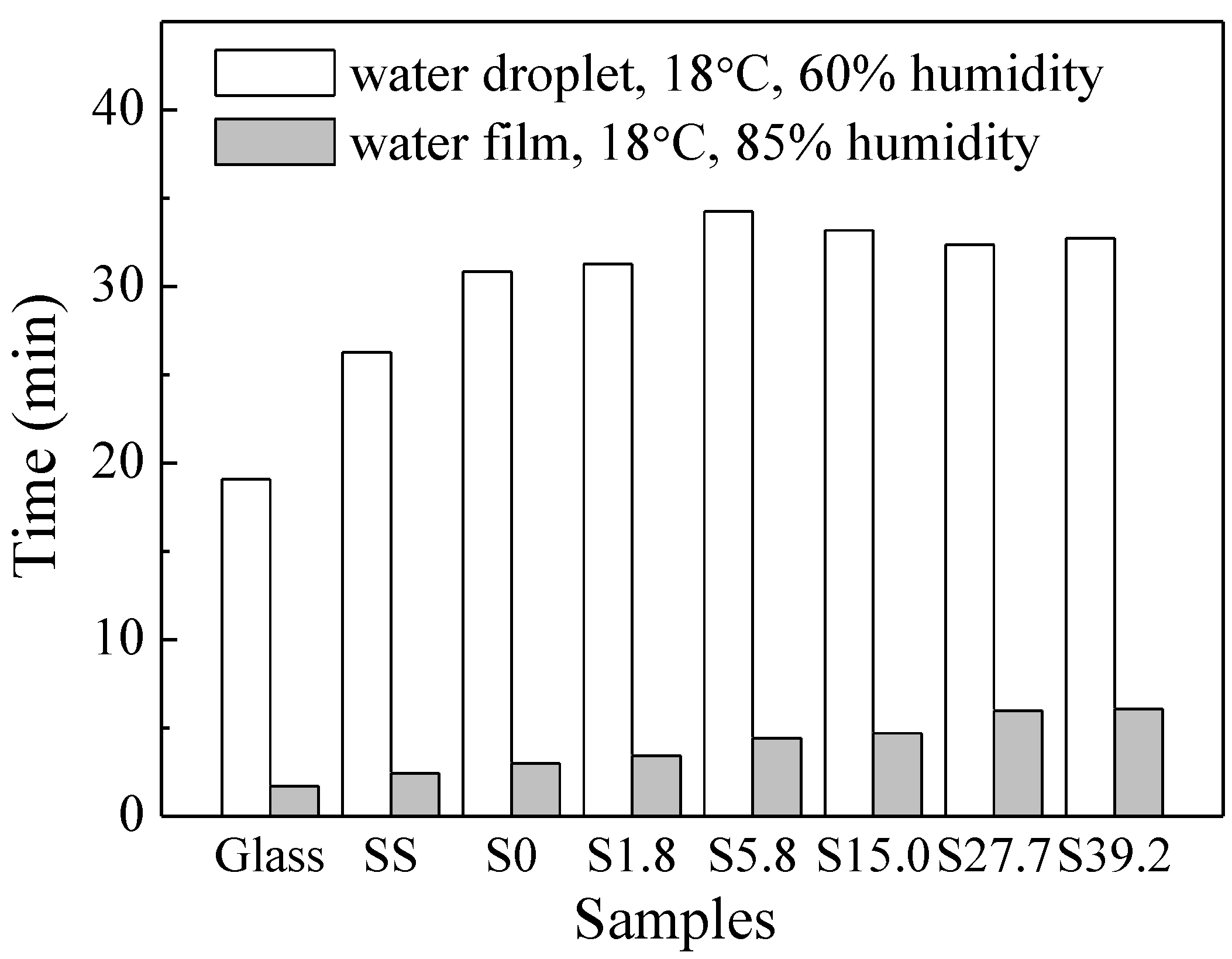
3.2. Hardness and Wettability Tests

The hardness of the films on silicon substrate was measured by a microhardness tester. The hardness of the pure Cr film is 994 HV0.001, and the hardness of the Cr–Ag composite films ranges from 628 to 968 HV0.001. These hardness values are much higher than those of stainless steel (~250 HV) and pure Ag film (240 HV). The hard Cr–Ag films will enhance the wear resistance of the coated stainless steel substrate.
The wettability of a material is determined by its chemical nature, surface groups, surface roughness and other factors. The Cr–Ag film samples are hydrophobic, with high water contact angles (100.5–120.1°, Table 1). Vapor deposited Cr-based films are often reported to exhibit hydrophobic behavior together with low surface energy [23,24]. The silver film sample is also hydrophobic (91.6°), agreeing with the literature report [25]. The E. coli cells suspension and glycol have also high contact angles at the surface of both kinds of films (Table 1).
The calculated surface energy values are shown in Figure 3. The hydrophilic slide glass has high surface energy with the polar component being dominant, and the hydrophobic stainless steel and coating samples have relatively low values with the dispersive one being dominant. It was reported that the hydrophobic surface with low surface energy is beneficial for anti-fouling and antimicrobial properties [15,26,27], while a hydrophilic surface with high surface energy is favorable for cell attachment [28].
Liquid evaporation is a common phenomenon in different industries. Water vaporizing rate at the surface, W can be described as W = a t b v d / p c H e by the multivariate nonlinear regression analysis modeling [29], in which t is temperature, v is air flow rate, p is pressure, H is humidity, and ae are the regression coefficients. Therefore, a water droplet dries more quickly at higher temperatures and lower humidity. The drying process of a 2 μL water droplet on the sample surface was observed with an optical microscope at constant ambient conditions (Figure 4a,b). Due to the smaller surface area of the droplets on Cr–Ag films, they dried more slowly than that on the glass substrate (Figure 5).
A layer of water film was sprayed on the surface of glass and film samples to simulate the drying process of droplets on materials in the cases of sneezing and coughing [2,3], where the liquid droplets are commonly smaller than 100 μm. Under the same preparation conditions, large water droplets (220–350 μm) are formed on the glass surface, while many fine water dots (70–160 μm, largely < 100 μm) are formed on the film surface (Figure 4c,d). The water layers on the film surface have a longer drying time than that on the glass surface, which is consistent with the observation of a single droplet (Figure 5). Because the sprayed water droplets are much smaller than the single 2 μL droplet, the drying time of sprayed water layers is significantly shorter than that of the 2 μL droplet. The drying time of sprayed water layer is thus dependent on material wettability, water droplet size and ambient conditions.

3.3. Antibacterial Test

The antibacterial property of the coating samples was evaluated against E. coli by the agar plate counting method. The bacteria grow well on glass samples, and the number of bacterial colonies is reduced for the Cr film sample. The antibacterial ratio of S0 is calculated to be 24% by (NgNf)/Ng at an action time of 3 h, where Ng and Nf are the colony numbers of glass and Cr film samples, respectively (Figure 6). This reveals a certain degree of antibacterial property of Cr elements [6,30]. Bacterial colonies are rarely observed for samples S1.8–S27.7, leading to an antibacterial ratio of 100% (Figure 6a). This is consistent with the report that the electrodeposited Cr–Ag nanocomposite coatings had high antibacterial activity (nearly 100%) against E. coli and Staphylococcus aureus [20]. E. coli cells can survive at a shorter action time (e.g., 30 min) and lower silver content. However, no colonies are observed on the agar plates if the inoculated glass and film samples were exposed to open air in the clean bench for 15 and 30 min during the action period (Figure 6b), due to the short drying time of bacterial suspension. This indicates that ambient conditions, material features and microdroplet hydration state have a great influence on the viability of bacteria and even coronaviruses at the surfaces [31,32,33]. These factors would be considered in controlling microbial transmission, especially for the public environmental facilities.

4. Conclusions

The Cr–Ag films with Ag content up to 39.2 at.% were deposited by the magnetron sputtering technique. The films consist of Cr and Ag composite and they are relatively flat, without serious silver segregation at the surface. The films are hard, hydrophobic with low surface energy and exhibit excellent antibacterial activity. These features would favor antimicrobial applications of the sputter-deposited Cr–Ag films.

Author Contributions

Conceptualization, P.S., Y.H. and T.F.; methodology, L.W. and Y.W.; validation, Y.W.; formal analysis, T.F. and Y.H.; investigation, T.F.; resources, T.F., Y.H. and P.S.; data curation, T.F.; writing—original draft preparation, T.F. and Y.H.; writing—review and editing, T.F.; supervision, P.S.; project administration, T.F.; funding acquisition, T.F. All authors have read and agreed to the published version of the manuscript.

Funding

This research was funded by the Natural Science Foundation of Shaanxi Province, grant number 2021JM-036.

Institutional Review Board Statement

Not applicable.

Informed Consent Statement

Not applicable.

Data Availability Statement

The data presented in this study are available on request from the corresponding author.

Acknowledgments

This study was supported by the Natural Science Fund of Shaanxi Province (No. 2021JM-036). The authors would thank Instrumental Analysis Center of Xi’an Jiaotong University for the SEM-EDX analyses.

Conflicts of Interest

We declare that we have no conflict of interest.

References

  1. Chang, T.; Sepati, M.; Herting, G.; Leygraf, C.; Rajarao, G.K.; Butina, K.; Richter-Dahlfors, A.; Blomberg, E.; Wallinder, I.O. A novel methodology to study antimicrobial properties of high-touch surfaces used for indoor hygiene applications-A study on Cu metal. PLoS ONE 2021, 16, e0247081. [Google Scholar] [CrossRef] [PubMed]
  2. Fontes, D.; Reyes, J.; Ahmed, K.; Kinzel, M. A study of fluid dynamics and human physiology factors driving droplet dispersion from a human sneeze. Phys. Fluids 2020, 32, 111904. [Google Scholar] [CrossRef] [PubMed]
  3. Redfern, J.; Tucker, J.; Simmons, L.M.; Askew, P.; Stephan, I.; Verran, J. Environmental and experimental factors affecting efficacy testing of nonporous plastic antimicrobial surfaces. Methods Protoc. 2018, 1, 36. [Google Scholar] [CrossRef] [Green Version]
  4. Connelly, M.C.; Reddy, G.S.; Nadagouda, M.N.; Sekhar, J.A. Antimicrobial and anticorrosive efficacy of inorganic nanoporous surfaces. Clean Technol. Environ. Policy 2017, 19, 845–857. [Google Scholar] [CrossRef]
  5. Kawakami, H.; Yoshida, K.; Nishida, Y.; Kikuchi, Y.; Sato, Y. Antibacterial properties of metallic elements for alloying evaluated with application of JIS Z 2801:2000. ISIJ Int. 2008, 48, 1299–1304. [Google Scholar] [CrossRef] [Green Version]
  6. Cazalini, E.M.; Miyakawa, W.; Teodoro, G.R.; Sobrinho, A.S.S.; Matieli, J.E.; Massi, M.; Koga-Ito, C.Y. Antimicrobial and anti-biofilm properties of polypropylene meshes coated with metal-containing DLC thin films. J. Mater. Sci. Mater. Med. 2017, 28, 97. [Google Scholar] [CrossRef] [Green Version]
  7. Nguyen, V.Q.; Ishihara, M.; Kinoda, J.; Hattori, H.; Nakamura, S.; Ono, T.; Miyahira, Y.; Matsui, T. Development of antimicrobial biomaterials produced from chitin-nanofiber sheet/silver nanoparticle composites. J. Nanobiotechnol. 2014, 12, 49. [Google Scholar] [CrossRef] [Green Version]
  8. Zhao, Y.; Xu, J.; Li, Z.; Fu, T.; Jiang, S. In vitro antibacterial properties of MoO3/SiO2/Ag2O nanocomposite coating prepared by double cathode glow discharge technique. Surf. Coat. Technol. 2020, 397, 125992. [Google Scholar] [CrossRef]
  9. Dinca, P.; Butoi, B.; Lungu, M.; Porosnicu, C.; Jepu, I.; Staicu, C.; Lungu, C.P.; Niculescu, A.; Burducea, I.; Trusca, O.; et al. Antibacterial efficiency of stainless-steel grids coated with Cu-Ag by thermionic vacuum arc method. Coatings 2020, 10, 322. [Google Scholar] [CrossRef] [Green Version]
  10. Wojcieszak, D.; Mazur, M.; Kaczmarek, D.; Mazur, P.; Szponar, B.; Domaradzki, J.; Kepinski, L. Influence of the surface properties on bactericidal and fungicidal activity of magnetron sputtered Ti-Ag and Nb-Ag thin films. Mater. Sci. Eng. C 2016, 62, 86–95. [Google Scholar] [CrossRef] [PubMed]
  11. Alias, R.; Mahmoodian, R.; Genasan, K.; Vellasamy, K.M.; Abd Shukor, M.H.; Kamarul, T. Mechanical, antibacterial, and biocompatibility mechanism of PVD grown silver-tantalum-oxide-based nanostructured thin film on stainless steel 316L for surgical applications. Mater. Sci. Eng. C 2020, 107, 110304. [Google Scholar] [CrossRef] [PubMed]
  12. Ferreri, I.; Calderon, S.V.; Escobar Galindo, R.; Palacio, C.; Henriques, M.; Piedade, A.P.; Carvalho, S. Silver activation on thin films of Ag-ZrCN coatings for antimicrobial activity. Mater. Sci. Eng. C 2015, 55, 547–555. [Google Scholar] [CrossRef] [PubMed] [Green Version]
  13. Wang, L.J.; Zhang, F.; Fong, A.; Lai, K.M.; Shum, P.W.; Zhou, Z.F.; Gao, Z.F.; Fu, T. Effects of silver segregation on sputter deposited antibacterial silver-containing diamond-like carbon films. Thin Solid Film. 2018, 650, 58–64. [Google Scholar] [CrossRef]
  14. Cloutier, M.; Turgeon, S.; Busby, Y.; Tatoulian, M.; Pireaux, J.J.; Mantovani, D. Controlled distribution and clustering of silver in Ag-DLC nanocomposite coatings using a hybrid plasma approach. ACS Appl. Mater. Interfaces 2016, 8, 21020–21027. [Google Scholar] [CrossRef] [PubMed]
  15. Swiatek, L.; Olejnik, A.; Grabarczyk, J.; Jedrzejczak, A.; Sobczyk-Guzenda, A.; Kaminska, M.; Jakubowski, W.; Szymanski, W.; Bociaga, D. Multi-doped diamond like-carbon coatings (DLC-Si/Ag) for biomedical applications fabricated using the modified chemical vapour deposition method. Diam. Relat. Mater. 2016, 67, 54–62. [Google Scholar] [CrossRef]
  16. Rashid, S.; Sebastiani, M.; Mughal, M.Z.; Daniel, R.; Bemporad, E. Influence of the silver content on mechanical properties of Ti-Cu-Ag thin films. Nanomaterials 2021, 11, 435. [Google Scholar] [CrossRef]
  17. Bai, L.; Hang, R.; Gao, A.; Zhang, X.; Huang, X.; Wang, Y.; Tang, B.; Zhao, L.; Chu, P.K. Nanostructured titanium-silver coatings with good antibacterial activity and cytocompatibility fabricated by one-step magnetron sputtering. Appl. Surf. Sci. 2015, 355, 32–44. [Google Scholar] [CrossRef]
  18. Wang, L.J.; Zhang, F.; Fong, A.; Lai, K.M.; Shum, P.W.; Zhou, Z.F.; Fu, T.; Ning, P.; Yang, S.Y. Tungsten film as a hard and compatible carrier for antibacterial agent of silver. J. Mater. Sci. 2018, 53, 10640–10652. [Google Scholar] [CrossRef]
  19. Karami, A.; Zhang, H.; Pederick, V.G.; McDevitt, C.A.; Kabir, M.S.; Xu, S.; Munroe, P.; Zhou, Z.; Xie, Z. Cr-Ag coatings: Synthesis, microstructure and antimicrobial properties. Surf. Eng. 2019, 35, 596–603. [Google Scholar] [CrossRef]
  20. Mendez-Albores, A.; Gonzalez-Arellano, S.G.; Reyes-Vidal, Y.; Torres, J.; Talu, S.; Cercado, B.; Trejo, G. Electrodeposited chrome/silver nanoparticle (Cr/AgNPs) composite coatings: Characterization and antibacterial activity. J. Alloys Compd. 2017, 710, 302–311. [Google Scholar] [CrossRef]
  21. Campos, M.D.; Zucchi, P.C.; Phung, A.; Leonard, S.N.; Hirsch, E.B. The activity of antimicrobial surfaces varies by testing protocol utilized. PLoS ONE 2016, 11, e0160728. [Google Scholar] [CrossRef]
  22. Ma, X.Y. Evaporating Process of Nanofluid Sessile Droplet and Deposition Patterns of Nanoparticles; Tianjin University of Commerce: Tianjin, China, 2016. (In Chinese) [Google Scholar]
  23. Hsu, C.H.; Huang, D.H.; Ho, W.Y.; Huang, L.T.; Chang, C.L. Characteristics and performance of Cr2O3/CrN double-layered coatings deposited by cathodic arc plasma deposition. Mater. Sci. Eng. A 2006, 429, 212–218. [Google Scholar] [CrossRef]
  24. Cheng, W.L.; Zhou, Z.F.; Shum, P.W.; Li, K.Y. Effect of Ni addition on the structure and properties of Cr-Ni-N coatings deposited by closed-field unbalanced magnetron sputtering ion plating. Surf. Coat. Technol. 2013, 229, 84–89. [Google Scholar] [CrossRef]
  25. Rebelo, R.; Calderon, S.V.; Fangueiro, R.; Henriques, M.; Carvalho, S. Influence of oxygen content on the antibacterial effect of Ag-O coatings deposited by magnetron sputtering. Surf. Coat. Technol. 2016, 305, 1–10. [Google Scholar] [CrossRef] [Green Version]
  26. Lan, W.-C.; Ou, S.-F.; Lin, M.-H.; Ou, K.-L.; Tsai, M.-Y. Development of silver-containing diamond-like carbon for biomedical applications. Part I: Microstructure characteristics, mechanical properties and antibacterial mechanisms. Ceram. Int. 2013, 39, 4099–4104. [Google Scholar] [CrossRef]
  27. Cunliffe, D.; Smart, C.A.; Alexander, C.; Vulfson, E.N. Bacterial adhesion at synthetic surfaces. Appl. Environ. Microbiol. 1999, 65, 4995–5002. [Google Scholar] [CrossRef] [PubMed] [Green Version]
  28. Echeverry-Rendon, M.; Galvis, O.; Aguirre, R.; Robledo, S.; Guillermo Castano, J.; Echeverria, F. Modification of titanium alloys surface properties by plasma electrolytic oxidation (PEO) and influence on biological response. J. Mater. Sci. Mater. Med. 2017, 28, 169. [Google Scholar] [CrossRef]
  29. Geng, J.T.; Li, X.Y.; Xing, G.L.; Sun, G.Q.; Yi, B.K. An experimental study on water surface evaporation rate. Chin. J. Power Sources 2010, 34, 470–472. (In Chinese) [Google Scholar]
  30. Jelinek, M.; Kocourek, T.; Zemek, J.; Miksovsky, J.; Kubinova, S.; Remsa, J.; Kopecek, J.; Jurek, K. Chromium-doped DLC for implants prepared by laser-magnetron deposition. Mater. Sci. Eng. C. 2015, 46, 381–386. [Google Scholar] [CrossRef]
  31. Aboubakr, H.A.; Sharafeldin, T.A.; Goyal, S.M. Stability of SARS-CoV-2 and other coronaviruses in the environment and on common touch surfaces and the influence of climatic conditions: A review. Transbound. Emerg. Dis. 2021, 68, 296–312. [Google Scholar] [CrossRef] [PubMed]
  32. Niu, L.; Liang, W.; Wang, X.; Mu, Y.; Wang, J.; Wu, D.; Zhao, X. Analysis of factors affecting virus survival on object surface and in air. Res. Environ. Sci. 2020, 33, 1589–1595. [Google Scholar]
  33. Fedorenko, A.; Grinberg, M.; Orevi, T.; Kashtan, N. Survival of the enveloped bacteriophage Phi6 (a surrogate for SARS-CoV-2) in evaporated saliva microdroplets deposited on glass surfaces. Sci. Rep. 2020, 10, 22419. [Google Scholar] [CrossRef] [PubMed]
Figure 1. XRD patterns of the Cr–Ag film samples with glass substrate.
Coatings 11 01153 g001
Figure 2. SEM surface images of the Cr-Ag films with glass substrate: (a) S0, (b) S15.0, (c) S27.7, (d) S39.2.
Coatings 11 01153 g002
Figure 3. Surface energy of the glass, stainless steel, Cr–Ag and Ag film samples.
Coatings 11 01153 g003
Figure 4. Representative top view images of 2 μL water droplet and sprayed water film on (a,c) glass and (b,d) film sample S5.8, respectively (bar = 1 mm).
Coatings 11 01153 g004
Figure 5. The drying time of 2 μL water droplet and the sprayed water film on glass and Cr–Ag film samples.
Coatings 11 01153 g005
Figure 6. Antibacterial ratio of the film samples tested by agar plate counting method at 20 °C: (a) closed wet conditions, (b) open-air conditions.
Coatings 11 01153 g006
Table 1. Silver target current (IAg), deposition time (t), deposition rate (R), silver content ([Ag]), grain size (d) and contact angle (CA) of the samples.
SampleIAg
(A)
t
(min)
R +
(nm/min)
[Ag] +
at.%
d *
(nm)
CA (°) *
WaterSuspensionGlycol
Glass/////49.1 ± 2.344.7 ± 1.544.5 ± 3.8
Steel/////91.1 ± 3.189.4 ± 2.872.7 ± 0.5
S003021.7027107.2 ± 0.395.8 ± 1.281.7 ± 2.1
S1.80.43026.31.818.9117.9 ± 0.9118.8 ± 2.397.3 ± 2.3
S5.80.63027.35.818.7120.1 ± 0.1121.4 ± 0.289.9 ± 3.6
S15.01.02532.015.012.7113.8 ± 1.3117.2 ± 1.494.6 ± 5.2
S27.71.52037.027.79.5100.5 ± 0.9105.4 ± 0.476.3 ± 1.3
S39.202.01560.739.29.2102.9 ± 2.2110.3 ± 1.681.5 ± 3.4
Silver2.03025.0100/91.6 ± 2.896.3 ± 2.267.3 ± 0.8
+: samples with Si(100) substrate, *: samples with glass substrate, suspension: Escherichia coli cells suspension (105 cfu/mL).
Publisher’s Note: MDPI stays neutral with regard to jurisdictional claims in published maps and institutional affiliations.

