Investigation of Ancient Architectural Painting from the Taidong Tomb in the Western Qing Tombs, Hebei, China


Abstract
:1. Introduction
2. Materials and Experiment
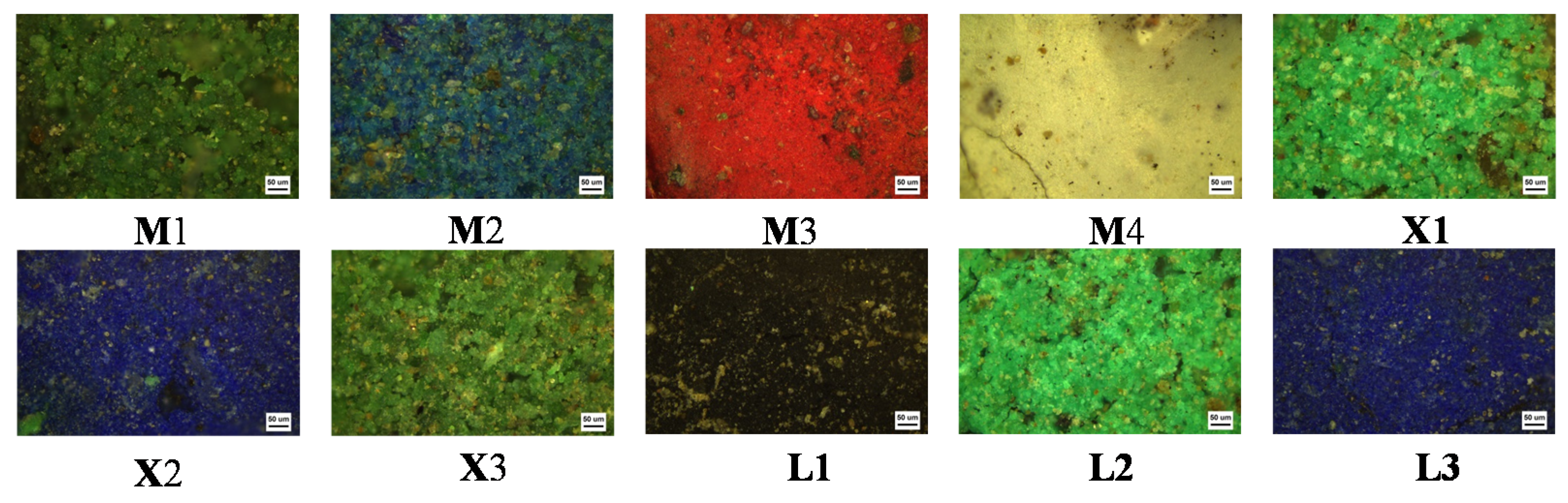
2.1. Sample Information
2.2. Sample Preparation
2.2.1. Cross-Section Preparation
2.2.2. Fiber Preparation
2.2.3. Preparation of Pigment Sample
2.2.4. Preparation of Mortar Sample
2.3. Instrument
3. Results and Discussion
3.1. Cross-Section
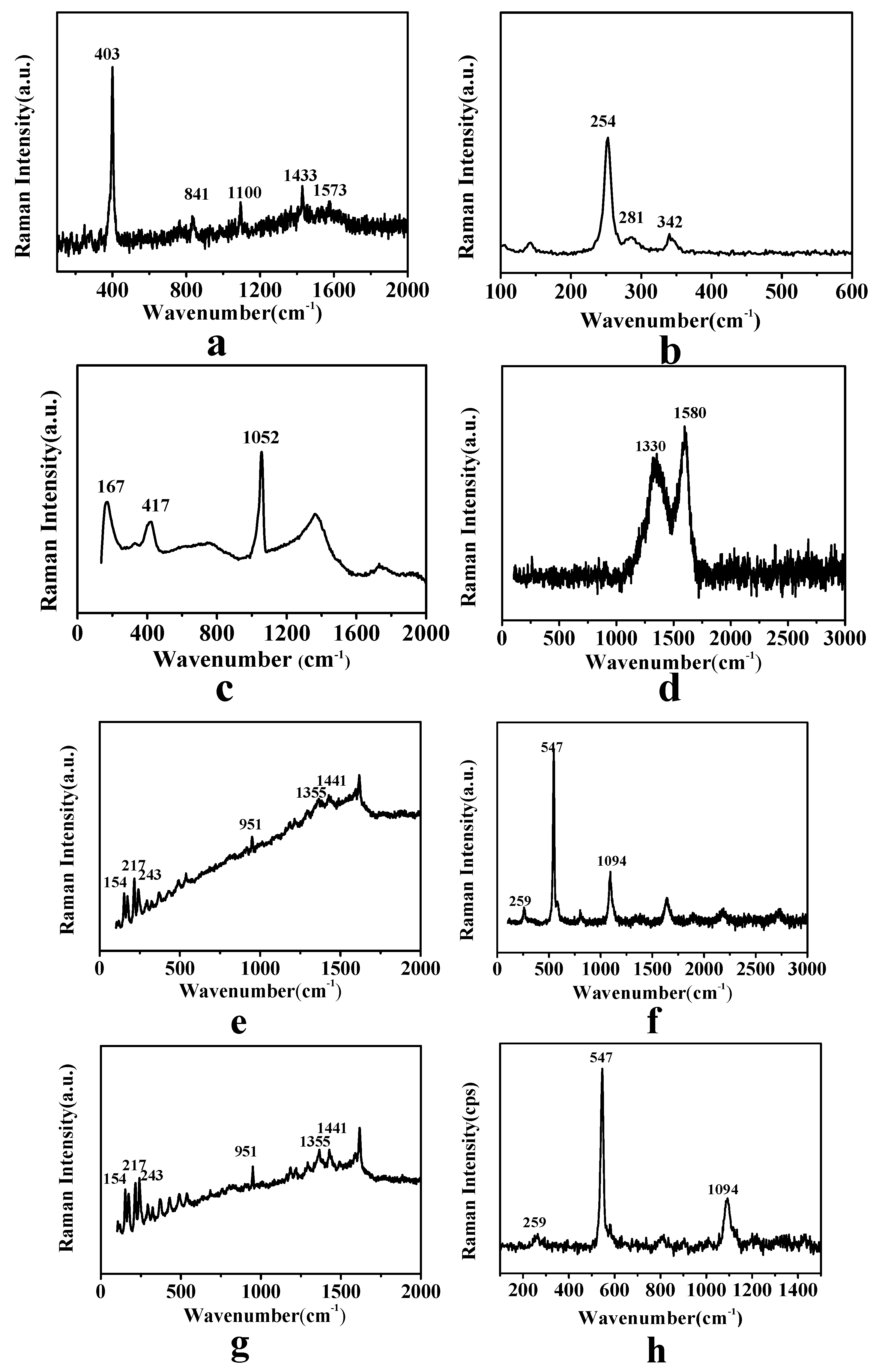
3.2. Pigments
3.2.1. Green Pigment
3.2.2. Blue Pigment
3.2.3. Red Pigment
3.2.4. Black Pigment
3.2.5. White Pigment
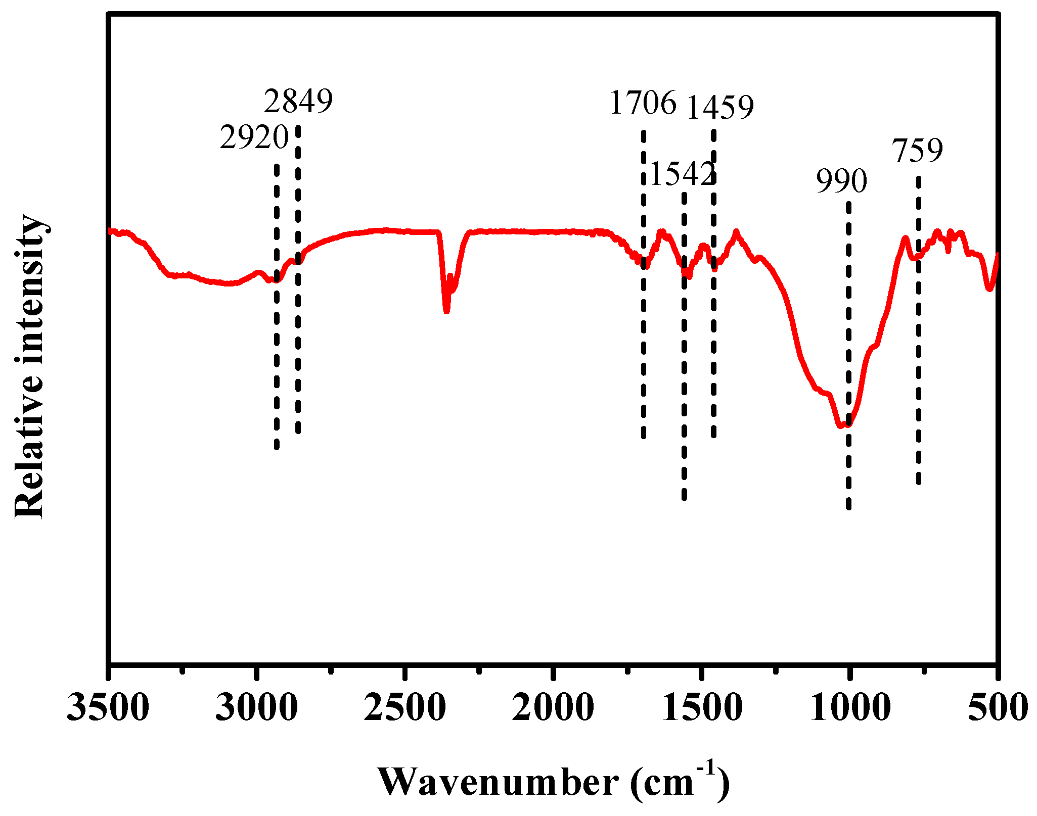
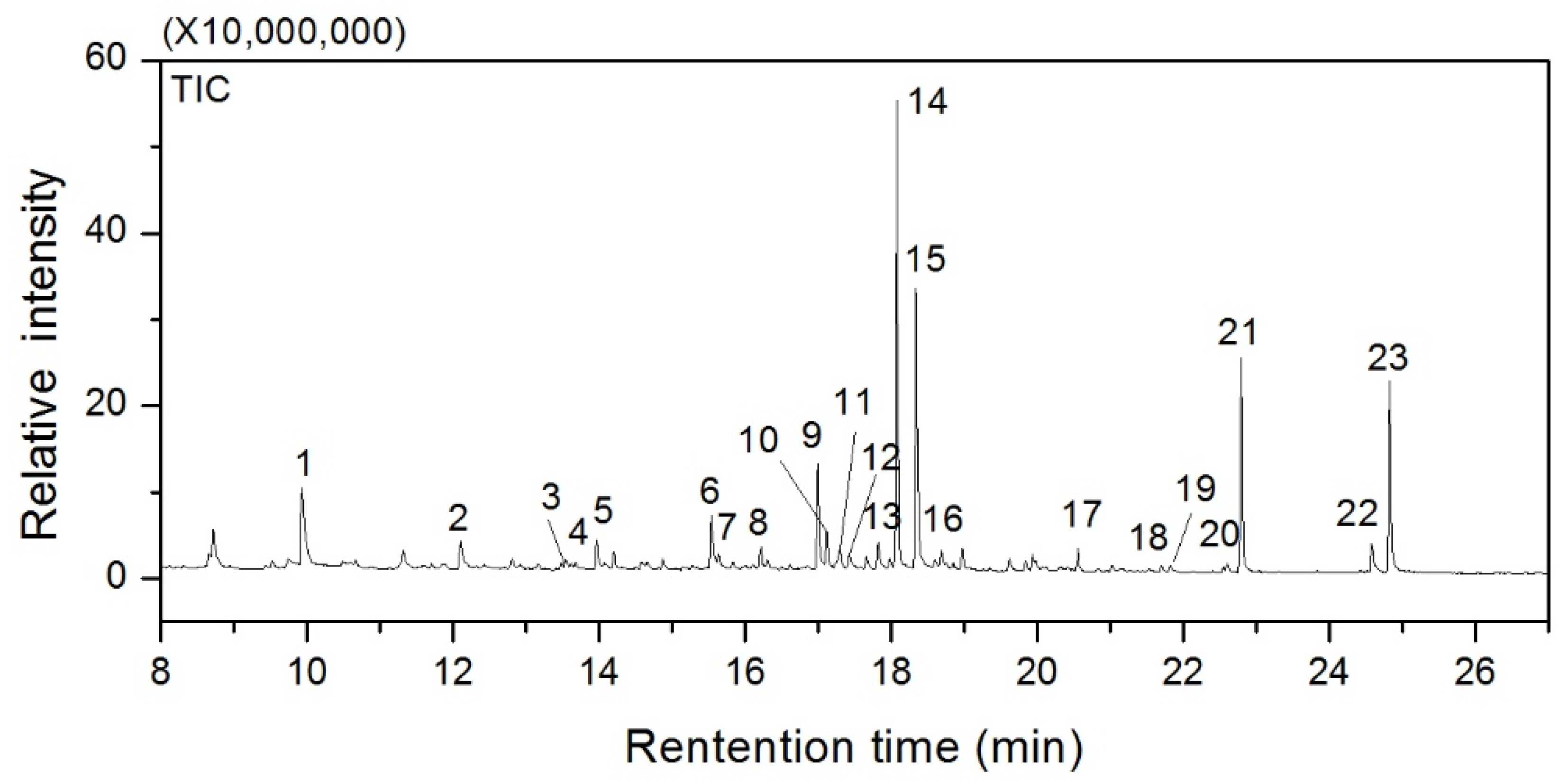
3.3. Mortar and Fiber
4. Conclusions
Author Contributions
Funding
Acknowledgments
Conflicts of Interest
References
- Kugler, V.; Bean, S.; Spring, M. Quantitative edx analysis of smalt pigment in sixteenth and eighteenth century paintings. Microsc. Microanal. 2013, 19, 1428–1429. [Google Scholar] [CrossRef] [Green Version]
- Samanian, K. Identification of green pigment used in persian wall paintings (AD 1501–1736) using plm, ft-ir, sem/edx and gc-ms techniques. Archaeometry 2015, 57, 740–758. [Google Scholar] [CrossRef]
- Bell, I.M.; Clark, R.J.H.; Gibbs, P.J. Raman spectroscopic library of natural and synthetic pigments. Spectrochim. Acta A 1997, 53, 2159–2179. [Google Scholar] [CrossRef]
- Burgio, L.; Melessanaki, K.; Doulgeridis, M.; Clark, R.J.H.; Anglos, D. Pigment identification in paintings employing laser induced breakdown spectroscopy and raman microscopy. Spectrochim. Acta B 2001, 56, 905–913. [Google Scholar] [CrossRef]
- Perez-Alonso, M.; Castro, K.; Madariaga, J.M. Investigation of degradation mechanisms by portable raman spectroscopy and thermodynamic speciation: The wall painting of santa maria de lemoniz (basque country, north of spain). Anal. Chim. Acta 2006, 571, 121–128. [Google Scholar] [CrossRef]
- Stanzani, E.; Bersani, D.; Lottici, P.P.; Colomban, P. Analysis of artist’s palette on a 16th century wood panel painting by portable and laboratory raman instruments. Vib. Spectrosc. 2016, 85, 62–70. [Google Scholar] [CrossRef] [Green Version]
- Gil, M.; Carvalho, M.L.; Seruya, A.; Ribeiro, I.; Queralt, I.; Candeias, A.E.; Mirão, J. Limewashing paintings in alentejo urban heritage: Pigment characterization and differentiation by wdxrf and xrd. Appl. Phys. A 2007, 90, 49–54. [Google Scholar] [CrossRef]
- Hochleitner, B.; Desnica, V.; Mantler, M.; Schreiner, M. Historical pigments: A collection analyzed with x-ray diffraction analysis and x-ray fluorescence analysis in order to create a database. Spectrochim. Acta B 2003, 58, 641–649. [Google Scholar] [CrossRef]
- Peifan, Q.; Deqi, Y.; Qi, M.; Aijun, S.; Jingqi, S.; Zengjun, Z.; Jianwei, H. Study and restoration of the yi ma wu hui layer of the ancient coating on the putuo zongcheng temple. Int. J. Herit. 2020. [Google Scholar] [CrossRef]
- Song, Y.; Gao, F.; Nevin, A.; Guo, J.; Zhou, X.; Wei, S.; Li, Q. A technical study of the materials and manufacturing process used in the gallery wall paintings from the jokhang temple, tibet. Herit. Sci. 2018, 6, 18. [Google Scholar] [CrossRef] [Green Version]
- Shi, J.; Li, T. Technical investigation of 15th and 19th century chinese paper currencies: Fiber use and pigment identification. J. Raman Spectrosc. 2013, 44, 892–898. [Google Scholar] [CrossRef]
- Petrova, O.I.; Pankin, D.V.; Povolotckaia, A.V.; Borisov, E.V.; Beznosova, M.O.; Krivul’ko, T.A.; Kurochkin, A.V. Identification of pigments in colored layers of a painting by raman spectroscopy. Opt. Spectrosc. 2018, 123, 965–969. [Google Scholar] [CrossRef]
- Rosi, F.; Miliani, C.; Borgia, I.; Brunetti, B.; Sgamellotti, A. Identification of nineteenth century blue and green pigments by in situ x-ray fluorescence and micro-raman spectroscopy. J. Raman Spectrosc. 2004, 33, 610–615. [Google Scholar] [CrossRef]
- Castillejo, M.; Martín, M.; Silva, D.; Stratoudaki, T.; Anglos, D.; Burgio, L.; Clark, R.J.H. Laser-induced breakdown spectroscopy and raman microscopy for analysis of pigments in polychromes. J. Cult. Herit. 2000, 1, 297–302. [Google Scholar] [CrossRef]
- Shen, A.G.; Wang, X.H.; Xie, W.; Shen, J.; Li, H.Y.; Liu, Z.A.; Hu, J.M. Pigment identification of colored drawings from wuying hall of the imperial palace by micro-raman spectroscopy and energy dispersive x-ray spectroscopy. J. Raman Spectrosc. 2006, 37, 230–234. [Google Scholar] [CrossRef]
- Thomas, R.; Pott, D.T. Atacamite pigment at tell abraq in the early iron age. Arab. Archaeol. Epigr. 1996, 7, 13–26. [Google Scholar] [CrossRef]
- Şerifaki, K.; Böke, H.; Yalçın, Ş.; İpekoğlu, B. Characterization of materials used in the execution of historic oil paintings by xrd, sem-eds, tga and libs analysis. Mater. Charact. 2009, 60, 303–311. [Google Scholar] [CrossRef] [Green Version]
- Bicchieri, M.; Nardone, M.; Russo, P.A.; Sodo, A.; Corsi, M.; Cristoforetti, G.; Palleschi, V.; Salvetti, A.; Tognoni, E. Characterization of azurite and lazurite based pigments by laser induced breakdown spectroscopy and micro-raman spectroscopy. Spectrochim. Acta B 2001, 56, 915–922. [Google Scholar] [CrossRef]
- Petrova, O.; Pankin, D.; Povolotckaia, A.; Borisov, E.; Krivul’ko, T.; Kurganov, N.; Kurochkin, A. Pigment palette study of the xix century plafond painting by raman spectroscopy. J. Cult. Herit. 2019, 37, 233–237. [Google Scholar] [CrossRef]
- Želinská, J.; Kopecká, I.; Svobodová, E.; Milovská, S.; Hurai, V. Stratigraphic em-eds, xrf, raman and ft-ir analysis of multilayer paintings from the main altar of the st. James church in levoča (slovakia). J. Cult. Herit. 2018, 33, 90–99. [Google Scholar] [CrossRef]
- Zhu, T.; Chen, J.; Hui, R.; Gong, L.; Zhang, W.; Zhang, Y. Spectroscopic characterization of the architectural painting from the cizhong catholic church of yunnan province, china. Anal. Lett. 2013, 46, 2253–2264. [Google Scholar] [CrossRef]
- Torres-Ruiz, J.; Pesquera, A.; Gil-Crespo, P.P.; Delgado, A. Exotic cu-mineralization in triassic red beds from navas de san juan (jaén, spain). Ore Geol. Rev. 2020, 119, 103399. [Google Scholar] [CrossRef]
- Van der Weerd, J.; Smith, G.D.; Firth, S.; Clark, R.J.H. Identification of black pigments on prehistoric southwest american potsherds by infrared and raman microscopy. J. Archaeol. Sci. 2004, 31, 1429–1437. [Google Scholar] [CrossRef]
- Mazzeo, R.; Cam, D.; Chiavari, G.; Fabbri, D.; Ling, H.; Prati, S. Analytical study of traditional decorative materials and techniques used in ming dynasty wooden architecture. The case of the drum tower in xi’an, p.R. Of china. J. Cult. Herit. 2004, 5, 273–283. [Google Scholar] [CrossRef]
- Zhu, T.; Li, T.; Liu, N.; Chen, J.; Huang, H.; Fu, Q.; Zhang, S. Hexi painting on xitian fanjing, a qing imperial buddhist temple in beijing, china: Technology revealed by analytical approaches (an initial report). Herit. Sci. 2016, 4, 42. [Google Scholar] [CrossRef] [Green Version]
- Taglieri, G.; Rigaglia, D.; Arrizza, L.; Daniele, V.; Macera, L.; Rosatelli, G.; Romè, V.; Musolino, G. Microanalytical investigations on a byzantine fresco of the dormitio virginis from sicily. J. Cult. Herit. 2019, 40, 155–162. [Google Scholar] [CrossRef]
- Schonemann, A.; Edwards, H.G. Raman and ftir microspectroscopic study of the alteration of chinese tung oil and related drying oils during ageing. Anal. Bioanal. Chem. 2011, 400, 1173–1180. [Google Scholar] [CrossRef]
- Otero, V.; Sanches, D.; Montagner, C.; Vilarigues, M.; Carlyle, L.; Lopes, J.A.; Melo, M.J. Characterisation of metal carboxylates by raman and infrared spectroscopy in works of art. J. Raman Spectrosc. 2014, 45, 1197–1206. [Google Scholar] [CrossRef]
- Tamburini, D.; Sardi, D.; Spepi, A.; Duce, C.; Tinè, M.R.; Colombini, M.P.; Bonaduce, I. An investigation into the curing of urushi and tung oil films by thermoanalytical and mass spectrometric techniques. Polym. Degrad. Stabi. 2016, 134, 251–264. [Google Scholar] [CrossRef] [Green Version]
- Wang, N.; He, L.; Zhao, X.; Simon, S. Comparative analysis of eastern and western drying-oil binding media used in polychromic artworks by pyrolysis-gas chromatography/mass spectrometry under the influence of pigments. Microchem. J. 2015, 123, 201–210. [Google Scholar] [CrossRef]


| Number | Sample Positon |
|---|---|
| M1 |  Ceiling of Minglou |
| M2 | |
| M3 | |
| M4 | |
| X1 |  Xipei Hall |
| X2 | |
| X3 | |
| L1 |  Long’en Hall |
| L2 | |
| L3 |
| Sample Number | Cross-Section Micrograph | Serial Number | Composition | Thickness | Total Number of Layers |
|---|---|---|---|---|---|
| X3 |  | a | Green pigment | 66 µm | 3 |
| b | Mortar | 969 µm | |||
| c | Fiber | 1200 µm | |||
| L2 |  | a | Green pigment | 32 µm | 3 |
| b | Mortar | 2200 µm | |||
| c | Fiber | >1000 µm |
| Sample Number | Main Elements of EDX Spectrum (wt.%) | ||||
|---|---|---|---|---|---|
| M1 | Cu (62.91) | Cl (13.82) | Si (12.11) | Ca (4.73) | K (2.16) |
| M2 | Cu (58.13) | Ca (20.04) | Si (18.37) | S (3.37) | Fe (2.21) |
| M3 | Hg (47.52) | S (36.73) | Si (7.42) | K (4.04) | Ca (2.80) |
| M4 | Pb (64.85) | Ca (19.09) | Si (7.43) | S (6.30) | Fe (1.11) |
| X1 | Cu (23.81) | As (20.84) | Si (16.36) | Ca (14.57) | S (8.45) |
| X2 | Ca (20.60) | Si (20.03) | Al (16.38) | K (15.68) | S (11.04) |
| X3 | Cu (42.91) | Cl (15.82) | Pb (14.11) | Si (10.73) | Ca (10.16) |
| L1 | Ca (31.71) | Si (29.01) | S (19.7) | K (4.55) | Fe (4.51) |
| L2 | Cu (36.95) | As (36.01) | Si (8.06) | Ba (7.53) | S (3.03) |
| L3 | Si (27.99) | Ca (26.13) | Al (20.88) | S (14.89) | K (6.47) |
| Sample Number | Main Elements of EDX Spectrum | Composition |
|---|---|---|
| Ceiling of Minglou (M1) | green pigment | atacamite |
| Ceiling of Minglou (M2) | blue pigment | azurite |
| Ceiling of Minglou (M3) | red pigment | vermilion |
| Ceiling of Minglou (M4) | white pigment | lead white |
| Xipei Hall (X1) | green pigment | Paris green |
| Xipei Hall (X2) | blue pigment | ultramarine |
| Xipei Hall (X3) | green pigment | Atacamite + anglesite |
| Long’en Hall (L1) | black pigment | carbon black |
| Long’en Hall (L2) | green pigment | Paris green |
| Long’en Hall (L3) | blue pigment | ultramarine |
| Peak Number | Retention/Time (min) | Characteristic Components and Typical Segments in Mass Spectrometry | Area (%) |
|---|---|---|---|
| 1 | 9.92 | butanedioic acid, dimethyl ester | 3.38 |
| 2 | 12.10 | pentanedioic acid, dimethyl ester | 0.58 |
| 3 | 13.54 | butanoic acid, 4-(dimethylamino)-3-hydroxy- | 0.12 |
| 4 | 13.67 | nonanoic acid, methyl ester | 0.49 |
| 5 | 13.96 | hexanedioic acid, dimethyl ester | 0.21 |
| 6 | 15.53 | heptanedioic acid, dimethyl ester | 2.35 |
| 7 | 15.63 | 10-undecenoic acid, methyl ester | 0.16 |
| 8 | 16.20 | 2,3,6-tri-o-methyl-d-glucopyranose | 0.22 |
| 9 | 16.99 | octanedioic acid, dimethyl ester | 4.98 |
| 10 | 17.11 | dimethyl phthalate | 0.54 |
| 11 | 17.29 | hexanedioic acid, 3-methoxy-, dimethyl ester | 0.27 |
| 12 | 17.4 | methyl alpha-d-mannopyranoside | 0.21 |
| 13 | 17.81 | 1,4-benzenedicarboxylic acid, dimethyl ester | 0.24 |
| 14 | 18.07 | dodecanoic acid, methyl ester | 28.95 |
| 15 | 18.33 | nonanedioic acid (azelaic acid), dimethyl ester | 19.51 |
| 16 | 18.68 | octanedioic acid, 4-methoxy-, dimethyl ester | 0.23 |
| 17 | 20.55 | methyl tetradecanoate | 0.26 |
| 18 | 21.70 | pentadecanoic acid, methyl ester | 0.11 |
| 19 | 21.81 | 1,2,4-benzenetricarboxylic acid, trimethyl ester | 0.13 |
| 20 | 22.59 | 1,3,5-benzenetricarboxylic acid, trimethyl ester | 0.11 |
| 21 | 22.78 | palmitic acid, methyl ester | 18.49 |
| 22 | 24.57 | 9-octadecenoic acid, methyl ester | 0.55 |
| 23 | 24.81 | stearic acid, methyl ester | 17.91 |
© 2020 by the authors. Licensee MDPI, Basel, Switzerland. This article is an open access article distributed under the terms and conditions of the Creative Commons Attribution (CC BY) license (http://creativecommons.org/licenses/by/4.0/).
Share and Cite
Fu, P.; Teri, G.-L.; Li, J.; Li, J.-X.; Li, Y.-H.; Yang, H. Investigation of Ancient Architectural Painting from the Taidong Tomb in the Western Qing Tombs, Hebei, China. Coatings 2020, 10, 688. https://doi.org/10.3390/coatings10070688
Fu P, Teri G-L, Li J, Li J-X, Li Y-H, Yang H. Investigation of Ancient Architectural Painting from the Taidong Tomb in the Western Qing Tombs, Hebei, China. Coatings. 2020; 10(7):688. https://doi.org/10.3390/coatings10070688
Chicago/Turabian StyleFu, Peng, Ge-Le Teri, Jing Li, Jia-Xin Li, Yu-Hu Li, and Hong Yang. 2020. "Investigation of Ancient Architectural Painting from the Taidong Tomb in the Western Qing Tombs, Hebei, China" Coatings 10, no. 7: 688. https://doi.org/10.3390/coatings10070688