Article

Design Strategy for Self-Healing Epoxy Coatings

1
Department of Macromolecular Science and Engineering, Case Western Reserve University, Cleveland, OH 44106, USA
2
Department of Chemistry and Chemical Biology, Rensselaer Polytechnic Institute, Troy, NY 12180, USA
*
Author to whom correspondence should be addressed.
Coatings 2020, 10(1), 50; https://doi.org/10.3390/coatings10010050
Submission received: 25 November 2019 / Revised: 27 December 2019 / Accepted: 3 January 2020 / Published: 6 January 2020
(This article belongs to the Special Issue Self-Healing Coatings and Films)

Abstract

:
Self-healing strategies including intrinsic and extrinsic self-healing are commonly used for polymeric materials to restore their appearance and properties upon damage. Unlike intrinsic self-healing tactics where recovery is based on reversible chemical or physical bonds, extrinsic self-healing approaches rely on a secondary phase to acquire the self-healing functionality. Understanding the impacts of the secondary phase on both healing performance and matrix properties is important for rational system design. In this work, self-healing coating systems were prepared by blending a bio-based epoxy from diglycidyl ether of diphenolate esters (DGEDP) with thermoplastic polyurethane (TPU) prepolymers. Such systems exhibit polymerization induced phase separation morphology that controls coating mechanical and healing properties. Structure–property analysis indicates that the degree of phase separation is controlled by tuning the TPU prepolymer molecular weight. Increasing the TPU prepolymer molecular weight results in a highly phase separated morphology that is preferable for mechanical performances but undesirable for healing functionality. In this case, diffusion of TPU prepolymers during healing is restricted by the epoxy network rigidity and chain entanglement. Low molecular weight TPU prepolymers tend to phase mix with the epoxy matrix during curing, resulting in the formation of a flexible epoxy network that benefits TPU flow while decreasing Tg and mechanical properties. This work describes a rational strategy to develop self-healing coatings with controlled morphology to extend their functions and tailor their properties for specific applications.

Graphical Abstract

1. Introduction

Motivated by extending material lifetime through restoring performance upon damage, research aimed at developing self-healing polymers that self-repair is growing [1,2,3,4]. Besides prolonging material service life, using the self-healing concept also helps minimize cost during damage repair. To obtain the self-healing functionality, a “mobile phase” that can close the crack should be generated [5,6]. The mobile phase can be used to distinguish between extrinsic and intrinsic self-healing polymers. Intrinsic self-healing systems repair cracks by chain rearrangement via the formation of primary or secondary reversible chemical/physical bonds [7,8,9,10,11,12,13]. While intrinsic healing materials offer the advantage of repeated restoration at the same damage site [14], the complex chemistry, processing difficulties and weak mechanical properties due to the presence of dynamic reversible bonds makes them impractical for commercial applications [15,16]. In contrast, extrinsic self-healing systems require the incorporation of external healing components such as catalysts or thermoplastic additives in the system [17,18,19,20,21,22]. These healing agents can be encapsulated or form blends during material manufacturing. To achieve the self-healing functionality, the external healing component must diffuse to the damage site to mend the crack. Since the extrinsic self-healing approach can be deployed in a wide range of material formulations, it has been used for a broad range of material applications such as aerospace, coatings, and structural materials [23,24,25,26].
Polymer coatings applied on metal substrates can form a thin barrier layer that prevents corrosion by impeding direct exposure to the environment [27,28,29]. Therefore, self-healing coatings, which can provide unassisted repair to prevent exposure of the underlying substrate to the environment, are highly desired solutions of considerable commercial interest. Strategies like dispersion of a microencapsulated catalyst and healing agents in the coating matrix [30,31,32], loading the healing agent in a core-shell bead-on-string morphology [33], or blending thermoplastics with polymerization-induced phase separation (PIPS) morphology [34] were reported to achieve self-healing coatings [35]. Although researchers have shown the effectiveness of introducing a secondary phase for self-healing, the impacts of the secondary phase on the engineering performances of the polymer system have rarely been studied [36,37,38,39,40,41]. Therefore, for rational material design it is important to understand the underlying mechanisms by which the secondary phase function (i.e., microstructure to macro properties) and elucidate its effects on system properties.
In a previous work, a highly transparent self-healing coating with facile process ability and good mechanical properties was obtained by blending epoxy with thermoplastic polyurethane (TPU) prepolymers [42]. TPU prepolymers form domains in the epoxy matrix through PIPS that, upon heating, can flow to the damaged area to mend the crack. We have previously observed that modifying the TPU prepolymer molecular weight results in changes in the curing kinetics and system mechanical properties.
In this work, a bio-based epoxy resin synthesized from n-alkyl esters of diphenolic acid was used in place of the petroleum based epoxy derived from bisphenol A (BPA) that has been associated with health risks [43,44]. The healing behavior and mechanical performance as a function of TPU prepolymer molecular weight were studied. TPU prepolymer diffusion was related to PIPS morphology by scanning electron microscopy (SEM) and chemorheology analyses. The self-healing mechanism was analyzed through correlations with epoxy matrix relaxation behavior and TPU chain diffusion.

2. Experimental Section

2.1. Materials

The diglycidyl ether of diphenolate butyl ester (DGEDP) epoxy (see Figure S1) was synthesized by methods described in previous work [45]. Hardener poly(propylene glycol) bis(2-aminopropyl ether) (Jeffamine D230) was purchased from Sigma-Aldrich. Polyurethane prepolymers were synthesized by reacting 1,4-phenylene diisocyanate (PPDI) with polycaprolactone diol (Mw~2000 g/mol) in different ratios. Abbreviations for compositional ratios of PPDI-to-polycaprolactone diol ([NCO]/[OH]) are as follows: “TPU 0.2”, “TPU 0.4”, “TPU 0.6”, “TPU 0.8”. The molecular weight and crystallinity of the prepolymers were determined in an earlier study by our group and the values are listed in Table S1 [42].

2.2. Preparation of Self-Healing Coatings

70 phr (parts per hundred) DGEDP monomers were mixed with 30 phr of TPU prepolymers at 80 °C until a clear homogenous blend formed. Once the mixture cooled down to room temperature, the blend was mixed with a stoichiometric amount of hardener Jeffamine D230 (weight ratio of DGEDP/Jeffamine 2.88:1) and stirred for 3 min until fully mix. Vacuum was applied to the mixture to remove air bubbles formed during mixing. Subsequently, the mixture was applied on a steel substrate (40.0 mm length × 20.0 mm width × 0.2 mm thickness) using a doctor blade to form a uniform coating layer with ~250 μm thickness. Then, the samples were cured first at room temperature for 48 h, then at 60 °C for 4 h. and at 80 °C for 4 h.

2.3. Characterization

2.3.1. Microscopy Characterization

To observe the damages before and after self-healing, SEM (Nova NanoLab 200, FEI Co, Hillsboro, USA) was used with operating voltage of 15 kV. Cracks were generated by using a home-made scratch machine in which a coating sample was placed in the sample holder and a razor blade was mounted and positioned at 45° to the sample. By controlling the distance between the blade and the coating substrate and the movement of the blade, cracks labeled as “mildly” (depth of the crack is half of the coating thickness) or “severely” (cracks penetrated the entire coating thickness) were made. Coatings with cracks were heated at 80 °C for 10 min to obtain the healed sample. All samples were coated with gold prior to observations.
The morphology of the coatings was observed utilizing SEM (Nova NanoLab 200) with an operating voltage of 5 kV. The cryo-fractured sample was immersed in tetrahydrofuran (THF) and kept under sonication in a water bath for 20 min to remove the soluble TPU prepolymers on the fractured surface. Specimens were sputter coated evenly with a thin layer of gold.

2.3.2. Thermal Characterization

The coatings were characterized for their dynamic mechanical performance with a dynamic mechanical analyzer (DMA, Q800, TA Instruments, New Castle, DE, USA) in single cantilever mode. Samples with rectangular shape (38.5 mm length × 4.7 mm width × 1.8 mm thickness) were loaded at 0.05% strain oscillation amplitude and analyzed over a temperature range from 25 to 150 °C, at a heating rate of 3 °C/min with frequency of 1 Hz.
Stress relaxation was carried out in a tensile mode by DMA (Q800, TA Instruments, New Castle, DE, USA) testing machine. Samples were annealed at 150 °C for 24 h to remove the residual stress before the test. The test was carried out by first equilibrating the sample at Tg + 50 °C for 30 min then loading with 0.5% constant strain and monitoring the stress decrease as a function of time.

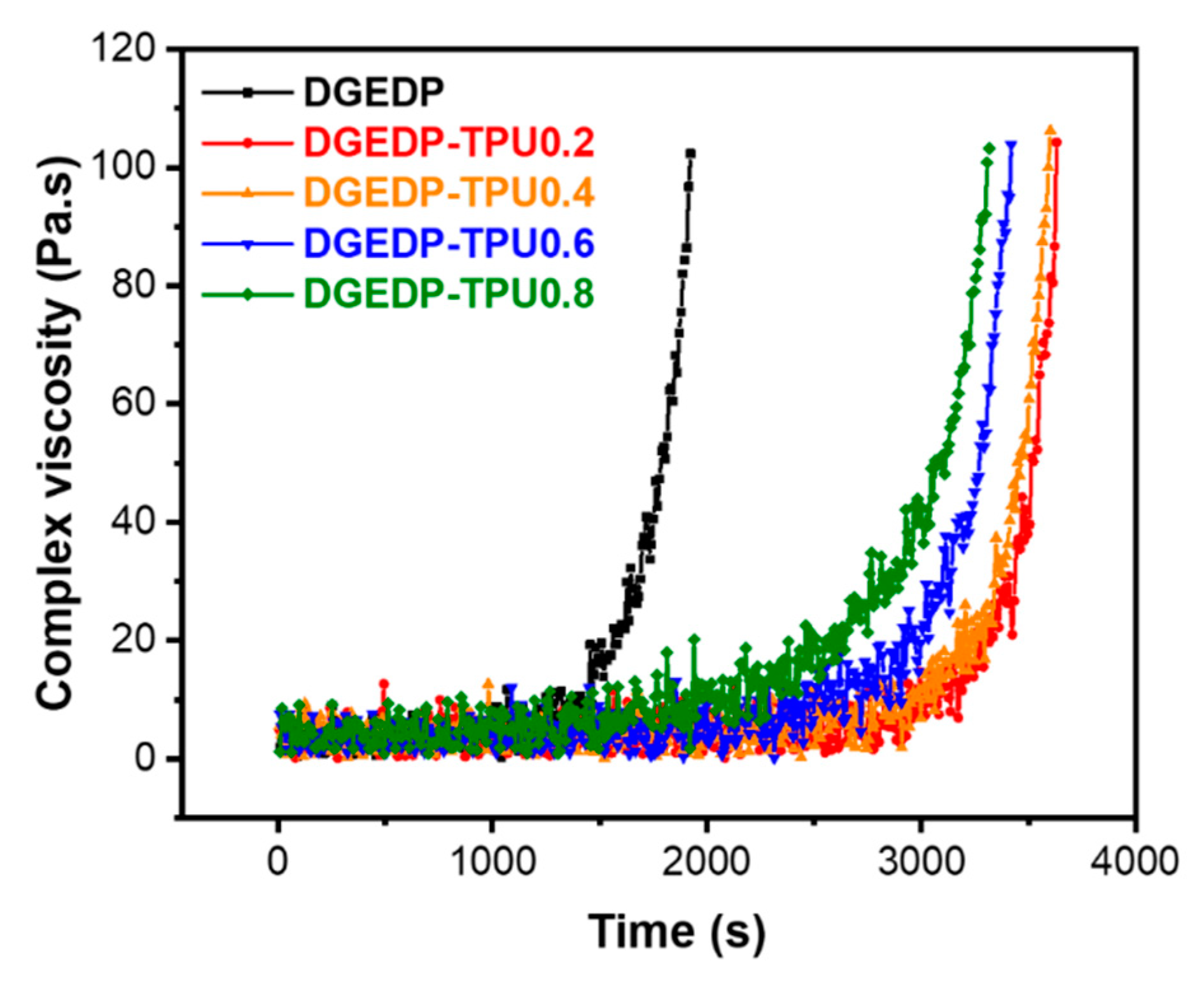
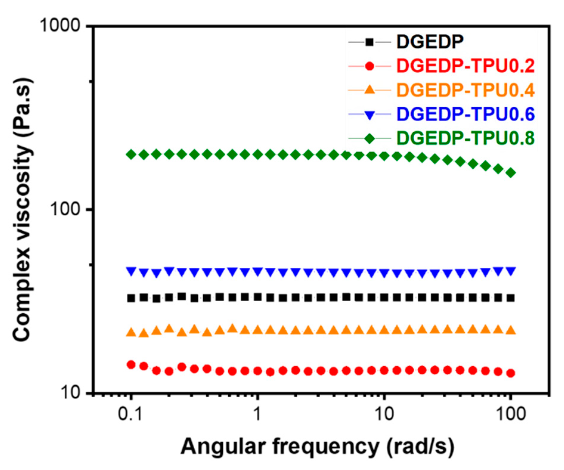
2.3.3. Rheological Characterization

The rheological testing was conducted using a rheometer (ARES G2, TA Instruments, New Castle, DE, USA) with 25 mm parallel plate geometry. The viscosity of the DGEDP/TPU prepolymer blends was measured in flow testing mode at 25 °C. The shear rate was changed from 100 to 0.1 s−1 during the test. Oscillation time sweep mode was conducted for the chemorheology study at 85 °C. The oscillation strain was fixed at 0.1% with constant 1 Hz frequency. All tests were conducted within the linear viscoelastic regime.

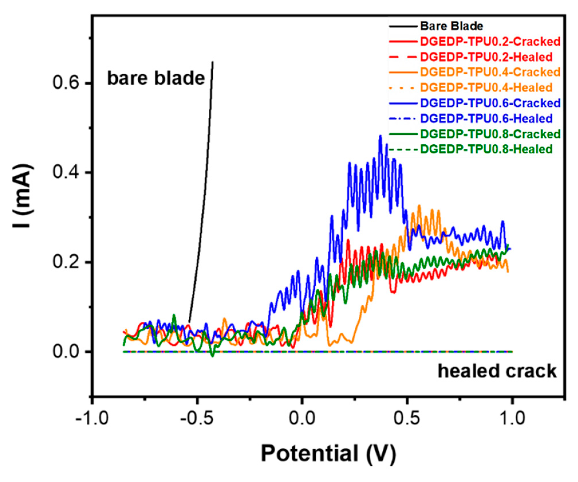
2.3.4. Anti-Corrosion Behavior

The anti-corrosion behavior of the cracked coating before and after healing was examined by an electrochemical test using linear sweep voltammetry. A steel blade with one side coated with the polymer film was exposed to 5 wt.% NaCl/H2O solutions by loading on an electrochemical cell equipped with three-electrode (Bio-Logic Science Instruments). In this setup, Ag/AgCl in 3 M KCl was used as reference electrode and Pt was used as counter electrode. The coating substrate acted as the working electrode. A linear scanned voltage from −0.8 to 1.0 V was applied and the electrical current data was recorded. After testing, the coating layer was peeled from the substrate using a razor blade allowing to observe the effect of the electrochemical treatment on the steel. Optical microscopy (Olympus BX51, Olympus Co, Tokyo, Japan) was used for the observation.

3. Results and Discussion

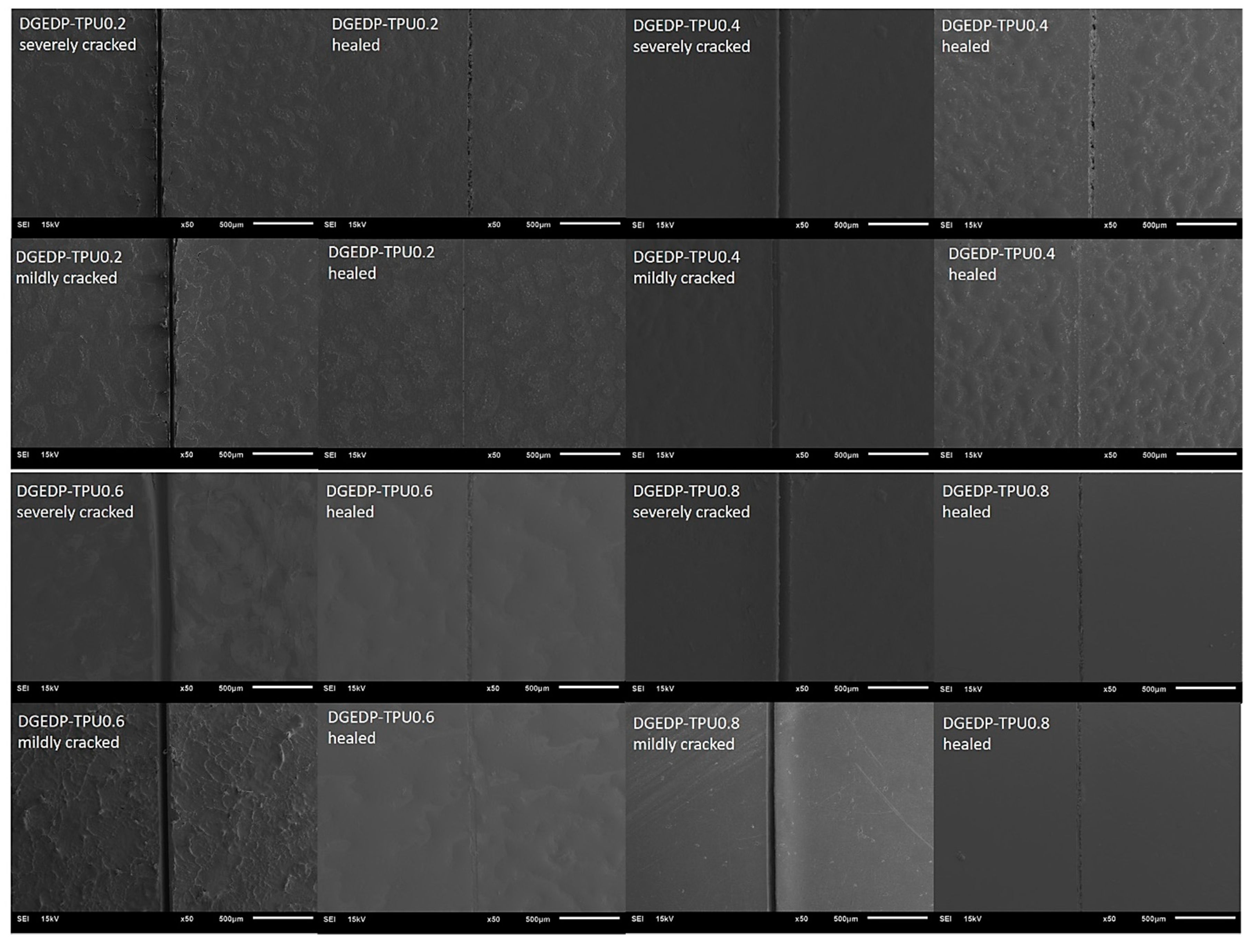
3.1. Self-Healing Performance of the Coatings

The self-healing capability of DGEDP blends with different molecular weight TPU prepolymers was visualized using SEM. Damages of different degrees were introduced on the coating by a razor blade followed by healing at 80 °C for 10 min. Cracks before and after healing were observed. Figure 1 shows that the crack width decreased upon heating, indicating that the epoxy matrix has the tendency to bring the crack surfaces in spatial proximity due to the shape memory response to heat. Furthermore, upon heating the molten TPU chains are able to flow to rebind the crack as shown in the healed sample. This shape memory assisted self-healing (SMASH) concept was also reported in other epoxy based self-healing coating systems [46,47]. Wider cracks were observed for severely damaged coatings by comparison with the mildly damaged ones due to the permanent material loss during scratching. For the severely damaged coatings, upon healing, only minor detectable separations were observed in the cracks. The mildly damaged coatings experience mostly plastic deformation; thus, the cracks were completely closed upon healing. In this case, only barely visible scars remained on the surface for the DGEDP-TPU 0.2, 0.4 and 0.6 systems. Overall, the DGEDP-TPU 0.8 sample shows lower self-healing efficiency in comparison with the other systems. Factors that influence the diffusion behavior of TPU prepolymers will be discussed later.
The function of polymer coatings on a metal substrate is to form an organic layer that protects the substrate from corrosion. Thus, it is essential to examine if the healed coatings are able to rebond and block the substrate from corrosive agents. The anti-corrosion behavior of severely damaged coatings before and after healing was studied by an electrochemical test, as shown in Figure 2. A high electrical current was observed in the bare blade under the voltage sweep, resulting in the formation of rust on the blade (see Figure S2) due to the electrochemical treatment. Relatively large electrical currents were observed in the DGEDP-TPU cracked coatings, indicating that the uncovered area on the substrate was undergoing active corrosion. No current was detected for the healed samples, showing that the barrier function was completely restored upon heating. After the electrochemical test, the substrate with the coating layer peeled off was observed by optical microscopy in Figure S2. The formation of rust was observed for all cracked samples, but no corrosion was detected in the healed samples.

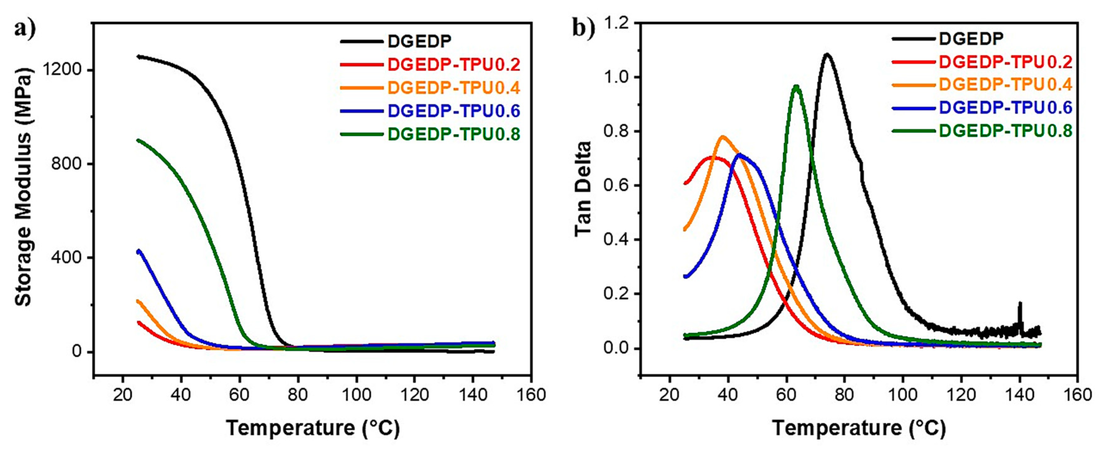
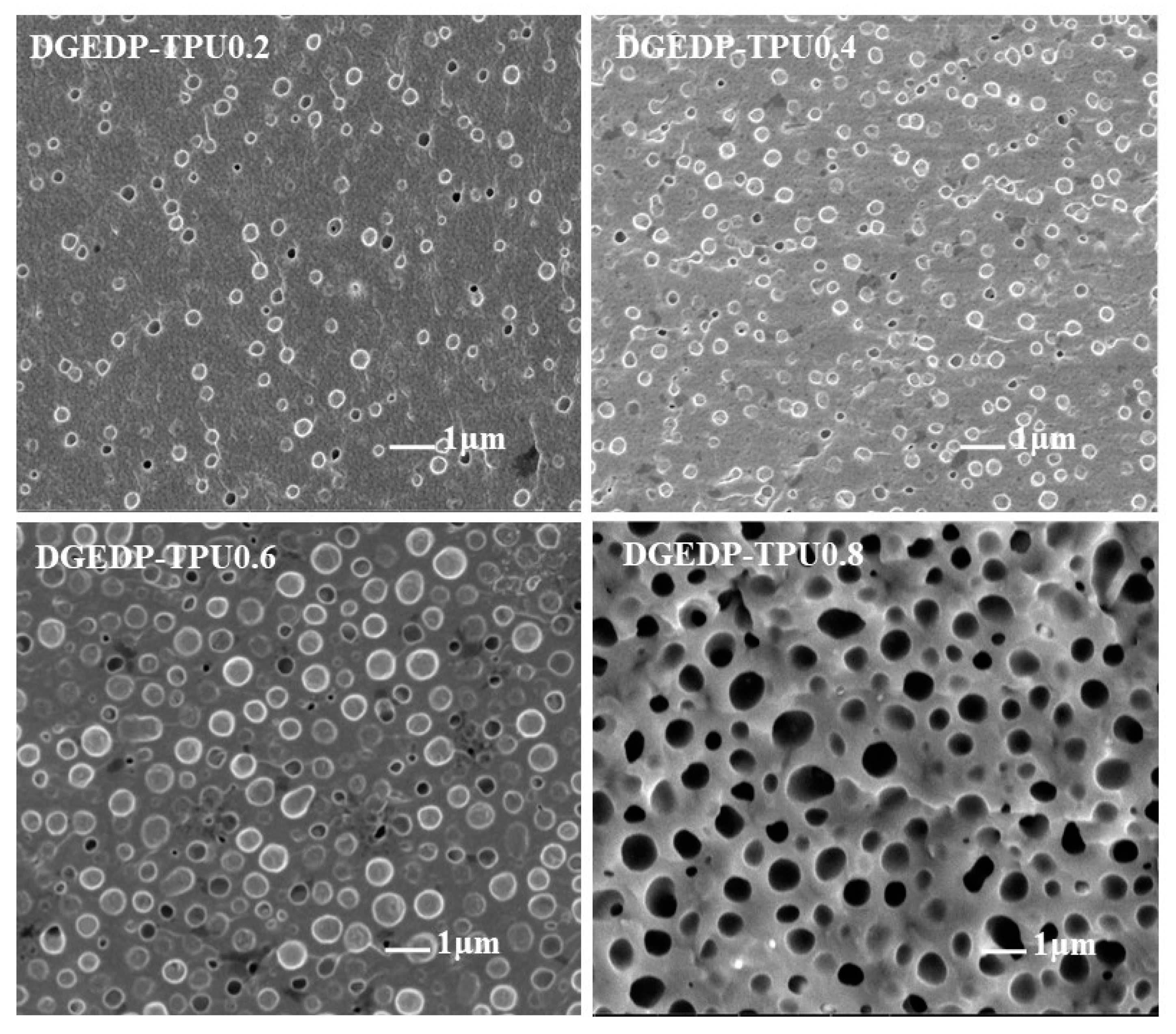
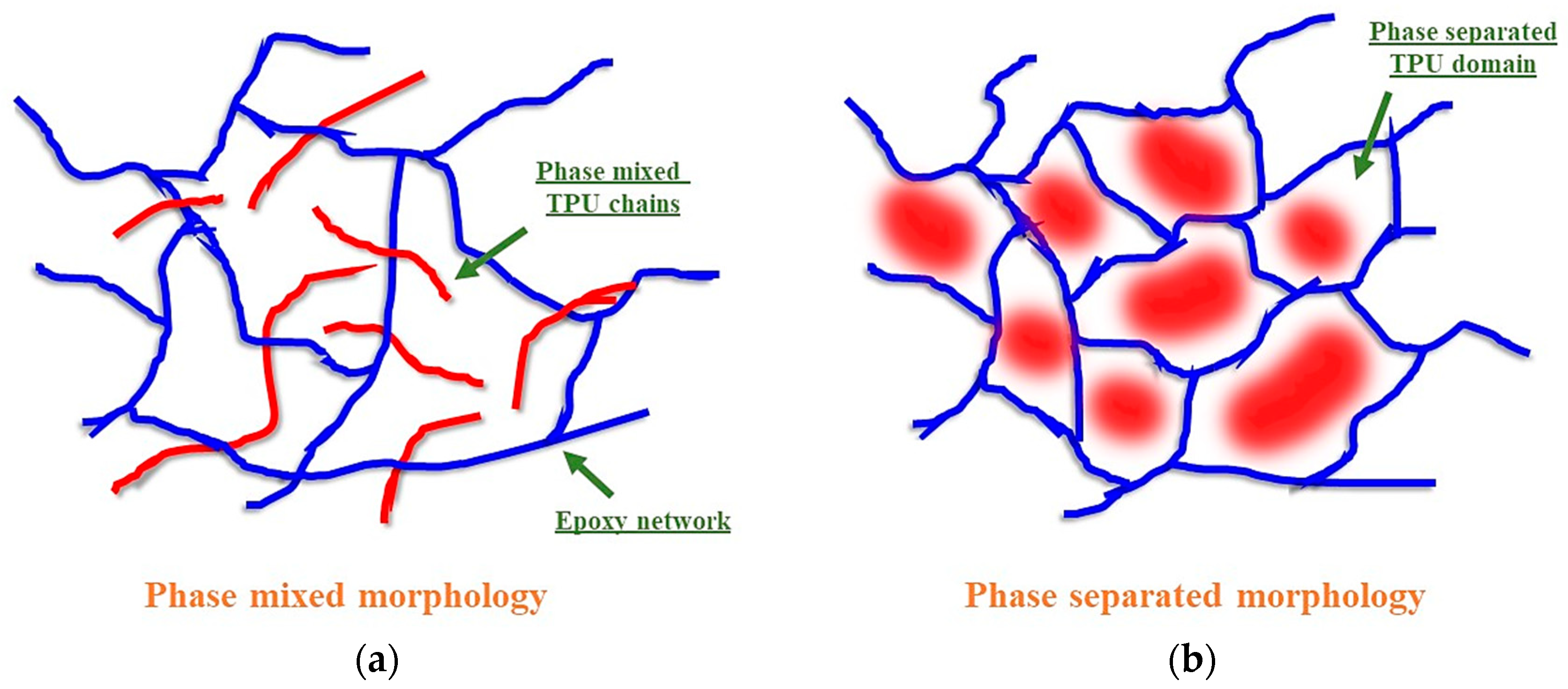
3.2. Structure–Property Relationship for the Coatings

To investigate structure–property relationships for the coatings, dynamic mechanical analysis was performed and the results are presented in Figure 3. The neat DGEDP shows the highest storage modulus and glass transition temperature (74 °C) due to the nature of the highly crosslinked network. The addition of TPU prepolymers into the system results in decreased modulus and Tg values due to PIPS lowering the crosslinking density [48,49]. Interestingly, the phase separation effect follows a trend in which DGEDP-TPU 0.2 shows the lowest storage modulus and Tg and the values increase by increasing the molecular weight of TPU prepolymers. DGEDP-TPU 0.8 shows the highest storage modulus (901 MPa at 25 °C) and Tg (64 °C) among all the mixed systems and these values are close to those of neat DGEDP.
The differences in mechanical properties for the DGEDP-TPU systems can be explained in terms of the morphology observed by SEM (Figure 4). The cryo-fractured surface was immersed in THF under sonication to remove the soluble TPU prepolymers. After etching, only the insoluble epoxy network remains for observation. This method has been widely used to observe the morphology of blend systems comprising both linear and crosslinked polymers [50,51]. Figure 4 indicates that the TPU prepolymers were etched out leaving behind spherical cavities. The micrographs indicate that the TPU prepolymer is phase separated within the epoxy matrix to different degrees: (a) the fully phase separated TPU forms dispersed spherical particles (see black holes left behind upon etching) (b) the partially phase separated TPU forms a TPU-rich phase (see light spherical domains after etching). DGEDP-TPU 0.2, 0.4 and 0.6 show mixtures of black holes and light spherical domains, inferring the existence of both phase mixing and phase separation in these systems. Both the particle size and the number of black hole particles increased by increasing the molecular weight of TPU prepolymers, showing an enhanced degree of phase separation from DGEDP-TPU 0.2 to 0.6. Figure S3 provides a detail analysis of particle size and size distribution in different DGEDP-TPU systems. The phase mixed TPU acts as a plasticizer reducing reactions between the epoxy resin and hardener resulting in decreased crosslink density. Therefore, as the degree of phase separation is enhanced from DGEDP-TPU 0.2 to 0.6, the corresponding Tg and storage modulus values increase. Interestingly, DGEDP-TPU 0.8 shows a unique morphology with only large size black holes in the fractured surface, indicative of a fully phase separated structure resulting in the highest Tg and storage modulus. However, this morphology is not preferable for the self-healing functionality as there is less materials flow after healing (Figure 1), indicating that TPU 0.8 chain motion is severely restricted in this system.
The morphologies formed by these systems with different levels of phase separation due to PIPS [52,53], demonstrate reduced compatibility of epoxy and TPU prepolymer as the curing progresses. The progress of the curing reaction in different DGEDP-TPU prepolymer systems was studied by chemorheology. Figure 5 shows a delay to differing degrees in curing as a function of the TPU prepolymer added. In general, adding 30 phr TPU to the system delays the reaction between epoxy and hardener extending the processing window in comparison with the neat epoxy [54,55]. However, phase separation of the TPU creates an epoxy-hardener rich phase where the reaction rate is locally enhanced. Therefore, the increase in reaction rate from DGEDP-TPU 0.2 to 0.8, observed by chemorheology, can be explained in terms of the increased degree of phase separation from TPU 0.2 to 0.8 during curing. This is in agreement with the morphology displayed in Figure 4 for the cured samples.

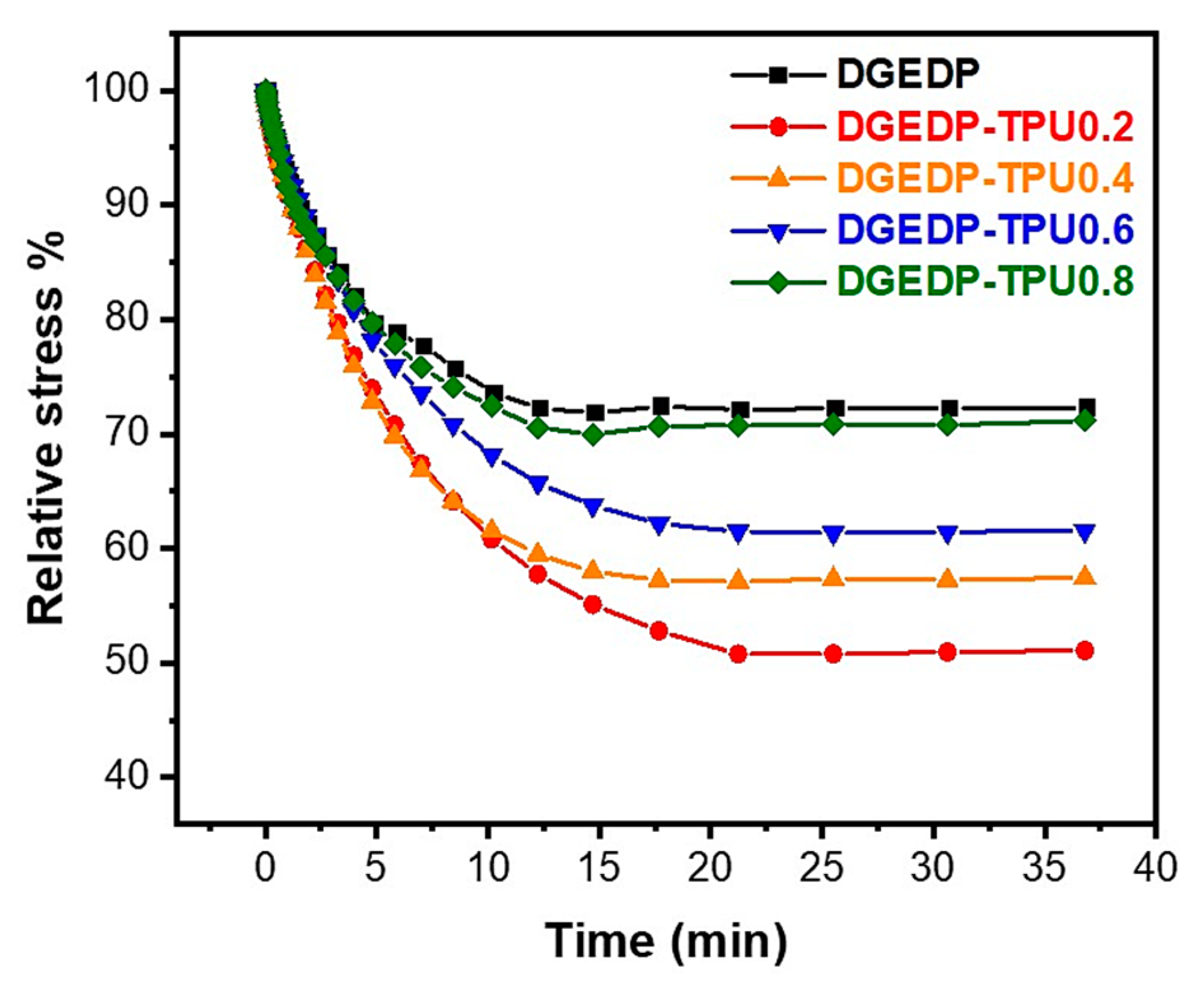
3.3. Stress Relaxation and Self-Diffusion

The requirement for extrinsic self-healing materials is that the secondary phase must diffuse to generate direct mass transport to the damage site. To further investigate the healing mechanism of this system, two factors that influence the chain diffusion of the TPU prepolymers are addressed: (i) the relaxation behavior of the epoxy matrix and (ii) the self-diffusion ability of the TPU prepolymers.
The stress relaxation behavior of polymers deformed above their Tg is related to the ability of segmental motion to locally reorient one or more components within the material to accommodate the applied stress [56,57]. Hence, it is important to examine the stress relaxation behavior of the epoxy network since the ability of the network to reorient impacts the self-healing functionality. Stress relaxation experiments were conducted at Tg + 50 °C for all samples. As shown in Figure 6, DGEDP-TPU 0.2 relaxes the most stress relative to the other samples, indicating higher chain mobility in this system due to the relatively low crosslinked network density. The relaxation behavior is reduced by increasing the TPU prepolymer molecular weight. DGEDP-TPU 0.8 relaxes the least amount of stress and the relaxation behavior is similar to neat DGEDP. This dense and rigid network is preferable for mechanical performance while the impeded segmental motion restricts TPU prepolymer flow, resulting in reduced healing efficiency as shown in Figure 1.
It is evident from morphological characterizations that different degrees of phase separation result as a function of TPU prepolymer molecular weight. The schematics in Figure 7 display both the phase mixing and phase separation morphologies. The phase mixed TPU prepolymers chains (left) dilute the reaction between the epoxy and hardener during curing, causing the formation of a loose network with increased molecular weight between crosslinked sites. In contrast, increased phase separation occurs by incorporating TPU prepolymers with relatively higher molecular weight (right) which form domains dispersed in a denser epoxy network, due to the diminished dilution effect during curing reactions.
The diffusion ability of the TPU prepolymer plays an important role in the self-healing functionality. Figure 8 shows the complex viscosity η∗ as a function of frequency at 25 °C for the different epoxy–TPU prepolymer mixtures. Noticeable shear thinning behavior was observed for the DGEDP-TPU 0.8 at high frequency, indicating chain entanglement. These entanglements impede chain diffusion such that relatively less material transfer can occur for the DGEDP-TPU 0.8 healed sample. No shear thinning that corresponds with chain entanglement was observed in the other systems indicating relatively easier flow upon melting.

4. Conclusions

In summary, self-healing epoxy coating systems with polymerization induced phase separated morphology were obtained by blending a bio-based epoxy DGEDP with TPU prepolymers. This work reveals that the epoxy–TPU morphology, which determines both mechanical and self-healing performance, can be rationally tuned by the selection of a TPU prepolymer with the appropriate molecular weight. We demonstrated that relatively higher molecular weight TPU prepolymers tend to phase separate to a greater degree within the epoxy matrix. This high degree of phase separation accelerates the epoxy curing rate by comparison with a phase mixed system, giving the system high crosslinking density. It therefore endows the coating with high glass transition temperature and mechanical performance but reduced healing efficiency due to restricted TPU chain mobility. In contrast, reduction in the TPU prepolymer molecular weight results in a lower degree of phase separation and lower epoxy network crosslinking. A reduction in the crosslink density allows the TPU prepolymers to more readily flow upon heating to mend cracks. However, these coatings with lower networking crosslinking have reduced mechanical performance. This work describes a rational design strategy to control the morphology of self-healing coatings so that they can be tailored for specific applications by balancing the mechanical properties and self-healing performance.

Supplementary Materials

The following are available online at https://www.mdpi.com/2079-6412/10/1/50/s1, Figure S1: Chemical structure of DGEDP-butyl, Figure S2: Optical microscopy images of the steel substrate after electrochemical treatment, Figure S3: Pore diameter distribution of cryo-fractured DGEDP-TPU system surfaces washed with THF, Table S1: Molecular weight of synthesized TPU prepolymers (OH end capped).

Author Contributions

Conceptualization, I.M.-Z., A.P. and D.Y.; methodology, D.Y., A.P. and V.S.B.; software, D.Y.; validation, R.A.G. and I.M.-Z.; formal analysis, I.M.-Z.; investigation, D.Y.; resources, V.S.B. and T.Y.; data curation, D.Y.; writing—original draft preparation, D.Y.; writing—review and editing, R.A.G. and I.M.-Z.; visualization, D.Y.; supervision, I.M.-Z.; project administration, D.Y.; funding acquisition, R.A.G. and I.M.-Z. All authors have read and agree to the published version of the manuscript.

Funding

This research was funded by National Science Foundation, grant number PIRE 1243313.

Conflicts of Interest

The authors declare no conflict of interest.

References

  1. Hager, M.D.; Greil, P.; Leyens, C.; Van Der Zwaag, S.; Schubert, U.S. Self-healing materials. Adv. Mater. 2010, 22, 5424–5430. [Google Scholar] [CrossRef] [PubMed]
  2. Nakahata, M.; Takashima, Y.; Yamaguchi, H.; Harada, A. Redox-responsive self-healing materials formed from host–guest polymers. Nat. Commun. 2011, 2, 511. [Google Scholar] [CrossRef] [PubMed] [Green Version]
  3. Wu, D.Y.; Meure, S.; Solomon, D. Self-healing polymeric materials: A review of recent developments. Prog. Polym. Sci. 2008, 33, 479–522. [Google Scholar] [CrossRef]
  4. Wool, R.P. Self-healing materials: A review. Soft Matter 2008, 4, 400–418. [Google Scholar] [CrossRef]
  5. Van der Kooij, H.M.; Susa, A.; García, S.J.; van der Zwaag, S.; Sprakel, J. Imaging the Molecular Motions of Autonomous Repair in a Self-Healing Polymer. Adv. Mater. 2017, 29, 1701017. [Google Scholar] [CrossRef] [PubMed]
  6. Geitner, R.; Legesse, F.-B.; Kuhl, N.; Bocklitz, T.W.; Zechel, S.; Vitz, J.; Hager, M.; Schubert, U.S.; Dietzek, B.; Schmitt, M.; et al. Do You Get What You See? Understanding Molecular Self-Healing. Chem. A Eur. J. 2018, 24, 2493–2502. [Google Scholar] [CrossRef]
  7. Canadell, J.; Goossens, H.; Klumperman, B. Self-Healing Materials Based on Disulfide Links. Macromolecules 2011, 44, 2536–2541. [Google Scholar] [CrossRef]
  8. Ying, H.; Zhang, Y.; Cheng, J. Dynamic urea bond for the design of reversible and self-healing polymers. Nat. Commun. 2014, 5, 3218. [Google Scholar] [CrossRef]
  9. Imato, K.; Nishihara, M.; Kanehara, T.; Amamoto, Y.; Takahara, A.; Otsuka, H. Self-Healing of Chemical Gels Cross-Linked by Diarylbibenzofuranone-Based Trigger-Free Dynamic Covalent Bonds at Room Temperature. Angew. Chem. Int. Ed. 2012, 51, 1138–1142. [Google Scholar] [CrossRef]
  10. Cromwell, O.R.; Chung, J.; Guan, Z. Malleable and Self-Healing Covalent Polymer Networks through Tunable Dynamic Boronic Ester Bonds. J. Am. Chem. Soc. 2015, 137, 6492–6495. [Google Scholar] [CrossRef]
  11. Cordier, P.; Tournilhac, F.; Soulié-Ziakovic, C.; Leibler, L. Self-healing and thermoreversible rubber from supramolecular assembly. Nature 2008, 451, 977–980. [Google Scholar] [CrossRef] [PubMed]
  12. Chen, Y.; Guan, Z. Multivalent hydrogen bonding block copolymers self-assemble into strong and tough self-healing materials. Chem. Commun. 2014, 50, 10868–10870. [Google Scholar] [CrossRef] [PubMed]
  13. Yuan, D.; Delpierre, S.; Ke, K.; Raquez, J.-M.; Dubois, P.; Manas-Zloczower, I. Biomimetic Water-Responsive Self-Healing Epoxy with Tunable Properties. ACS Appl. Mater. Interfaces 2019, 11, 17853–17862. [Google Scholar] [CrossRef] [PubMed]
  14. Delpierre, S.; Willocq, B.; Manini, G.; Lemaur, V.; Goole, J.; Gerbaux, P.; Cornil, J.; Dubois, P.; Raquez, J.-M. A Simple Approach for Self-Healable and Stiff Polymer Network from Iminoboronate-Based Boroxine Chemistry. Chem. Mater. 2019. [Google Scholar] [CrossRef]
  15. Guimard, N.K.; Oehlenschlaeger, K.K.; Zhou, J.; Hilf, S.; Schmidt, F.G.; Barner-Kowollik, C. Current trends in the field of self-healing materials. Macromol. Chem. Phys. 2012, 213, 131–143. [Google Scholar] [CrossRef]
  16. Garcia, S.J. Effect of polymer architecture on the intrinsic self-healing character of polymers. Eur. Polym. J. 2014, 53, 118–125. [Google Scholar] [CrossRef] [Green Version]
  17. Van Tittelboom, K.; De Belie, N.; Van Loo, D.; Jacobs, P. Self-healing efficiency of cementitious materials containing tubular capsules filled with healing agent. Cem. Concr. Compos. 2011, 33, 497–505. [Google Scholar] [CrossRef]
  18. Cho, S.H.; Andersson, H.M.; White, S.R.; Sottos, N.R.; Braun, P.V. Polydimethylsiloxane-Based Self-Healing Materials. Adv. Mater. 2006, 18, 997–1000. [Google Scholar] [CrossRef]
  19. Jones, A.S.; Rule, J.D.; Moore, J.S.; White, S.R.; Sottos, N.R. Catalyst Morphology and Dissolution Kinetics of Self-Healing Polymers. Chem. Mater. 2006, 18, 1312–1317. [Google Scholar] [CrossRef]
  20. Lutterman, D.A.; Surendranath, Y.; Nocera, D.G. A Self-Healing Oxygen-Evolving Catalyst. J. Am. Chem. Soc. 2009, 131, 3838–3839. [Google Scholar] [CrossRef]
  21. Pingkarawat, K.; Wang, C.H.; Varley, R.J.; Mouritz, A.P. Self-healing of delamination cracks in mendable epoxy matrix laminates using poly [ethylene-co-(methacrylic acid)] thermoplastic. Compos. Part A Appl. Sci. Manuf. 2012, 43, 1301–1307. [Google Scholar] [CrossRef]
  22. Pingkarawat, K.; Wang, C.H.; Varley, R.J.; Mouritz, A.P. Mechanical properties of mendable composites containing self-healing thermoplastic agents. Compos. Part A Appl. Sci. Manuf. 2014, 65, 10–18. [Google Scholar] [CrossRef]
  23. Zhu, D.Y.; Rong, M.Z.; Zhang, M.Q. Self-healing polymeric materials based on microencapsulated healing agents: From design to preparation. Prog. Polym. Sci. 2015, 49–50, 175–220. [Google Scholar] [CrossRef]
  24. Pang, J.W.C.; Bond, I.P. A hollow fibre reinforced polymer composite encompassing self-healing and enhanced damage visibility. Compos. Sci. Technol. 2005, 65, 1791–1799. [Google Scholar] [CrossRef]
  25. Toohey, K.S.; Sottos, N.R.; Lewis, J.A.; Moore, J.S.; White, S.R. Self-healing materials with microvascular networks. Nat. Mater. 2007, 6, 581–585. [Google Scholar] [CrossRef] [PubMed]
  26. Williams, G.; Trask, R.; Bond, I. A self-healing carbon fibre reinforced polymer for aerospace applications. Compos. Part A Appl. Sci. Manuf. 2007, 38, 1525–1532. [Google Scholar] [CrossRef]
  27. Mansfeld, F. Use of electrochemical impedance spectroscopy for the study of corrosion protection by polymer coatings. J. Appl. Electrochem. 1995, 25, 187–202. [Google Scholar] [CrossRef]
  28. Tan, C.K.; Blackwood, D.J. Corrosion protection by multilayered conducting polymer coatings. Corros. Sci. 2003, 45, 545–557. [Google Scholar] [CrossRef]
  29. Grundmeier, G.; Schmidt, W.; Stratmann, M. Corrosion protection by organic coatings: Electrochemical mechanism and novel methods of investigation. Electrochim. Acta 2000, 45, 2515–2533. [Google Scholar] [CrossRef]
  30. Cho, S.H.; White, S.R.; Braun, P.V. Self-healing polymer coatings. Adv. Mater. 2009, 21, 645–649. [Google Scholar] [CrossRef]
  31. Samadzadeh, M.; Boura, S.H.; Peikari, M.; Kasiriha, S.M.; Ashrafi, A. A review on self-healing coatings based on micro/nanocapsules. Prog. Org. Coat. 2010, 68, 159–164. [Google Scholar] [CrossRef]
  32. Huang, M.; Yang, J. Facile microencapsulation of HDI for self-healing anticorrosion coatings. J. Mater. Chem. 2011, 21, 11123–11130. [Google Scholar] [CrossRef]
  33. Park, J.H.; Braun, P.V. Coaxial electrospinning of self-healing coatings. Adv. Mater. 2010, 22, 496–499. [Google Scholar] [CrossRef]
  34. Luo, X.; Mather, P.T. Shape Memory Assisted Self-Healing Coating. ACS Macro Lett. 2013, 2, 152–156. [Google Scholar] [CrossRef]
  35. Andreeva, D.V.; Fix, D.; Möhwald, H.; Shchukin, D.G. Self-Healing Anticorrosion Coatings Based on pH-Sensitive Polyelectrolyte/Inhibitor Sandwichlike Nanostructures. Adv. Mater. 2008, 20, 2789–2794. [Google Scholar] [CrossRef]
  36. Doan, T.Q.; Leslie, L.S.; Kim, S.Y.; Bhargava, R.; White, S.R.; Sottos, N.R. Characterization of core-shell microstructure and self-healing performance of electrospun fiber coatings. Polymer 2016, 107, 263–272. [Google Scholar] [CrossRef] [Green Version]
  37. Kumar, G.S. Self-Healing Materials: Fundamentals, Design Strategies, and Applications; Wiley-vch: Weinheim, Germany, 2009; ISBN 9783527318292. [Google Scholar]
  38. Yang, T.; Du, Y.; Li, Z.M.; Wang, C.H. Mechanical Properties of Self-Healing Carbon Fiber-Epoxy Composite Stitched with Mendable Polymer Fiber. Polym. Polym. Compos. 2014, 22, 329–336. [Google Scholar] [CrossRef]
  39. Varley, R.J.; Craze, D.A.; Mouritz, A.P.; Wang, C.H. Thermoplastic Healing in Epoxy Networks: Exploring Performance and Mechanism of Alternative Healing Agents. Macromol. Mater. Eng. 2013, 298, 1232–1242. [Google Scholar] [CrossRef]
  40. Grigoriev, D.; Shchukina, E.; Shchukin, D.G. Nanocontainers for Self-Healing Coatings. Adv. Mater. Interfaces 2017, 4, 1600318. [Google Scholar] [CrossRef]
  41. Wei, H.; Wang, Y.; Guo, J.; Shen, N.Z.; Jiang, D.; Zhang, X.; Yan, X.; Zhu, J.; Wang, Q.; Shao, L.; et al. Advanced micro/nanocapsules for self-healing smart anticorrosion coatings. J. Mater. Chem. A 2015, 3, 469–480. [Google Scholar] [CrossRef]
  42. Yuan, D.; Bonab, V.S.; Patel, A.; Manas-Zloczower, I. Self-healing epoxy coatings with enhanced properties and facile processability. Polymer 2018, 147, 196–201. [Google Scholar] [CrossRef]
  43. Chrysanthos, M.; Galy, J.; Pascault, J.-P. Preparation and properties of bio-based epoxy networks derived from isosorbide diglycidyl ether. Polymer 2011, 52, 3611–3620. [Google Scholar] [CrossRef]
  44. Baroncini, E.A.; Kumar Yadav, S.; Palmese, G.R.; Stanzione, J.F., III. Recent advances in bio-based epoxy resins and bio-based epoxy curing agents. J. Appl. Polym. Sci. 2016, 133. [Google Scholar] [CrossRef] [Green Version]
  45. Maiorana, A.; Spinella, S.; Gross, R.A. Bio-Based Alternative to the Diglycidyl Ether of Bisphenol A with Controlled Materials Properties. Biomacromolecules 2015, 16, 1021–1031. [Google Scholar] [CrossRef] [PubMed]
  46. Luo, X.; Mather, P.T. Shape Memory Assisted Self-Healing (SMASH) Coating. Macromolecules 2010, 6, 2146. [Google Scholar]
  47. Rodriguez, E.D.; Luo, X.; Mather, P.T. Linear/Network Poly(ε-caprolactone) Blends Exhibiting Shape Memory Assisted Self-Healing (SMASH). ACS Appl. Mater. Interfaces 2011, 3, 152–161. [Google Scholar] [CrossRef]
  48. Siddhamalli, S.K.; Kyu, T. Toughening of thermoset/thermoplastic composites via reaction-induced phase separation: Epoxy/phenoxy blends. J. Appl. Polym. Sci. 2000, 77, 1257–1268. [Google Scholar] [CrossRef]
  49. Fernández, B.; Corcuera, M.A.; Marieta, C.; Mondragon, I. Rheokinetic variations during curing of a tetrafunctional epoxy resin modified with two thermoplastics. Eur. Polym. J. 2001, 37, 1863–1869. [Google Scholar] [CrossRef]
  50. Siddhamalli, S.K. Toughening of epoxy/polycaprolactone composites via reaction induced phase separation. Polym. Compos. 2000, 21, 846–855. [Google Scholar] [CrossRef]
  51. Chen, J.-L.; Chang, F.-C. Phase Separation Process in Poly(ε-caprolactone)—Epoxy Blends. Macromolecules 1999, 32, 5348–5356. [Google Scholar] [CrossRef]
  52. Wang, M.; Yu, Y.; Wu, X.; Li, S. Polymerization induced phase separation in poly (ether imide)-modified epoxy resin cured with imidazole. Polymer 2004, 45, 1253–1259. [Google Scholar] [CrossRef]
  53. Yu, Y.; Wang, M.; Gan, W.; Tao, Q.; Li, S. Polymerization-Induced Viscoelastic Phase Separation in Polyethersulfone-Modified Epoxy Systems. J. Phys. Chem. B 2004, 108, 6208–6215. [Google Scholar] [CrossRef] [PubMed]
  54. Martinez, I.; Martin, M.D.; Eceiza, A.; Oyanguren, P.; Mondragon, I. Phase separation in polysulfone-modified epoxy mixtures. Relationships between curing conditions, morphology and ultimate behavior. Polymer 2000, 41, 1027–1035. [Google Scholar] [CrossRef]
  55. Aizpurua, B.; Franco, M.; Corcuera, M.A.; Riccardi, C.C.; Mondragon, I. Chemorheology and ultimate behavior of epoxy-amine mixtures modified with a liquid oligomer. J. Appl. Polym. Sci. 2000, 76, 1269–1279. [Google Scholar] [CrossRef]
  56. Villani, M.; Deshmukh, Y.S.; Camlibel, C.; Esteves, A.C.C.; de with, G. Superior relaxation of stresses and self-healing behavior of epoxy-amine coatings. RSC Adv. 2016, 6, 245–259. [Google Scholar] [CrossRef]
  57. Yu, K.; Taynton, P.; Zhang, W.; Dunn, M.L.; Qi, H.J. Influence of stoichiometry on the glass transition and bond exchange reactions in epoxy thermoset polymers. RSC Adv. 2014, 4, 48682–48690. [Google Scholar] [CrossRef]
Figure 1. SEM images of DGEDP-TPU cracked coatings and the coatings after self-healing.
Coatings 10 00050 g001
Figure 2. Voltage (V) versus current (I) plots measured in linear sweep voltammetry experiments for bare steel and DGEDP-TPU prepolymer coatings cracked and healed.
Coatings 10 00050 g002
Figure 3. DMA (a) storage modulus (b) tan delta of DGEDP and DGEDP-TPU prepolymer coatings.
Coatings 10 00050 g003
Figure 4. SEM images of DGEDP-TPU systems cryo-fractured and surface washed with THF.
Coatings 10 00050 g004
Figure 5. Evolution of the complex viscosity as a function of curing time at 85 °C.
Coatings 10 00050 g005
Figure 6. Stress relaxation behavior of DGEDP-TPU systems as a function of time.
Coatings 10 00050 g006
Figure 7. Schematics of phase mixed (a) and phase separated morphologies resulting from differing TPU prepolymer molecular weight (b).
Coatings 10 00050 g007
Figure 8. Complex viscosity η∗ as function of frequency at 25 °C.
Coatings 10 00050 g008

