Article

Composite Magnetic Photocatalyst Bi5O7I/MnxZn1−xFe2O4: Hydrothermal-Roasting Preparation and Excellent Photocatalytic Activity

1
State Key Laboratory of Coal Mine Disaster Dynamics and Control, Chongqing University, Chongqing 400044, China
2
College of Chemistry and Chemical Engineering, Chongqing University, Chongqing 400044, China
*
Author to whom correspondence should be addressed.
Nanomaterials 2019, 9(1), 118; https://doi.org/10.3390/nano9010118
Submission received: 17 December 2018 / Revised: 13 January 2019 / Accepted: 15 January 2019 / Published: 18 January 2019

Abstract

:
A new composite magnetic photocatalyst, Bi5O7I/MnxZn1−xFe2O4, prepared by a hydrothermal-roasting method was studied. The photocatalytic properties of Bi5O7I/MnxZn1−xFe2O4 were evaluated by degradation of Rhodamine B (RhB) under simulated sunlight irradiation, and the structures and properties were characterized by X-ray diffraction (XRD), Fourier-transform infrared spectroscopy (FTIR), scanning electron microscopy (SEM), transmission electron microscopy (TEM), ultraviolet-visible light (UV-Vis) diffuse reflectance spectra (DRS), and a vibrating sample magnetometer (VSM). The results indicated that Bi5O7I/MnxZn1−xFe2O4 was an orthorhombic crystal, which was similar to that observed for Bi5O7I. Bi5O7I/MnxZn1−xFe2O4 consisted of irregularly shaped nanosheets that were 40–60 nm thick. The most probable pore size was 24.1 nm and the specific surface area was 7.07 m2/g. Bi5O7I/MnxZn1−xFe2O4 could absorb both ultraviolet and visible light, and the energy gap value was 3.22 eV. The saturation magnetization, coercivity and residual magnetization of Bi5O7I/MnxZn1−xFe2O4 were 3.9 emu/g, 126.6 Oe, and 0.7 emu/g respectively, which could help Bi5O7I/MnxZn1−xFe2O4 be separated and recycled from wastewater under the action of an external magnetic field. The recycling experiments revealed that the average recovery rate of the photocatalyst was 90.1%, and the photocatalytic activity was still more than 81.1% after five cycles.

1. Introduction

In recent years, with the progress of nanotechnology and the application of photocatalytic technology in environmental pollution treatment [1,2,3], lots of semiconducting metal-oxide nanostructures were widely used for water purification due to their great photocatalytic performance [4,5]. Grottrup [6] applied Bi for doping ZnO, which significantly enhanced its ability in the photocatalytic degradation of methylene blue. Huang [7] indicated that the rate constant of degradation of 17 β-estradiol over N-doped Bi2O3 was 6.3 times that obtained over Bi2O3. Oppong [8] pointed out that the reason for the better photocatalytic performance of Gd–TiO2–graphene oxide (GO) nanocomposites compared to pure TiO2 was because GO sheets and Gd3+ ions are excellent co-catalysts and their presence promotes the reaction sites. Currently, bismuth-based nanometer semiconductors are one of the research hotspots in the field of photocatalytic materials due to their unique electronic structure and excellent absorption ability of ultraviolet and visible light [9,10,11,12]. Bi5O7I, an oxygen-rich bismuth-based nanometer semiconductor [13,14,15], is composed of a Bi 6p orbital at the bottom of the conduction band and Bi 6s, O 2p, and I 5p orbitals at the top of the valence band [16,17]. The Bi 6s and O 2p orbitals can form several dispersed hybrid valence bands, while the I 5p orbital disperses the valence bands further [18,19]. Consequently, the above results accelerate the migration of photo-generated holes and promote the occurrence of oxidation reactions [20,21]. Sun et al. thought that the Bi5O7 and I sections form a unique hierarchical structure successively along the c-axis orderly. As an accelerator for the separation of photo-generated electron–hole pairs, the permanent electrostatic field between the layers can improve the photocatalytic activity of Bi5O7I [22]. Xia et al. prepared sheets of Bi5O7I using the calcining method. However, the photocatalytic degradation rate of Bi5O7I (0.1 g) for Rhodamine B (RhB) solution (100 mL, 10mg/L) was only 52% in 120 min under simulated sunlight irradiation [23]. In order to enhance the photocatalytic activity of Bi5O7I, some means were mentioned, such as doping and compounding [24,25,26,27]. The photocatalytic degradation rate of Eu (3%)/Bi5O7I microspheres was 2.8 times that of Bi5O7I [28], and the photocatalytic degradation efficiency of a graphitic carbon nitride (g-C3N4) (10%)–Bi5O7I nanocomposite was 1.4 times that of Bi5O7I [29].
Most organic pollutants in wastewater can be degraded using photocatalytic technology, with good degradation effects and no secondary pollution. Nevertheless, the complex process, large energy consumption, and low recovery rate are the main disadvantages of the common recovery method, because photocatalytic materials disperse in wastewater uniformly [30]. MnxZn1−xFe2O4 is a soft magnetic ferrite material with its own benefits, such as high saturation magnetization, high permeability, low coercive force, low loss, strong stability, and so on [31]. Therefore, composite magnetic photocatalytic materials prepared with MnxZn1−xFe2O4 as a magnetic matrix can achieve the goal of magnetic recovery, and the heterojunction formed between MnxZn1−xFe2O4 and the photocatalyst is conducive to enhancing the photocatalytic activity of the composite. For instance, Zhang et al. [32] synthesized a MnxZn1−xFe2O4/α-Bi2O3 composite magnetic photocatalyst using the dip-calcination method. However, the recovery rate was only 84.1% under the action of an external magnet, and the degradation time was more than four hours when the degradation rate attained 86.2% after five recoveries. The energy consumption of the preparation process is tremendous, because the precursor of MnxZn1−xFe2O4 must be calcined at 1200 °C for three hours. In addition, the synthesis of composite magnetic photocatalytic materials using Bi5O7I as a photocatalyst and MnxZn1−xFe2O4 as the magnetic matrix is rarely reported.
To overcome these shortcomings, a Bi5O7I/MnxZn1−xFe2O4 composite magnetic photocatalyst was prepared using a hydrothermal-roasting method, and the structures and properties were characterized by X-ray diffraction (XRD), Fourier-transform infrared spectroscopy (FTIR), scanning electron microscopy (SEM), transmission electron microscopy (TEM), ultraviolet-visible light (UV-Vis) diffuse reflectance spectra (DRS), and a vibrating sample magnetometer (VSM). In the meantime, the activity and stability of Bi5O7I/MnxZn1−xFe2O4 were evaluated through the degradation of RhB under simulated sunlight irradiation.

2. Materials and Methods

Analytical reagents of Bi(NO3)3·5H2O, KI, C2H6O2, Fe2(SO4)3, MnSO4·H2O, ZnSO4·7H2O, NaOH, C28H31ClN2O3, and HNO3 were used as raw materials for sample preparation, and were provided by Chengdu Kelong Chemical Ltd (Chengdu, China).

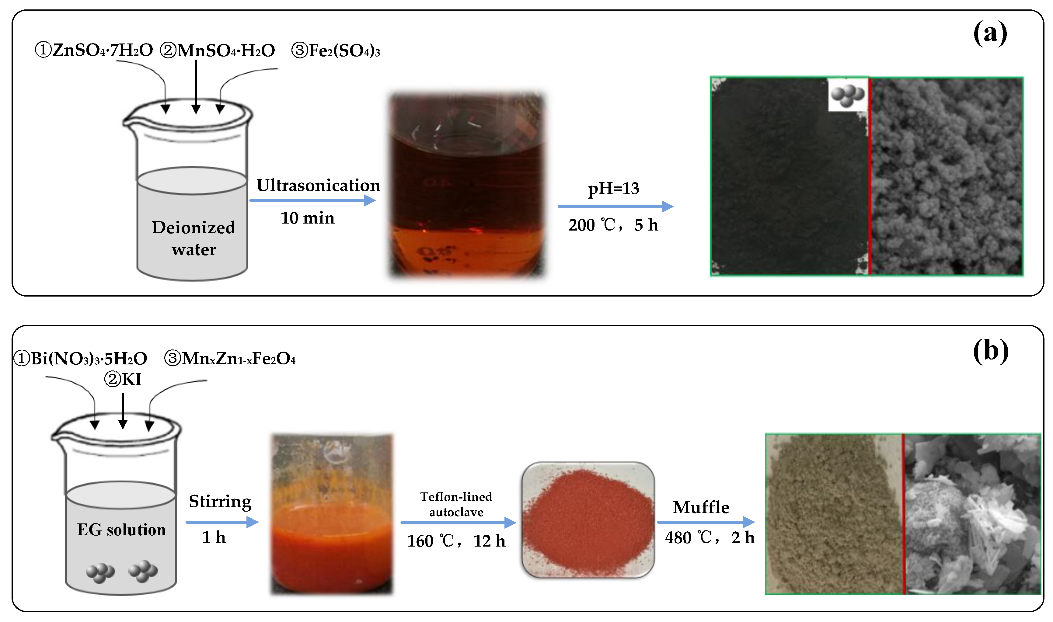
2.1. Preparation of Bi5O7I/MnxZn1−xFe2O4

MnxZn1−xFe2O4 was prepared using a hydrothermal method. Firstly, according to the molar ratio of n(MnO):n(ZnO):n(Fe2O3) = 3.28:1.33:5.39, ZnSO4·7H2O, MnSO4·H2O, and Fe2(SO4)3 were dissolved in deionized water. Then, 5 M NaOH aqueous solution was added dropwise to adjust the pH value of the solution to 13 under vigorous stirring. Subsequently, the solution was transferred into a Teflon-lined autoclave for reaction at 200 °C for 5 h. Afterward, the resulting precipitates were washed with deionized water, as well as dilute sulfuric acid, several times, before being dried at 80 °C for 12 h.
Bi5O7I/MnxZn1−xFe2O4 was prepared using a hydrothermal-roasting method. Firstly, 5 mL of ethylene glycol (EG) was dissolved in 35 mL of deionized water with stirring for 10 min to gain solution A. Then, 2 mmol Bi (NO3)3·5H2O and 2 mmol KI were continuously dissolved in solution A while stirring to acquire suspension liquid B. Then, 10 wt.% as-prepared MnxZn1−xFe2O4 was added into suspension liquid B with continuous stirring for 60 min. Afterward, the mixed turbid solution was transferred into a Teflon-lined autoclave for reaction at 160 °C for 12 h, and the resulting precipitates were washed with deionized water several times. Finally, the precipitates were dried at 80 °C for 5 h and roasted at 480 °C for 2 h in a muffle, obtaining Bi5O7I/MnxZn1−xFe2O4. The synthetic process is displayed in Scheme 1.

2.2. Characterization

The structures of the as-prepared products were characterized by X-ray diffraction (XRD; Shimadzu, XRD-6000, Shimadzu, Kyoto, Japan) and Fourier-transform infrared spectroscopy (FTIR; Nicolet iS50, Thermo Fisher Scientific, Waltham, MA, USA). The morphologies and microstructures of the products were observed using scanning electron microscopy (SEM; S4800, Hitachi, Tokyo, Japan) and transmission electron microscopy (TEM; Tecnai G2F20, FEI, Hillsboro, OR, USA). The surfaces and apertures of the products were measured using an automatic multistation surface and aperture analyzer (Quadrasorb 2MP, Quantachrome, Boynton Beach, FL, USA). The element contents in the composite were analyzed by X-ray photoelectron spectroscopy (XPS, ESCALAB250Xi, Thermo Fisher Scientific, Waltham, MA, USA). The optical absorption ability and magnetic performance of the products were identified with UV-Vis diffuse reflectance spectra (UV-Vis DRS; TU1901, Beijing, China) and a vibrating sample magnetometer (VSM; 7410, Lake Shore, Westerville, OH, USA) respectively.

2.3. Photocatalytic Evaluation

The photocatalytic properties of the samples were evaluated through the degradation of Rhodamine B (RhB) under simulated sunlight irradiation. Firstly, 0.1 g of photocatalyst and 100 mL of RhB aqueous solution (10 mg·L−1) were put into a beaker and stirred for 30 min in the dark to reach the adsorption balance. Then, the mixtures were irradiated with a xenon lamp (CEL-HXF3000, AULTT) of 300 W, and the ultraviolet-visible emission spectrum. Then, 4 mL of the solution was withdrawn at set time intervals, before being centrifuged at 4000 rpm for 5 min to get the supernatant. Finally, the characteristic absorbance of RhB was measured using a UV-Vis spectrophotometer.

3. Results and Discussion

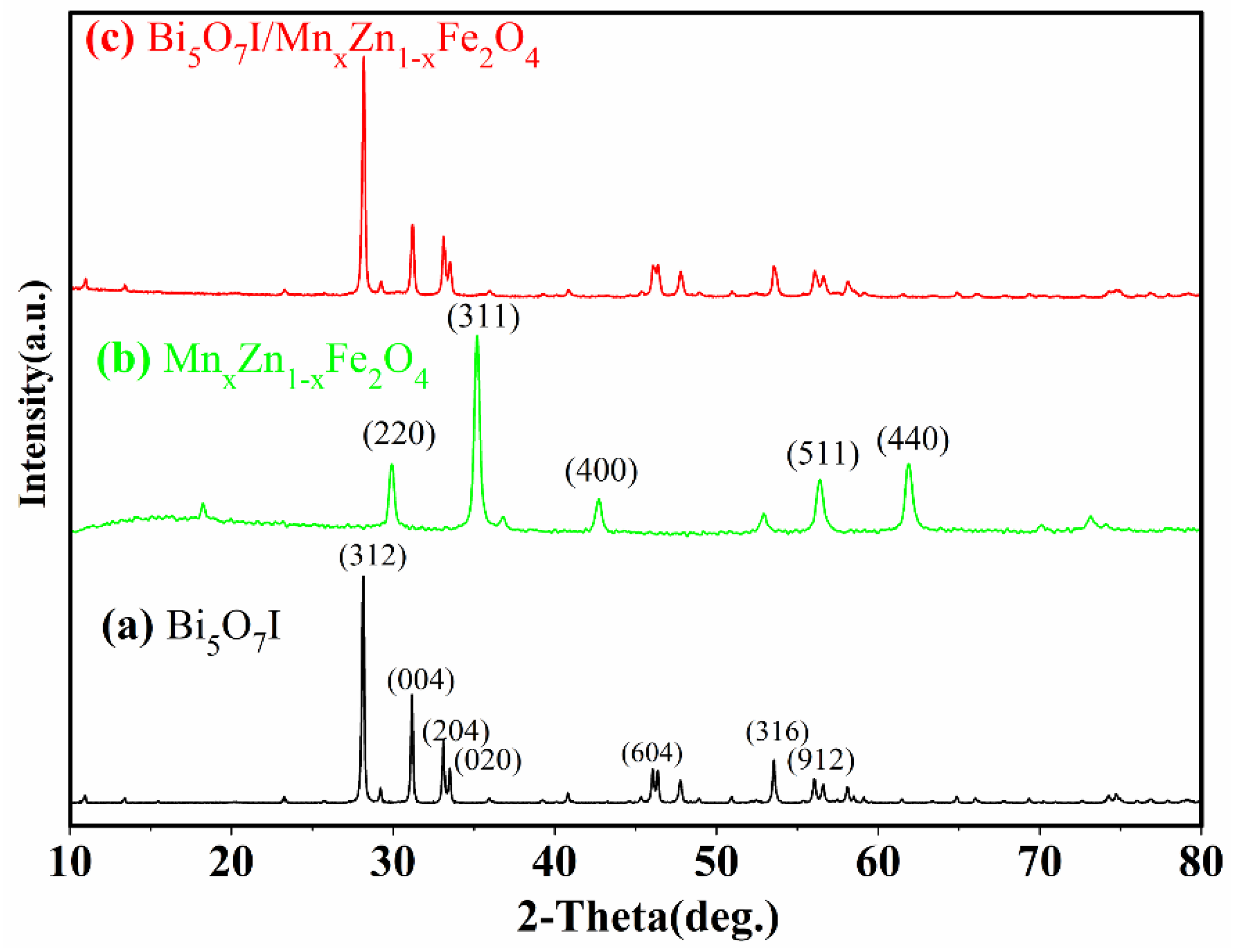
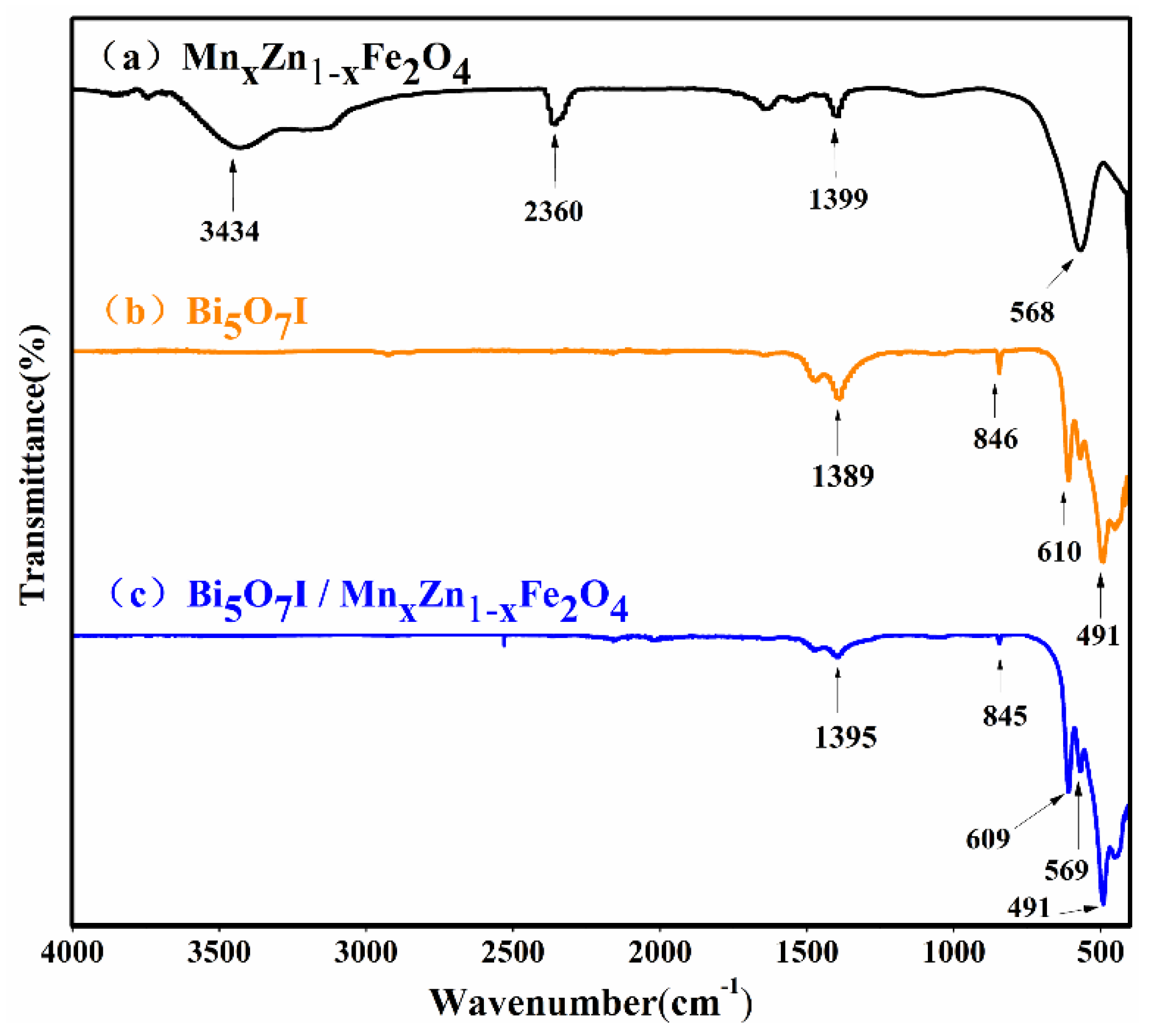
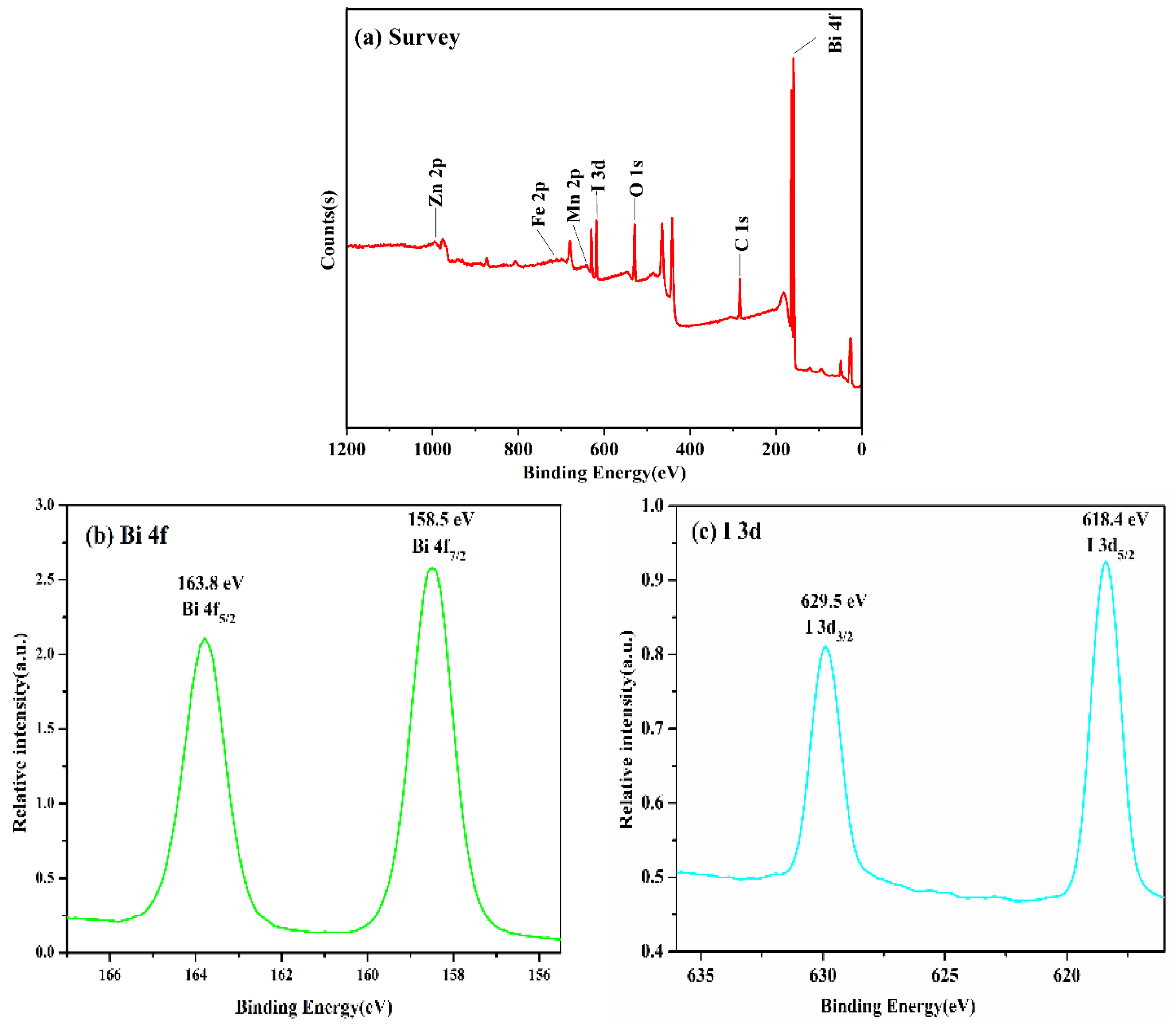
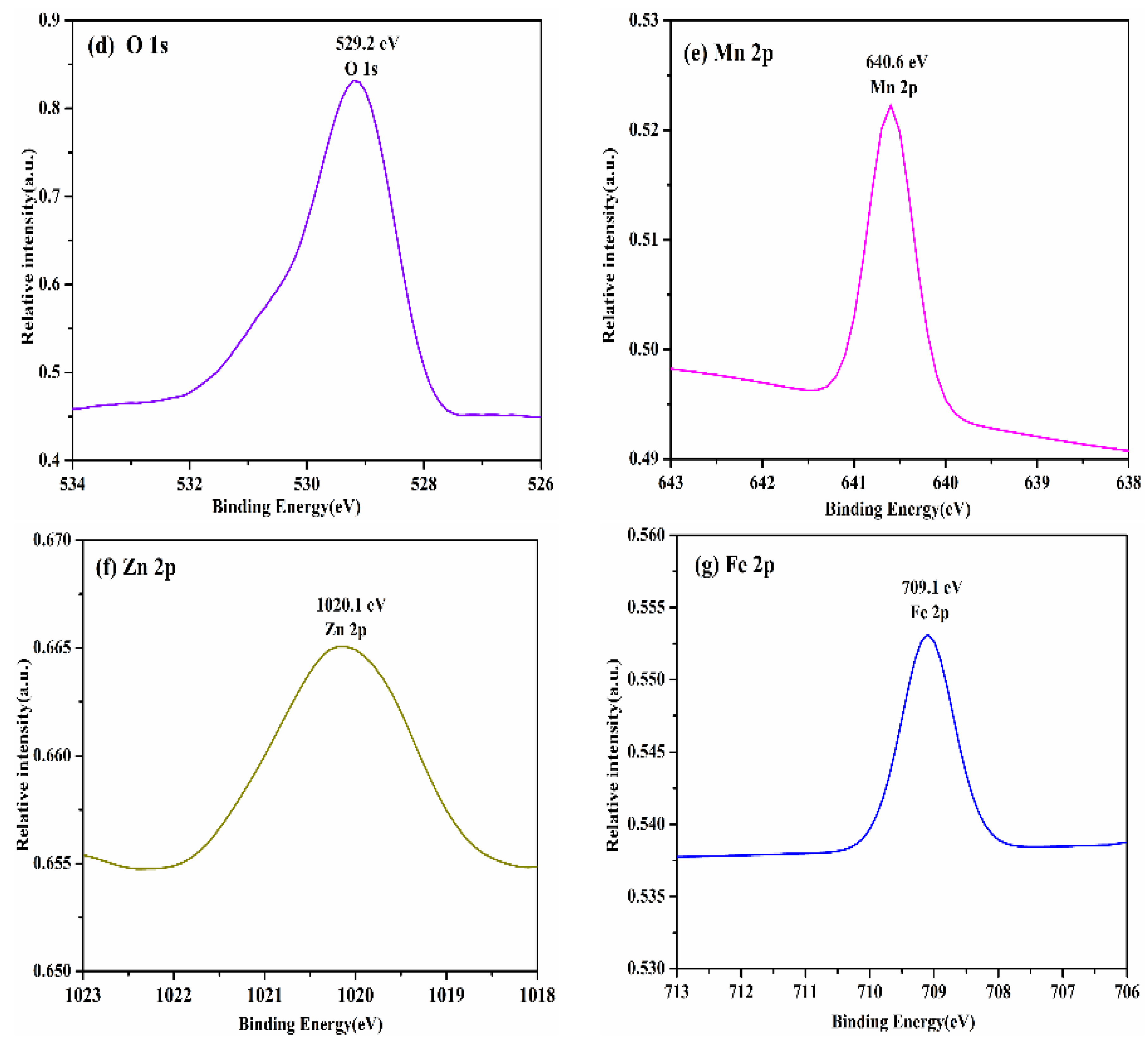
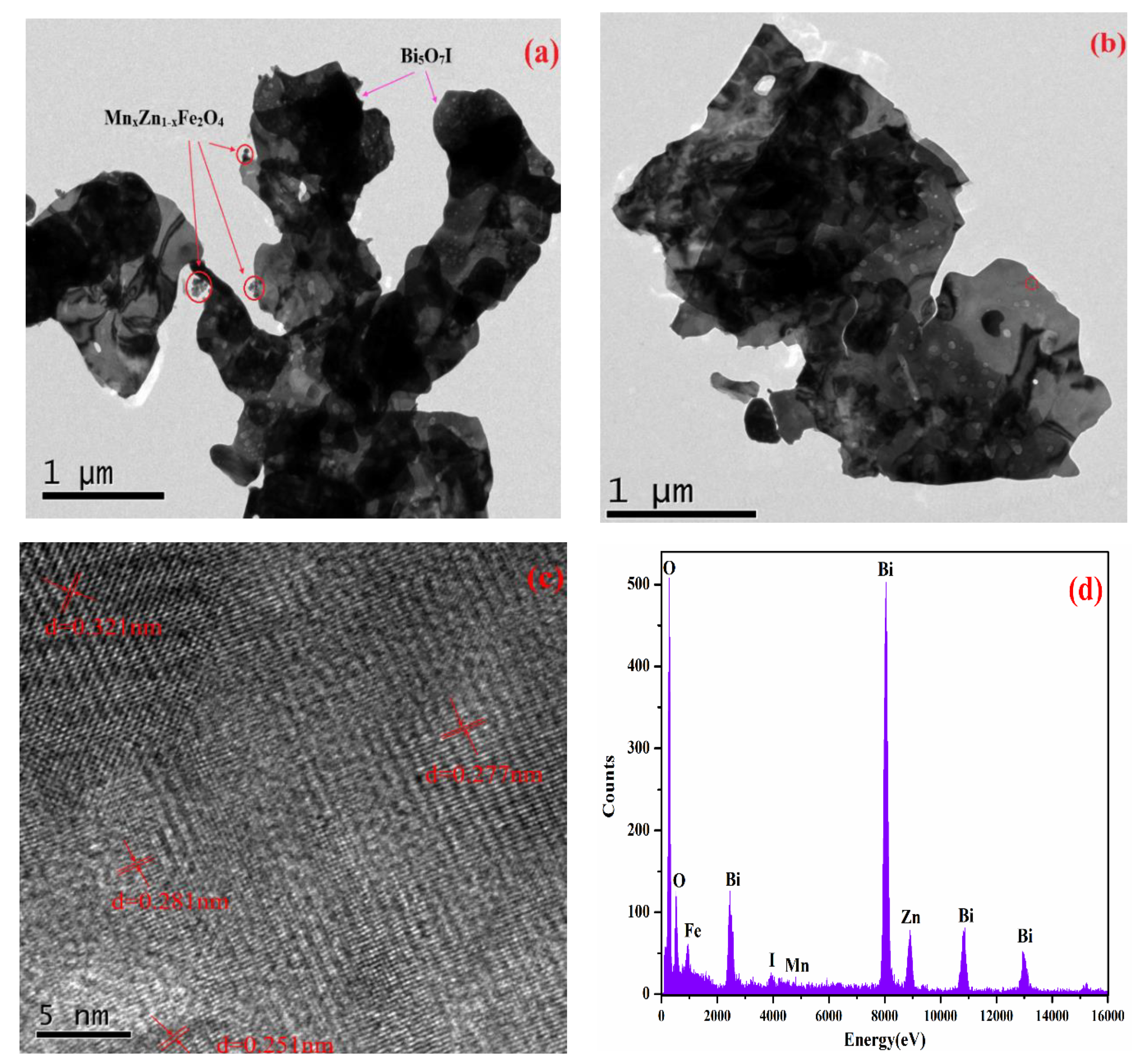
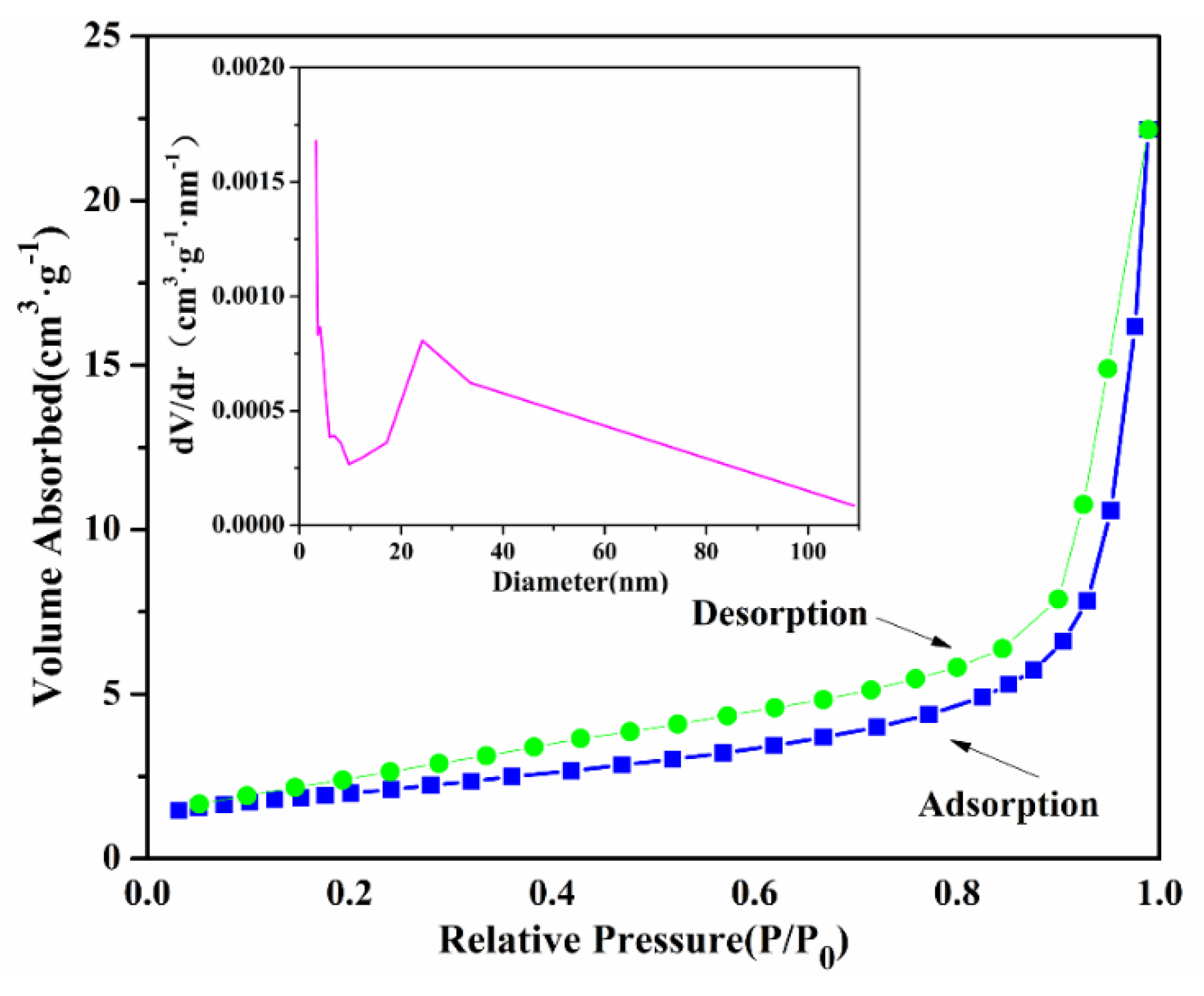
3.1. Structure Characteristics

Figure 1 presents the XRD patterns of MnxZn1−xFe2O4, Bi5O7I, and Bi5O7I/MnxZn1−xFe2O4. As displayed, the diffraction peaks, located at 28.24°, 31.23°, 33.14°, 33.55°, 47.82°, 53.60°, and 56.12°, were assigned to the (312), (004), (204), (020), (224), (316), and (912) planes of orthorhombic Bi5O7I (JCPDS file No. 40-0538). Neither the preferred growth direction nor the crystal structure of Bi5O7I were changed by MnxZn1−xFe2O4, because the characteristic peaks of Bi5O7I/MnxZn1−xFe2O4 were basically consistent with Bi5O7I. Interestingly, the peak of the (912) plane of Bi5O7I/MnxZn1−xFe2O4 overlapped with the (511) plane of the MnxZn1−xFe2O4. The average crystalline size of Bi5O7I/MnxZn1−xFe2O4 was 71.3 nm, as calculated using the Scherrer formula. The cell parameters were as follows: a = 16.8036 Å, b = 5.0721 Å, c = 11.7316 Å, α = β = γ = 90°.
The FTIR spectra of MnxZn1−xFe2O4, Bi5O7I, and Bi5O7I/MnxZn1−xFe2O4 are shown in Figure 2. The characteristic peaks at 3434 cm−1 and 2360 cm−1 were attributed to the stretching vibration and bending vibration of the hydroxyl group (–OH) from surface-adsorbed water, respectively [33]. Typical Raman bands of the Fe–O–Fe bond and the stretching vibration of the Zn–O bond were located at 1399 cm−1 and 568 cm−1, respectively [34]. The intensive signals around 1389 cm−1, 846 cm−1, and 610 cm−1 referred to the bending vibration of the Bi–O bond, and 491 cm−1 referred to the stretching vibration of the Bi–O bond [35]. Figure 2c indicates that the hydroxyl group in MnxZn1−xFe2O4 disappeared because of the roasting process. There was a slight blueshift about the bending vibration of the Bi–O bond (from 610 cm−1 to 609 cm−1) and the stretching vibration of the Fe–O–Fe bond (from 1399 cm−1 to 1395 cm−1). Moreover, the stretching vibration of Zn–O bond (569 cm−1) could be seen in Bi5O7I/MnxZn1−xFe2O4.
The chemical composition and state of the Bi5O7I/MnxZn1−xFe2O4 sample were investigated using XPS. The survey spectra in Figure 3a reveal that the elements of Bi, I, O, Mn, Zn, and Fe exist in Bi5O7I/MnxZn1−xFe2O4. The atomic percentages of Bi, I, O, Mn, Zn, and Fe were 27.4%, 3.74%, 58.13%, 6.51%, 0.35%, and 3.87%, respectively. Figure 3b describes that the two characteristic peaks of Bi 4f5/2 and Bi 4f7/2 were located at 163.8 eV and 158.5 eV in the high-resolution spectra. The I 3d3/2 and I 3d5/2 peaks in Figure 3c could be fitted with two peaks at 629.6 eV and 618.4 eV. As can be seen from Figure 3d–g, the signals at 529.2 eV, 640.6 eV, 1020.4eV, and 709.1eV were attributed to the O 1s, Mn 2p, Zn 2p, and Fe 2p orbitals, respectively.
In order to observe the morphology of the materials, the samples were characterized by SEM. As seen from Figure 4, Bi5O7I was composed of irregularly shaped nanosheets, while MnxZn1−xFe2O4 was a spherical particle. The layer thickness of the nanosheets was around 40–60 nm, as shown in Figure 4c. The energy-dispersive X-ray spectroscopy (EDS) spectrum confirmed that the particles in Figure 4c were MnxZn1−xFe2O4, demonstrating the successful creation of the MnxZn1−xFe2O4 and Bi5O7I compound.
Figure 5a,b display that lots of irregular holes on the sample surface existed. At the same time, many particles of MnxZn1−xFe2O4 were in contact with the gray shells of Bi5O7I. The high-resolution TEM (HRTEM) image of the circular region in Figure 5b reveals that Bi5O7I/MnxZn1−xFe2O4 was polycrystalline due to the different orientation of the crystal surface. The three fringe spacings of 0.321 nm, 0.277 nm, and 0.281 nm between neighboring crystal lattices corresponded to the crystal surface (312), (204), and (004) of the Bi5O7I crystal, and the fringe spacing of 0.251 nm corresponded to the crystal surface (311) of MnxZn1−xFe2O4. The EDS result proved the existence of Bi, I, O, Mn, Zn, and Fe elements, consistent with the XPS investigation.
A specific surface analyzer was utilized to research the specific surface area and pore diameter distribution of Bi5O7I/MnxZn1−xFe2O4. From Figure 6, according to the Brunauer isotherm classification method, the adsorption–desorption isotherm belonged to class IV. The pore diameter distribution curve described that the most probable pore size of Bi5O7I/MnxZn1−xFe2O4 was 24.1 nm. Furthermore, the specific surface area of the Bi5O7I/MnxZn1−xFe2O4 sample calculated using the Brunauer–Emmett–Teller (BET) model was 7.07 m2/g.

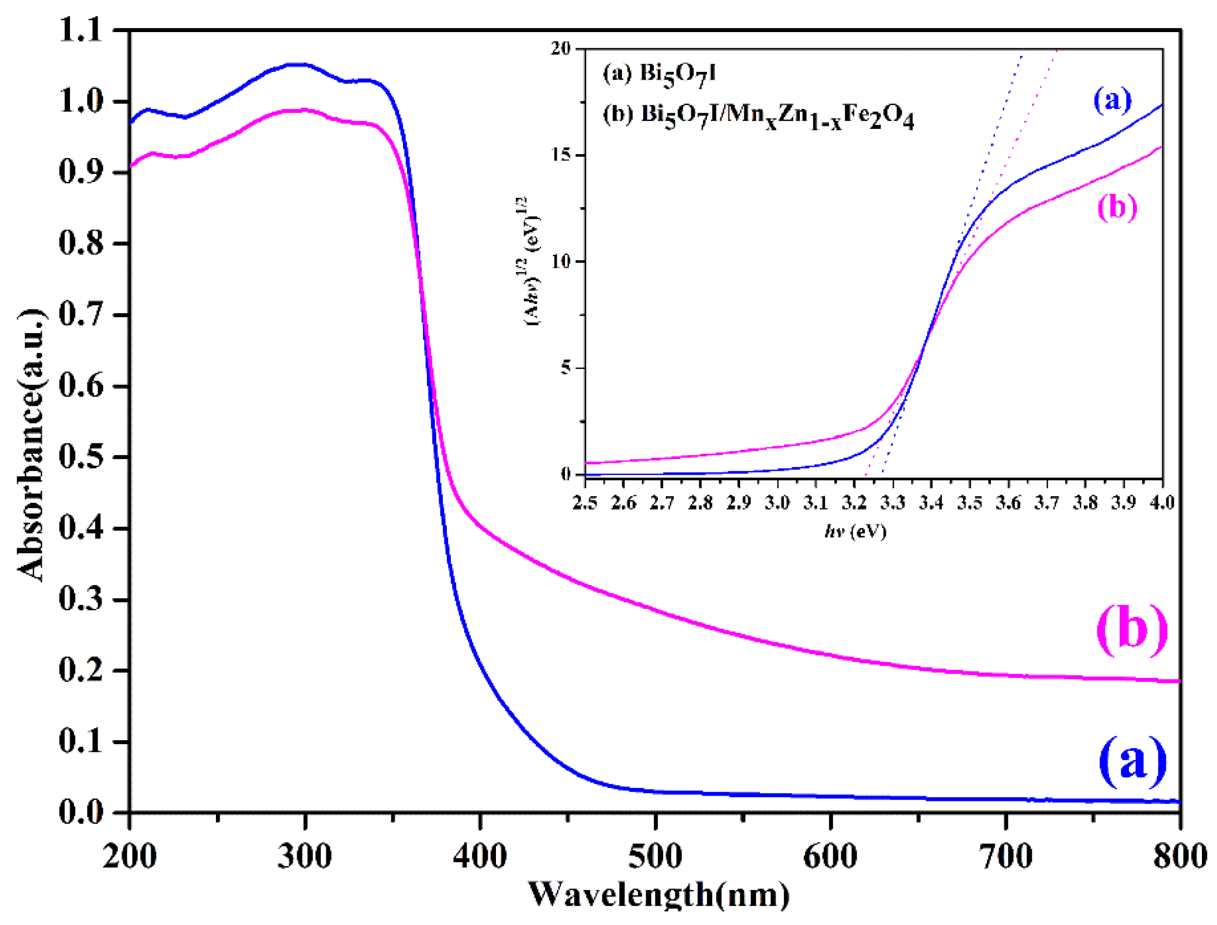
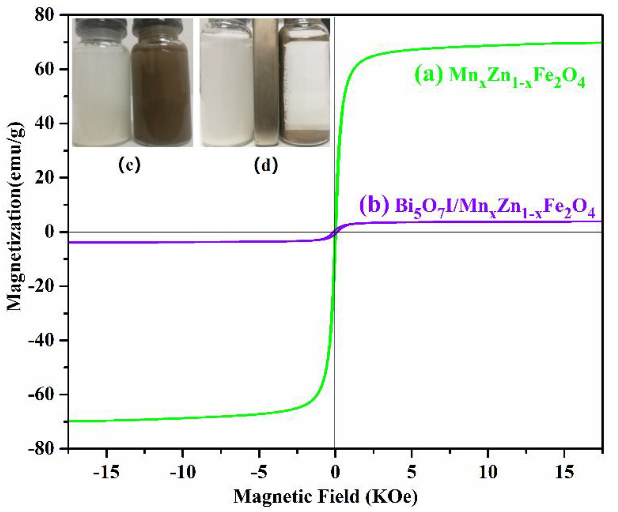
3.2. Absorption Light Ability and Magnetic Properties

The UV-Vis DRS and the (ahv)1/2hv curve are shown in Figure 7. The largest absorption wavelengths of Bi5O7I and Bi5O7I/MnxZn1−xFe2O4 were 470 nm and 600 nm, respectively, illustrating that both could absorb ultraviolet and visible light, and that adding MnxZn1−xFe2O4 extended the range of absorption light. Moreover, the band-gap energy of the samples could be obtained using Equation (1) [22].
a h ν = A ( h ν E g ) n 2
where α, h, v, and Eg are the absorption coefficient, Planck constant, light frequency, and bandgap width, respectively. A is a constant, and n depends on the transition type of the semiconductor optical carriers (direct transition, n = 1; indirect transition, n = 4). According to the plots of (ahv)1/2hv, the band-gap energies of Bi5O7I and Bi5O7I/MnxZn1−xFe2O4 were determined to be 3.27 eV and 3.22 eV, respectively.
In general, the magnetic performance of catalysts determines the recovery efficiency. Therefore, the magnetic hysteresis loops of the samples were measured. Figure 8b depicts that the saturation magnetization (Ms), coercivity (Hc), and residual magnetization (Mr) of Bi5O7I/MnxZn1−xFe2O4 were 3.9 emu/g, 126.6 Oe, and 0.7 emu/g, respectively. Compared with MnxZn1−xFe2O4, Ms declined because the mass ratio of magnetic materials in the photocatalyst was only 10%. It is worth noting that Bi5O7I/MnxZn1−xFe2O4 is easy to magnetize or demagnetize, and its hysteresis loss was small in the alternating magnetic field. Figure 8d shows that the particles of Bi5O7I/MnxZn1−xFe2O4 in the suspension (the right bottle) moved to the magnet rapidly when a magnet was placed close to the bottle, and the suspension became clear after 4 min. However, the suspension containing Bi5O7I (the left bottle) did not show any obvious change under the same conditions. From the above analysis, it was determined that the composite photocatalyst has great magnetic separation capabilities.

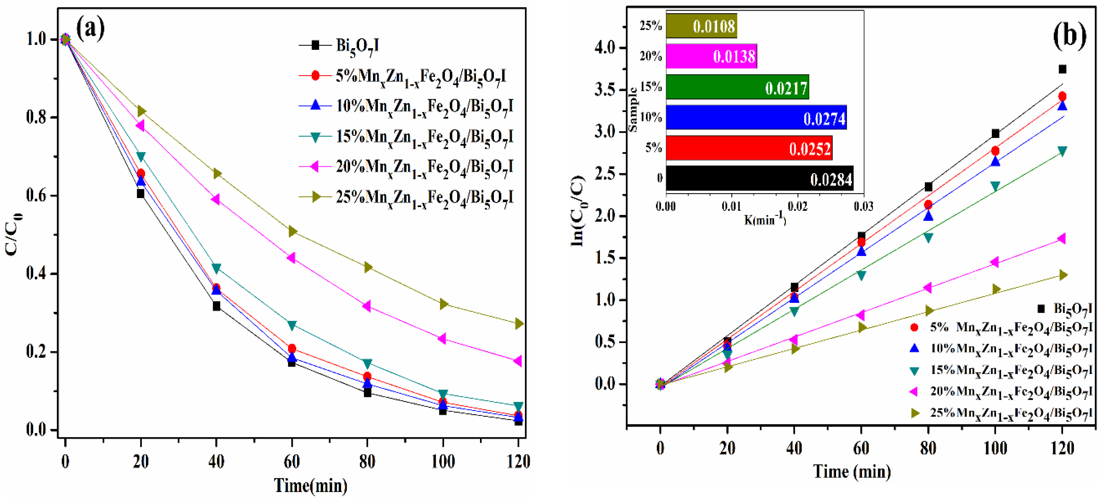
3.3. Photocatalytic Activity

The photocatalytic activity of the as-prepared samples was investigated through photocatalytic degradation experiments of RhB. From Figure 9a, pure Bi5O7I presented the highest photocatalytic activity, whereby 97.6% of RhB was degraded within 120 min. The degradation properties of 5%, 10%, 15%, 20%, and 25% MnxZn1−xFe2O4/Bi5O7I were 96.3%, 96.7%, 93.8%, 82.3%, and 72.7%, respectively. In addition, the first-order kinetics model given by equation ln(C0/C) = kt was applied to quantitatively understand the reaction kinetics, where C0 (mg·L−1) is the initial concentration of RhB solution, C (mg·L−1) is the concentration in aqueous solution at time t, and k (min−1) is the apparent first-order kinetic constant [36]. The degradation constants (in Figure 9b) were calculated to be 0.0284, 0.0252, 0.0274, 0.0217, 0.0138, and 0.0108 min−1 for pure Bi5O7I, and 5%, 10%, 15%, 20%, and 25% MnxZn1−xFe2O4/Bi5O7I samples, respectively. Compared with Bi5O7I, the photocatalytic activity of the compounds declined for two reasons. On one hand, MnxZn1−xFe2O4 became the recombination center of the photogenerated electron (e) and hole (h+), which reduced the lifetime of the photogenerated carriers. On the other hand, adding MnxZn1−xFe2O4 decreased the amount of catalyst in the compound.

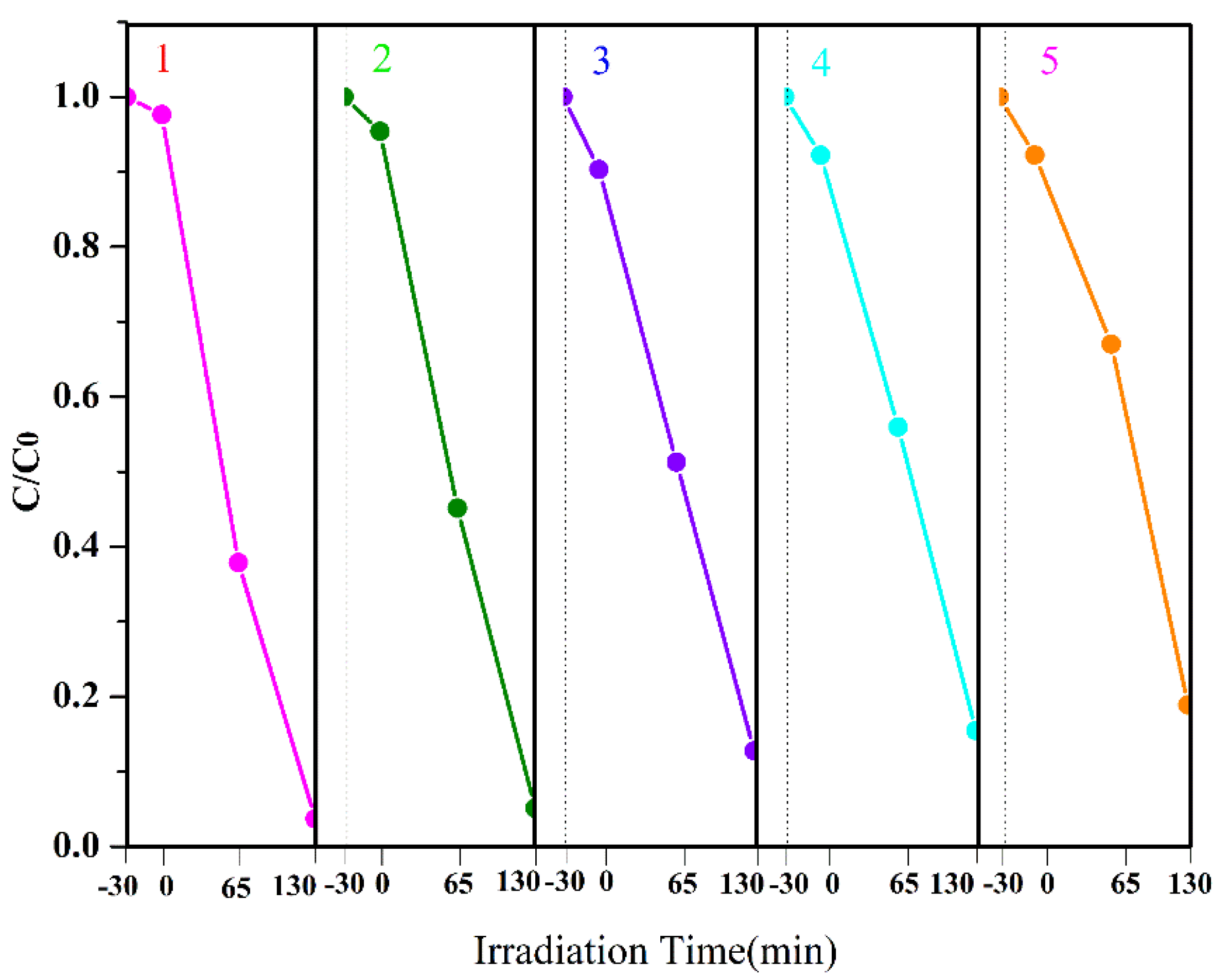
3.4. Stability and Recycling Ability

The stability and the recycling ability of Bi5O7I/MnxZn1−xFe2O4 were studied through recycling experiments. After each reaction, the photocatalyst was separated by an external magnet and then washed with deionized water before being dried at 80 °C for 3 h. The recycling experiments show that the average recovery rate was 90.1%. Figure 10 shows that the degradation rate was still more than 81.1% after five reuses. The experimental results indicate that the composite magnetic photocatalyst can be reused several times with excellent stability.

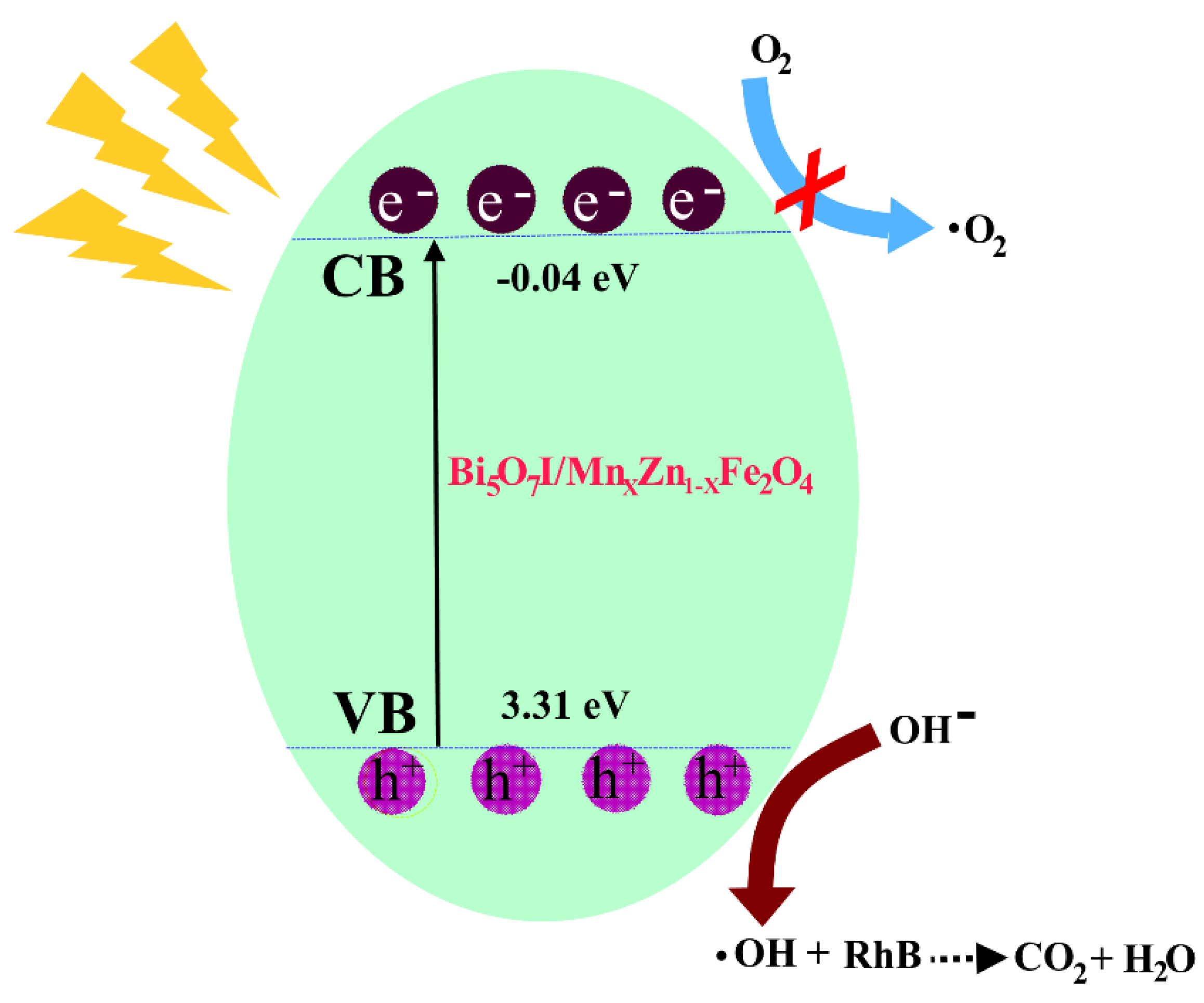
3.5. Photocatalytic Mechanism

There are a few reasons why the composite magnetic photocatalyst has good photocatalytic activity. Firstly, the existence of a porous structure is helpful for improving the transfer efficiency of the photogenerated electron and hole. In this structure, the distance from where photogenerated charge is generated to the semiconductor surface is shortened, which can effectively reduce the recombination of the photogenerated electron and hole [37].
Secondly, the band structure of the photocatalyst is beneficial for producing a hydroxyl radical (•OH). Figure 11 illustrates the proposed mechanism for the photocatalytic activity of Bi5O7I/MnxZn1−xFe2O4 with RhB. As is known, •OH is the major active substance in the photocatalytic degradation of organic pollutants [38]. The adsorbed water is oxidized to •OH by a hole when the valence band top has more positive redox potential than that of •OH/H2O (+2.27 eV). The position of the conduction band bottom (ECB) can be obtained using Equation (2) [39].
E C B = X E C 0.5 E g ,
where X is the absolute electronegativity of the semiconductor oxide, EC is the potential energy of the free electron in a standard hydrogen electrode (~4.5 eV), and Eg is the band gap of the semiconductor oxide. The position of the conduction band bottom for the photocatalyst was determined to be 0.04 eV. Therefore, the position of the valence band top was 3.31 eV, which is sufficient to turn OH into •OH through oxidation.

4. Conclusions

The composite magnetic photocatalyst Bi5O7I/MnxZn1−xFe2O4 was prepared using a hydrothermal-roasting method. This is convenient for mass production in the future because of its simple process and low cost. According to the analysis results of XRD, FTIR, XPS, SEM, and TEM, Bi5O7I and MnxZn1−xFe2O4 were successfully combined. Bi5O7I/MnxZn1−xFe2O4 is a mesoporous material, able to absorb ultraviolet and visible light. Meanwhile, Bi5O7I/MnxZn1−xFe2O4 is a soft magnetic material with great magnetic induction intensity. The photocatalytic degradation and recycling experiments revealed that Bi5O7I/MnxZn1−xFe2O4 has good photocatalytic activity and stability.

Author Contributions

Conceptualization, H.W. and L.X.; Data Curation, H.W.; Formal Analysis, Y.L. and Q.F.; Investigation, T.W. and R.W.; Methodology, H.W.; Resources, L.X.; Supervision, C.L.; Writing—Original Draft, H.W.; Writing—Review and Editing, H.W. and L.X.

Funding

This research was funded by the Chongqing Basic Science and Advanced Technology Research Program (CSTC2015jcyjBX0015) and the Natural Science Foundation of China (51374259).

Acknowledgments

The authors thank Yahan Yang and Xiang Wu for their contributions to data processing.

Conflicts of Interest

The authors declare no conflicts of interest.

References

  1. Kumar, A.; Sharma, S.K.; Sharma, G. Wide spectral degradation of Norfloxacin by Ag@BiPO4/BiOBr/BiFeO3 nano-assembly: Elucidating the photocatalytic mechanism under different light sources. J. Hazard. Mater. 2019, 364, 429–440. [Google Scholar] [CrossRef] [PubMed]
  2. Guo, F.; Shi, W.L.; Li, M.Y.; Wen, H. 2D/2D Z-scheme heterojunction of CuInS2/g-C3N4 for enhanced visible-light-driven photocatalytic activity towards the degradation of tetracycline. Sep. Purific. Technol. 2019, 210, 608–615. [Google Scholar] [CrossRef]
  3. Li, S.J.; Mo, L.Y.; Liu, Y.P.; Zhang, H.; Ge, Y.; Zhou, Y. Ag2CO3 Decorating BiOCOOH Microspheres with Enhanced Full-Spectrum Photocatalytic Activity for the Degradation of Toxic Pollutants. Nanomaterials. 2018, 8, 914. [Google Scholar] [CrossRef] [PubMed]
  4. Mishra, Y.K.; Adelung, R. ZnO tetrapod materials for functional applications. Mater. Today 2018, 21, 631–651. [Google Scholar] [CrossRef]
  5. Sharma, M.; Joshi, M.; Nigam, S.; Shree, S.; Avasthi, D.K.; Adelung, R.; Srivastava, S.K.; Mishra, Y.K. ZnO tetrapods and activated carbon based hybrid composite: Adsorbents for enhanced decontamination of hexavalent chromium from aqueous solution. Chem. Eng. J. 2019, 358, 540–551. [Google Scholar] [CrossRef]
  6. Grottrup, J.; Schutt, F.; Smazna, D.; Lupan, O.; Adelung, R.; Mishra, Y.K. Porous ceramics based on hybrid inorganic tetrapodal networks for efficient photocatalysis and water purification. Ceram. Int. 2017, 43, 14915–14922. [Google Scholar] [CrossRef]
  7. Huang, D.; Long, Y.J.; Luo, L.J.; Li, L.; Zhang, S.; Wang, L.; Jiang, F. Synthesis of N-doped Bi2O3 and its excellent visible light photocatalytic performance for the degradation of 17 beta-estradiol. Sci. Adv. Mater. 2019, 11, 105–111. [Google Scholar] [CrossRef]
  8. Oppong, S.O.B.; Opoku, F.; Govender, P.P. Tuning the electronic and structural properties of Gd-TiO2-GO nanocomposites for enhancing photodegradation of IC dye: The role of Gd3+ ion. Appl. Catal. B Environ. 2019, 243, 106–120. [Google Scholar] [CrossRef]
  9. Han, G.; Li, D.Y.; Zheng, Y.F. Enhanced visible-light-responsive photocatalytic properties of Bi2MoO6-BiOCl nanoplate composites. J. Nanosci. Nanotechnol. 2018, 18, 5575–5581. [Google Scholar] [CrossRef]
  10. Wang, Y.; Tan, G.; Ren, H. Synthesis of BiVO4 with surface heterojunction for enhancing photocatalytic activity by low temperature aqueous method. Mater. Lett. 2018, 229, 308–311. [Google Scholar] [CrossRef]
  11. Wang, A.J.; Zhang, J.; Zhao, W.; Zhu, W.; Zhong, Q. Porphyrin decorated Bi2O2CO3 nanocomposites with efficient difunctional properties of photocatalysis and optical nonlinearity. J. Alloy. Compd. 2018, 748, 929–937. [Google Scholar] [CrossRef]
  12. Wu, Y.; Li, M.; Yuan, J. A facile pechini method to synthesize novel Bi12SiO20-Bi2SiO5 heterostructure photocatalysts with enhanced visible light photocatalytic activity. J. Mater. Sci. Mater. Electron. 2018, 29, 4503–4508. [Google Scholar] [CrossRef]
  13. Ketterer, J.; Keller, E.; Kramer, V. Crystal-structure of bismuth oxide iodide, beta-Bi5O7I. Z. Kristallorgr. 1985, 172, 63–70. [Google Scholar] [CrossRef]
  14. Peng, Y.; Mao, Y.G.; Liu, T. Synthesis of one-dimensional Bi2O3-Bi5O7I heterojunctions with high interface quality. Crystengcomm. 2018, 20, 4771–4780. [Google Scholar] [CrossRef]
  15. Liang, C.; Niu, C.G.; Zhang, L.; Wen, X.J.; Yang, S.F.; Guo, H.; Zeng, G.M. Construction of 2D heterojunction system with enhanced photocatalytic performance: Plasmonic Bi and reduced graphene oxide co-modified Bi5O7I with high-speed charge transfer channels. J. Hazard. Mater. 2019, 361, 245–258. [Google Scholar] [CrossRef] [PubMed]
  16. Huang, W.L. Electronic structures and optical properties of BiOX (X = F, Cl, Br, I) via DFT Calculations. J. Comput. Chem. 2009, 30, 1882–1891. [Google Scholar] [CrossRef] [PubMed]
  17. Sangita, D.; Tilak, D.; Soumendu, D. Impact of bi-axial strain on the structural, electronic and optical properties of photo-catalytic bulk bismuth oxyhalides. Phys. Chem. Chem. Phys. 2017, 20, 103–111. [Google Scholar]
  18. Yang, N.; Lv, X.; Zhong, S.T.; Qian, D.; Han, S.; Li, D.; Geng, X.; Fang, H.; Jiang, W. Preparation of Z-scheme AgI/Bi5O7I plate with high visible light photocatalytic performance by phase transition and morphological transformation of BiOI microspheres at room temperature. Dalton Trans. 2018, 47, 11420–11428. [Google Scholar] [CrossRef]
  19. Fu, H.; Pan, C.; Yao, W.; Zhu, Y. Visible-light-induced degradation of rhodamine B by nanosized Bi2WO6. J. Phys. Chem. B 2005, 109, 22432–22439. [Google Scholar] [CrossRef]
  20. Fu, J.-Y.; Chen, L.-W.; Dai, Y.-M.; Liu, F.-Y.; Huang, S.-T.; Chen, C.-C. BiOmFn/BiOxIy/GO Nanocomposites: Synthesis, characterization, and photocatalytic activity. Mol. Catal. 2018, 455, 214–223. [Google Scholar] [CrossRef]
  21. Kudo, A.; Omori, K.; Kato, H. A novel aqueous process for preparation of crystal form-controlled and highly crystalline BiVO4 powder from layered vanadates at room temperature and its photocatalytic and photophysical properties. J. Am. Chem. Soc. 1999, 121, 11459–11467. [Google Scholar] [CrossRef]
  22. Sun, S.M.; Wang, W.Z.; Zhang, L.; Zhou, L.; Yin, W.; Shang, M. Visible light-induced efficient contaminant removal by Bi5O7I. Environ. Sci. Technol. 2009, 43, 2005–2010. [Google Scholar] [CrossRef] [PubMed]
  23. Xia, Y.; He, Z.; Su, J.; Tang, B.; Liu, Y. Enhanced photocatalytic performance of Z-scheme Cu2O/Bi5O7I nanocomposites. J. Mater. Sci. Mater. Electron. 2018, 29, 15271–15281. [Google Scholar] [CrossRef]
  24. Wang, C.; Zhu, L.; Chang, C.; Fu, Y.; Chu, X. Preparation of magnetic composite photocatalyst Bi2WO6/CoFe2O4 by two-step hydrothermal method and itsphotocatalytic degradation of bisphenol A. Catal. Commun. 2013, 37, 92–95. [Google Scholar] [CrossRef]
  25. Liu, Y.B.; Zhu, G.Q.; Gao, J.Z.; Zhu, R.; Hojamberdiev, M.; Wang, C.; Wei, X.; Liu, P. A novel synergy of Er3+/Fe3+ co-doped porous Bi5O7I microspheres with enhanced photocatalytic activity under visible-light irradiation. Appl. Catal. B Environ. 2017, 205, 421–432. [Google Scholar] [CrossRef]
  26. Chen, X.J.; Dai, Y.Z.; Wang, X.Y.; Guo, J.; Liu, T.H.; Li, F.F. Synthesis and characterization of Ag3PO4 immobilized with graphene oxide (GO) for enhanced photocatalytic activity and stability over 2,4-dichlorophenol under visible light irradiation. J. Hazard. Mater. 2015, 292, 9–18. [Google Scholar] [CrossRef]
  27. Wang, S.; Chen, Y.; Long, Y.J.; Li, L.; Wang, L.; Zhang, S.; Jiang, F. Room Temperature Synthesis of BiOI/Bi5O7I p-n Heterojunction with Enhanced Photocatalytic Activity for 17 alpha-Ethynylestradiol. ChemistrySelect 2018, 3, 8095–8105. [Google Scholar] [CrossRef]
  28. Zhang, Y.F.; Zhu, G.Q.; Gao, J.Z; Zhu, R.; Hojamberdiev, M.; Wang, C.; Liu, P. Superior-performance spherical-like Eu-doped Bi5O7I photocatalysts for the removal of organic pollutants under visible-light irradiation. J. Mater. Sci. Mater. Electron. 2017, 28, 11034–11045. [Google Scholar] [CrossRef]
  29. Geng, X.Q.; Chen, S.; Lv, X.; Jiang, W.; Wang, T. Synthesis of g-C3N4/Bi5O7I microspheres with enhanced photocatalytic activity under visible light. Appl. Surf. Sci. 2018, 462, 18–28. [Google Scholar] [CrossRef]
  30. Kermani, M.; Kakavandi, B.; Farzadkia, M. Catalytic ozonation of high concentrations of catechol over TiO2@Fe3O4 magnetic core-shell nanocatalyst: Optimization, toxicity and degradation pathway studies. J. Clean. Prod. 2018, 192, 597–607. [Google Scholar] [CrossRef]
  31. Gimenes, R.; Baldissera, M.R.; da Silva, M.R.A.; da Silveira, C.A.; Soares, D.A.W.; Perazolli, L.A.; da Silva, M.R.; Zaghete, M.A. Structural and magnetic characterization of MnxZn1−xFe2O4 (x = 0.2; 0.35; 0.65; 0.8; 1.0) ferrites obtained by the citrate precursor method. Ceram. Int. 2012, 38, 741–746. [Google Scholar] [CrossRef]
  32. Zhang, Z.D.; Xu, L.J.; Liu, C.L. Preparation and characterization of composite magnetic photocatalyst MnxZn1−xFe2O4/beta-Bi2O3. RSC Adv. 2015, 5, 79997–80004. [Google Scholar] [CrossRef]
  33. Liu, Z.; Xu, W.; Fang, J. Decoration of BiOI quantum size nanoparticles with reduced graphene oxide in enhanced visible-light-driven photocatalytic studies. Appl. Surf. Sci. 2012, 259, 441–447. [Google Scholar] [CrossRef]
  34. Xie, T.P.; Li, H.; Liu, C.L.; Yang, J.; Xiao, T.; Xu, L. Magnetic Photocatalyst BiVO4/Mn-Zn ferrite/Reduced Graphene Oxide: Synthesis Strategy and Its Highly Photocatalytic Activity. Nanomaterials 2018, 8, 380. [Google Scholar] [CrossRef] [PubMed]
  35. Sun, Y.Y.; Wu, J.; Ma, T.J.; Wang, P.; Cui, C.; Ma, D. Synthesis of C@Bi2MoO6 nanocomposites with enhanced visible light photocatalytic activity. Appl. Surf. Sci. 2017, 403, 141–150. [Google Scholar] [CrossRef]
  36. Chang, M.J.; Cui, W.N.; Wang, H.; Liu, J.; Li, H.L.; Du, H.L.; Peng, L.G. Recoverable magnetic CoFe2O4/BiOI nanofibers for efficient visible light photocatalysis. Colloid Surf. A 2019, 562, 127–135. [Google Scholar] [CrossRef]
  37. Yang, J.; Xu, L. J.; Liu, C.L.; Xie, T. Preparation and photocatalytic activity of porous Bi5O7I nanosheets. Appl. Surf. Sci. 2014, 319, 265–271. [Google Scholar] [CrossRef]
  38. Chen, L.; Huang, R. Room-temperature synthesis of flower-like BiOX (X = Cl, Br, I) hierarchical structures and their visible-light photocatalytic activity. Inorg. Chem. 2013, 52, 11118–11125. [Google Scholar] [CrossRef]
  39. Babu, V.J.; Bhavatharinib, R.S.R. Electrospun BiOI nano/microtectonic plate-like structure synthesis and UV-light assisted photodegradation of ARS dye. RSC Adv. 2014, 4, 19251–19256. [Google Scholar] [CrossRef] [Green Version]
Scheme 1. Schematic illustration of the synthetic process for (a) MnxZn1−xFe2O4 and (b) Bi5O7I/MnxZn1−xFe2O4.
Nanomaterials 09 00118 sch001
Figure 1. (a) X-ray diffraction (XRD) patterns of Bi5O7I; (b) XRD patterns of MnxZn1−xFe2O4; (c) XRD patterns of Bi5O7I/MnxZn1−xFe2O4.
Nanomaterials 09 00118 g001
Figure 2. (a) Fourier-transform infrared (FTIR) spectrum of MnxZn1−xFe2O4; (b) FTIR spectrum of Bi5O7I; (c) FTIR spectrum of Bi5O7I/MnxZn1−xFe2O4.
Nanomaterials 09 00118 g002
Figure 3. (a) X-ray photoelectron spectroscopy (XPS) survey spectra; (bg) corresponding high-resolution XPS spectra of each element.
Nanomaterials 09 00118 g003aNanomaterials 09 00118 g003b
Figure 4. (a) SEM image of Bi5O7I; (b) SEM image of MnxZn1−xFe2O4; (c) SEM image of Bi5O7I/MnxZn1−xFe2O4; (d) energy-dispersive X-ray spectroscopy (EDS) spectrum of MnxZn1−xFe2O4.
Nanomaterials 09 00118 g004
Figure 5. (a,b) TEM image of Bi5O7I/MnxZn1−xFe2O4; (c) high-resolution TEM (HRTEM) image of Bi5O7I/MnxZn1−xFe2O4; (d) EDS spectrum of Bi5O7I/MnxZn1−xFe2O4.
Nanomaterials 09 00118 g005
Figure 6. The N2 adsorption–desorption isotherm of Bi5O7I/MnxZn1−xFe2O4. The pore diameter distribution curve is shown in the inset image.
Nanomaterials 09 00118 g006
Figure 7. (a) Ultraviolet-visible light (UV-Vis) diffuse reflectance spectrum of Bi5O7I; (b) UV-Vis diffuse reflectance spectrum of Bi5O7I/MnxZn1−xFe2O4. The (ahv)1/2hv curve is shown in the inset image.
Nanomaterials 09 00118 g007
Figure 8. (a) The magnetic hysteresis loop of MnxZn1−xFe2O4; (b) the magnetic hysteresis loop of Bi5O7I/MnxZn1−xFe2O4. In the inset images, (c) the suspension of Bi5O7I and Bi5O7I/MnxZn1−xFe2O4, and (d) the suspension of Bi5O7I and Bi5O7I/MnxZn1−xFe2O4 are shown under the magnetic field.
Nanomaterials 09 00118 g008
Figure 9. (a) Photocatalytic degradation and (b) kinetic linear simulation curves of Rhodamine B (RhB) over Bi5O7I and Bi5O7I/MnxZn1−xFe2O4.
Nanomaterials 09 00118 g009
Figure 10. Degradation rate of RhB on Bi5O7I/MnxZn1−xFe2O4 after being recycled.
Nanomaterials 09 00118 g010
Figure 11. Schematic of the possible reaction mechanism of the photocatalytic procedure.
Nanomaterials 09 00118 g011

