Article

Enhanced Catalytic Reduction of 4-Nitrophenol Driven by Fe3O4-Au Magnetic Nanocomposite Interface Engineering: From Facile Preparation to Recyclable Application

1
College of Physics, Jilin Normal University, Siping 136000, China
2
Key Laboratory of Functional Materials Physics and Chemistry of the Ministry of Education, Jilin Normal University, Changchun 130103, China
3
School of Materials Science and Engineering, Changchun University of Science and Technology, Changchun 130022, China
4
Technology Development Department, GLOBALFOUNDRIES (Singapore) Pte. Ltd., 60 Woodlands Industrial Park D, Street 2, Singapore 738406, Singapore
5
School of Environment and Safety Engineering, Jiangsu University, Zhenjiang 212013, China
6
United Microelect Corp. Ltd., 3 Pasir Ris Dr 12, Singapore 519528, Singapore
*
Authors to whom correspondence should be addressed.
These authors contributed equally to this work.
Nanomaterials 2018, 8(5), 353; https://doi.org/10.3390/nano8050353
Submission received: 23 April 2018 / Revised: 18 May 2018 / Accepted: 18 May 2018 / Published: 22 May 2018
(This article belongs to the Special Issue Alleviating Climate Change and Pollution with Nanomaterials)

Abstract

:
In this work, we report the enhanced catalytic reduction of 4-nitrophenol driven by Fe3O4-Au magnetic nanocomposite interface engineering. A facile solvothermal method is employed for Fe3O4 hollow microspheres and Fe3O4-Au magnetic nanocomposite synthesis via a seed deposition process. Complementary structural, chemical composition and valence state studies validate that the as-obtained samples are formed in a pure magnetite phase. A series of characterizations including conventional scanning/transmission electron microscopy (SEM/TEM), Mössbauer spectroscopy, magnetic testing and elemental mapping is conducted to unveil the structural and physical characteristics of the developed Fe3O4-Au magnetic nanocomposites. By adjusting the quantity of Au seeds coating on the polyethyleneimine-dithiocarbamates (PEI-DTC)-modified surfaces of Fe3O4 hollow microspheres, the correlation between the amount of Au seeds and the catalytic ability of Fe3O4-Au magnetic nanocomposites for 4-nitrophenol (4-NP) is investigated systematically. Importantly, bearing remarkable recyclable features, our developed Fe3O4-Au magnetic nanocomposites can be readily separated with a magnet. Such Fe3O4-Au magnetic nanocomposites shine the light on highly efficient catalysts for 4-NP reduction at the mass production level.

1. Introduction

Nowadays, numerous nitroaromatic compounds have been discharged into rivers with the overuse of dyes, explosives and pesticides in industry, causing serious water pollution [1]. Regrettably, 4-nitrophenol (4-NP) is the most typical toxic and refractory organic pollutant, attacking the eco-systems of both humans, as well as animals and consequently promoting various diseases [2]. Therefore, the degradation of 4-NP into non-toxic small molecules and in turn preventing harm has become a research hotspot in recent years [3,4].
Although it is difficult to degrade 4-NP due to the high stability and low solubility of 4-NP in water, many semiconductor nanomaterials (such as, ZnO [5], Cu2O [6] and TiO2 [7]) and various methods (e.g., typical photocatalysis and chemical catalysis [8]) have been developed to solve the pollution problems of 4-NP. Generally, the catalytic conversion of 4-NP to 4-aminophenol (4-AP) is an imperative process, using excess sodium borohydride as the reducing agent, not only because 4-AP is less toxic than 4-NP, but also because 4-AP is in demand on the market in many industrial fields [9]. Unfortunately, the reduction process of 4-NP to 4-AP is very slow by NaBH4 alone. Therefore, the use of catalysts is necessary to enable electron transfer from donor BH4 to acceptor 4-NP [10].
Most recently, noble metal nanocrystals have received both fundamental and practical attention owing to their potential applications in many fields such as ultrasensitive biosensing [11], imaging agents [12], photothermal therapy [13], catalysts [14], etc. Especially, Au nanoparticles (NPs) have been intensely explored because of their unique and tunable optical properties and high catalytic activities [15,16,17,18,19]. Compared to the semiconductor photocatalysts, noble metal catalysts possess higher catalytic activity with respect to organic pollutants [20,21,22,23,24]. Typically, Au NPs have been intensely employed for the catalytic reduction of a variety of organic pollutants [25]. However, Au NPs tend to agglomerate to form clusters due to their high surface area, reducing their intrinsic high catalytic activities [26,27]. To restrain the aggregation of Au NPs, considerable research efforts have been devoted to immobilization of Au NPs onto various supporting materials [28]. Several excellent reviews discussed that the stability of Au NPs could be significantly improved through solid supporting materials, such as carbon, silica, magnetic materials, and so on [29,30,31]. Among the various above-mentioned supports, iron oxides have received much more attention because they can be readily separated with a magnet and thereby possess the advantage of being magnetically recoverable and recyclable [32,33,34,35,36].
Theoretically, the shape and the size of the Fe3O4 play a deterministic role in defining the chemical and physical properties of Fe3O4 owing to the shape anisotropy [37]. For instance, the nanomaterials with a hollow structure have potential application prospects as catalysis and in biotechnology due to their higher load efficiency compared to other solid structures [38]. Therefore, Fe3O4 hollow microspheres have become the focus of study owing to their large specific surface area, excellent magnetic properties and hollow structural characteristics, allowing multiple different molecules to be loaded [39]. Notably, it is hard to directly attach Au NPs to the surfaces of the Fe3O4 hollow microspheres owing to the dissimilar nature of the two species’ surfaces. The use of the mediating “glue” layer of polymers supplying a certain kind of functional group is necessary, which not only can combine Au NPs with Fe3O4 hollow microspheres, but also enhance the stability, water solubility and the biocompatibility of nanomaterials [40]. For example, Wang et al. [41] reported that polyethyleneimine (PEI) could self-assemble on the surfaces of Fe3O4 to form a polymer shell, thus adhering to Au NPs via the abundant amine groups. However, the weak electrostatic interactions among positively-charged amino groups and negatively-charged Au NPs are unreliable, resulting in the partial separation of Au NPs from the polymers. Yan et al. [42] prepared bifunctional Fe3O4/Au nanocomposites by the direct reduction of HAuCl4 in the presence of carboxylate-functionalized Fe3O4 particles. Liu and co-workers [43] recently discovered that the bidentate ligands with two chelating sulfur groups such as the dithiocarbamates (DTCs) featuring carbodithioate (CS2) groups were more stable than other common ligands such as thiol and amino groups in adsorbing onto the surfaces of Au NPs, indicating that the PEI-DTC polymers may boost the Au NPs’ adhesive force onto Fe3O4 hollow microspheres’ surfaces.
In this work, by adjusting the addition rounds of Au seeds, the amount of Au seeds coating the modified surfaces of Fe3O4 hollow microspheres can be well controlled, and the corresponding optical, magnetic and catalytic performances are investigated. Fe3O4-Au magnetic nanocomposites consisting of Fe3O4 hollow microspheres, PEI-DTC polymers and Au NPs are successfully utilized as effective nanocatalysts for the reduction of 4-NP to 4-AP by NaBH4. The aim of this study is to provide a new generation of magnetic nanocomposites with high catalytic efficiency and recyclable application to organic pollutants. The preparation and catalytic process to 4-NP of the Fe3O4-Au magnetic nanocomposites are shown in Figure 1.

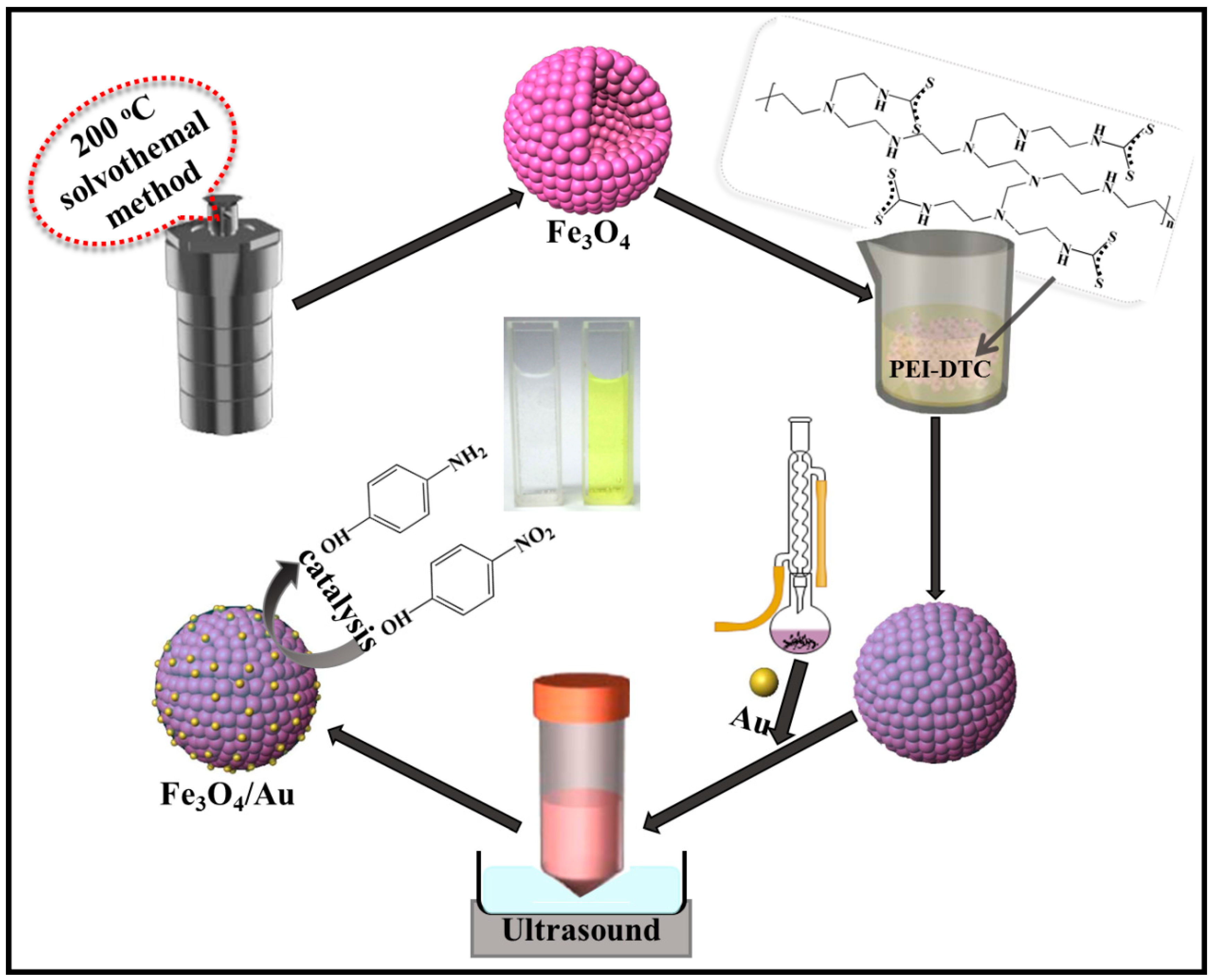
2. Experimental Section

2.1. Materials’ Development

Chemicals, including methanol, polyethyleneimine (PEI, branched, MW ≈ 25,000 g/mol) and carbon disulfide (CS2), were purchased from Aladdin industrial Co., Ltd. (Shanghai, China). Iron chloride hydrate (FeCl3·6H2O) was purchased from Shanghai Macklin Biochemical Co., Ltd. (Shanghai, China). Ethylene glycol (EG), sodium dodecyl sulfate (SDS), sodium acetate (NaAc·3H2O), potassium hydroxide (KOH), sodium citrate dihydrate (Na3C6H5O7·2H2O), 4-nitrophenol (4-NP) and gold (III) chloride hydrate (HAuCl4·3H2O), were purchased from Sinopharm Chemical Reagent Co., Ltd. (Shanghai, China). The aforementioned reagents were analytical grade and were utilized without further purification.

2.2. Synthesis of Fe3O4 Hollow Microspheres

In a typical synthesis process for the Fe3O4 hollow microspheres, 1.62 g FeCl3·6H2O were dispersed in 60 mL EG into a beaker with mechanical stirring at room temperature. After 30 min, 2.70 g NaAc·3H2O and 0.1839 g SDS were added to the mixture. The mixture solution was vigorously stirred for 1 h until it became homogeneous. Then, the mixture was transferred to the Teflon-lined stainless steel autoclave, and it was kept at 200 °C for 12 h. After the samples were cooled to room temperature naturally, the products were obtained by centrifuging, sequentially rinsed with ethanol and deionized water for five times and subsequently dried under vacuum at 60 °C over night to obtain the Fe3O4 hollow microspheres.

2.3. Gold Seeds Synthesis

Approximately 80 mg Na3C6H5O7·2H2O were dissolved in 8 mL deionized water. Two milliliters of 58 mM HAuCl4·3H2O were diluted into 198 mL of deionized water with vigorous stirring. The mixture was subsequently heated under reflux at 105 °C. The well-prepared sodium citrates were steadily added dropwise. The reaction mixtures were sustained for 15 min under stirring and reflux, leading to a buff to burgundy color change. The mixture was eventually cooled down to room temperature naturally.

2.4. PEI-DTC Synthesis

Five hundred milligrams of PEI (0.02 mmol) and 650 mg KOH (11.6 mmol) were dissolved in 50 mL methanol under magnetic stirring with completely dissolved KOH. The solution was then purged with N2 for 15 s in order to exhaust the oxygen thoroughly, and 695 μL of CS2 (11.6 mmol) was added by dripping slowly into the mixed solution. PEI-DTC was obtained with stirring for 10 min with a solution color change to light yellow.

2.5. Fe3O4@PEI-DTC-Au Seeds’ Synthesis

In this step, the surfaces of Fe3O4 hollow microspheres were functionalized with PEI-DTC. Ten milligrams of Fe3O4 hollow microspheres were washed with methanol three times. PEI-DTC solution droplets were injected with a pipette under vortexing. The developed mixture was then kept for 1 h. The precipitate was gathered with a dedicated external magnet and rinsed with deionized water three times to remove the unnecessary PEI-DTC. Finally, Fe3O4@PEI-DTC NPs were re-dispersed in deionized water.
In comparison with counterpart bulk composites, equipped with a large surface area, the nanostructured materials demonstrate superior optical, magnetic and electrical characteristics [44,45,46,47,48]. The preparation of Fe3O4@PEI-DTC-Au seed nanocomposites is as follows. The as-obtained 10 mL of gold seed colloids were added dropwise into the 4 mL of Fe3O4@PEI-DTC NPs. Then, Fe3O4@PEI-DTC-Au nanocomposites were rinsed with deionized water and the same process repeated, and the sample was named as Fe3O4-Au 20 mL. Additional samples were prepared with the same conditions as Fe3O4-Au 20 mL, while the amount of gold seed colloids was changed to 40 and 60 mL, named as Fe3O4-Au 40 mL and Fe3O4-Au 60 mL, respectively.

2.6. Application of Fe3O4-Au Magnetic Nanocomposites for Catalytic Reduction of 4-NP

The catalytic ability of Fe3O4-Au magnetic nanocomposites was performed in a cuvette with 1 mg of Fe3O4-Au for the reduction of 4-NP (0.005 mol/L, 1 mL) in the presence of NaBH4 (0.2 mol/L, 1 mL). The UV-Vis spectrophotometer was used to record the catalytic reduction rate at different time intervals in the scanning range of 200–500 nm.
In order to study the recyclability of the prepared Fe3O4-Au magnetic nanocomposites, the samples were collected with a magnet from the reaction solution when the reduction process was completed. The obtained magnetic nanocomposites were rinsed with deionized water and ethanol three times and then repeated in the next reaction. The same catalytic reduction process to 4-NP was repeated six times.

2.7. Characterizations

The structure and morphology of the samples were characterized by X-ray diffractometer (XRD) (Rigaku D/Max-2500, Rigaku Corporation, Tokyo, Japan), Mössbauer spectrum (FAST Comtec Mössbauer system, FAST Comtec, Oberhaching, Germany), X-ray photoelectron spectroscopy (XPS) (Thermo Scientific ESCALAB 250Xi, Thermo Fisher Scientific, Waltham, MA, USA), field-emission scanning electron microscopy (FESEM) (JEOL JSM-7800F, JEOL Ltd., Tokyo, Japan) and transmission electron microscope (TEM) (JEOL 2100, JEOL Ltd., Tokyo, Japan). Ultraviolet-visible spectroscopy (UV-Vis) spectra and magnetic properties were measured with a Shimadzu UV 3600 spectrophotometer (Shimadzu Corporation, Tokyo, Japan) and a quantum design MPMS3 superconducting quantum interference device (SQUID) magnetometer (Quantum Design, Inc., San Diego, CA, USA), respectively.

3. Results and Discussion

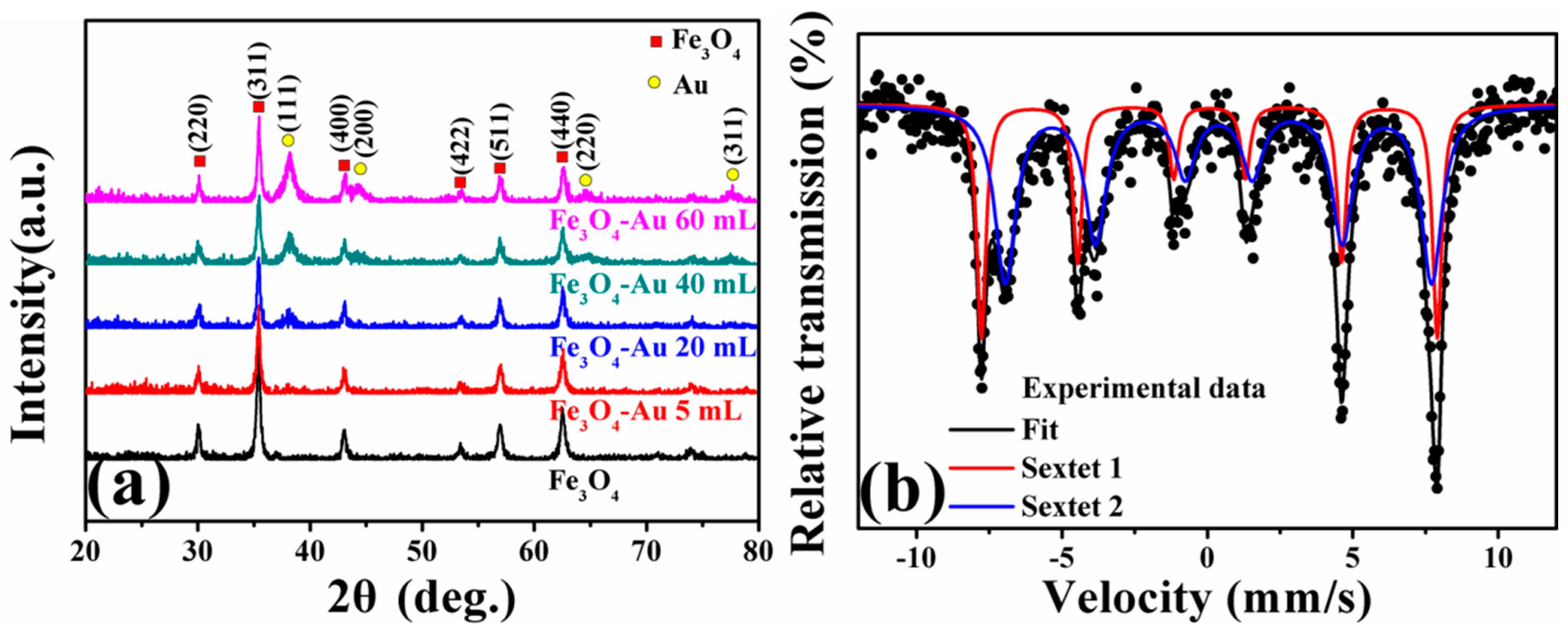
3.1. X-ray Diffraction of the Fe3O4-Au Magnetic Nanocomposites

Figure 2a shows XRD patterns of the as-obtained pure Fe3O4 hollow microspheres and Fe3O4-Au magnetic nanocomposites with the various amounts of gold seed colloids added (Fe3O4-Au 5 mL, Fe3O4-Au 20 mL, Fe3O4-Au 40 mL and Fe3O4-Au 60 mL). As for the pure Fe3O4 hollow microspheres, the diffraction peaks located at 30.4°, 35.5°, 43.4°, 53.4°, 57.3° and 62.8° were indexed to (220), (311), (400), (422), (511) and (440) of Fe3O4 (Joint Committee on Powder Diffraction Standards, JCPDS 19-0629), respectively [21,49]. The average crystallite size of the pure Fe3O4 hollow microspheres was about 576.5 nm calculated by the Debye–Scherrer equation [50]. Four additional peaks of Fe3O4-Au magnetic nanocomposites at about 38.4°, 44.5°, 64.7° and 77.5° could be assigned into the (111), (200), (220) and (311) of Au (JCPDS 04-0784), respectively, suggesting the coexistence of Fe3O4 and Au [51,52]. Figure S1 presents the Pawley refinement of the XRD pattern of the pure Fe3O4 hollow microspheres. The residual weighted profile R-factor (Rwp) of the sample is 15.74%, which indicates that the sample is in agreement with the standard magnetite Fe3O4. In addition, it is well established that the area of the diffraction peak of XRD patterns is proportional to the contents of that phase in the mixture. Increasing addition times of gold seed colloids enhanced the diffraction peaks of Au, implying that the number of the gold seeds coating the surfaces of Fe3O4 hollow microspheres increases.
Because magnetite Fe3O4 and maghemite γ-Fe2O3 have nearly the same crystal structure of an inverse spinel type, it is difficult to differentiate them only based on the XRD results. The Mössbauer spectrum is an available characterization technique to distinguish magnetite from maghemite [53,54]. Mössbauer spectra of Fe3O4 hollow microspheres fitted with two sextets (Zeeman splitting patterns) are shown in Figure 2b. The acute and strong lines of the magnetic sextets show a characteristic double six-peak structure of magnetite Fe3O4 [55]. The hyperfine field is 48.5 and 45.5 Tesla, and the isomer shift is 0.603 and 0.287 mm/s, corresponding to Fe2+ and Fe3+ ions at octahedral interstitial sites and Fe3+ ions at tetrahedral interstitial sites, respectively. Table 1 lists the fitted Mössbauer parameters. All these results testify that the as-obtained sample is a pure magnetite Fe3O4 instead of maghemite γ-Fe2O3 and consistent with the results reported by Ghosh et al. [56]. In fact, the oxidation product of the magnetite Fe3O4 is either maghemite γ-Fe2O3 or hematite α-Fe2O3, which strongly depends on the oxidation temperature. Generally, a high temperature heat treatment is necessary to realize the phase transition of magnetite Fe3O4, which indirectly suggests that the structure of magnetite Fe3O4 is stable at room temperature [57,58].

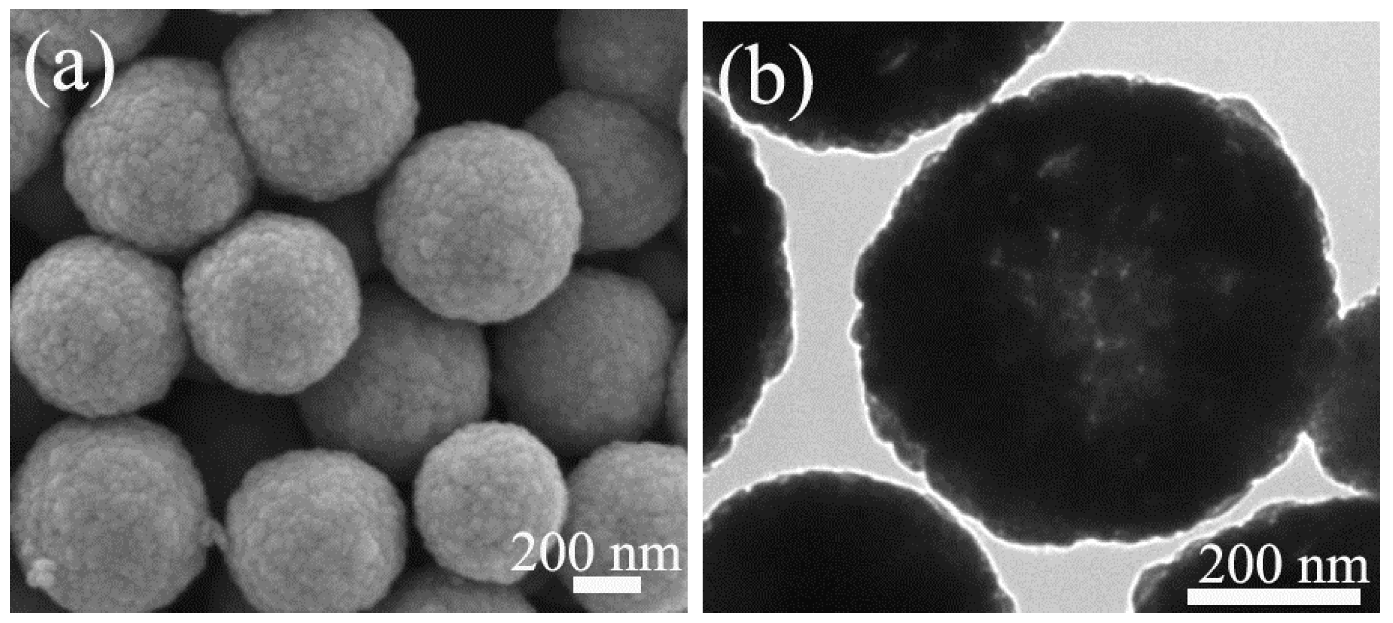
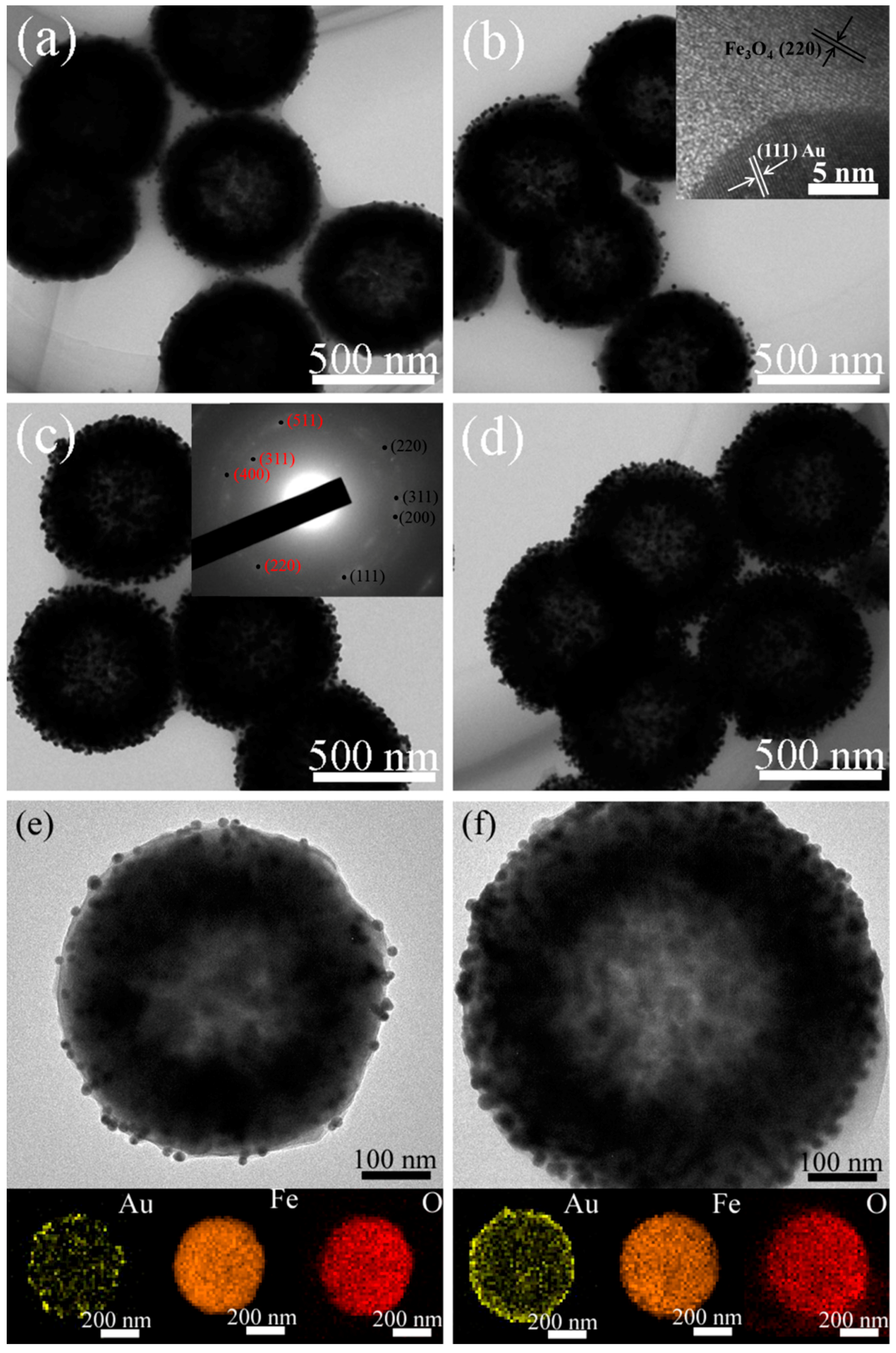
3.2. Morphology of the Fe3O4-Au Magnetic Nanocomposites

The specific structure and morphology of the developed Fe3O4-Au magnetic nanocomposites were studied by TEM and SEM. In particular, TEM measurement is an effective methodology for structure research in materials science and materials engineering [59,60,61,62,63,64,65,66,67,68,69,70,71]. Figure 3 displays SEM and TEM images of pure Fe3O4 hollow microspheres. It can be observed that the pure Fe3O4 hollow microspheres are spherical shape. The average crystallite size is about 576 nm, and the samples show the narrow size distribution, as shown in Figure S2. TEM and SEM images of Fe3O4-Au 5 mL, Fe3O4-Au 20 mL, Fe3O4-Au 40 mL and Fe3O4-Au 60 mL are presented in Figure 4 and Figure S3. The as-prepared Fe3O4-Au magnetic nanocomposites consist of the Fe3O4 hollow microspheres and the gold seeds of about 16 nm in diameter. Clearly, the gold seeds are randomly and homogeneously attached to the surfaces of the Fe3O4 hollow microspheres. Gold seeds are seen black, and Fe3O4 hollow microspheres present a light color in the TEM images, attributed to the higher electron density of Au compared to that of Fe3O4 [72]. It can be easily observed from the high-resolution TEM (HRTEM) image in the inset of Figure 4b that the interplanar distance values are 0.204 and 0.217 nm, matching the (220) lattice plane of Fe3O4 and the (111) lattice plane of Au, respectively [73]. The selected area electron diffraction (SAED) pattern of Fe3O4-Au 40 mL is presented in the inset of Figure 4c, consisting of both the (220), (311), (400) and (511) diffraction rings of Fe3O4 and the (111), (200), (220) and (311) diffraction rings of Au [43]. The HRTEM and SAED results further verify the coexistence of Fe3O4 and Au. An interesting finding is that when increasing Au seed colloids’ addition times, the gold seed contents on Fe3O4 surfaces are further boosted, which is consistent with the aforementioned XRD data. This is supported by the following elemental mapping images.
Figure 4e,f shows the TEM images of single Fe3O4-Au 5 mL and Fe3O4-Au 60 mL microspheres and corresponding energy-dispersive X-ray (EDS) mapping images of Au, Fe and O elements [74,75]. It could be clearly found that the Fe3O4 hollow microspheres have hollow internal structures, and Au seeds are homogeneously attached to the surfaces of the Fe3O4 hollow microspheres. In addition, compared with Fe3O4-Au 5 mL, the amount of gold seeds on the surfaces of Fe3O4 for Fe3O4-Au 60 mL increases significantly.

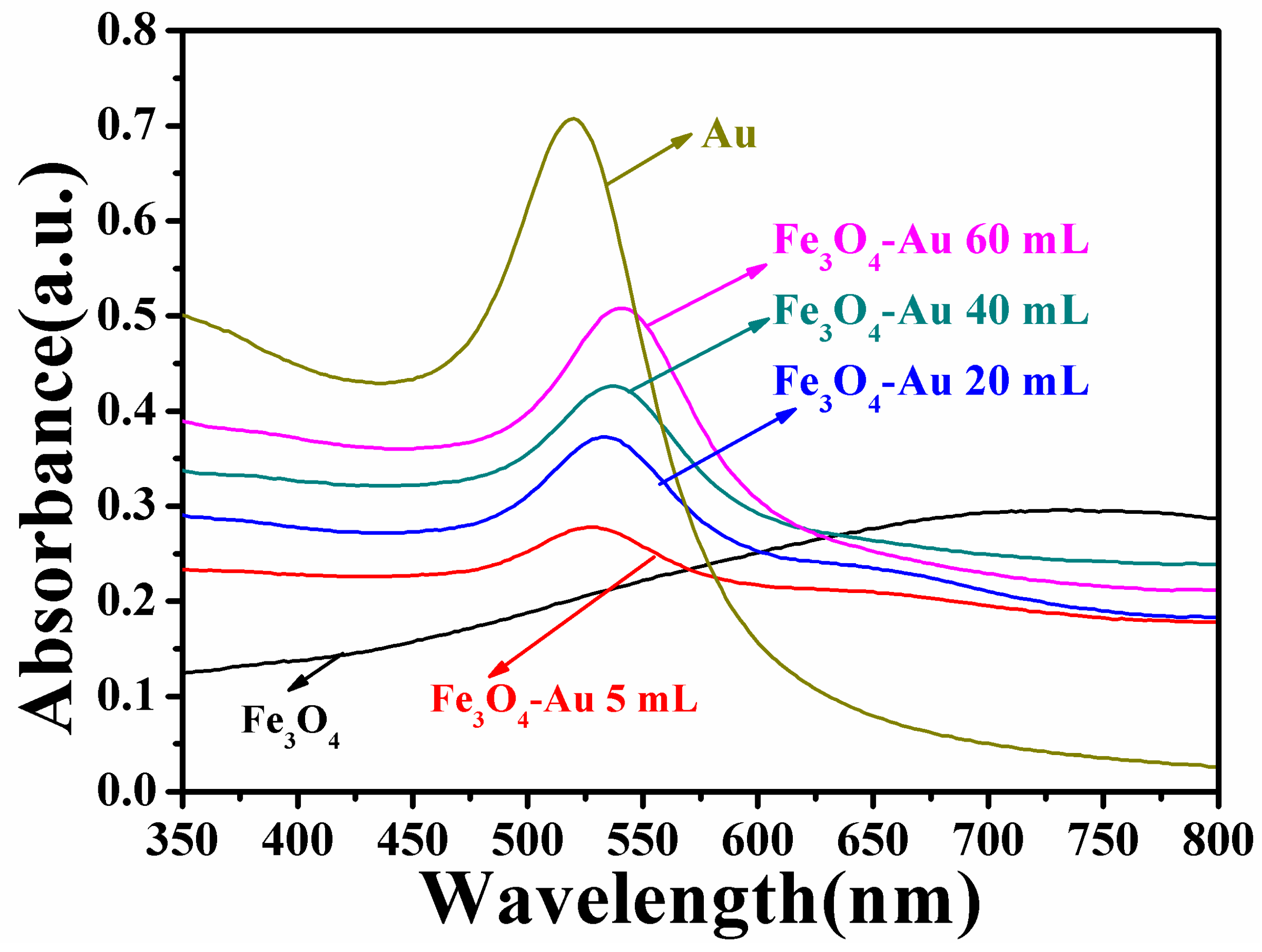
3.3. Optical Properties of the Fe3O4-Au Magnetic Nanocomposites

UV-Vis spectroscopy studies are carried out to investigate the optical properties of pure Fe3O4 hollow microspheres and Fe3O4-Au magnetic nanocomposites. Figure 5 presents the UV-Vis spectra of pure Fe3O4 hollow microspheres, Au seed colloids, Fe3O4-Au 5 mL, Fe3O4-Au 20 mL, Fe3O4-Au 40 mL and Fe3O4-Au 60 mL magnetic nanocomposites. No absorption peak is observed for the pure Fe3O4 hollow microspheres in the visible wavelength span [34,76]. The Au gold seed colloids display a characteristic surface plasmon resonance (SPR) peak at ~520 nm on account of the coherent excitation of the free electrons within the conduction band [77]. In addition, all the Fe3O4-Au magnetic nanocomposites show a clear characteristic SPR band of gold nanostructures. Interestingly, the absorption peak of Fe3O4-Au magnetic nanocomposites is broadened and shows a slight red shift compared with that of pure Au seed colloids. The more Au seeds are deposited on Fe3O4 hollow microspheres’ surfaces, the more evident the red shift of the absorption peak is. The absorption band shows a red-shift from 526 nm for Fe3O4-Au 5 mL to 540 nm for Fe3O4-Au 60 mL. This red shift of the SPR absorption band can be ascribed to plasmon hybridization and the surface plasmon coupling of neighboring Au seeds deposited on Fe3O4 hollow microspheres [52,78].

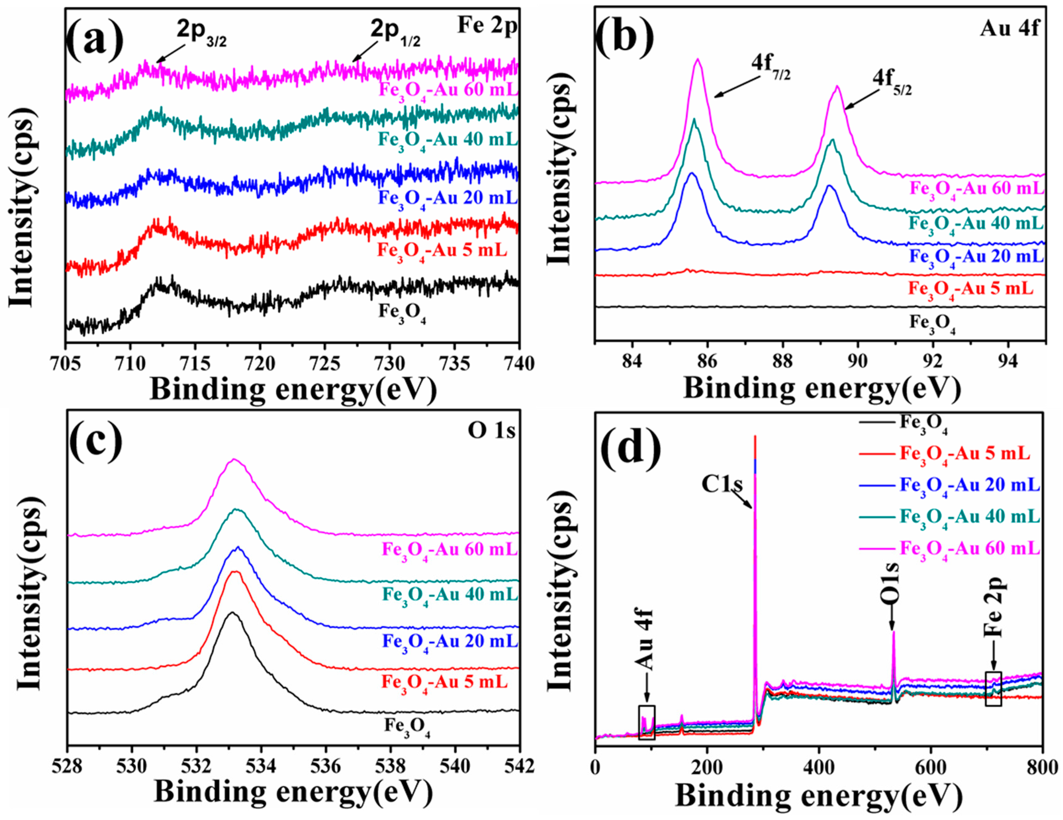
3.4. XPS of the Fe3O4-Au Magnetic Nanocomposites

XPS was applied to characterize the elemental chemical states and chemical compositions of the pure Fe3O4 hollow microspheres and Fe3O4-Au magnetic nanocomposites. The C 1s peak at 284.8 eV is applied as the reference for charge correction [79]. The XPS results of Fe3O4, Fe3O4-Au 5 mL, Fe3O4-Au 20 mL, Fe3O4-Au 40 mL and Fe3O4-Au 60 mL are shown in Figure 6. Figure 6a is the high-resolution scans of the XPS spectra for Fe 2p. The binding energy of pure Fe3O4 hollow microspheres is assessed at 711.2 and 724.5 eV by using Gaussian-Lorentzian fitting and assigned to the Fe 2p3/2 and Fe 2p1/2 of magnetite Fe3O4, respectively [80]. As for Fe3O4-Au 20 mL (Figure 6b), the peak doublet appearing at 83.9 and 87.9 eV with a spin orbit splitting of ~4 eV is assigned to Au 4f7/2 and Au 4f5/2, respectively [81]. It is important to highlight that with the increase of the quantity of the Au seeds on the Fe3O4 surfaces, the intensity of Fe 2p decreases and the intensity of Au 4f increases, because the intensity of XPS spectra is proportional to the atomic concentration and the atomic sensitivity factor [82]. Meanwhile, Fe 2p-associated peaks shift toward lower binding energy, while Au 4f peaks shift toward the opposite binding energy side. The shift in binding energy provides evidence of an interaction between Au and Fe3O4. The chemical shifts in the Fe 2p peaks and Au 4f peaks are probably caused by the transfer of charge carriers at the metal-oxide interfaces [83,84,85]. As shown in Figure 6c, the O 1s spectrum of pure Fe3O4 exhibits only a single peak at 532.8 eV, and no obvious shift of peak position is observed [86]. The survey scan results demonstrate that all the indexed peaks correspond to those of Fe, Au, O and C (Figure 6d).

3.5. Magnetic Properties of the Fe3O4-Au Magnetic Nanocomposites

The magnetic properties of the obtained samples were measured using SQUID. The magnetization versus magnetic field (M-H) loops of pure Fe3O4 hollow microspheres and Fe3O4-Au magnetic nanocomposites are presented in Figure 7. The M-H loops of all the samples are S-shaped curves and exhibit negligible coercivity and remanence, which is the typical characteristic of superparamagnetic nanomaterials [87]. Moreover, in order to confirm the superparamagnetism of the as-obtained sample, the temperature-dependent magnetization (M-T) on pure Fe3O4 hollow microspheres was measured under zero-field-cooled (ZFC) and 1000 Oe field-cooled (FC) conditions (Figure S4). With the increase of temperature, the magnetization increases to a maximum at the blocking temperature and then decreases, consistent with the superparamagnetic behavior [88]. The saturation magnetization (Ms) of pure Fe3O4 hollow microspheres reaches up to 93.2 emu/g, but the Ms value of Fe3O4-Au 5 mL reduces to 88.1 emu/g. Further coating with the gold seeds results in a continuous decrease of the Ms value. As for Fe3O4-Au 60 mL, the Ms value is only 68.6 emu/g. The gradual decrease in the Ms value can be ascribed to the increase in the weight ratio of Au seeds to Fe3O4 hollow microspheres or the diamagnetic contribution of the Au seeds deposited onto the surfaces of the Fe3O4 hollow microspheres [89,90,91]. It must also be mentioned that although Ms values decrease owing to the introduction of Au seeds, the Fe3O4-Au magnetic nanocomposites retain the strong magnetic responsivity and can be easily magnetically collected from aqueous solution by the magnet (as shown in the inset of Figure 7), which is beneficial to their economic and reusable applications.

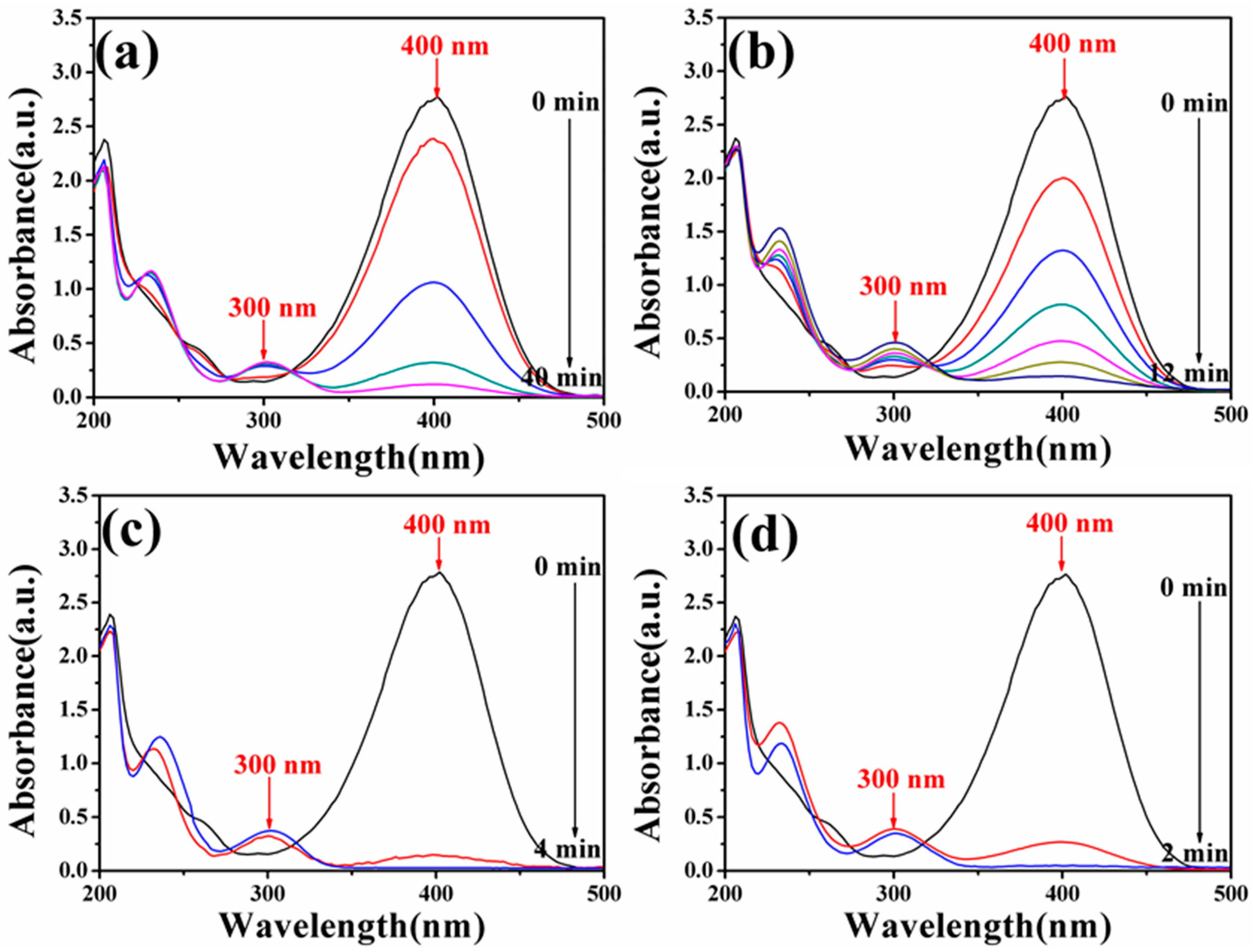
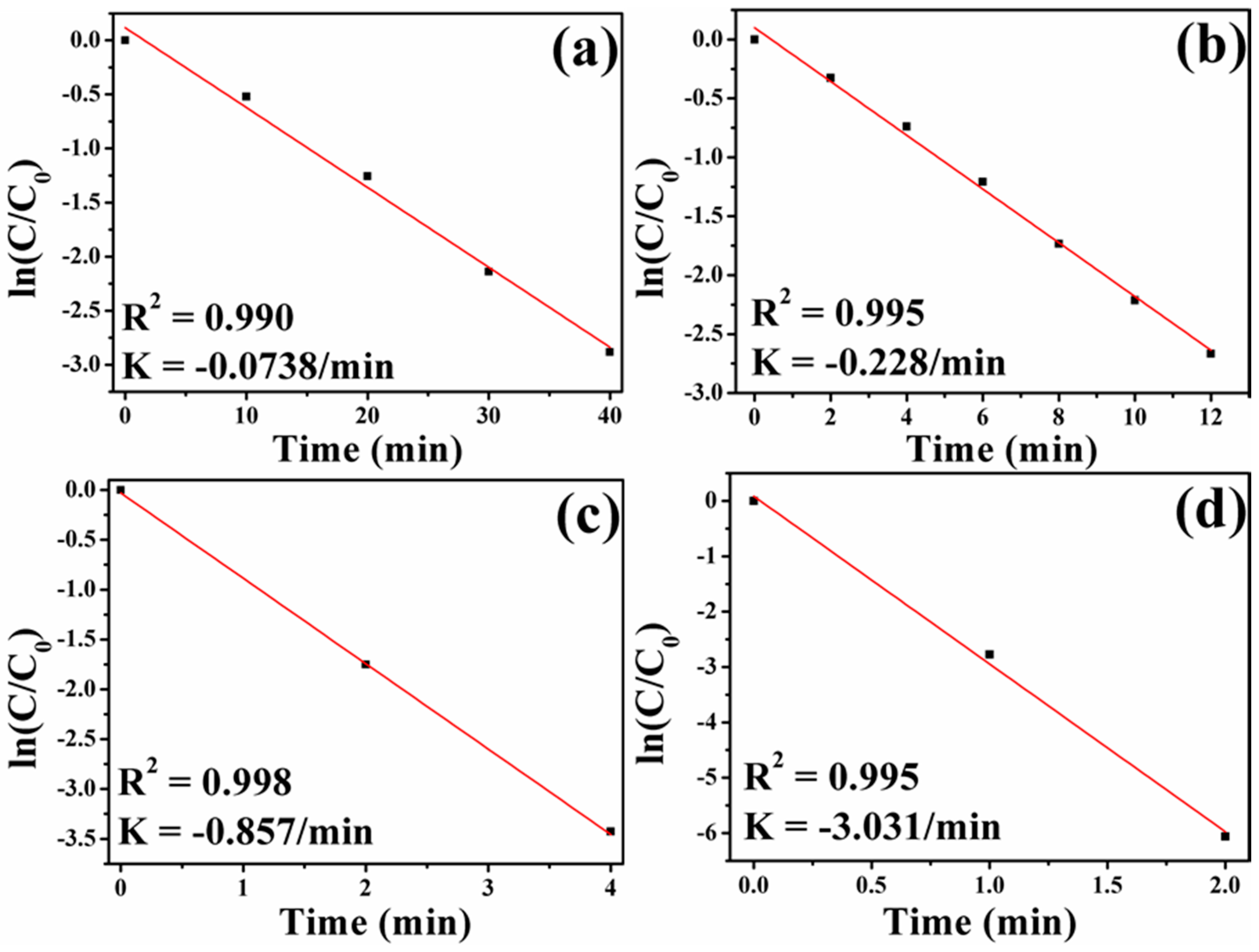
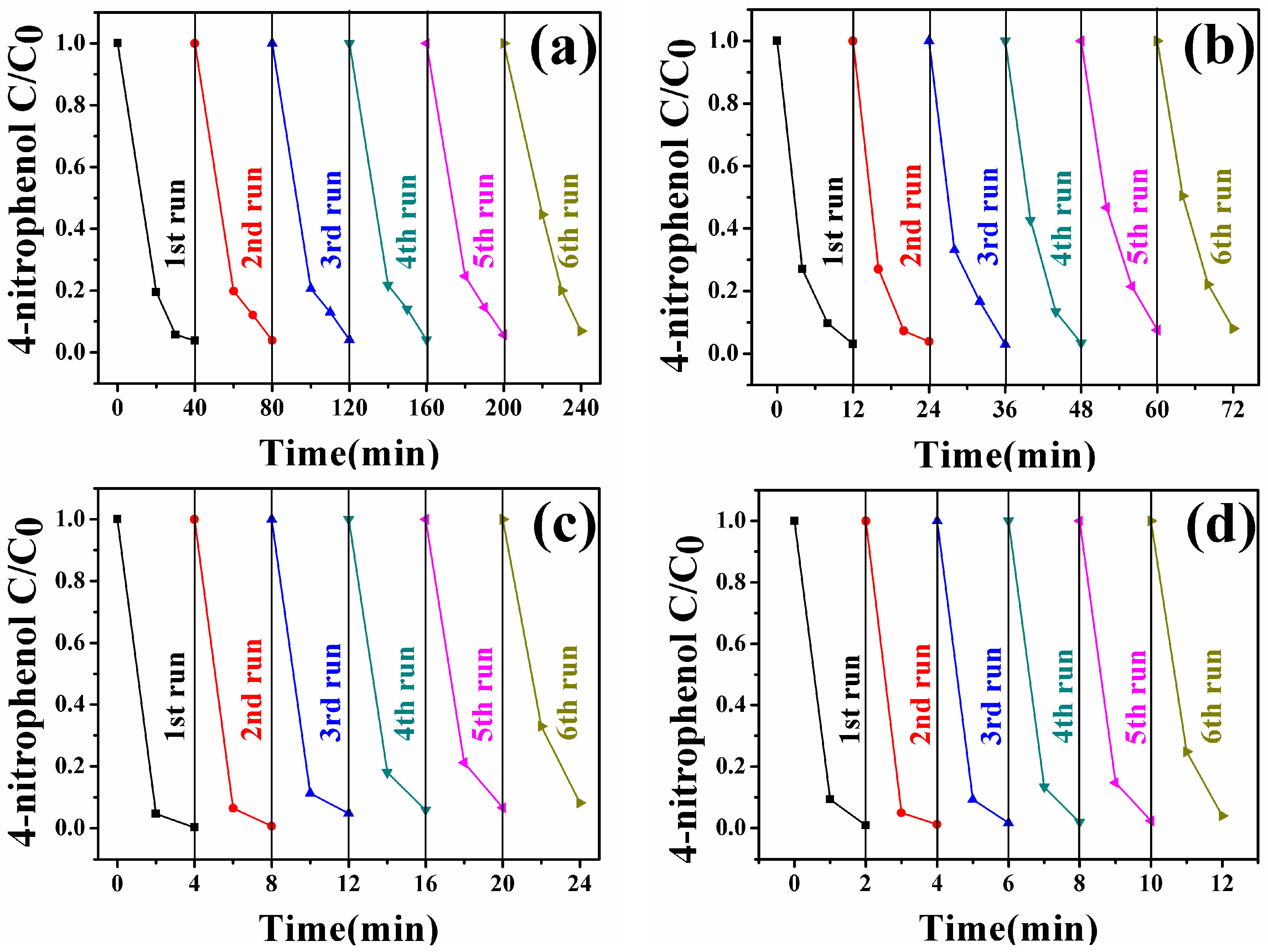
3.6. Catalytic Activity of Fe3O4-Au Magnetic Nanocomposites to 4-NP

To study the catalytic performance of Fe3O4-Au magnetic nanocomposites to organic pollutants, the catalytic reduction of 4-NP in the presence of excess NaBH4 was selected as a model reaction. As shown in Figure S5, the original aqueous 4-NP solution has a maximum absorption peak at 317 nm. If the catalysts are absent, the absorption peak at 317 nm is unchanged even after a couple of days [20]. Once freshly obtained NaBH4 solution is added, the absorption peak at 317 nm shifts to 400 nm due to the formation of 4-nitrophenolate ions, and meanwhile, the color of the mixture changes from light yellow to dark yellow [92]. However, the reduction reaction cannot be produced without gold seeds even in the presence of excess NaBH4, as can be testified in Figure S6. There is almost no change in the intensity at 400 nm after 480 min, indicating that the addition of pure Fe3O4 hollow microspheres has little influence on the reduction of 4-NP. Figure 8 exhibits UV-Vis absorption spectra of 4-NP catalyzed by the different Fe3O4-Au magnetic nanocomposites (Fe3O4-Au 5 mL, Fe3O4-Au 20 mL, Fe3O4-Au 40 mL and Fe3O4-Au 60 mL) in the presence of NaBH4 at a certain time interval. When Fe3O4-Au magnetic nanocomposites are introduced into the reaction system, the absorption peak of all the samples at 400 nm decreases in intensity. At the same time, a new peak located at 300 nm appears and increases concomitantly in intensity with the reaction time, which is attributed to the formation of 4-AP, the corresponding product in the reduction of 4-NP. The whole peak at 400 nm disappears after 40, 12, 4 and 2 min catalyzed by Fe3O4-Au 5 mL, Fe3O4-Au 20 mL, Fe3O4-Au 40 mL and Fe3O4-Au 60 mL, respectively. The color of the solution transforms from yellow to colorless, demonstrating that the 4-NP is completely changed into 4-AP and no other byproducts are generated. The corresponding pseudo-first-order plots of ln(C/C0) versus reaction time over different Fe3O4-Au magnetic nanocomposites are portrayed in Figure 9. The concentration of NaBH4 is identified as a constant because it is much more excessive compared to that of 4-NP. The ratio of C (the concentration of 4-NP at reaction time t) to C0 (the initial concentration of 4-NP) is obtained from the relative intensity of respective absorbance (At/A0) at 400 nm. The linear relationship of ln(C/C0) against reaction time represents that the reduction of 4-NP by the Fe3O4-Au magnetic nanocomposites follows the pseudo first order kinetics. The rate constant (k) can be calculated by a liner plot of ln(C/C0) vs. reduction time. The rate constants of 4-NP are 0.0738, 0.228, 0.857 and 3.031/min using Fe3O4-Au 5 mL, Fe3O4-Au 20 mL, Fe3O4-Au 40 mL and Fe3O4-Au 60 mL as nanocatalysts, respectively. Obviously, the rate constants for 4-NP reduction increase when increasing Au seed addition rounds. It is reasonable that if the noble metal nanomaterials’ loading is increased, the mass fraction of the noble metal nanomaterials is higher in the final samples, and thus, a better catalytic performance should be accomplished [93]. We thus propose that the catalytic capability of Fe3O4-Au magnetic nanocomposites could be greatly improved when more gold seeds cover the surfaces of the Fe3O4 hollow microspheres.
To implement the practical application of Fe3O4-Au magnetic nanocomposites, their stability and the recyclability are essential. All the Fe3O4-Au magnetic nanocomposites were separated magnetically and reused after catalytic reduction of 4-NP. As shown in Figure 10, all the samples can be successfully reused for at least six reaction runs for the catalytic reduction of 4-NP, elaborating that the Fe3O4-Au magnetic nanocomposites possess the excellent stability and can serve as recoverable and efficient nanocatalysts of the organic pollutants.

4. Conclusions

In summary, Fe3O4-Au magnetic nanocomposites were prepared by a well-developed seed deposition method. Structural analyses confirmed that the as-prepared Fe3O4-Au magnetic nanocomposites possessed a high surface area and that the gold seeds were homogeneously deposited onto the Fe3O4 hollow microspheres’ surfaces. The increase of the quantity of the gold seeds attached to the surfaces of the Fe3O4 hollow microspheres results in Fe 2p peaks shifting toward a lower binding energy and Au 4f peaks shifting towards a higher binding energy. Although the Ms value decreases with the increase of the gold seed amount deposited on the Fe3O4 surfaces, the samples retain the strong magnetic responsivity and can be easily collected by a magnet. The pseudo-first-order kinetics are used to calculate the rate constant of 4-NP, and the rate constants for the 4-NP reduction increase when increasing the added amount of gold seeds. Furthermore, Fe3O4-Au magnetic nanocomposites can serve as recyclable nanocatalysts for 4-NP. Therefore, Fe3O4-Au magnetic nanocomposites can be excellent nanocatalysts for the catalytic reduction of organic pollutants in the treatment of waste water.

Supplementary Materials

The following are available online at https://www.mdpi.com/2079-4991/8/5/353/s1. Figure S1: Pawley refinement of the XRD pattern of the pure Fe3O4 hollow microspheres. Red dots, blue lines and olive bars represent the experimental data, the calculated date and the peak position of the sample, respectively. The bottom line in black shows the different experimental-calculated data, Figure S2: Particle size distribution obtained from the analysis of the TEM images for pure Fe3O4 hollow microspheres, Figure S3: SEM and corresponding EDS images of the as-prepared Fe3O4-Au nanocomposites with the different addition quantities of the gold seed colloids (Fe3O4 (a, b and c), Fe3O4-Au 5 mL (d, e and f), Fe3O4-Au 20 mL (g, h and i), Fe3O4-Au 40 mL (j, k and l) and Fe3O4-Au 60 mL (m, n and o), Figure S4: ZFC and FC curves of pure Fe3O4 hollow microspheres under an applied field of 1000 Oe, Figure S5: UV-Vis absorption spectra of 4-NP before (red line) and after adding NaBH4 solution (black line), Figure S6: UV-Vis absorption spectra of 4-NP catalyzed by pure Fe3O4 hollow microspheres in presence of NaBH4 before and after 480 min.

Author Contributions

Conceptualization, Y.L. and S.G.X.; Software, D.W.; Validation, Z.L. and L.C.; Formal Analysis, Y.Z.; Data Curation, Q.K.; Writing-Original Draft Preparation, Y.C. and D.H.; Writing-Review & Editing, Y.S., Y.Z. and Y.W.; Supervision, J.Y.; Funding Acquisition, Y.L. and S.G.X.

Funding

This research was funded by the National Natural Science Foundation of China (Grant Numbers 21676115, 51609100, 61705020, 61675090 and 61575080), Program for the development of Science and Technology of Jilin province (Grant Numbers 20160101287JC and 20150519024JH), Guangdong Province Science and Technology Projects (Grant Numbers 2015A030310342 and 2015A010103004), and Technology of Education Department of Jilin Province (Grant Number JJKH20170374KJ).

Conflicts of Interest

The authors declare no conflict of interest.

References

  1. Zhang, X.F.; Zhu, X.Y.; Feng, J.J.; Wang, A.J. Solvothermal synthesis of N-doped graphene supported PtCo nanodendrites with highly catalytic activity for 4-nitrophenol reduction. Appl. Surf. Sci. 2018, 428, 798–808. [Google Scholar] [CrossRef]
  2. Bordbar, M.; Negahdar, N.; Nasrollahzadeh, M. Melissa Officinalis L. leaf extract assisted green synthesis of CuO/ZnO nanocomposite for the reduction of 4-nitrophenol and Rhodamine B. Sep. Purif. Technol. 2018, 191, s295–s300. [Google Scholar] [CrossRef]
  3. Nag, S.; Pramanik, A.; Chattopadhyay, D.; Bhattacharyya, M. Green-fabrication of gold nanomaterials using Staphylococcus warneri from Sundarbans estuary: An effective recyclable nanocatalyst for degrading nitro aromatic pollutants. Environ. Sci. Pollut. Res. 2017, 24, 1–19. [Google Scholar] [CrossRef] [PubMed]
  4. Meziane, D.; Benadda-Kordjani, A.; Nezzal, G.; Benammar, S.; Djadoun, A. Para-nitrophenol reduction on solvothermally prepared cobalt@silica core-shell catalysts. React. Kinet. Mech. Catal. 2017, 122, 1145–1158. [Google Scholar] [CrossRef]
  5. He, L.L.; Tong, Z.F.; Wang, Z.H.; Chen, M.; Huang, N.; Zhang, W. Effects of calcination temperature and heating rate on the photocatalytic properties of ZnO prepared by pyrolysis. J. Colloid Interface Sci. 2017, 509, 448–456. [Google Scholar] [CrossRef] [PubMed]
  6. Liu, L.; Yang, W.; Sun, W.; Li, Q.; Shang, J.K. Creation of Cu2O@TiO2 composite photocatalysts with p-n heterojunctions formed on exposed Cu2O facets, their energy band alignment study, and their enhanced photocatalytic activity under visible light illumination. ACS Appl. Mater. Interfaces 2015, 7, 1465–1476. [Google Scholar] [CrossRef] [PubMed]
  7. Andreou, D.; Iordanidou, D.; Tamiolakis, I.; Armatas, G.S.; Lykakis, I.N. Reduction of nitroarenes into aryl amines and n-aryl hydroxylamines via activation of NaBH4 and ammonia-borane complexes by Ag/TiO2 catalyst. Nanomaterials 2016, 6, 54. [Google Scholar] [CrossRef] [PubMed]
  8. Zhao, X.; Wang, Y.; Feng, W.H.; Lei, H.T.; Li, J. Preparation of Cu(II) porphyrin-TiO2 composite in one-pot method and research on photocatalytic property. RSC Adv. 2017, 7, 52738–52746. [Google Scholar] [CrossRef]
  9. Liu, Y.; Zhang, Y.Y.; Kou, Q.W.; Chen, Y.; Han, D.L.; Wang, D.D.; Lu, Z.Y.; Chen, L.; Yang, J.H.; Xing, S. Eco-friendly seeded Fe3O4-Ag nanocrystals: A new type of highly efficient and low cost catalyst for methylene blue reduction. RSC Adv. 2018, 8, 2209–2218. [Google Scholar] [CrossRef]
  10. Shah, M.T.; Balouch, A.; Sirajuddin; Pathan, A.A.; Abdullah; Mahar, A.M.; Sabir, S.; Khattak, R.; Umar, A.A. SiO2 caped Fe3O4 nanostructures as an active heterogeneous catalyst for 4-nitrophenol reduction. Microsyst. Technol. 2017, 23, 5745–5758. [Google Scholar] [CrossRef]
  11. Svedendahl, M.; Verre, R.; Kall, M. Refractometric Biosensing Based on Optical Phase Flips in Sparse and Short-Range-Ordered Nanoplasmonic Layers. Light Sci. Appl. 2014, 3, e220. [Google Scholar] [CrossRef]
  12. Zhu, Z.; Bai, B.; You, O.; Li, Q.; Fan, S. Fano resonance boosted cascaded optical field enhancement in a plasmonic nanoparticle-in-cavity nanoantenna array and its SERS application. Light Sci. Appl. 2015, 4, e296. [Google Scholar] [CrossRef]
  13. Wang, P.; Wang, Y.; Tong, L. Graphene-doped polymer nanofibers for low-threshold nonlinear optical waveguiding. Light Sci. Appl. 2013, 2, e102. [Google Scholar] [CrossRef]
  14. Li, X.; Xing, L.; Zheng, K.; Wei, P.; Du, L.; Shen, M.; Shi, X. Formation of gold nanostar-coated hollow mesoporous silica for tumor multimodality imaging and photothermal therapy. ACS Appl. Mater. Interfaces 2017, 9, 5817–5827. [Google Scholar] [CrossRef] [PubMed]
  15. Aslam, U.; Chavez, S.; Linic, S. Controlling energy flow in multimetallic nanostructures for plasmonic catalysis. Nat Nano 2017, 12, 1000–1005. [Google Scholar] [CrossRef] [PubMed]
  16. Zhang, Q.; Wang, H. Facet-Dependent Catalytic Activities of Au Nanoparticles Enclosed by High-Index Facets. ACS Catal. 2014, 4, 4027–4033. [Google Scholar] [CrossRef]
  17. Karabchevsky, A.; Mosayyebi, A.; Kavokin, A.V. Tuning the chemiluminescence of a luminol flow using plasmonic nanoparticles. Light Sci. Appl. 2016, 5, 1–7. [Google Scholar] [CrossRef]
  18. Linnenbank, H.; Grynko, Y.; Forstner, J.; Linden, S. Second harmonic generation spectroscopy on hybrid plasmonic/dielectric nanoantennas. Light Sci. Appl. 2016, 5, e16013. [Google Scholar] [CrossRef]
  19. Blum, O.; Shaked, N.T. Prediction of photothermal phase signatures from arbitrary plasmonic nanoparticles and experimental verification. Light Sci. Appl. 2015, 4, e322. [Google Scholar] [CrossRef]
  20. Gangarapu, M.; Sarangapany, S.; Veerabhali, K.K.; Devipriya, S.P.; Arava, V.B.R. A high-performance catalytic and recyclability of phyto-synthesized silver nanoparticles embedded in natural polymer. J. Clust. Sci. 2017, 28, 3127–3138. [Google Scholar] [CrossRef]
  21. Jin, C.J.; Qu, Y.; Wang, M.G.; Han, J.; Hu, Y.M.; Guo, R. Aqueous solution-based Fe3O4 seed-mediated route to hydrophilic Fe3O4-Au janus nanoparticles. Langmuir 2016, 32, 4595–4601. [Google Scholar] [CrossRef] [PubMed]
  22. Navalón, S.; García, H. Nanoparticles for Catalysis. Nanomaterials 2016, 6, 123. [Google Scholar] [CrossRef] [PubMed]
  23. Maham, M.; Nasrollahzadeh, M.; Sajadi, S.M.; Nekoei, M. Biosynthesis of Ag/reduced graphene oxide/Fe3O4 using Lotus Garcinii leaf extract and its application as a recyclable nanocatalyst for the reduction of 4-nitrophenol and organic dyes. J. Colloid Interface Sci. 2017, 497, 33–42. [Google Scholar] [CrossRef] [PubMed]
  24. Jia, L.; Zhou, T.; Xu, J.; Li, F.H.; Xu, Z.Q.; Zhang, B.B.; Guo, S.L.; Shen, X.K.; Zhang, W.S. AuPd bimetallic nanocrystals embedded in magnetic halloysite nanotubes: Facile synthesis and catalytic reduction of nitroaromatic compounds. Nanomaterials 2017, 7, 333. [Google Scholar] [CrossRef] [PubMed]
  25. Xiao, Z.Y.; Zhai, S.R.; Ma, X.P.; Zhao, Z.Y.; Wang, X.; Bai, H.; An, Q.D. Monolithic Cu/C hybrid beads with well developed porosity for reduction of 4-nitrophenol to 4-aminophenol. New J. Chem. 2017, 41, 13230–13234. [Google Scholar] [CrossRef]
  26. Zhang, K.H.; Wang, C.W.; Rong, Z.; Xiao, R.; Zhou, Z.; Wang, S.Q. Silver coated magnetic microflowers as an efficient and recyclable Catalyst for catalytic reduction. New J. Chem. 2017, 41, 14199–141208. [Google Scholar] [CrossRef]
  27. Sun, J.; Chen, L. Superparamagnetic POT/Fe3O4 nanoparticle composites with supported Au nanoparticles as recyclable high-performance nanocatalysts. Mater. Today Chem. 2017, 5, 43–51. [Google Scholar] [CrossRef]
  28. Wang, D.M.; Duan, H.C.; Lü, J.H.; Lü, C.L. Fabrication of thermo-responsive polymer functionalized reduced graphene oxide@Fe3O4@Au magnetic nanocomposites for enhanced catalytic applications. J. Mater. Chem. A 2017, 5, 5088–5097. [Google Scholar] [CrossRef]
  29. Rath, P.C.; Saikia, D.; Mishra, M.; Kao, H.M. Exceptional catalytic performance of ultrafine Cu2O nanoparticles confined in cubic mesoporous carbon for 4-nitrophenol reduction. Appl. Surf. Sci. 2018, 427, 1217–1226. [Google Scholar] [CrossRef]
  30. Meng, G.H.; Zhang, X.Y.; Liu, C.; Wu, J.N.; Guo, X.H.; Liu, Z.Y. Ag quantum dot/montmorillonite compositeswith fluorescent properties: An efficient catalyst. Res. Chem. Intermed. 2017, 43, 7137–7145. [Google Scholar] [CrossRef]
  31. Govan, J.; Gun’ko, Y.K. Recent advances in the application of magnetic nanoparticles as a support for homogeneous catalysts. Nanomaterials 2014, 4, 222–241. [Google Scholar] [CrossRef] [PubMed]
  32. Liu, Y.; Zhang, Y.Y.; Kou, Q.W.; Chen, Y.; Sun, Y.T.; Han, D.L.; Wang, D.D.; Lu, Z.Y.; Chen, L.; Yang, J.H.; Xing, S.G.Z. Highly Efficient, Low-Cost, and Magnetically Recoverable FePt–Ag Nanocatalysts Towards Green Reduction of Organic Dyes. Nanomaterials 2018, 8, 329. [Google Scholar] [CrossRef] [PubMed]
  33. Wysocka, I.; Kowalska, E.; Trzciński, K.; Łapiński, M.; Nowaczyk, G.; Zielińska-Jurek, A. UV-Vis-Induced Degradation of Phenol over Magnetic Photocatalysts Modified with Pt, Pd, Cu and Au Nanoparticles. Nanomaterials 2018, 2, 28. [Google Scholar] [CrossRef] [PubMed]
  34. Pardo, I.R.; Pons, M.R.; Heredia, A.A.; Usagre, J.V.; Ribera, A.; Galian, R.E.; Prieto, J.P. Fe3O4@Au@mSiO2 as enhancing nanoplatform for rose bengal photodynamic activity. Nanoscale 2017, 9, 10388–10396. [Google Scholar] [CrossRef] [PubMed]
  35. Wang, Y.S.; Wang, Y.; Xia, H.; Wang, G.; Zhang, Z.Y.; Han, D.D.; Lv, C.; Feng, J.; Sun, H.B. Preparation of Fe3O4-Au-GO nanocomposite for simultaneous treatment of oil/water separation and dye decomposition. Nanoscale 2016, 8, 17451–17457. [Google Scholar] [CrossRef] [PubMed]
  36. Guo, R.; Jiao, T.F.; Xing, R.R.; Chen, Y.; Guo, W.C.; Zhou, J.X.; Zhang, L.X.; Peng, Q.M. Hierarchical AuNPs-Loaded Fe3O4/Polymers Nanocomposites Constructed by Electrospinning with Enhanced and magnetically recyclable catalytic capacities. Nanomaterials 2017, 7, 317. [Google Scholar] [CrossRef] [PubMed]
  37. Tedsree, K.; Temnuch, N.; Sriplai, N.; Pinitsoontorn, S. Ag modified Fe3O4@TiO2 magnetic core-shell nanocomposites for photocatalytic degradation of methylene blue. Mater. Today 2017, 4, 6576–6584. [Google Scholar] [CrossRef]
  38. Yang, J.H.; Kou, Q.W.; Liu, Y.; Wang, D.D.; Lu, Z.Y.; Chen, L.; Zhang, Y.Y.; Wang, Y.X.; Zhang, Y.J.; Han, D.L.; et al. Effects of amount of benzyl ether and reaction time on the shape and magnetic properties of Fe3O4 nanocrystals. Powder Technol. 2017, 319, 53–59. [Google Scholar] [CrossRef]
  39. Duan, L.F.; Jia, S.S.; Wang, T.H.; Xue, B.; Wang, Y.Q.; Zhao, L.J. Synthesis and Characterization of hollow Fe3O4 submicrospheres by a Simple Solvothermal Synthesis. Met. Mater. Int. 2011, 17, 801–804. [Google Scholar] [CrossRef]
  40. Hu, Y.; Yang, J.; Wei, P.; Li, J.C.; Ding, L.; Zhang, G.X.; Shi, X.Y.; Shen, M.W. Facile synthesis of hyaluronic acid-modified Fe3O4/Au composite nanoparticles for targeted dual mode MR/CT imaging of tumors. J. Mater. Chem. B 2015, 3, 9098–9108. [Google Scholar] [CrossRef]
  41. Wang, C.W.; Li, P.; Wang, J.F.; Rong, Z.; Pang, Y.F.; Xu, J.W.; Dong, P.T.; Xiao, R.; Wang, S.Q. Polyethylenimine-interlayered core-shell-satellite 3D magnetic microspheres as versatile SERS substrate. Nanoscale 2015, 7, 18694–18707. [Google Scholar] [CrossRef] [PubMed]
  42. Yan, F.; Sun, R.Y. Facile Synthesis of Bifunctional Fe3O4/Au Nanocomposite and Their Application in Catalytic Reduction of 4-Nitrophenol. Mater. Res. Bull. 2014, 57, 293–299. [Google Scholar] [CrossRef]
  43. Liu, Y.; Kou, Q.W.; Wang, D.D.; Chen, L.; Sun, Y.T.; Lu, Z.Y.; Zhang, Y.Y.; Wang, Y.X.; Yang, J.H.; Xing, S. Rational synthesis and tailored optical and magnetic Characteristics of Fe3O4-Au composite nanoparticles. J. Mater. Sci. 2017, 52, 10163–10174. [Google Scholar] [CrossRef]
  44. Xing, G.Z.; Wang, Y.; Wong, J.I.; Shi, Y.M.; Huang, Z.X.; Li, S.; Yang, H.Y. Hybrid CuO/SnO2 nanocomposites: Towards cost-effective and high performance binder free lithium ion batteries anode materials. Appl. Phys. Lett. 2014, 105, 143905. [Google Scholar] [CrossRef]
  45. Xing, G.Z.; Wang, D.D.; Cheng, C.J.; He, M.; Li, S.; Wu, T. Emergent ferromagnetism in ZnO/Al2O3 core-shell nanowires: Towards oxide spinterfaces. Appl. Phys. Lett. 2013, 103, 022402. [Google Scholar] [CrossRef]
  46. Sun, Y.; Li, Q. Research of zinc oxide quantum dot light-emitting diodes based on preparation of chemical solutions. Chin. J. Liq. Cryst. Disp. 2016, 31, 635–642. [Google Scholar] [CrossRef]
  47. Chen, X.Y.; Tian, Z. Recent progress in terahertz dynamic modulation based on graphene. Chin. Opt. 2017, 10, 86–97. [Google Scholar] [CrossRef]
  48. Li, T.; Zhang, M.-L.; Wang, F.; Zhang, D.-M.; Wang, G.-P. Fabrication of optical waveguide amplifiers based on bonding-type NaYF4: Er nanoparticles-polymer. Chin. Opt. 2017, 10, 219–225. [Google Scholar] [CrossRef]
  49. Yang, J.H.; Pan, M.Q.; Shi, R.X.; Yang, L.L.; Wang, J.; Kong, X.W.; Yang, W.Q.; Wang, D.D.; Zhou, Z. Novel Fe3O4 hollow microspheres: Nontemplate hydrothermal synthesis, superparamagnetism and biocompatibility. Nanosci. Nanotech. Let. 2017, 9, 109–117. [Google Scholar] [CrossRef]
  50. Shah, S.T.; Yehye, W.A.; Saad, O.; Simarani, K.; Chowdhury, Z.Z.; Alhadi, A.A.; Al-Ani, L.A. Surface functionalization of iron oxide nanoparticles with gallic acid as potential antioxidant and antimicrobial agents. Nanomaterials 2017, 7, 306. [Google Scholar] [CrossRef] [PubMed]
  51. Jin, C.J.; Han, J.; Chu, F.Y.; Wang, X.X.; Guo, R. Fe3O4@PANI hybrid shell as multifunctional support for au nanocatalysts with remarkably improved catalytic performance. Langmuir 2017, 33, 4520–4527. [Google Scholar] [CrossRef] [PubMed]
  52. Miao, P.; Tang, Y.G.; Wang, L. DNA modified Fe3O4@Au magnetic nanoparticles as selective probes for simultaneous detection of heavy metal ions. ACS Appl. Mater. Interfaces 2017, 9, 3940–3947. [Google Scholar] [CrossRef] [PubMed]
  53. Baskakov, A.O.; Solov’eva, A.Y.; Ioni, Y.V.; Starchikov, S.S.; Lyubutin, I.S.; Khodos, I.I.; Avilov, A.S.; Gubin, S.P. Magnetic and interface properties of the core-shell Fe3O4/Au nanocomposites. Appl. Surf. Sci. 2017, 422, 638–644. [Google Scholar] [CrossRef]
  54. Li, X.A.; He, Y.Y.; Sui, H.; He, L. One-step fabrication of dual responsive lignin coated Fe3O4 nanoparticles for efficient removal of cationic and anionic dyes. Nanomaterials 2018, 8, 162. [Google Scholar] [CrossRef] [PubMed]
  55. Wang, W.T.; Tang, B.T.; Wu, S.L.; Gao, Z.M.; Ju, B.Z.; Teng, X.X.; Zhang, S.F. Controllable 5-sulfosalicylic acid assisted solvothermal synthesis of monodispersed superparamagnetic Fe3O4 nanoclusters with tunable size. J. Magn. Magn. Mater. 2017, 423, 111–117. [Google Scholar] [CrossRef]
  56. Ghosh, R.; Pradhan, L.; Devi, Y.P.; Meena, S.S.; Tewari, R.; Kumar, A.; Sharma, S.; Gajbhiye, N.S.; Vatsa, R.K.; Pandey, B.N.; et al. Induction heating studies of Fe3O4 magnetic nanoparticles capped with oleic acid and polyethylene glycol for hyperthermia. J. Mater. Chem. 2011, 21, 13388–13398. [Google Scholar] [CrossRef]
  57. Ayyappan, S.; Thoguluva, R.R.; John, P. Superior thermal stability of polymer capped Fe3O4 magnetic nanoclusters. J. Am. Ceram. Soc. 2018, 101, 483–491. [Google Scholar] [CrossRef]
  58. Kazeminezhad, I.; Mosivand, S. Phase Transition of Electrooxidized Fe3O4 to γ and α-Fe2O3 Nanoparticles Using Sintering Treatment. Acta Phys. Pol. A 2014, 125, 1210–1214. [Google Scholar] [CrossRef]
  59. Zhang, M.; Xia, P.; Wang, L.; Zheng, J.; Wang, Y.; Xu, J.; Wang, L. Synthesis and fabrication of CNTs/Fe3O4@Pdop@Au nanocables by a facile approach. RSC Adv. 2014, 4, 44423–44426. [Google Scholar] [CrossRef]
  60. Matioli, E.; Brinkley, S.; Kelchner, K.M.; Hu, Y.L.; Nakamura, S.; DenBaars, S.; Speck, J.; Weisbuch, C. High-brightness polarized light-emitting diodes. Light Sci. Appl. 2012, 1, 479–482. [Google Scholar] [CrossRef]
  61. Li, L.; Guo, W.; Yan, Y.; Lee, S.; Wang, T. Label-free super-resolution imaging of adenoviruses by submerged microsphere optical nanoscopy. Light Sci. Appl. 2013, 2, e104. [Google Scholar] [CrossRef]
  62. Wang, D.D.; Wang, W.L.; Huang, M.Y.; Lek, A.; Lam, J.; Mai, Z.H. Failure mechanism analysis and process improvement on time-dependent dielectric breakdown of Cu/ultra-low-k dielectric based on complementary Raman and FTIR spectroscopy study. AIP Adv. 2014, 4, 107–110. [Google Scholar] [CrossRef]
  63. Xing, G.; Wang, D.; Yi, J.; Yang, L.; Gao, M.; He, M.; Yang, J.; Ding, J.; Sum, T.C.; Wu, T. Correlated d0 ferromagnetism and photoluminescence in undoped ZnO nanowires. Appl. Phys. Lett. 2010, 96, 112511. [Google Scholar] [CrossRef]
  64. Cheng, X.; Zhang, J.; Ding, T.; Wei, Z.; Li, H.; Wang, Z. The effect of an electric field on the thermomechanical damage of nodular defects in dielectric multilayer coatings irradiated by nanosecond laser pulses. Light Sci. Appl. 2013, 2, e80. [Google Scholar] [CrossRef]
  65. Wang, D.D.; Xing, G.Z.; Yan, F.; Yan, Y.S.; Li, S. Ferromagnetic (Mn, N)-codoped ZnO nanopillars array: Experimental and computational insights. Appl. Phys. Lett. 2014, 104, 022412. [Google Scholar] [CrossRef]
  66. Pincella, F.; Isozaki, K.; Miki, K. A visible light-driven plasmonic photocatalyst. Light Sci. Appl. 2014, 3, 110–118. [Google Scholar] [CrossRef] [Green Version]
  67. Xing, G.Z.; Fang, X.S.; Zhang, Z.; Wang, D.D.; Huang, X.; Guo, J.; Liao, L.; Zheng, Z.; Xu, H.R.; Yu, T. Ultrathin single-crystal ZnO nanobelts: Ag-catalyzed growth and field emission property. Nanotechnology 2010, 21, 255701. [Google Scholar] [CrossRef] [PubMed]
  68. Wang, B.; Qu, S. Absorption spectra and near-electric field enhancement effects of Au- and Ag-Fe3O4 dimers. Appl. Surf. Sci. 2014, 292, 1002–1008. [Google Scholar] [CrossRef]
  69. Zhang, X.; Song, L.; Cai, L.; Tian, X.; Zhang, Q.; Qi, X.; Zhou, W.; Zhang, N.; Yang, F.; Fan, Q.; et al. Optical visualization and polarized light absorption of the single-wall carbon nanotube to verify intrinsic thermal applications. Light Sci. Appl. 2015, 4, e318. [Google Scholar] [CrossRef]
  70. Xing, G.Z.; Yi, J.B.; Wang, D.D.; Liao, L.; Yu, T.; Shen, Z.X.; Huan, C.H.A.; Sum, T.C.; Ding, J.; Wu, T. Strong correlation between ferromagnetism and oxygen deficiency in Cr-doped In2O3−δ nanostructures. Phys. Rev. B 2009, 79, 174406. [Google Scholar] [CrossRef]
  71. Xing, G.Z.; Yi, J.B.; Tao, J.G.; Liu, T.; Wong, L.M.; Zhang, Z.; Li, G.P.; Wang, S.J.; Ding, J.; Sum, T.C.; et al. Comparative study of room-temperature ferromagnetism in cu-doped zno nanowires enhanced by structural inhomogeneity. Adv. Mater. 2008, 20, 3521–3527. [Google Scholar] [CrossRef]
  72. Tran, V.T.; Zhou, H.J.; Lee, S.; Hong, S.C.; Kim, J.; Jeong, S.; Lee, J. Magnetic-assembly mechanism of superparamagnetoplasmonic nanoparticles on a charged surface. ACS Appl. Mater. Interfaces 2015, 7, 8650–8658. [Google Scholar] [CrossRef] [PubMed]
  73. Ye, M.; Wei, Z.; Hu, F.; Wang, J.X.; Ge, G.L.; Hu, Z.Y.; Shao, M.W.; Lee, S.; Liu, J. Fast assembling microarrays of superparamagnetic Fe3O4@Au nanoparticle clusters as reproducible substrates for surface-enhanced Raman scattering. Nanoscale 2015, 7, 13427–13437. [Google Scholar] [CrossRef] [PubMed]
  74. Du, J.J.; Xu, J.W.; Sun, Z.L.; Jing, C.Y. Au nanoparticles grafted on Fe3O4 as effective SERS substrates for label-free detection of the 16 EPA priority polycyclic aromatic hydrocarbons. Anal. Chim. Acta 2016, 915, 81–89. [Google Scholar] [CrossRef] [PubMed]
  75. Chen, J.; Pang, S.; He, L.; Nugen, S.R. Highly sensitive and selective detection of nitrite ions using Fe3O4@SiO2/Au magnetic nanoparticles by surface-enhanced raman spectroscopy. Biosens. Bioelectron. 2016, 85, 726–733. [Google Scholar] [CrossRef] [PubMed]
  76. Li, F.; Yu, Z.F.; Zhao, L.Y.; Xue, T. Synthesis and application of homogeneous Fe3O4 core/Au shell nanoparticles with strong SERS effect. RSC Adv. 2016, 6, 10352–10357. [Google Scholar] [CrossRef]
  77. Jiang, G.M.; Huang, Y.X.; Zhang, S.; Zhu, H.Y.; Wu, Z.B.; Sun, S.H. Controlled synthesis of Au-Fe heterodimer nanoparticles and their conversion into Au Fe3O4 heterostructured nanoparticles. Nanoscale 2016, 8, 17947–17952. [Google Scholar] [CrossRef] [PubMed]
  78. Freitas, M.C.C.; Couto, M.M.; Barroso, M.F.; Pereira, C.; Delos-Santos-Álvarez, N.; Miranda-Ordieres, A.J.; Lobo-Castañón, M.J.; Delerue-Matos, C. Highly monodisperse Fe3O4@Au superparamagnetic nanoparticles as reproducible platform for genosensing genetically modified organisms. ACS Sens. 2016, 1, 1044–1053. [Google Scholar] [CrossRef]
  79. Abdullaalmamun, M.; Kusumoto, Y.; Zannat, T.; Horieand, Y.; Manaka, H. Au-ultrathin functionalized core–shell (Fe3O4@Au) monodispersed nanocubes for a combination of magnetic/plasmonic photothermal cancer cell killing. RSC Adv. 2013, 3, 7816–7827. [Google Scholar] [CrossRef]
  80. Zhao, H.Y.; Liu, S.; He, J.; Pan, C.C.; Li, H.; Zhou, Z.Y.; Ding, Y.; Huo, D.; Hu, Y. Synthesis and application of strawberry-like Fe3O4-Au nanoparticles as CT-MR dual-modality contrast agents in accurate detection of the progressive liver disease. Biomaterials 2015, 51, 194–207. [Google Scholar] [CrossRef] [PubMed]
  81. Wang, L.Y.; Luo, J.; Fan, Q.; Suzuki, M.; Suzuki, I.S.; Engelhard, M.H.; Lin, Y.H.; Kim, N.; Wang, J.Q.; Zhong, C.J. Monodispersed Core-Shell Fe3O4@Au Nanoparticles. J. Phys. Chem. B 2005, 109, 21593–21601. [Google Scholar] [CrossRef] [PubMed]
  82. Hong, L.R.; Zhao, J.; Lei, Y.M.; Yuan, R.; Zhuo, Y. Efficient electrochemiluminescence from Ru(bpy)32+ enhanced by three-layer porous Fe3O4@SnO2@Au nanoparticles for label-free and sensitive bioanalysis. Electrochim. Acta 2017, 241, 291–298. [Google Scholar] [CrossRef]
  83. Wang, Y.; Li, H.; Zhang, J.J.; Yan, X.Y.; Chen, Z.X. Fe3O4 and Au nanoparticles dispersed on graphene support as a highly active catalyst toward the reduction of 4-nitrophenol. Phys. Chem. Chem. Phys. 2016, 18, 615–623. [Google Scholar] [CrossRef] [PubMed]
  84. Xia, Q.D.; Fu, S.S.; Ren, G.J.; Chai, F.; Jiang, J.J.; Qu, F.Y. Fabrication of Fe3O4@Au hollow spheres with recyclable and efficient catalytic properties. New J. Chem. 2016, 40, 818–824. [Google Scholar] [CrossRef]
  85. Yu, L.; Zhang, Y.T.; Chen, R.; Zhang, D.H.; Wei, X.H.; Chen, F.; Wang, J.X.; Xu, M.T. A highly sensitive resonance light scattering probe for Alzheimer’s amyloid-β peptide based on Fe3O4@Au composites. Talanta 2015, 131, 475–479. [Google Scholar] [CrossRef] [PubMed]
  86. Hou, Y.H.; Yuan, H.L.; Chen, H.; Shen, J.H.; Li, L.C. Controlled fabrication and microwave absorbing mechanism of hollow Fe3O4@C microspheres. Sci. China Chem. 2017, 60, 740–747. [Google Scholar] [CrossRef]
  87. Kwizera, E.A.; Chaffin, E.; Wang, Y.M.; Huang, X.H. Synthesis and properties of magnetic-optical core-shell nanoparticles. RSC Adv. 2017, 7, 17137–17153. [Google Scholar] [CrossRef] [PubMed]
  88. Mitra, A.; Mohapatra, J.; Meena, S.S.; Tomy, C.V.; Aslam, M. Verwey Transition in Ultrasmallsized Octahedral Fe3O4 Nanoparticles. J. Phys. Chem. C 2012, 118, 19356–19362. [Google Scholar] [CrossRef]
  89. Zhu, Y.M.; Zhou, X.W.; Chen, D.S.; Li, F.; Xue, T.; Farag, A.S. Ternary Fe3O4@PANI@Au nanocomposites as a magnetic catalyst for degradation of organic dyes. Sci. China Technol. Sci. 2017, 60, 749–757. [Google Scholar] [CrossRef]
  90. Zhang, X.; Ding, S.N. Sandwich-structured electrogenerated chemiluminescence immunosensor based on dual-stabilizers-capped CdTe quantum dots as signal probes and Fe3O4-Au nanocomposites as magnetic separable carriers. Sensor. Actuators B Chem. 2017, 240, 1123–1133. [Google Scholar] [CrossRef]
  91. Hu, R.; Zheng, M.X.; Wu, J.C.; Li, C.; Shen, D.Q.; Yang, D.; Li, L.; Ge, M.F.; Chang, Z.M.; Dong, W.F. Core-shell magnetic gold nanoparticles for magnetic field-enhanced radio-photothermal therapy in cervical cancer. Nanomaterials 2017, 7, 111. [Google Scholar] [CrossRef] [PubMed]
  92. Wang, Z.Y.; Su, R.N.; Wang, D.; Wang, J.X.; Pu, Y.; Chen, J.F. Sulfurized graphene as efficient metal-free catalysts for reduction of 4-nitrophenol to 4-aminophenol. Ind. Eng. Chem. Res. 2017, 56, 13610–13617. [Google Scholar] [CrossRef]
  93. Liu, X.; Li, Y.; Xing, Z.; Zhao, X.H.; Liu, N.N.; Chen, F.Y. Monolithic carbon foam-supported Au nanoparticles with excellent catalytic performance in a fixed-bed system. New J. Chem. 2017, 41, 15027–15032. [Google Scholar] [CrossRef]
Figure 1. Schematic illustration of the fabrication process and catalytic application to 4-nitrophenol (4-NP) of Fe3O4-Au magnetic nanocomposites. PEI-DTC, polyethyleneimine-dithiocarbamate.
Nanomaterials 08 00353 g001
Figure 2. XRD patterns of the as-prepared pure Fe3O4 hollow microspheres and Fe3O4-Au magnetic nanocomposites with the different addition quantities of the gold seed colloids (Fe3O4-Au 5 mL, Fe3O4-Au 20 mL, Fe3O4-Au 40 mL and Fe3O4-Au 60 mL) (a); Mössbauer spectra of pure Fe3O4 hollow microsphere (b).
Nanomaterials 08 00353 g002
Figure 3. SEM images (a) and TEM images (b) of pure Fe3O4 hollow microspheres.
Nanomaterials 08 00353 g003
Figure 4. TEM images of Fe3O4-Au 5 mL (a) and Fe3O4-Au 20 mL with the HRTEM image (inset) (b); Fe3O4-Au 40 mL with the SAED pattern (inset) (c) and Fe3O4-Au 60 mL (d); TEM images of single Fe3O4-Au 5 mL (e) and Fe3O4-Au 60 mL (f) microspheres and corresponding EDS elemental mapping images (Au, Fe and O).
Nanomaterials 08 00353 g004
Figure 5. UV-Vis absorption spectra of pure Fe3O4 hollow microspheres, Au seed colloids, Fe3O4-Au 5 mL, Fe3O4-Au 20 mL, Fe3O4-Au 40 mL and Fe3O4-Au 60 mL magnetic nanocomposites.
Nanomaterials 08 00353 g005
Figure 6. XPS spectra of the as-obtained Fe3O4, Fe3O4-Au 5 mL, Fe3O4-Au 20 mL, Fe3O4-Au 40 mL and Fe3O4-Au 60 mL: the Fe 2p binding energies (a); the Au 4f binding energies (b); the O1s binding energies (c) and XPS survey spectra (d).
Nanomaterials 08 00353 g006
Figure 7. Magnetic hysteresis (M-H) loops of Fe3O4, Fe3O4-Au 5 mL, Fe3O4-Au 20 mL, Fe3O4-Au 40 mL and Fe3O4-Au 60 mL. The inset is the photograph of pure Fe3O4 hollow microspheres and Fe3O4-Au 60 mL magnetic nanocomposites in deionized water after using a magnet.
Nanomaterials 08 00353 g007
Figure 8. UV-Vis absorption spectra of 4-NP after reduction catalyzed by Fe3O4-Au 5 mL (a); Fe3O4-Au 20 mL (b); Fe3O4-Au 40 mL (c) and Fe3O4-Au 60 mL (d).
Nanomaterials 08 00353 g008
Figure 9. Plots of ln(C/C0) against reaction time: Fe3O4-Au 5 mL (a); Fe3O4-Au 20 mL (b); Fe3O4-Au 40 mL (c) and Fe3O4-Au 60 mL (d).
Nanomaterials 08 00353 g009
Figure 10. Reusability of Fe3O4-Au 5 mL (a); Fe3O4-Au 20 mL (b); Fe3O4-Au 40 mL (c) and Fe3O4-Au 60 mL (d) for catalytic reduction of 4-NP.
Nanomaterials 08 00353 g010
Table 1. Mössbauer spectrum parameters of pure Fe3O4 hollow microspheres: IS is the isomer shift, QS the quadrupole splitting, HIN the hyperfine field, HWHM the half width at half maximum and AREA the relative absorption area.
CompositionIS (mm/s)QS (mm/s)HIN (T)HWHM (mm/s)AREA (%)
A0.2870.01548.50.18634.3
B0.6030.01245.50.43365.7

