
Evaluation of Sputtering Processes in Strontium Iridate Thin Films
 , , and
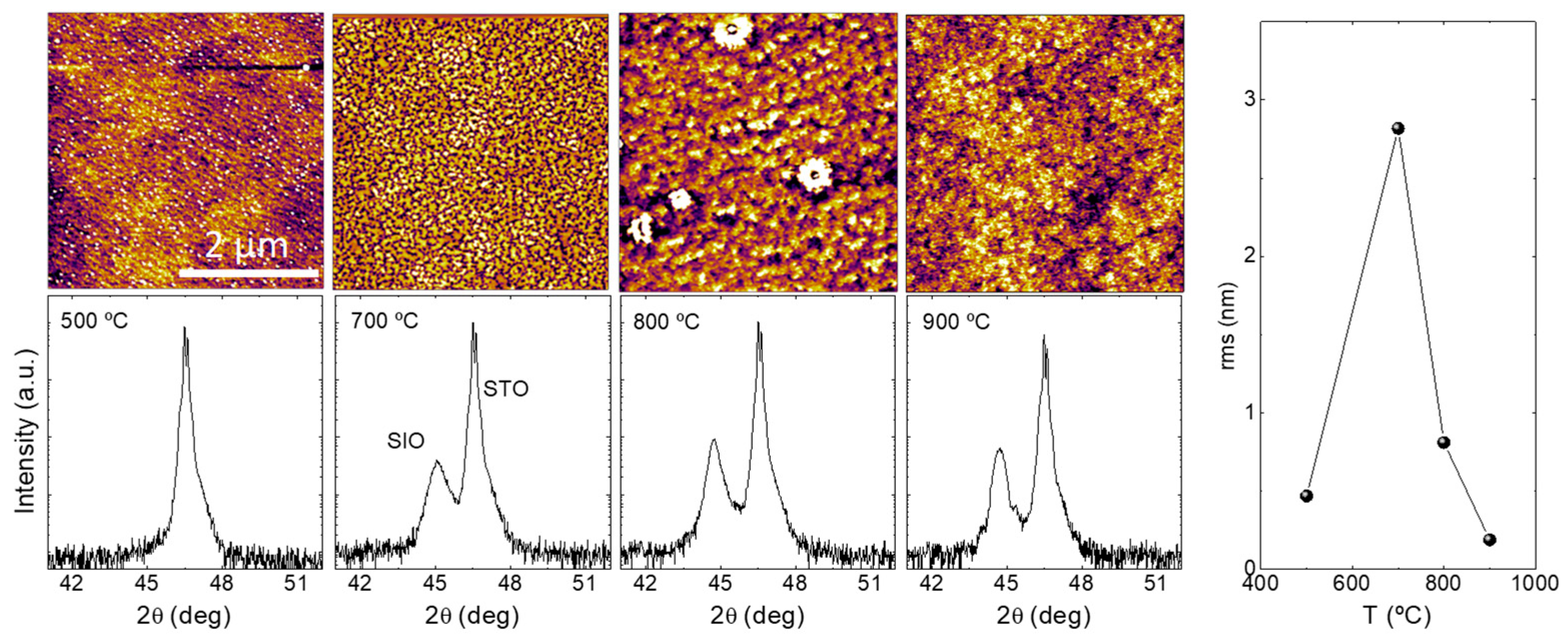
Abstract
:1. Introduction
2. Materials and Methods
2.1. Film Growth
2.2. Structural and Physical Characterization
3. Results and Discussion
3.1. Influence of Growth Conditions
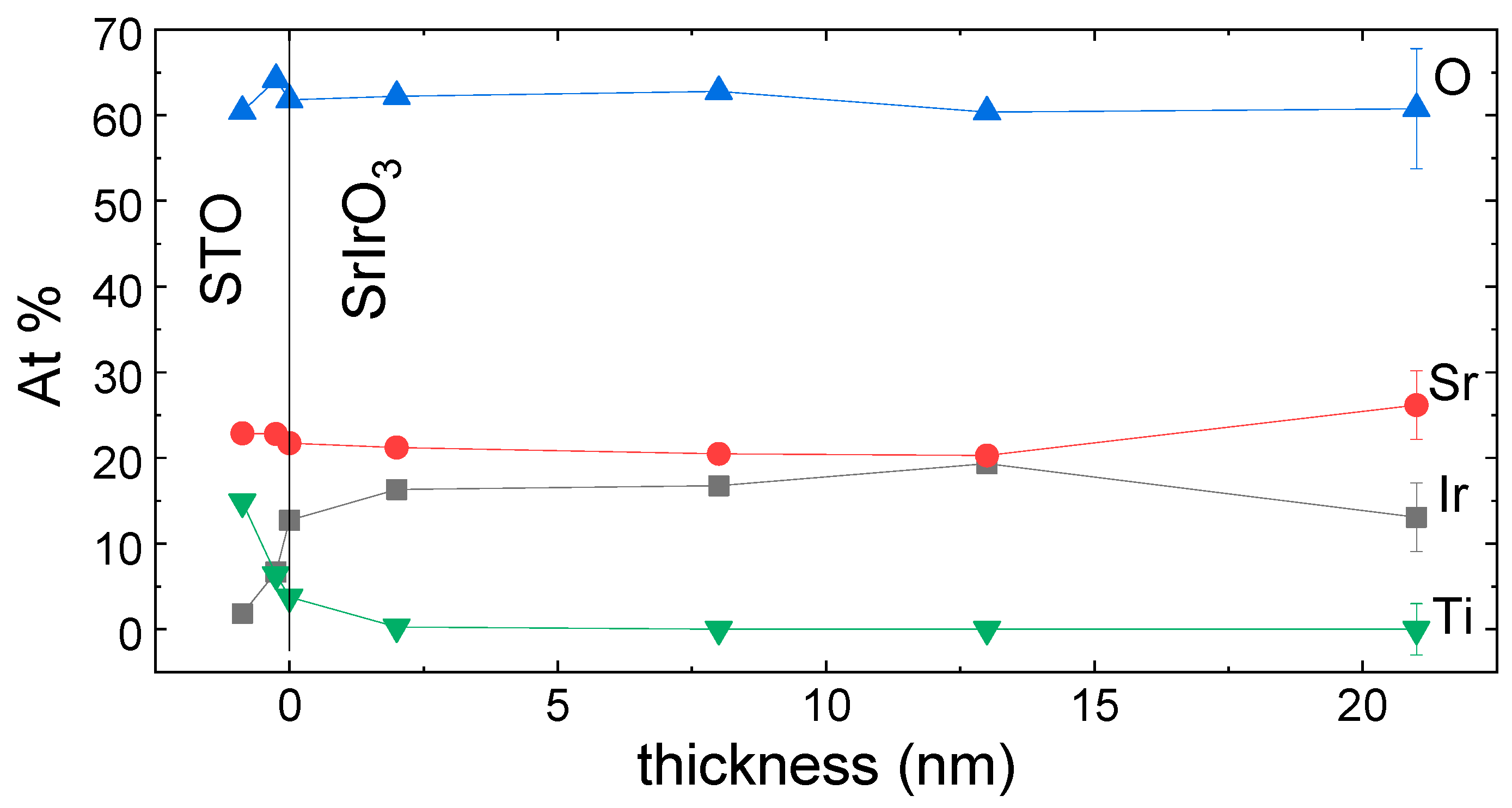
3.2. Film Stoichiometry
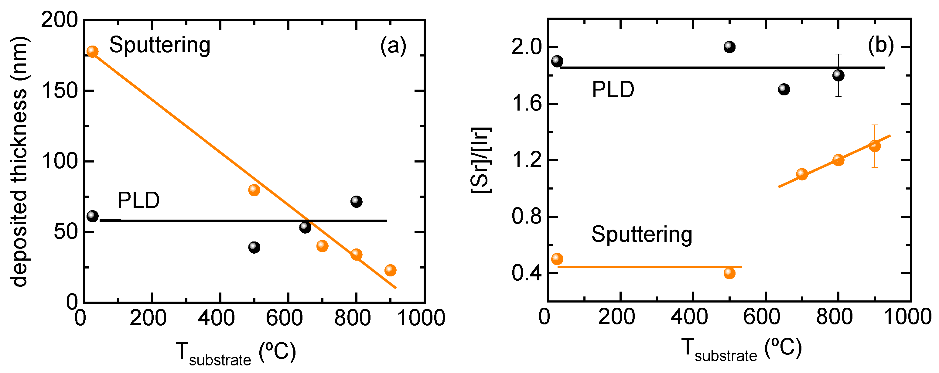
3.3. Sputtering Processes
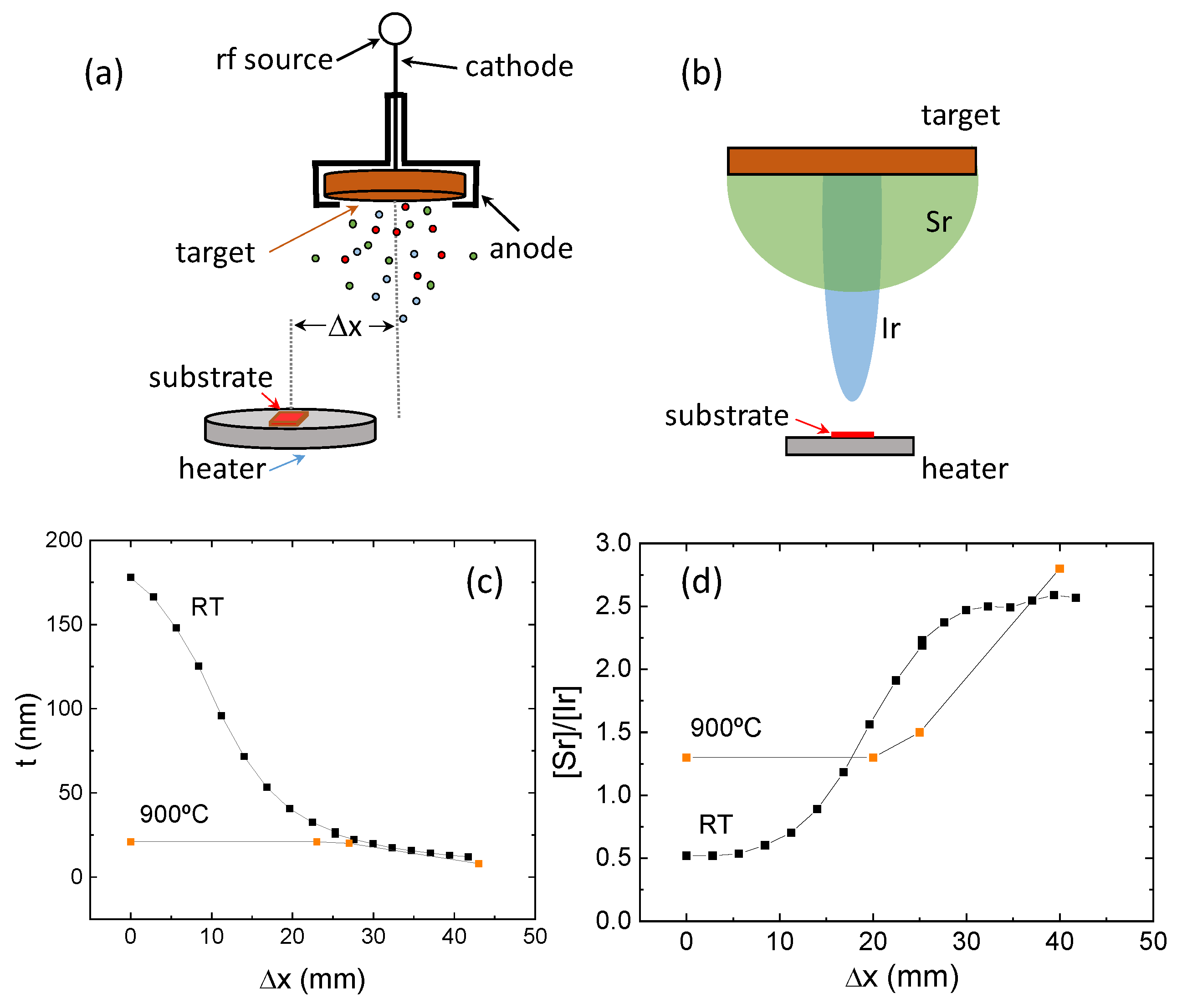
3.3.1. Backsputtering Phenomenon
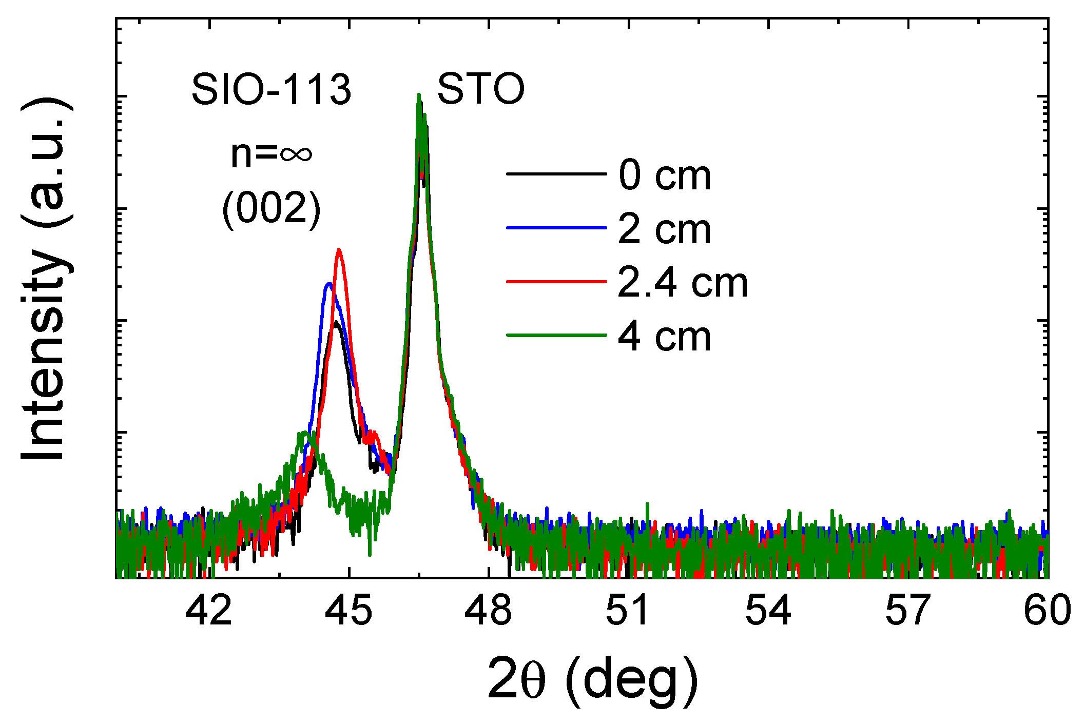
3.3.2. Angular Distribution
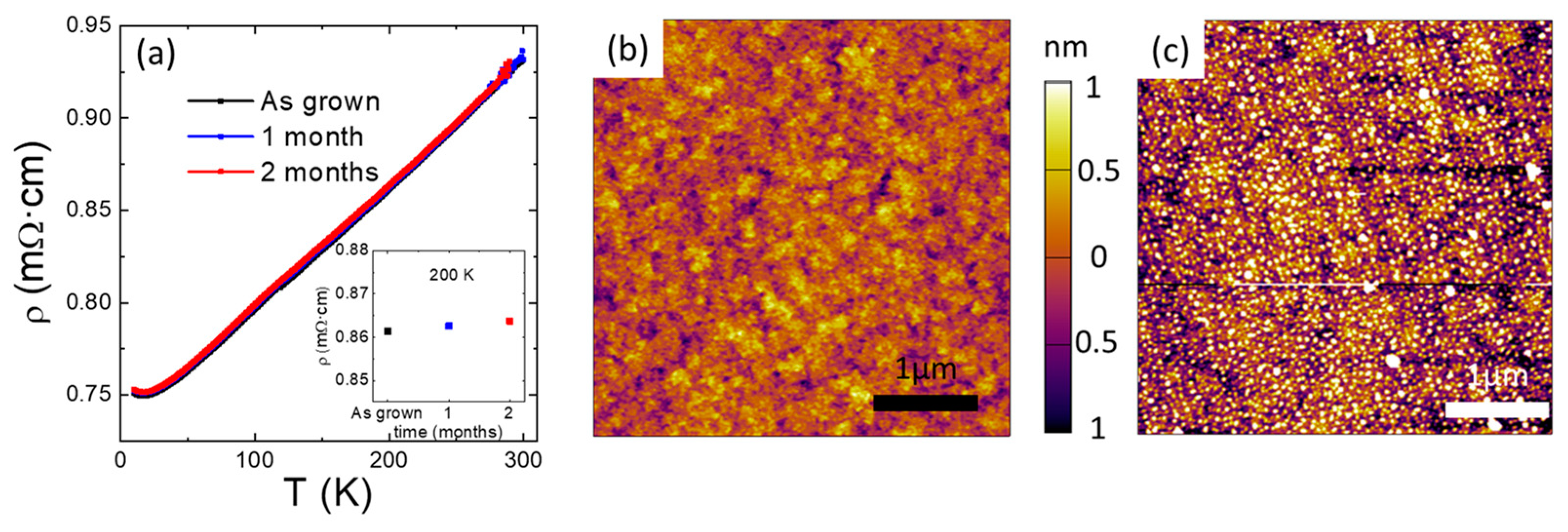
3.4. Thin-Film Stability
4. Conclusions
Supplementary Materials
Author Contributions
Funding
Data Availability Statement
Acknowledgments
Conflicts of Interest
References
- Tarancón, A.; Pryds, N. Functional Oxide Thin Films for Advanced Energy and Information Technology. Adv. Mater. Interfaces 2019, 6, 1900990. [Google Scholar] [CrossRef]
- Wang, Q.; Gu, Y.; Chen, C.; Pan, F.; Song, C. Oxide Spintronics as a Knot of Physics and Chemistry: Recent Progress and Opportunities. J. Phys. Chem. Lett. 2022, 13, 10065. [Google Scholar] [CrossRef]
- MacManus-Driscoll, J.L.; Wells, M.P.; Yun, C.; Lee, J.-W.; Eom, C.-B.; Schlom, D.G. New approaches for achieving more perfect transition metal oxide thin films. APL Mater. 2020, 8, 040904. [Google Scholar] [CrossRef]
- Hwang, H.Y.; Iwasa, Y.; Kawasaki, M.; Keimer, B.; Nagaosa, N.; Tokura, Y. Emergent phenomena at oxide interfaces. Nat. Mater. 2012, 11, 103–113. [Google Scholar] [CrossRef]
- Kaul, A.; Gorbenko, O.; Novojilov, M.; Kamenev, A.; Bosak, A.; Mikhaylov, A.; Boytsova, O.; Kartavtseva, M. Epitaxial stabilization—A tool for synthesis of new thin film oxide materials. J. Cryst. Growth 2005, 275, e2445–e2451. [Google Scholar] [CrossRef]
- Cao, G.; Schlottmann, P. The challenge of spin-orbit-tuned ground states in iridates: A key issues review. Rep. Prog. Phys. 2018, 81, 042502. [Google Scholar] [CrossRef]
- Wu, X.; Li, Z.; Yi, X.; Jia, C.; Zhang, W. Enhanced synaptic performances in SrIrO3 thin films by a ferroelectric layer. Appl. Phys. Lett. 2023, 123, 012104. [Google Scholar] [CrossRef]
- Seitz, L.C.; Dickens, C.F.; Nishio, K.; Hikita, Y.; Montoya, J.; Doyle, A.; Kirk, C.; Vojvodic, A.; Hwang, H.Y.; Norskov, J.K.; et al. A highly active and stable IrOx/SrIrO3 catalyst for the oxygen evolution reaction. Science 2016, 353, 1011–1014. [Google Scholar] [CrossRef]
- Fuentes, V.; Vasić, B.; Konstantinović, Z.; Martínez, B.; Balcells, L.; Pomar, A. Resistive Switching in Semimetallic SrIrO3 Thin Films. ACS Appl. Electron. Mater. 2019, 1, 1981. [Google Scholar] [CrossRef]
- Fuentes, V.; Vasić, B.; Konstantinović, Z.; Martínez, B.; Balcells, L.; Pomar, A. Resistive switching in Strontium iridate based thin films. J Magn. Magn. Mater. 2020, 501, 166419. [Google Scholar] [CrossRef]
- Nelson, J.N.; Schreiber, N.J.; Georgescu, A.B.; Goodge, B.H.; Faeth, B.D.; Parzyck, C.T.; Zeledon, C.; Kourkoutis, L.F.; Millis, A.J.; Georges, A.; et al. Interfacial charge transfer and persistent metallicity of ultrathin SrIrO3/SrRuO3 heterostructures. Sci. Adv. 2022, 8, eabj0481. [Google Scholar] [CrossRef] [PubMed]
- Sen, K.; Fuchs, D.; Heid, R.; Kleindienst, K.; Wolff, K.; Schmalian, J.; Le Tacon, M. Strange semimetal dynamics in SrIrO3. Nat. Commun. 2020, 11, 4270. [Google Scholar] [CrossRef] [PubMed]
- Skoropata, E.; Nichols, J.; Ok, J.M.; Chopdekar, R.V.; Choi, E.S.; Rastogi, A.; Sohn, C.; Gao, X.; Yoon, S.; Farmer, T.; et al. Interfacial tuning of chiral magnetic interactions for large topological Hall effects in LaMnO3/SrIrO3 heterostructures. Sci. Adv. 2020, 6, eaaz3902. [Google Scholar] [CrossRef] [PubMed]
- Matsuno, J.; Ogawa, N.; Yasuda, K.; Kagawa, F.; Koshibae, W.; Nagaosa, N.; Tokura, Y.; Kawasaki, M. Interface-driven topological Hall effect in SrRuO3-SrIrO3 bilayer. Sci. Adv. 2016, 2, e1600304. [Google Scholar] [CrossRef] [PubMed]
- Yoo, M.-W.; Tornos, J.; Sander, A.; Lin, L.-F.; Mohanta, N.; Peralta, A.; Sanchez-Manzano, D.; Gallego, F.; Haskel, D.; Freeland, J.W.; et al. Large intrinsic anomalous Hall effect in SrIrO3 induced by magnetic proximity effect. Nat. Commun. 2021, 12, 3283. [Google Scholar] [CrossRef]
- Suraj, T.S.; Müller, M.; Gelder, S.; Geprägs, S.; Opel, M.; Weiler, M.; Sethupathi, K.; Huebl, H.; Gross, R.; Rao, M.S.R.; et al. Effect of interfacial oxidation layer in spin pumping experiments on Ni80Fe20/SrIrO3 heterostructures. J. Appl. Phys. 2020, 128, 083903. [Google Scholar] [CrossRef]
- Crossley, S.; Swartz, A.G.; Nishio, K.; Hikita, Y.; Hwang, H.Y. All-oxide ferromagnetic resonance and spin pumping with SrIrO3. Phys. Rev. B 2019, 100, 115163. [Google Scholar] [CrossRef]
- Nan, T.; Anderson, T.J.; Gibbons, J.; Hwang, K.; Campbell, N.; Zhou, H.; Dong, Y.Q.; Kim, G.Y.; Shao, D.F.; Paudel, T.R.; et al. Anisotropic spin-orbit torque generation in epitaxial SrIrO3 by symmetry design. Proc. Natl. Acad. Sci. USA 2019, 116, 16186. [Google Scholar] [CrossRef]
- Lao, B.; Liu, P.; Zheng, X.; Lu, Z.; Li, S.; Zhao, K.; Gong, L.; Tang, T.; Wu, K.; Shi, Y.-G.; et al. Anisotropic linear and nonlinear charge-spin conversion in topological semimetal SrIrO3. Phys. Rev. B 2022, 106, L220409. [Google Scholar] [CrossRef]
- Martin-Rio, S.; Konstantinovic, Z.; Pomar, A.; Balcells, L.; Pablo-Navarro, J.; Ibarra, M.R.; Magén, C.; Mestres, N.; Frontera, C.; Martínez, B. Spin-to-Charge Conversion in All-Oxide La2/3Sr1/3MnO3/SrIrO3 Heterostructures. ACS Appl. Mater. Interfaces 2023, 15, 37038. [Google Scholar] [CrossRef]
- Longo, J.; Kafalas, J.; Arnott, R. Structure and properties of the high and low pressure forms of SrIrO3. J. Solid. State Chem. 1971, 3, 174. [Google Scholar] [CrossRef]
- Zhao, J.G.; Yang, L.X.; Yu, Y.; Li, F.Y.; Yu, R.C.; Fang, Z.; Chen, L.C.; Jin, C.Q. High-pressure synthesis of orthorhombic SrIrO3 perovskite and its positive magnetoresistance. J. Appl. Phys. 2008, 103, 103706. [Google Scholar] [CrossRef]
- Fruchter, L.; Brouet, V.; Brisset, F.; Moutaabbid, H.; Klein, Y. Growth facets of SrIrO3 thin films and single crystals. CrystEngComm 2019, 21, 3822. [Google Scholar] [CrossRef]
- Nishio, K.; Hwang, H.Y.; Hikita, Y. Thermodynamic guiding principles in selective synthesis of strontium iridate Ruddlesden-Popper epitaxial films. APL Mater. 2016, 4, 036102. [Google Scholar] [CrossRef]
- Liu, X.; Cao, Y.; Pal, B.; Middey, S.; Kareev, M.; Choi, Y.; Shafer, P.; Haskel, D.; Arenholz, E.; Chakhalian, J. Synthesis and electronic properties of Ruddlesden-Popper strontium iridate epitaxial thin films stabilized by control of growth kinetics. Phys. Rev. Mater. 2017, 1, 075004. [Google Scholar] [CrossRef]
- Gutiérrez-Llorente, A.; Iglesias, L.; Rodríguez-González, B.; Rivadulla, F. Epitaxial stabilization of pulsed laser deposited Srn+1IrnO3n+1 thin films: Entangled effect of growth dynamics and strain. APL Mater. 2018, 6, 091101. [Google Scholar] [CrossRef]
- Fruchter, L.; Schneegans, O.; Li, Z.Z. Anisotropy and interaction effects of strongly strained SrIrO3 thin films. J. Appl. Phys. 2016, 120, 075307. [Google Scholar] [CrossRef]
- Ovsyannikov, G.A.; Shaikhulov, T.A.; Stankevich, K.L.; Khaydukov, Y.; Andreev, N.V. Magnetism at an iridate/manganite interface: Influence of strong spin-orbit interaction. Phys. Rev. B 2020, 102, 144401. [Google Scholar] [CrossRef]
- Connell, J.G.; Isaac, B.J.; Ekanayake, G.B.; Strachan, D.R.; Seo, S.S.A. Preparation of atomically flat SrTiO3 surfaces using a deionized-water leaching and thermal annealing procedure. Appl. Phys. Lett. 2012, 101, 251607. [Google Scholar] [CrossRef]
- Wasa, K. Sputtering Phenomena. In Handbook of Sputtering Technology; Wasa, K., Kanno, I., Kotera, H., Eds.; William Andrew Publishing: Oxford, UK, 2012; pp. 41–75. [Google Scholar]
- Guan, D.; Ryu, G.; Hu, Z.; Zhou, J.; Dong, C.-L.; Huang, Y.-C.; Zhang, K.; Zhong, Y.; Komarek, A.C.; Zhu, M.; et al. Utilizing ion leaching effects for achieving high oxygen-evolving performance on hybrid nanocomposite with self-optimized behaviors. Nat. Commun. 2020, 11, 3376. [Google Scholar] [CrossRef] [PubMed]
- Seo, S.S.A.; Nichols, J.; Hwang, J.; Terzic, J.; Gruenewald, J.H.; Souri, M.; Thompson, J.; Connell, J.G.; Cao, G. Selective growth of epitaxial Sr2IrO4 by controlling plume dimensions in pulsed laser deposition. Appl. Phys. Lett. 2016, 109, 201901. [Google Scholar] [CrossRef]
- Choudhary, R.; Nair, S.; Yang, Z.; Lee, D.; Jalan, B. Semi-metallic SrIrO3 films using solid-source metal-organic molecular beam epitaxy. APL Mater. 2022, 10, 091118. [Google Scholar] [CrossRef]
- Groenendijk, D.J.; Autieri, C.; Girovsky, J.; Martinez-Velarte, M.C.; Manca, N.; Mattoni, G.; Monteiro, A.M.R.V.L.; Gauquelin, N.; Verbeeck, J.; Otte, A.F.; et al. Spin-Orbit Semimetal SrIrO3 in the Two-Dimensional Limit. Phys. Rev. Lett. 2017, 119, 256403. [Google Scholar] [CrossRef]
- Guan, D.; Zhong, J.; Xu, H.; Huang, Y.-C.; Hu, Z.; Chen, B.; Zhang, Y.; Ni, M.; Xu, X.; Zhou, W.; et al. A universal chemical-induced tensile strain tuning strategy to boost oxygen-evolving electrocatalysis on perovskite oxides. Appl. Phys. Rev. 2022, 9, 011422. [Google Scholar] [CrossRef]
- Zhang, L.; Liang, Q.; Xiong, Y.; Zhang, B.; Gao, L.; Li, H.; Chen, Y.B.; Zhou, J.; Zhang, S.-T.; Gu, Z.-B.; et al. Tunable semimetallic state in compressive-strained SrIrO3 films revealed by transport behavior. Phys. Rev. B 2015, 91, 035110. [Google Scholar] [CrossRef]
- Biswas, A.; Kim, K.-S.; Jeong, Y.H. Metal insulator transitions in perovskite SrIrO3 thin films. J. Appl. Phys. 2014, 116, 213704. [Google Scholar] [CrossRef]
- Gruenewald, J.H.; Nichols, J.; Terzic, J.; Cao, G.; Brill, J.W.; Seo, S.S. Compressive strain-induced metal–insulator transition in orthorhombic SrIrO3 thin films. J. Mater. Res. 2014, 29, 2491. [Google Scholar] [CrossRef]
- Wu, F.-X.; Zhou, J.; Zhang, L.Y.; Zhang, S.-T.; Gu, Z.-B.; Yao, S.-H.; Chen, Y.-F. Metal-insulator transition in SrIrO3 with strong spin-orbit interaction. J. Phys. Condens. Matter 2013, 25, 125604. [Google Scholar] [CrossRef]
- Manca, N.; Groenendijk, D.J.; Pallecchi, I.; Autieri, C.; Tang, L.M.K.; Telesio, F.; Mattoni, G.; McCollam, A.; Picozzi, S.; Caviglia, A.D. Balanced electron-hole transport in spin-orbit semimetal SrIrO3 heterostructures. Phys. Rev. B 2018, 97, 081105. [Google Scholar] [CrossRef]


Disclaimer/Publisher’s Note: The statements, opinions and data contained in all publications are solely those of the individual author(s) and contributor(s) and not of MDPI and/or the editor(s). MDPI and/or the editor(s) disclaim responsibility for any injury to people or property resulting from any ideas, methods, instructions or products referred to in the content. |
© 2024 by the authors. Licensee MDPI, Basel, Switzerland. This article is an open access article distributed under the terms and conditions of the Creative Commons Attribution (CC BY) license (https://creativecommons.org/licenses/by/4.0/).
Share and Cite
Fuentes, V.; Balcells, L.; Konstantinović, Z.; Martínez, B.; Pomar, A. Evaluation of Sputtering Processes in Strontium Iridate Thin Films. Nanomaterials 2024, 14, 242. https://doi.org/10.3390/nano14030242
Fuentes V, Balcells L, Konstantinović Z, Martínez B, Pomar A. Evaluation of Sputtering Processes in Strontium Iridate Thin Films. Nanomaterials. 2024; 14(3):242. https://doi.org/10.3390/nano14030242
Chicago/Turabian StyleFuentes, Víctor, Lluis Balcells, Zorica Konstantinović, Benjamín Martínez, and Alberto Pomar. 2024. "Evaluation of Sputtering Processes in Strontium Iridate Thin Films" Nanomaterials 14, no. 3: 242. https://doi.org/10.3390/nano14030242