Article

Narrow UVB-Emitted YBO3 Phosphor Activated by Bi3+ and Gd3+ Co-Doping

School of Materials Science and Engineering, Tianjin Key Lab for Rare Earth Materials and Applications, Nankai University, Tianjin 300350, China
*
Authors to whom correspondence should be addressed.
Nanomaterials 2023, 13(6), 1013; https://doi.org/10.3390/nano13061013
Submission received: 9 February 2023 / Revised: 24 February 2023 / Accepted: 8 March 2023 / Published: 11 March 2023

Abstract

:
Y0.9(GdxBi1−x)0.1BO3 phosphors (x = 0, 0.2, 0.4, 0.6, 0.8, and 1.0, YGB) were obtained via high-temperature solid-state synthesis. Differentiated phases and micro-morphologies were determined by adjusting the synthesis temperature and the activator content of Gd3+ ions, verifying the hexagonal phase with an average size of ~200 nm. Strong photon emissions were revealed under both ultraviolet and visible radiation, and the effectiveness of energy transfer from Bi3+ to Gd3+ ions was confirmed to improve the narrow-band ultraviolet-B (UVB) (6PJ8S7/2) emission of Gd3+ ions. The optimal emission was obtained from Y0.9Gd0.08Bi0.02BO3 phosphor annealed at 800 °C, for which maximum quantum yields (QYs) can reach 24.75% and 1.33% under 273 nm and 532 nm excitations, respectively. The optimal QY from the Gd3+-Bi3+ co-doped YGB phosphor is 75 times the single Gd3+-doped one, illustrating that these UVB luminescent phosphors based on co-doped YBO3 orthoborates possess bright UVB emissions and good excitability under the excitation of different wavelengths. Efficient photon conversion and intense UVB emissions indicate that the multifunctional Gd3+-Bi3+ co-doped YBO3 orthoborate is a potential candidate for skin treatment.

Graphical Abstract

1. Introduction

Skin treatment using artificial sources of ultraviolet (UV) radiation in controlled conditions is well established, and narrow-band ultraviolet-B (UVB) therapy has been demonstrated to be effective against skin diseases and disorders such as psoriasis, vitiligo and hyperbilirubinemia (commonly known as infant jaundice) [1,2,3,4]. Phototherapy with narrow-band UVB (310–313 nm) as photosensitizers is believed to result from the direct interaction between the light of certain frequencies and tissues, causing a change in immune response [5,6,7]. Furthermore, during phototherapy investigations, it was observed that light belonging to longer wavelengths of the UVB region was more effective, while that of the shorter wavelengths was much less effective or even harmful [8,9]. Rare-earth (RE) orthoborates (RE-BO3, RE = lanthanide, yttrium, and scandium) have aroused considerable interest due to their wide range of applications in plasma display panels and mercury-free fluorescent lamps [10,11]. In particular, YBO3 is an excellent host for UV phosphors due to its high-vacuum UV transparency, exceptional optical damage thresholds, strong absorption in the UV range, and good chemical inertness [12,13,14]. Additionally, the YBO3 phosphors exhibit a wide bandgap and high host-to-activator energy transfer efficiency at moderate RE3+ concentrations [15]. Therefore, it is of great interest to investigate RE-doped YBO3 orthoborates for UVB treatments.
Among the RE ions, lanthanide gadolinium (Gd3+) is of particular interest because of its ubiquitous nature (well known as U-spectrum) and the characteristic narrow-band UVB emission from 6PJ8S7/2 transitions [16,17]. The optical properties of Gd3+ ions have been widely studied, and many Gd-doped compounds can be used as efficient phosphors in the new generation of UV fluorescent lamps. Moreover, as a promising activator or sensitizer, the Bi3+ ion shows excellent emission and absorption ability in the UV region. Furthermore, the transitions of 8S7/26IJ (emission at ~270 nm) and 8S7/26PJ (at ~311 nm) of the Gd3+ ion overlap with the 3P11S0 (at ~260 nm) transition of the Bi3+ ion in YBO3 [18,19], which permits an efficient energy transfer from the Bi3+ to Gd3+ ions. Further research on improving the UVB emission has also been reported [18]. From a practical point of view, UV-emitting phosphors in well-defined regions are required for various applications. Keeping this in mind, we prepared a UVB-emitting Gd3+-Bi3+ co-doped YBO3 phosphor in this work, which can effectively achieve light conversion and UVB emissions. When UV fluorescence is irradiated on the surface of a skin wound, the activity of the mitochondrial catalase can increase in cells, which could promote the synthesis of proteins and the decomposition of adenosine triphosphate (ATP), ultimately healing the wound. A schematic representation of healing, which adopts phosphors as light conversion layers, is conceived in Figure 1.
In this work, narrow-band UVB-emitting phosphors of Gd3+-Bi3+ co-doped Y0.9(GdxBi1−x)0.1BO3 (YGB, x = 0, 0.2, 0.4, 0.6, 0.8 and 1.0) were fabricated by high-temperature solid-state synthesis. The samples with the hexagonal phase and well-dispersed particles were characterized by XRD and SEM techniques, manifesting a microsize of ~200 nm. The responses to UV and the visible (VIS) radiation of these YGB phosphors were compared, and sharp UVB luminescence was recorded with the adjustment of Gd3+ content. The sintering temperature indicated that co-doped Bi3+ ions enhanced the characteristic UVB luminescence from Gd3+ ions. The spectroscopic intensity parameters of YGB phosphors were derived from relative spectral power distributions, and the maximum quantum yields (QYs) at 313 nm were calculated at 24.75% and 1.33% under 273 nm and 532 nm excitations, respectively. YGB orthoborate phosphors with intense UVB emission could provide a viable approach for developing multifunctional composite materials for skin treatments.

2. Materials and Methods

The powders of Y0.9(GdxBi1−x)0.1BO3 (x = 0, 0.2, 0.4, 0.6, 0.8, and 1.0, marked as YGB-0, YGB-0.2, YGB-0.4, YGB-0.6 YGB-0.8, and YGB-1.0, respectively) phosphors were prepared using high-purity reagents Y2O3 (99.9%), Gd2O3 (99.9%), Bi2O3 (A.R.), and H3BO3 (A.R.) as raw materials. The original chemicals for YGB with different Bi3+ contents as the designed sensitizer were mixed by grinding them in an agate mortar according to the stoichiometric ratio. The raw powders were transferred into alumina crucibles and pre-sintered at 500 °C for 1 h and then sintered at 700 °C, 800 °C, 900 °C and 1000 °C for 5 h. Afterwards, the samples were ground thoroughly after cooling.
The phase and the crystal structure of powders were identified by an X-ray diffractometer (XRD, MiniFlex 600, Rigaku, Tokyo, Japan) using Cu Kα radiation. Morphologies of the powders were analyzed by a field emission scanning electron microscope (SEM, JSM-7800F, JEOL, Tokyo, Japan) equipped with energy dispersive spectroscopy (EDS, X-MaxN 50, Oxford, Oxford, UK) using an accelerating voltage of 15 kV. Particle size distributions were measured in a Nanoparticle Analyzer (Zetasizer Nano-ZS, Malvern, UK). Photoluminescence (PL) spectra were recorded using a Keithley 2010 multimeter and the monochromator (λ500, Zolix, Beijing, China) equipped with a Si detector (DSi200, Zolix, Beijing, China). A commercial Xe lamp and two solid-state lasers emitting at 266 nm and 532 nm were used as the excitation sources for different excitation wavelengths. A standard PTFE diffuse reflective white plate (reflectivity greater than 99.9%) was used as a reference. The schematic diagram of the experimental setup is depicted in Figure 2. The relative spectral power distribution was obtained and calibrated by the Optical Power Meter (1830-C, Newport, Newport County, RI, USA). All measurements were performed at room temperature.

3. Results

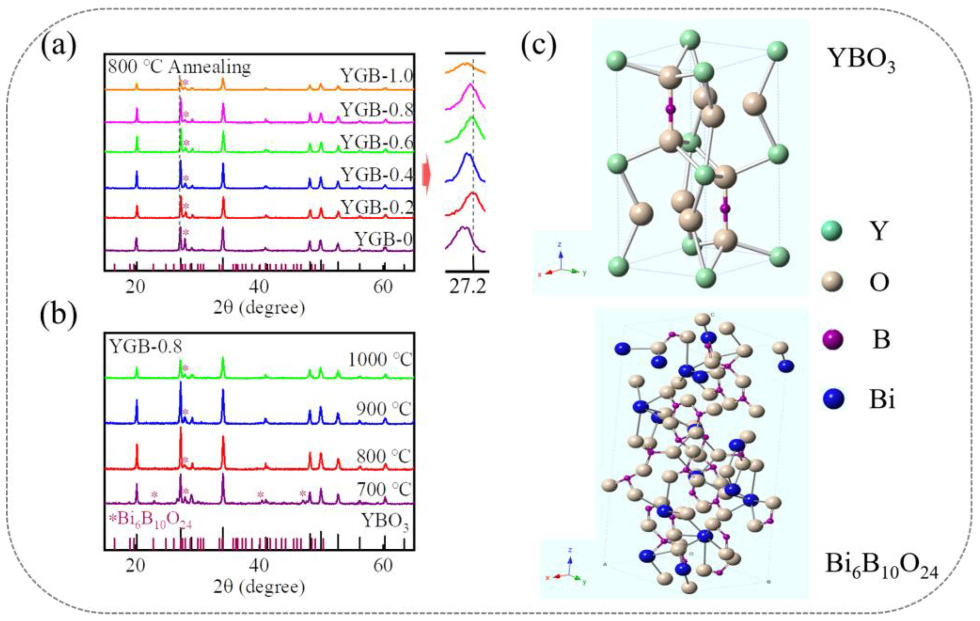
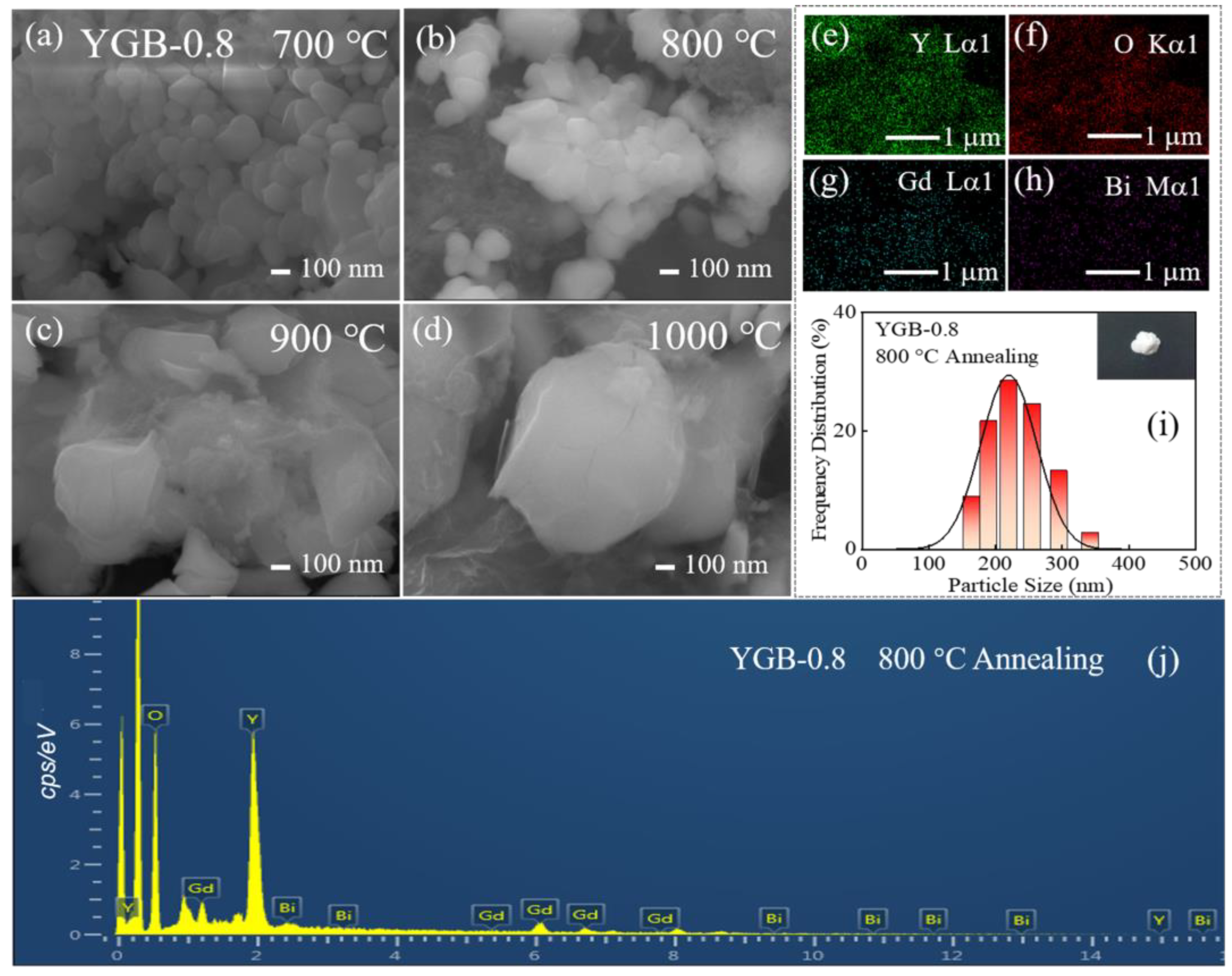
3.1. Structure and Morphology

Figure 3a shows the XRD patterns of YGB phosphors with different compositions annealed at 800 °C, the main peaks of which are in agreement with the JCPDS Card of YBO3 (PDF#16-0277). The phosphors were confirmed as polycrystalline materials that possess a hexagonal crystal structure with space group P63/m and the cell parameters of a = b = 3.778 Å and c = 8.81 Å, similarly to what has been previously reported [20]. Moreover, the well-defined sharp diffraction peaks imply that these samples have high crystallinity, illustrating that Gd3+ and Bi3+ ions are substituted within the host. Some small impurity peaks are identified as Bi6B10O24 (PDF#29-0228), which are attributed to the interaction of Bi2O3 and excess H3BO3 during the fabrication process [21]. The corresponding reaction equation is as follows: 2Bi2O3 + B2O3→Bi4B2O9 and 3Bi4B2O9 + 7B2O3→2Bi6B10O24 [22]. Here, the XRD peaks (2θ = ~27.2°) of YGB samples with slightly smaller angles, in comparison with the standard YBO3, should be attributed to the larger radius of Gd3+ (1.053 Å, 8-coordination) and Bi3+ (1.170 Å, 8-coordination) relative to that of Y3+ (1.019 Å, 8-coordination), according to Bragg’s law [23,24,25], while the shift in diffraction peaks resulted from the different Gd3+-Bi3+ contents in these phosphors. The slight change is also attributed to the surface charge redistribution of the crystal nucleus, induced by an inner-electron charge transfer between the doped ions and lattice cations [26,27]. Therefore, these results show that doped Gd3+ and Bi3+ ions do not affect the main crystal structure of YBO3 and should be completely dissolved into the host lattice.
As shown in Figure 3b, the impurities decrease, and the relative intensities of diffraction peaks initially increase and then decrease with the annealing temperature; this is attributed to the melting point of Bi6B10O24 and the selectivity of the growth in the solid-state synthesis process [25,28]. In addition, the YGB crystal is composed of 8-coordinated Y3+ and 4- coordinated B3+ ions, which is illustrated in Figure 3c. Here, the Y3+ ions are 8- coordinated with two nonequivalent environments, while the B3+ ions and two interconnected BO4 tetrahedral coordination form (BO3)3− groups [29]. Moreover, due to the similar ionic radii of the 8-coordinated Y3+, Bi3+, and Gd3+ ions, Gd3+ and Bi3+ ions can easily substitute the Y3+ sites and form a solid solution of (Y,Gd,Bi)BO3 crystals.
Take the Y0.9Gd0.08Bi0.02BO3 (YGB-0.8) phosphor as an example. The SEM images in Figure 4a–d show the typical morphologies of particles annealed at 700, 800, 900, and 1000 °C, revealing that the powders annealed at 800 °C and below possess an average size of ~200 nm and the regular morphology. To show this, the particle size distributions of the YGB-0.8 phosphor annealed at 800 °C are shown in Figure 4i. Here, the inset shows the macroscopic appearance of the sample exhibited under natural light irradiation. It can be observed that the particle size is mainly concentrated at ∼200 nm, which is consistent with the SEM images. As shown in the SEM images, the powders with a narrow particle size distribution have been synthesized at lower sintering temperatures, and they possess a large effective surface area and weak atomic binding energy, resulting in the lower local symmetry of the YO8 polyhedron and the surface defects of nanoparticles. When the annealing temperature exceeds a certain value, the crystal phase is gradually purified together with grain growth. Obviously, the morphology becomes more irregular in angularity, heterogeneity and compactness with the increase in sintering temperature, which is due to the changes in van der Waals attractions, while the small particle size may be caused by the distortion of anionic groups on the particle’s surface [30,31,32]. The size of spherical particles is significantly larger after sintering at 1000 °C, while compositional particles lose their spherical shape and undergo significant aggregation, which is attributed to the higher activity of atoms on the particle’s surface caused by the further decomposition of precursors. Under higher temperature annealing, the atoms could diffuse and combine with adjacent ones to form stable chemical bonds, leading to agglomeration [33,34]. No obvious changes in the morphology or particle size with various Gd3+ contents were observed at the same sintering temperature (not shown here), indicating that the doped Bi3+ and Gd3+ ions do not impact crystallization and grain growth. For the YGB-0.8 sample annealed at 800 °C, the homogeneous distributions of Gd, Bi, O, and Y elements are clearly observed by EDS, as shown in Figure 4e–h,j. The B element is undetected since its corresponding energy in the X-ray spectrum falls outside the scope. Moreover, high-packing densities, good slurry properties, and well-distributed particles in YGB systems are conducive to photon release.

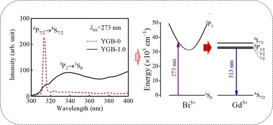
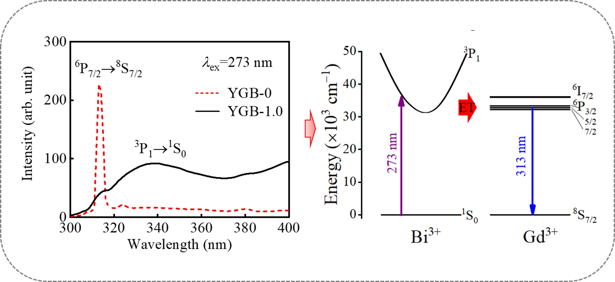
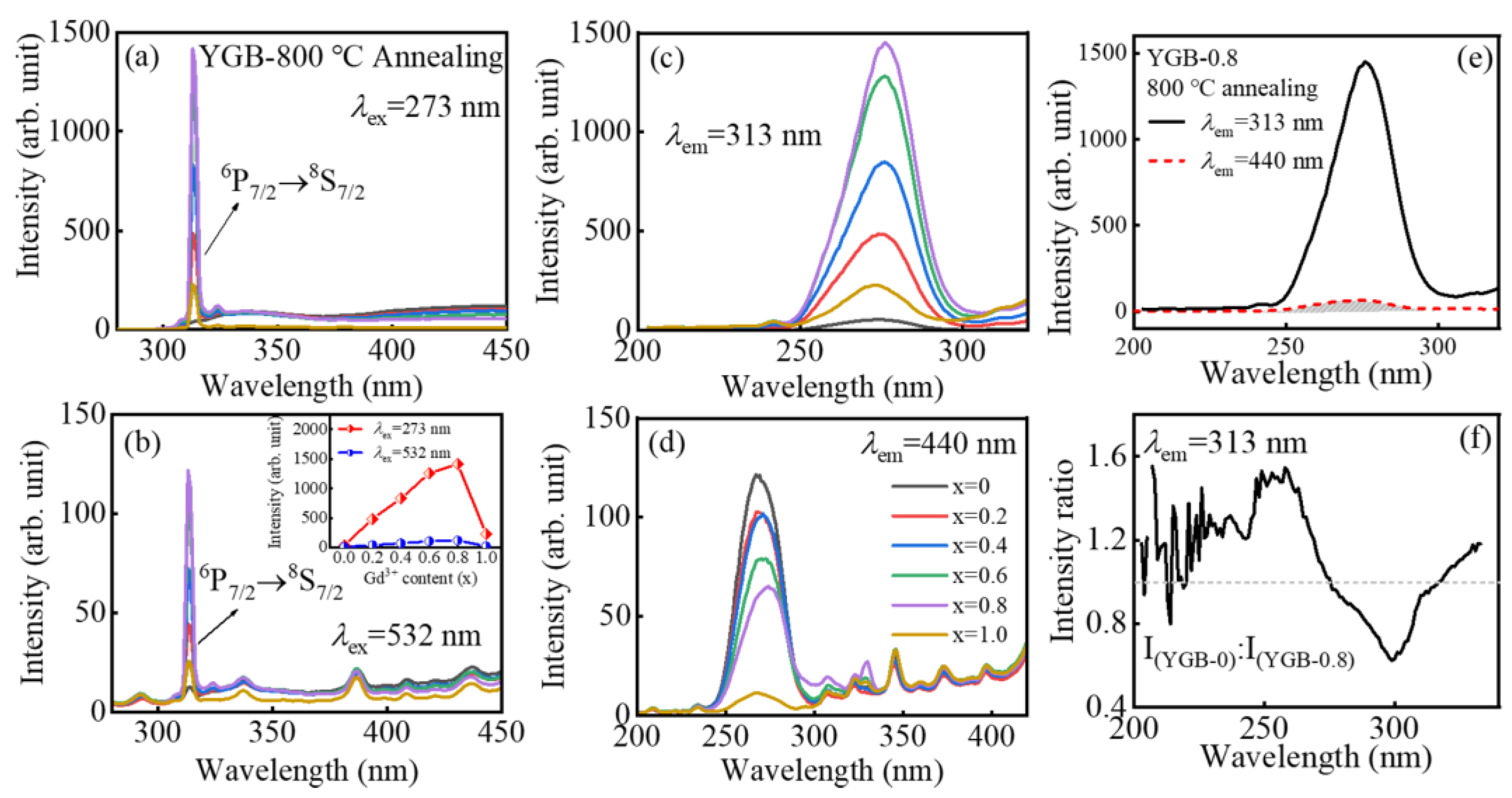
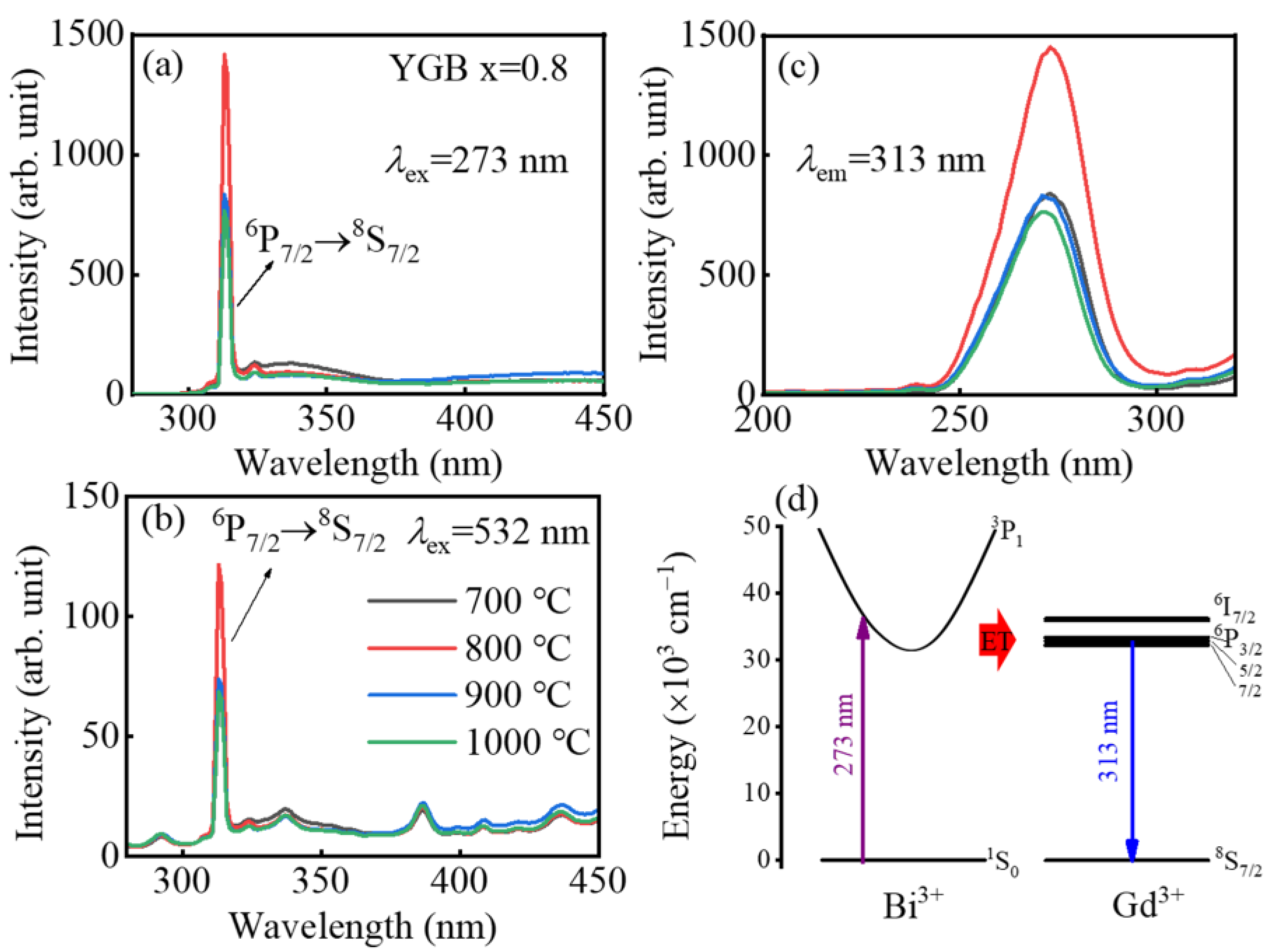
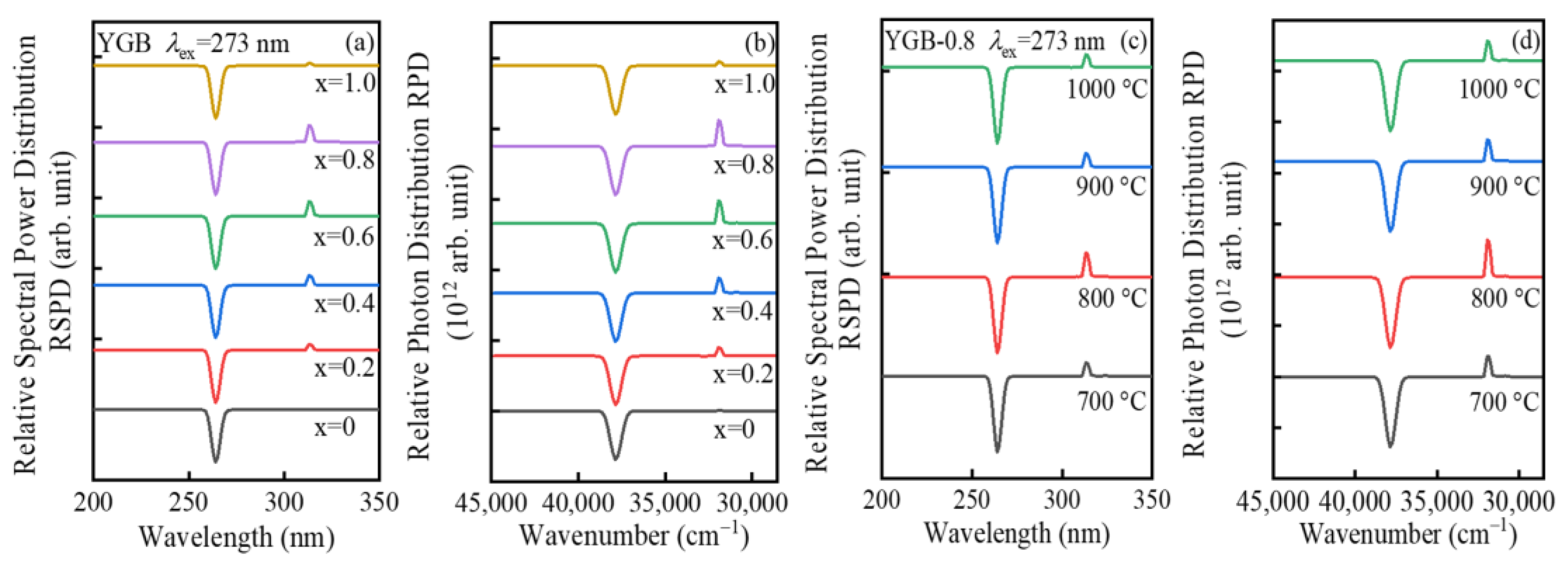
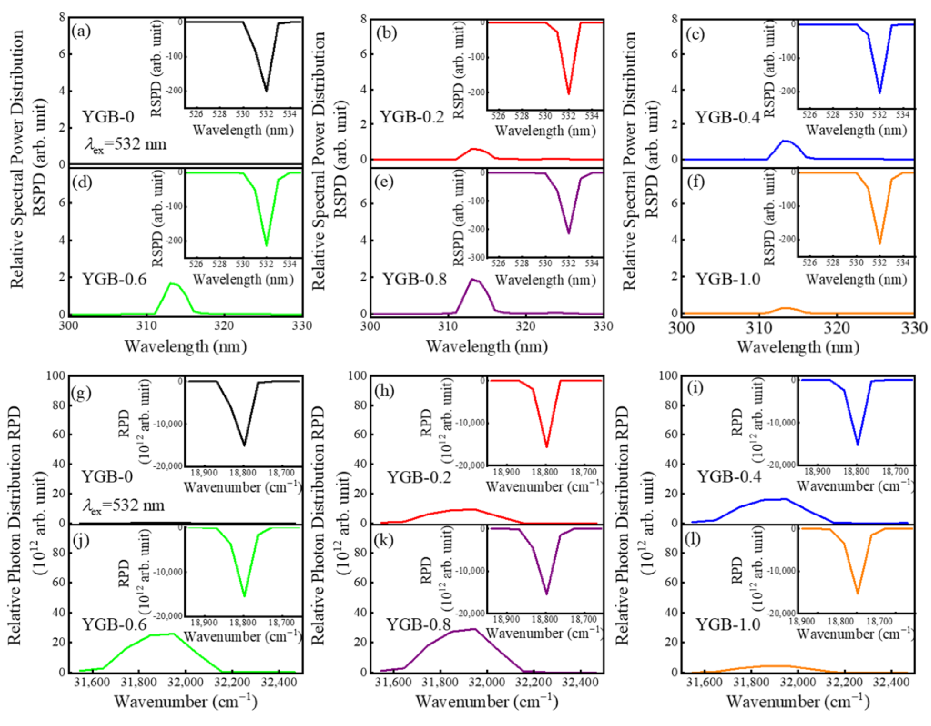
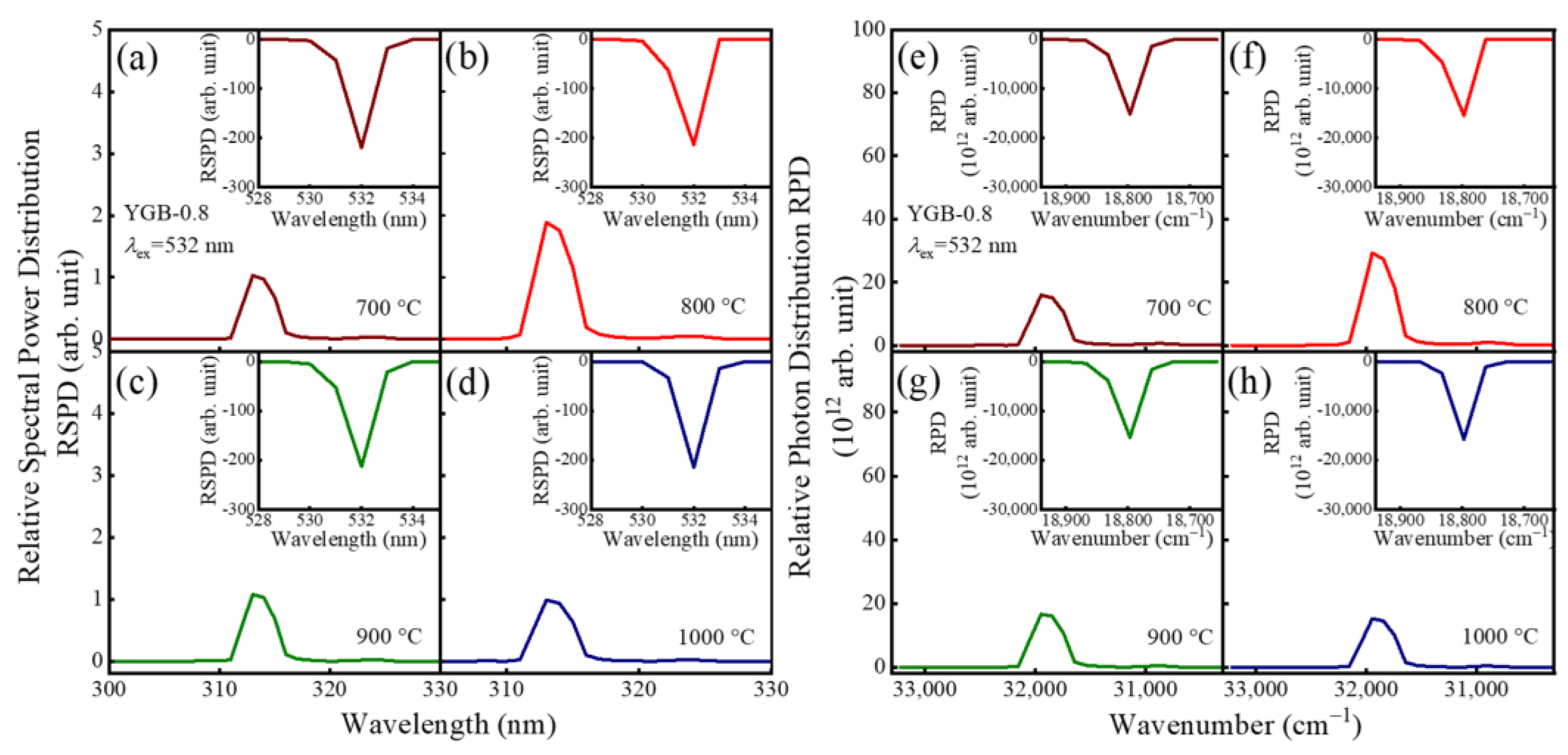
3.2. Fluorescence Behaviors of YGB Phosphor

Figure 5a,b show the typical emission spectra of YGB phosphors with different Bi3+/Gd3+ contents (x = 0, 0.2, 0.4, 0.6, 0.8 and 1.0) under UV (273 nm) and VIS (532 nm) excitations. Notably, strong UV emissions can also be obtained by up-conversion under the excitation of a 532 nm laser, and all samples show a narrow-band emission at 313 nm, which is attributed to the 6P7/28S7/2 transition of Gd3+ ions [6]. Emission intensity increases until the Gd3+ content exceeds x = 0.8, which results from the energy transfer between Bi3+ and Gd3+ ions; with the further increase in Gd3+ contents, concentration quenching occurs with the attenuation of emission intensity. In order to identify the change in spectral intensity more clearly, the dependence of the PL emission intensity at 313 nm on the Gd3+ content (x) in YGB phosphors under 273 nm and 532 nm excitations is illustrated in the inset of Figure 5b. Compared with the sample without Bi3+, the weaker wide emission located around 440 nm resulted from the 6s2→6s6p transitions of Bi3+ ions. According to the photoluminescence excitation (PLE) spectra of these YGB phosphors in Figure 5c, monitored at 313 nm, the strongest excitation band centered at 273 nm should be attributed to the 8S7/26IJ transition of the Gd3+ ion, which well overlaps with the 253.7 nm line of mercury lamps [35], while the emission peak at 440 nm was derived from the 3P11S0 transition of Bi3+ ions [36]. In particular, Figure 5d shows the PLE spectra of YGB samples with different Gd3+ contents in monitoring the 440 nm emission, the intensity of which decreases with the Gd3+ content, illustrating the energy transfer (ET) from Bi3+ to Gd3+ ions that consumes the excitation energy of Bi3+ ions.
The energy transfer depends on the overlap between the excitation band of the activator and the emission band of the sensitizer in the phosphors. Bi3+ ions have a 6s2 outer electronic configuration with a 1S0 ground state, and the excited state has the configuration of 6s6p with 3P0, 3P1, 3P2, and 1P1 splitting levels. Due to the forbidden transitions of 1S03P0 and 1S03P2 by the electronic selection rules, the 1S03P1 and 1S01P1 transitions of Bi3+ ions are usually observed [37]. For the sample doped with only Bi3+ ions, it would first relax and transit into the lowest 3P1 excited state and then return to the 1S0 ground state via radiation. However, when Bi3+ and Gd3+ ions were co-doped into the host, energy transfer would occur, since the 3P11S0 emission of Bi3+ effectively overlapped with the energy levels of Gd3+ (6P7/2, 6P5/2, and 6P3/2) [38]. For the Y0.9Gd0.08Bi0.02BO3 (YGB-0.8) sample annealed at 800 °C, the overlapped excitation spectra of Bi3+ and Gd3+ ions are shown in Figure 5e, confirming their efficient excitability in the short-wave UV region, which is advantageous for the resonance energy transfer from Bi3+ to Gd3+ ions. In order to further clarify the controversies over the ET from Bi3+ to Gd3+, Figure 5f presents the Gd3+ fluorescence intensity ratio between 800 °C annealed YGB-0 and YGB-0.8 phosphors, monitored at 313 nm, which demonstrates the sensitizing effect of Bi3+ on Gd3+ ions. Compared with the sample without Gd3+ ions, the excitation energy of Bi3+ in Bi3+-Gd3+ co-doped samples is transferred to the Gd3+ ion and leads to stronger fluorescence emissions from the Gd3+ ion in short-wave UVB radiation, resulting in increased excitability. In addition, the excitation peaks located at 258 nm are consistent with the characteristic excitation peaks of Bi3+ (1S01P1) [25,37], which further confirms the effectiveness of ET from Bi3+ to Gd3+ ions.
To further investigate the effect of sintering temperatures on luminous properties, the PL and PLE spectra measured at room temperature from the YGB-0.8 phosphors annealed at different temperatures are illustrated in Figure 6a–c. The intensity of the excitation peak at 313 nm increases with the annealing temperature until 800 °C. The grain size increases while the porosity decreases significantly with the increase in temperature, enhancing the luminous intensity. When the sintering temperature is higher than 800 °C, the decreased intensity is attributed to the accelerated volatilization of Bi3+ and the crystalline defects. Furthermore, the enhanced PL emission originates from the absorption of exciting UV light by co-doped Bi3+ ions, which transfer the energy to the Gd3+ ions [18,39]. This mechanism is schematically shown in Figure 6d. Firstly, phosphors absorb the UV light, which leads to the 1S03P1 transition of Bi3+ ions. The Bi3+ ions then transfer the energy non-radiatively to Gd3+ ions and ultimately realize UVB emissions from Gd3+ ions. Moreover, the smaller electronegativity of Gd3+ (1.20), compared to that of Y3+ (1.22) and Bi3+ ion (~2.02), allows an easier charge transfer, thus promoting PL emissions [19,40].
In order to evaluate the optical property of Gd3+-Bi3+ co-doped YGB phosphors with different Gd3+ contents and annealing temperatures, the relative spectral power distributions and relative photon distributions were determined and compared, as in Figure 7a–d. Under the 273 nm excitation, the sharp narrow-band UVB emission at 313 nm that originates from the 6PJ8S7/2 transition increases significantly with the increase in Gd3+ content and annealing temperature, which reaches the maximum value while the Gd3+ content is x = 0.8 and is annealed at 800 °C. This should be attributed to the increased energy transfer caused by the reduced distance among Gd3+-Gd3+ ion pairs [41]. The relative photon distribution provides fundamental information with respect to optical fields and relevant applications. Depending on the relative spectral power distribution P(λ), photon distribution N(ν) can be deduced by N ν = λ 3 h c P ( λ ) , where ν, λ, h, c, and P(λ) represent wavenumber, wavelength, Planck constant, vacuum light velocity, and spectral power distribution, respectively [42]. Here, the abscissae of the distribution spectra were converted to a wavenumber (cm−1) for accurate deconvolution. The net absorption and emission photon distribution curves of Gd3+-Bi3+ co-doped YBO3 phosphors were derived, as presented in Figure 7b,d, and their net emission and absorption intervals were selected at 30,300–33,300 cm−1 (corresponding to 313 nm) and 36,300–39,200 cm−1 (corresponding to 273 nm).
In addition, upon the excitation under 532 nm VIS light, the samples still emit the up-conversion UVB emission at 313 nm. The spectral power distribution and the photon number distribution of all samples with different Gd3+ contents (x = 0, 0.2, 0.4, 0.6, 0.8, and 1.0) are displayed in Figure 8. With the increase in Gd3+ contents, the emission intensity increases until Y0.9Gd0.08Bi0.02BO3, because the transfer probability is proportional to the interaction between the sensitizer (Bi3+) and the activator (Gd3+) in both non-radiative and radiative resonance energy transfers. When x > 0.8, the intensity significantly decreases due to prominent concentration quenching caused by the reduced distance among Gd3+-Gd3+ ions.
According to Blasse [43,44], the energy would transfer from one activator to another until all energy is consumed. This phenomenon is regarded as concentration quenching in fluorescence, which is due to the non-radiative energy transfer among identical ions. Thus, the critical distance (RC) is a parameter that is essential to understanding this phenomenon, which is calculated using the following equation: R c = 2 3 V 4 π x c N 1 3 , where V is the volume of the unit cell (in Å3), xc is critical concentration, and N is the number of Y3+/Bi3+/Gd3+ ions in the unit cell. Herein, the values are xc = 0.08, N= 6, and V = 108.90 Å3, and the critical distance RC of the YGB phosphor is calculated to be about 7.57 Å. Meanwhile, the corresponding spectral power distribution and photon number distribution of YGB-0.8 phosphors annealed from 700 °C to 1000 °C under the 532 nm excitation were also derived, and they are shown in Figure 9 to demonstrate the up-conversion emission monitored at 313 nm and the optimal annealing temperature of 800 °C. These results verify the effectiveness of Gd3+-Bi3+ co-doped phosphors in photon conversion and provide the theoretical basis for their application in skin treatments.
The spectral parameters could also provide external quantum yields (QYs) to assess luminescence and laser materials, which are used to calculate the utilization efficiency of the absorbed photons for desired emissions, defined as the photon number ratio of emission and absorption. Namely, QY = emitted photons/absorbed photons = Nem/Nabs. Here, the maximum QY is derived to be 24.75% in a Y0.9Gd0.08Bi0.02BO3 sample annealed at 800 °C under the 273 nm excitation, which is larger than that of other Gd3+ ions doped phosphors [45,46], and it is 75 times the single Gd3+-doped sample in this work. On the basis of these QYs, a higher photon release efficiency is achieved, which further exhibits the potential of Gd3+-Bi3+ co-doped YBO3 phosphors for UVB skin treatment and reflects the energy transfer effectiveness between Bi3+ and Gd3+ ions in these phosphors. Moreover, this phosphor maintains a unique up-conversion excitability in the VIS region with a QY of 1.33% under the excitation of 532 nm. The QY values for the different contents and annealing temperatures of these co-doped YGB phosphors, under the excitation of the 273 and 532 nm, are listed in Table 1. These results reveal that the Gd3+-Bi3+ activated YBO3 phosphors with up/down-conversion excitability exhibit excellent UVB emission performance.

4. Conclusions

UVB-emitting Y0.9(GdxBi1−x)0.1BO3 phosphors (x = 0, 0.2, 0.4, 0.6, 0.8, and 1.0) with the hexagonal phase and an average ~200 nm grain size were fabricated via the solid-state synthesis method. The enhanced PL emissions and the overlapped spectra verify the energy transfer between the Bi3+ and Gd3+ ions and a well-defined sharp and intense peak centered at 313 nm due to the 6P7/28S7/2 transitions of Gd3+ ions. The Y0.9Gd0.08Bi0.02BO3 phosphor annealed at 800 °C exhibits the highest QY values of 24.75% and 1.33% under the excitation of 273 nm and 532 nm, respectively, confirming that the system possesses excellent excitability in both UV and VIS regions. The optimal QY from the Gd3+-Bi3+ co-doped YBO3 phosphor is 75 times the single Gd3+-doped sample. Bright and narrow UVB emissions resulting from efficient photon conversion demonstrate the multifunctional applications of Gd3+-Bi3+-activated YBO3 phosphors and provide a new route for skin treatments.

Author Contributions

Conceptualization, Z.Y. and Y.Y.; methodology, Z.Y.; validation, Z.Y.; formal analysis, Z.Y. and Y.Y.; resources, Y.Y. and J.S.; data curation, Z.Y.; writing—original draft preparation, Z.Y.; writing—review and editing, Y.Y.; visualization, Z.Y. and Y.Y.; supervision, Y.Y. and J.S.; project administration, J.S.; funding acquisition, Y.Y. and J.S. All authors have read and agreed to the published version of the manuscript.

Funding

This research was funded by the National Natural Science Foundation of China (No. 62275132) and the Natural Science Foundation of Tianjin City (No. 21JCZDJC00020).

Institutional Review Board Statement

Not applicable.

Informed Consent Statement

Not applicable.

Data Availability Statement

Data will be made available upon request.

Conflicts of Interest

The authors declare no conflict of interest.

References

  1. Chauhan, A.; Palan, C.; Sawala, N.; Omanwar, S. Synthesis and Photoluminescence study of LaF3:Gd3+ phosphors for Phototherapy Application. J. Emerg. Technol. Innov. Res. 2022, 9, 382–387. [Google Scholar]
  2. Hönigsmann, H.; Brenner, W.; Rauschmeier, W.; Konrad, K.; Wolff, K. Photochemotherapy for cutaneous T cell lymphoma: A follow-up study. J. Am. Acad. Dermatol. 1984, 10, 238–245. [Google Scholar] [CrossRef] [PubMed]
  3. Scherschun, L.; Kim, J.J.; Lim, H.W. Narrow-band ultraviolet B is a useful and well-tolerated treatment for vitiligo. J. Am. Acad. Dermatol. 2001, 44, 999–1003. [Google Scholar] [CrossRef] [Green Version]
  4. Dani, C.; Martelli, E.; Reali, M.F.; Bertini, G.; Panin, G.; Rubaltelli, F. Fiberoptic and conventional phototherapy effects on the skin of premature infants. J. Pediatr. 2001, 138, 438–440. [Google Scholar] [CrossRef] [PubMed]
  5. Al Salman, M.; Ghiasi, M.; Farid, A.S.; Taraz, M.; Azizpour, A.; Mahmoudi, H. Oral simvastatin combined with narrowband UVB for the treatment of psoriasis: A randomized controlled trial. Dermatol. Ther. 2021, 34, e15075. [Google Scholar] [CrossRef] [PubMed]
  6. Cheng, Z.; Liu, T.; Shen, M.; Peng, Y.; Yang, S.; Khan, W.U.; Zhang, Y. Luminescence and energy transfer of Ce3+/Gd3+/Tb3+/Eu3+ doped hexagonal fluoride. J. Lumin. 2022, 241, 118477. [Google Scholar] [CrossRef]
  7. Wang, X.; Chen, Y.; Kner, P.A.; Pan, Z. Gd3+-activated narrowband ultraviolet-B persistent luminescence through persistent energy transfer. Dalton Trans. 2021, 50, 3499–3505. [Google Scholar] [CrossRef]
  8. Reich, A.; Mędrek, K. Effects of narrow band UVB (311 nm) irradiation on epidermal cells. Int. J. Mol. Sci. 2013, 14, 8456–8466. [Google Scholar] [CrossRef] [Green Version]
  9. Youssef, Y.E.; Eldegla, H.E.A.; Elmekkawy, R.S.M.; Gaballah, M.A. Evaluation of vitamin D receptor gene polymorphisms (ApaI and TaqI) as risk factors of vitiligo and predictors of response to narrowband UVB phototherapy. Arch. Dermatol. Res. 2022, 1–8. [Google Scholar] [CrossRef]
  10. Zhu, Q.; Wang, S.; Li, J.-G.; Li, X.; Sun, X. Compounds, Spherical engineering and space-group dependent luminescence behavior of YBO3: Eu3+ red phosphors. J. Alloys Compd. 2018, 731, 1069–1079. [Google Scholar] [CrossRef]
  11. Gao, Y.; Jiang, P.; Cong, R.; Yang, T. Photoluminescence of Bi3+ in LiCaY5(BO3)6 and color-tunable emission through energy transfer to Eu3+/Tb3+. J. Lumin. 2022, 251, 119161. [Google Scholar] [CrossRef]
  12. Wang, L.; Wang, Y. Enhanced photoluminescence of YBO3:Eu3+ with the incorporation of Sc3+, Bi3+ and La3+ for plasma display panel application. J. Lumin. 2007, 122, 921–923. [Google Scholar] [CrossRef]
  13. Chen, L.; Yang, G.; Liu, J.; Shu, X.; Jiang, Y.; Zhang, G. Photoluminescence properties of Eu3+ and Bi3+ in YBO3 host under VUV/UV excitation. J. Appl. Phys. 2009, 105, 013513. [Google Scholar] [CrossRef]
  14. Zeng, X.; Im, S.-J.; Jang, S.-H.; Kim, Y.-M.; Park, H.-B.; Son, S.-H.; Hatanaka, H.; Kim, G.-Y.; Kim, S.-G. Luminescent properties of (Y, Gd)BO3:Bi3+, RE3+ (RE = Eu, Tb) phosphor under VUV/UV excitation. J. Lumin. 2006, 121, 1–6. [Google Scholar] [CrossRef]
  15. Song, H.; Yu, H.; Pan, G.; Bai, X.; Dong, B.; Zhang, X.; Hark, S. Electrospinning preparation, structure, and photoluminescence properties of YBO3:Eu3+ nanotubes and nanowires. Chem. Mater. 2008, 20, 4762–4767. [Google Scholar] [CrossRef]
  16. Singh, V.; Sivaramaiah, G.; Rao, J.; Kim, S. Investigation of new UV-emitting, Gd-activated Y4Zr3O12 phosphors prepared via combustion method. J. Lumin. 2015, 157, 82–87. [Google Scholar] [CrossRef]
  17. Gupta, P.; Sahni, M.; Chauhan, S. Enhanced photoluminescence properties of rare earth elements doped Y0. 50Gd0. 50BO3 phosphor and its application in red and green LEDs. Optik 2021, 240, 166810. [Google Scholar] [CrossRef]
  18. Gawande, A.; Sonekar, R.; Omanwar, S. Combustion synthesis and energy transfer mechanism of Bi3+→Gd3+ and Pr3+→Gd3+ in YBO3. Combust. Sci. Technol. 2014, 186, 785–791. [Google Scholar] [CrossRef]
  19. Zhu, Q.; Wang, S.; Li, X.; Sun, X.; Li, J.-G. Well-dispersed (Y0.95−xGdxEu0.05)(B(OH)4)CO3 colloidal spheres as a novel precursor for orthoborate red phosphor and the effects of Gd3+ doping on structure and luminescence. CrystEngComm 2018, 20, 4546–4555. [Google Scholar] [CrossRef]
  20. Zhang, W.; Liu, S.; Hu, Z.; Liang, Y.; Feng, Z.; Sheng, X. Preparation of YBO3: Dy3+, Bi3+ phosphors and enhanced photoluminescence. Mater. Sci. Eng. B 2014, 187, 108–112. [Google Scholar] [CrossRef]
  21. Liang, F.; Zhou, Y.-L.; Wang, S.-Q.; Zhang, Q.-P. Self-propagating High-temperature Synthesis and Photoluminescence Properties of Bi3B5O12 Powders. Chem. Lett. 2015, 44, 571–573. [Google Scholar] [CrossRef]
  22. Sun, S.; Zhang, Q.; Dai, Y.; Pei, X. Enhanced microwave dielectric properties of Bi6B10O24 ceramics as ultra-low temperature co-fired ceramics materials. J. Mater. Sci.-Mater. Electron. 2022, 33, 13604–13613. [Google Scholar] [CrossRef]
  23. Pianassola, M.; Stand, L.; Loveday, M.; Chakoumako, B.C.; Koschan, M.; Melcher, C.L.; Zhuravleva, M. Czochralski growth and characterization of the multicomponent garnet (Lu1/4Yb1/4Y1/4Gd1/4)3Al5O12. Phys. Rev. Mater. 2021, 5, 083401. [Google Scholar] [CrossRef]
  24. Xu, H.; Wang, L.; Tan, L.; Wang, D.; Wang, C.; Shi, J. Sites occupancy preference of Bi3+ and white light emission through co-doped Sm3+ in LiGd5P2O8. J. Am. Ceram. Soc. 2018, 101, 3414–3423. [Google Scholar] [CrossRef]
  25. Accardo, G.; Audasso, E.; Yoon, S.P. Unravelling the synergistic effect on ionic transport and sintering temperature of nanocrystalline CeO2 tri-doped with Li Bi and Gd as dense electrolyte for solid oxide fuel cells. J. Alloys Compd. 2022, 898, 162880. [Google Scholar] [CrossRef]
  26. Wang, X.; Wang, Y.; Bu, Y.; Yan, X.; Wang, J.; Cai, P.; Vu, T.; Seo, H. Influence of doping and excitation powers on optical thermometry in Yb3+-Er3+ doped CaWO4. Sci. Rep. 2017, 7, 43383. [Google Scholar] [CrossRef] [PubMed]
  27. Wang, X.; Bu, Y.; Xiao, Y.; Kan, C.; Lu, D.; Yan, X. Size and shape modifications, phase transition, and enhanced luminescence of fluoride nanocrystals induced by doping. J. Mater. Chem. C 2013, 1, 3158–3166. [Google Scholar] [CrossRef]
  28. Yu, H.; Liu, J.; Zhang, W.; Zhang, S. Ultra-low sintering temperature ceramics for LTCC applications: A review. J. Mater. Sci.-Mater. Electron. 2015, 26, 9414–9423. [Google Scholar] [CrossRef]
  29. Chadeyron, G.; El-Ghozzi, M.; Mahiou, R.; Arbus, A.; Cousseins, J. Revised structure of the orthoborate YBO3. J. Solid State Chem. 1997, 128, 261–266. [Google Scholar] [CrossRef]
  30. Zou, D.; Ma, Y.; Qian, S.; Huang, B.; Zheng, G.; Dai, Z. Improved luminescent properties of novel nanostructured Eu3+ doped yttrium borate synthesized with carbon nanotube templates. J. Alloys Compd. 2014, 584, 471–476. [Google Scholar] [CrossRef]
  31. Armetta, F.; Saladino, M.L.; Martino, D.F.C.; Livreri, P.; Berrettoni, M.; Caponetti, E. Synthesis of yttrium aluminum garnet nanoparticles in confined environment II: Role of the thermal treatment on the composition and microstructural evolution. J. Alloys Compd. 2017, 719, 264–270. [Google Scholar] [CrossRef]
  32. Solgi, S.; Ghamsari, M.S.; Tafreshi, M.J.; Karevane, R. Synthesis condition effects on the emission enhancement of YBO3 powder. Optik 2020, 218, 165031. [Google Scholar] [CrossRef]
  33. Velchuri, R.; Kumar, B.V.; Devi, V.R.; Prasad, G.; Prakash, D.J.; Vithal, M. Preparation and characterization of rare earth orthoborates, LnBO3 (Ln = Tb, La, Pr, Nd, Sm, Eu, Gd, Dy, Y) and LaBO3:Gd, Tb, Eu by metathesis reaction: ESR of LaBO3:Gd and luminescence of LaBO3:Tb, Eu. Mater. Res. Bull. 2011, 46, 1219–1226. [Google Scholar] [CrossRef]
  34. Srivastava, S.; Behera, S.K.; Nayak, B. Effect of the Y:B ratio on phase purity and development of thermally stable nano-sized Eu3+-doped YBO3 red phosphor using sodium borohydride. Dalton Trans. 2015, 44, 7765–7769. [Google Scholar] [CrossRef] [PubMed]
  35. Mokoena, P.; Gohain, M.; Bezuidenhoudt, B.; Swart, H.; Ntwaeaborwa, O. Luminescent properties and particle morphology of Ca3(PO4)2: Gd3+, Pr3+ phosphor powder prepared by microwave assisted synthesis. J. Lumin. 2014, 155, 288–292. [Google Scholar] [CrossRef]
  36. Chen, L.; Zheng, H.; Cheng, J.; Song, P.; Yang, G.; Zhang, G.; Wu, C. Site-selective luminescence of Bi3+ in the YBO3 host under vacuum ultraviolet excitation at low temperature. J. Lumin. 2008, 128, 2027–2030. [Google Scholar] [CrossRef]
  37. Abo-Naf, S.M.; Abdel-Hameed, S.A.M.; Marzouk, M.A.; Elwan, R.L. Sol-gel synthesis, paramagnetism, photoluminescence and optical properties of Gd-doped and Bi-Gd-codoped hybrid organo-silica glasses. J. Mater. Sci.-Mater. Electron. 2015, 26, 2363–2373. [Google Scholar] [CrossRef]
  38. De Hair, J.T.W.; Konijnend, W.L. The intermediate role of Gd3+ in the energy transfer from a sensitizer to an activator (especially Tb3+). J. Electrochem. Soc. 1980, 127, 161. [Google Scholar] [CrossRef]
  39. Carnall, W.; Fields, P.; Rajnak, K. Electronic energy levels of the trivalent lanthanide aquo ions. II. Gd3+. J. Chem. Phys. 1968, 49, 4443–4446. [Google Scholar] [CrossRef]
  40. Xia, Z.; Dong, B.; Liu, G.; Liu, D.; Chen, L.; Fang, C.; Liu, L.; Liu, S.; Tang, C.; Yuan, S. Effect of electronegativity on transport properties of (La0.83M0.17)0.67Ca0.33MnO3 (M = Y, Bi). Phys. Status Solidi 2005, 202, 113–119. [Google Scholar] [CrossRef]
  41. Chen, L.; Luo, A.; Deng, X.; Xue, S.; Zhang, Y.; Liu, F.; Zhu, J.; Yao, Z.; Jiang, Y.; Chen, S. Luminescence and energy transfer in the Sb3+ and Gd3+ activated YBO3 phosphor. J. Lumin. 2013, 143, 670–673. [Google Scholar] [CrossRef]
  42. Yu, Z.; Luo, Z.; Liu, X.; Pun, E.Y.B.; Lin, H. Deagglomeration in Eu3+-activated Li2Gd4(MoO4)7 polycrystalline incorporated polymethyl methacrylate. Opt. Mater. 2019, 93, 76–84. [Google Scholar] [CrossRef]
  43. Blasse, G. Energy transfer between inequivalent Eu2+ ions. J. Solid State Chem. 1986, 62, 207–211. [Google Scholar] [CrossRef]
  44. Kwon, I.-E.; Yu, B.-Y.; Bae, H.; Hwang, Y.-J.; Kwon, T.-W.; Kim, C.-H.; Pyun, C.-H.; Kim, S.-J. Luminescence properties of borate phosphors in the UV/VUV region. J. Lumin. 2000, 87, 1039–1041. [Google Scholar] [CrossRef]
  45. Ajmal, M.; Atabaev, T.S. Facile fabrication and luminescent properties enhancement of bimodal Y2O3: Eu3+ particles by simultaneous Gd3+ codoping. Opt. Mater. 2013, 35, 1288–1292. [Google Scholar] [CrossRef]
  46. Igashira, T.; Kawano, N.; Okada, G.; Kawaguchi, N.; Yanagida, T. Photoluminescence and scintillation properties of Ce-doped Sr2(Gd1-xLux)8(SiO4)6O2 (x = 0.1, 0.2, 0.4, 0.5, 0.6) crystals. Opt. Mater. 2018, 79, 232–236. [Google Scholar] [CrossRef]
Figure 1. Scheme of light conversion layer for skin treatment using UVB emissions.
Nanomaterials 13 01013 g001
Figure 2. The schematic diagram of the experimental setup for the PL measurement.
Nanomaterials 13 01013 g002
Figure 3. XRD patterns of (a) Y0.9(GdxBi1−x)0.1BO3 (x = 0, 0.2, 0.4, 0.6, 0.8 and 1.0) phosphors annealed at 800 °C and (b) YGB-0.8 phosphors with different annealing temperatures ranging from 700 °C to 1000 °C. The ⁎ represents the diffraction peaks from Bi6B10O24 phase. (c) The crystal structure of YGB systems, showing the coordination environment of YBO3 and Bi6B10O24.
Nanomaterials 13 01013 g003
Figure 4. (ad) SEM images of YGB-0.8 samples with different annealing temperatures ranging from 700 °C to 1000 °C. (eh) Elemental mapping; (i) particle size distributions and (j) EDS spectrum of the YGB-0.8 sample annealed at 800 °C. Inset in (i): the macroscopic appearance of the sample exhibited under natural light irradiation.
Nanomaterials 13 01013 g004
Figure 5. PL spectra of YGB phosphors (x = 0, 0.2, 0.4, 0.6, 0.8, and 1.0) under the (a) 273 nm and (b) 532 nm excitations, and their PLE spectra monitoring at (c) 313 nm and (d) 440 nm. Inset in (b): The dependence of PL emission intensities at 313 nm on the Gd3+ content (x) in YGB phosphors under 273 nm and 532 nm excitations. (e) Excitation spectra of YGB-0.8 phosphor with the spectral overlap presented by the shade and (f) the fluorescence intensity ratio of the 800 °C annealed YGB-0.8 and YGB-0 phosphors monitored at 313 nm.
Nanomaterials 13 01013 g005
Figure 6. PL spectra of YGB-0.8 phosphors annealed from 700 °C to 1000 °C under (a) 273 nm and (b) 532 nm excitations. (c) Their PLE spectra monitored at 313 nm and (d) the schematic energy transfer mechanism from Bi3+ to Gd3+ ions.
Nanomaterials 13 01013 g006
Figure 7. (a,c) Relative spectral power distributions and (b,d) relative photon distributions of Y0.9(GdxBi1−x)0.1BO3 (x = 0, 0.2, 0.4, 0.6, 0.8 and 1.0) and YGB-0.8 samples with different annealing temperatures from 700 °C to 1000 °C under the 273 nm excitation.
Nanomaterials 13 01013 g007
Figure 8. (af) Relative spectral power distributions and (gl) relative photon distributions of Y0.9(GdxBi1−x)0.1BO3 (x = 0, 0.2, 0.4, 0.6, 0.8 and 1.0) phosphors annealed at 800 °C under the 532 nm excitation.
Nanomaterials 13 01013 g008
Figure 9. (af) Relative spectral power distributions and (eh) relative photon distributions of YGB-0.8 samples with different annealing temperatures from 700 °C to 1000 °C under the 532 nm excitation.
Nanomaterials 13 01013 g009
Table 1. Quantum yields in Gd3+-Bi3+ co-doped phosphors with different Gd3+ contents and sintering temperatures under 273 and 532 nm excitation.
Excitation Wavelength (nm)External Quantum Yield QY (%)
Gd3+ Content (x) 800 °C AnnealingSintering Temperature (°C)
00.20.40.60.81.07008009001000
2730.337.5314.8021.8124.753.9113.7024.7514.0212.83
5320.010.510.911.251.330.230.811.330.770.76
Disclaimer/Publisher’s Note: The statements, opinions and data contained in all publications are solely those of the individual author(s) and contributor(s) and not of MDPI and/or the editor(s). MDPI and/or the editor(s) disclaim responsibility for any injury to people or property resulting from any ideas, methods, instructions or products referred to in the content.

