

Cooking Delicacy with Ice—Nanobubble Isolation Switches Stewing to ‘BBQ’
 , and
, and
Abstract
:1. Introduction
2. Materials and Methods
2.1. Preparation of BNBs
2.2. Characterization of BNBs
2.3. Mutton Cooking
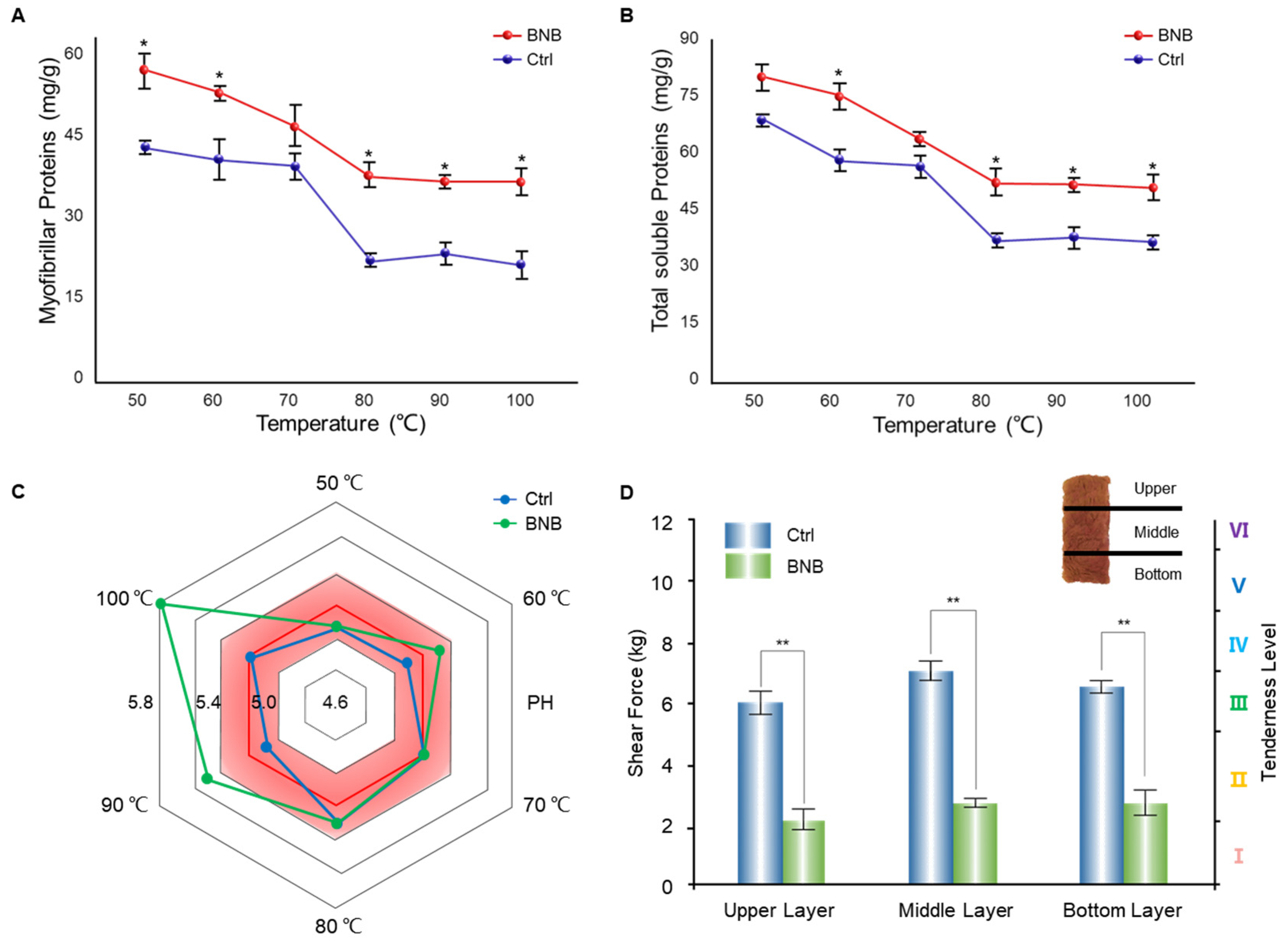
2.4. pH Measurement
2.5. Shear Force Measurement
2.6. Total Soluble Protein Content Measurement
2.7. Myosin Content Measurement
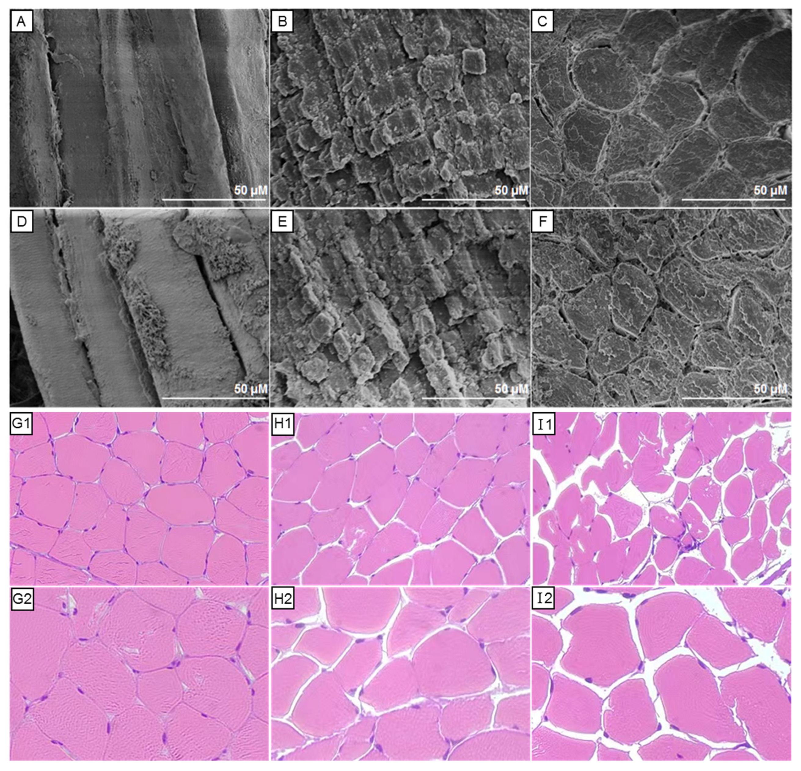
2.8. SEM and EDS
2.9. Histopathology
2.10. Statistical Analysis
3. Results
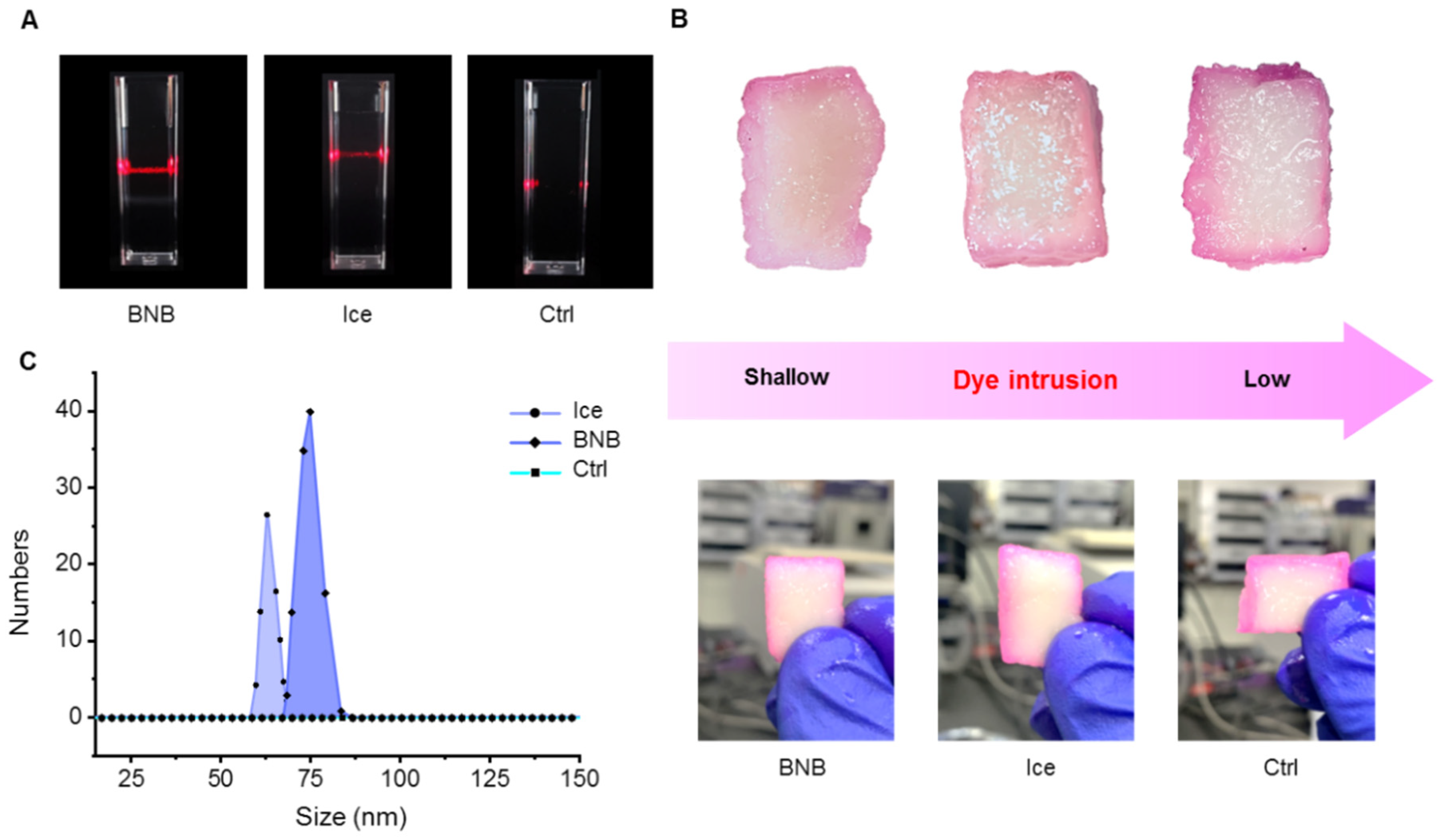
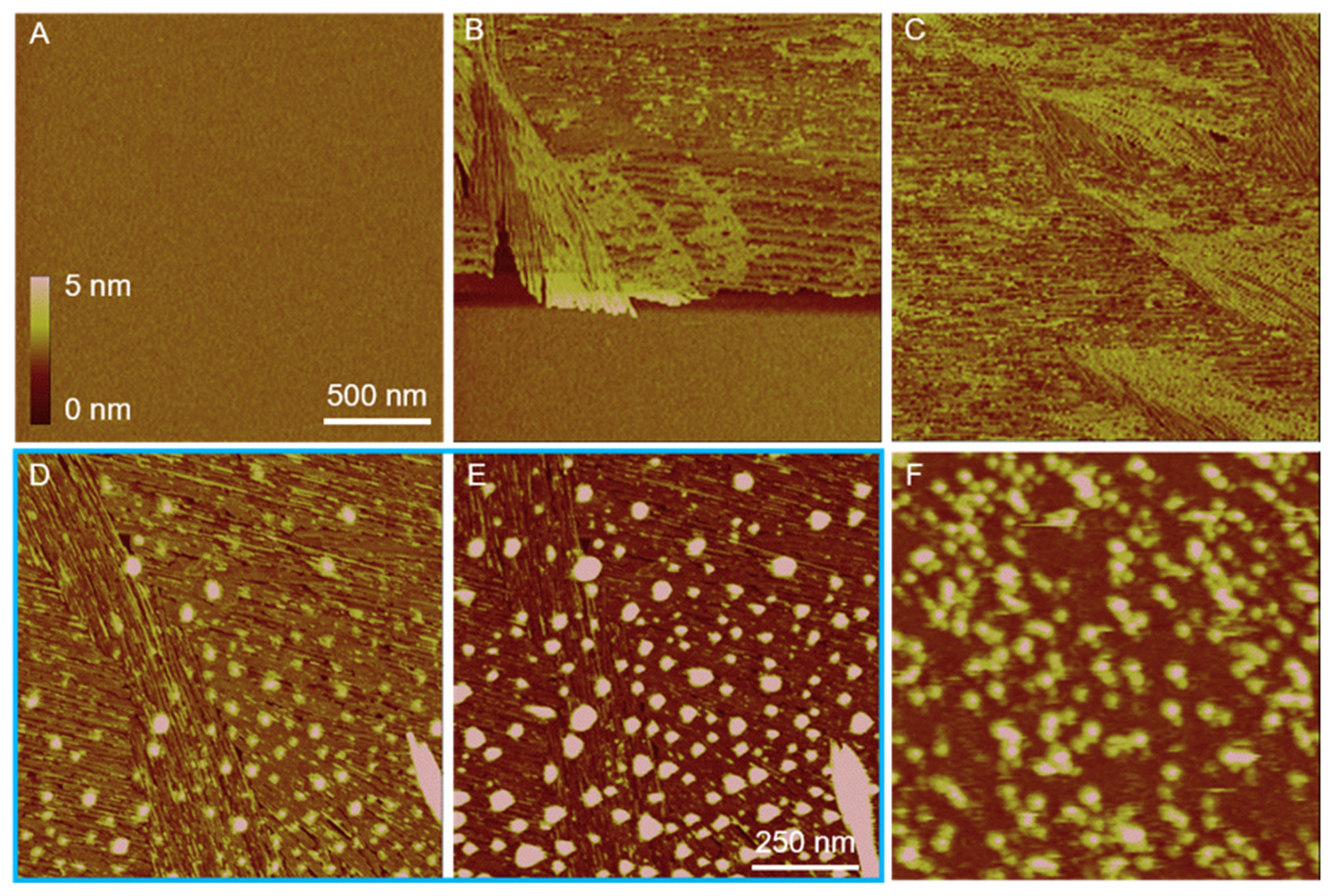
3.1. Freeze/Thaw-Induced BNBs
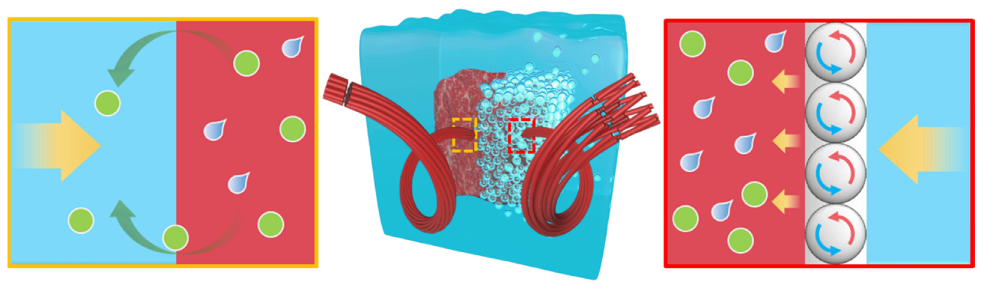
3.2. Protective Effect of Interfacial BNBs through ‘BNB Isolation Wall’
3.3. Analysis of Meat Microstructures
4. Discussion
Supplementary Materials
Author Contributions
Funding
Data Availability Statement
Acknowledgments
Conflicts of Interest
References
- Sevanto, S.; Holbrook, N.M.; Ball, M.C. Freeze/Thaw-induced embolism: Probability of critical bubble formation depends on speed of ice formation. Front. Plant Sci. 2012, 3, 107. [Google Scholar] [CrossRef] [Green Version]
- Parker, J.L.; Claesson, P.M.; Attard, P. Bubbles, cavities, and the long-ranged attraction between hydrophobic surfaces. J. Phys. Chem. 1994, 98, 8468–8480. [Google Scholar] [CrossRef]
- Temesgen, T.; Bui, T.T.; Han, M.; Kim, T.-I.; Park, H. Micro and nanobubble technologies as a new horizon for water-treatment techniques: A review. Adv. Colloid Interface Sci. 2017, 246, 40–51. [Google Scholar] [CrossRef] [PubMed]
- Borkent, B.M.; Dammer, S.M.; Schönherr, H.; Vancso, G.J.; Lohse, D. Superstability of surface nanobubbles. Phys. Rev. Lett. 2007, 98, 204502. [Google Scholar] [CrossRef] [PubMed] [Green Version]
- Seddon, J.R.; Kooij, E.S.; Poelsema, B.; Zandvliet, H.J.W.; Lohse, D. Surface bubble nucleation stability. Phys. Rev. Lett. 2011, 106, 056101. [Google Scholar] [CrossRef] [Green Version]
- Sun, Y.J.; Xie, G.Y.; Peng, Y.L.; Xia, W.; Sha, J. Stability theories of nanobubbles at solid-liquid interface: A review. Colloids Surf. A Physicochem. Eng. Asp. 2016, 495, 176–186. [Google Scholar] [CrossRef]
- Li, H.; Hu, L.; Xia, Z. Impact of groundwater salinity on bioremediation enhanced by micro-nano bubbles. Materials 2013, 6, 3676–3687. [Google Scholar] [CrossRef] [Green Version]
- Zhou, L.; Wang, X.; Shin, H.-J.; Wang, J.; Tai, R.; Zhang, X.; Fang, H.; Xiao, W.; Wang, L.; Wang, C.; et al. Ultrahigh Density of Gas Molecules Confined in Surface Nanobubbles in Ambient Water. J. Am. Chem. Soc. 2020, 142, 5583–5593. [Google Scholar] [CrossRef]
- Wang, Y.; Shen, Z.; Guo, Z.; Hu, J.; Zhang, Y. Effects of nanobubbles on peptide self-assembly. Nanoscale 2018, 10, 20007–20012. [Google Scholar] [CrossRef]
- Xie, X.; Yang, Y.; Lin, W.; Liu, H.; Liu, H.; Yang, Y.; Chen, Y.; Fu, X.; Deng, J. Cell-penetrating peptide-siRNA conjugate loaded YSA-modified nanobubbles for ultrasound triggered siRNA delivery. Colloids Surf. B Biointerfaces 2015, 136, 641–650. [Google Scholar] [CrossRef]
- Hernandez, C.; Gulati, S.; Fioravanti, G.; Stewart, P.L.; Exner, A.A. Cryo-EM Visualization of Lipid and Polymer-Stabilized Perfluorocarbon Gas Nanobubbles—A Step Towards Nanobubble Mediated Drug Delivery. Sci. Rep. 2017, 7, 13517. [Google Scholar] [CrossRef] [PubMed] [Green Version]
- Cavalli, R.; Soster, M.; Argenziano, M. Nanobubbles: A promising efficienft tool for therapeutic delivery. Ther. Deliv. 2016, 7, 117–138. [Google Scholar] [CrossRef] [PubMed]
- Gao, Y.; Hernandez, C.; Yuan, H.-X.; Lilly, J.; Kota, P.; Zhou, H.; Wu, H.; Exner, A.A. Ultrasound molecular imaging of ovarian cancer with CA-125 targeted nanobubble contrast agents. Nanomed. Nanotechnol. Biol. Med. 2017, 13, 2159–2168. [Google Scholar] [CrossRef] [PubMed]
- Bosca, F.; Bielecki, P.A.; Exner, A.A.; Barge, A. Porphyrin-Loaded Pluronic Nanobubbles: A New US-Activated Agent for Future Theranostic Applications. Bioconjug. Chem. 2018, 29, 234–240. [Google Scholar] [CrossRef]
- Yin, T.; Wang, P.; Li, J.; Zheng, R.; Zheng, B.; Cheng, D.; Li, R.; Lai, J.; Shuai, X. Ultrasound-sensitive siRNA-loaded nanobubbles formed by hetero-assembly of polymeric micelles and liposomes and their therapeutic effect in gliomas. Biomaterials 2013, 34, 4532–4543. [Google Scholar] [CrossRef]
- Jing, H.; Cheng, W.; Li, S.; Wu, B.; Leng, X.; Xu, S.; Tian, J. Novel cell-penetrating peptide-loaded nanobubbles synergized with ultrasound irradiation enhance EGFR siRNA delivery for triple negative Breast cancer therapy. Colloids Surf. B Biointerfaces 2016, 146, 387–395. [Google Scholar] [CrossRef] [PubMed]
- Bhat, Z.F.; Morton, J.D.; Mason, S.L.; Bekhit, A.E.D.A. Applied and emerging methods for meat tenderization: A comparative perspective. Compr. Rev. Food Sci. Food Saf. 2018, 17, 841–859. [Google Scholar] [CrossRef] [Green Version]
- Wang, X.; Wang, X.; Muhoza, B.; Feng, T.; Xia, S.; Zhang, X. Microwave combined with conduction heating effects on the tenderness, water distribution, and microstructure of pork belly. Innov. Food Sci. Emerg. Technol. 2020, 62, 102344. [Google Scholar] [CrossRef]
- Thomas, O.C.; Cavicchi, R.E.; Tarlov, M.J. Effect of surface wettability on fast transient microboiling behavior. Langmuir 2003, 19, 6168–6177. [Google Scholar] [CrossRef]
- Maheshwari, S.; van der Hoef, M.; Prosperetti, A.; Lohse, D. Dynamics of formation of a vapor nanobubble around a heated nanoparticle. J. Phys. Chem. C 2018, 122, 20571–20580. [Google Scholar] [CrossRef]
- Humphries, C. Cooking: Delicious science. Nature 2012, 486, S10–S11. [Google Scholar] [CrossRef]
- Barham, P.; Skibsted, L.H.; Bredie, W.L.; Bom Frøst, M.; Møller, P.; Risbo, J.; Snitkjær, P.; Mortensen, L.M. Molecular gastronomy: A new emerging scientific discipline. Chem. Rev. 2010, 110, 2313–2365. [Google Scholar] [CrossRef] [PubMed]
- Dhungana, P.; Bhandari, B. Development of a continuous membrane nanobubble generation method applicable in liquid food processing. Int. J. Food Sci. Technol. 2021, 56, 4268–4277. [Google Scholar] [CrossRef]
- Li, T.; Cui, Z.; Sun, J.; Jiang, C.; Li, G. Generation of Bulk Nanobubbles by Self-Developed Venturi-Type Circulation Hydrodynamic Cavitation Device. Langmuir 2021, 37, 12952–12960. [Google Scholar] [CrossRef] [PubMed]
- Marino, R.; Albenzio, M.; Della Malva, A.; Santillo, A.; Loizzo, P.; Sevi, A. Proteolytic pattern of myofibrillar protein and meat tenderness as affected by breed and aging time. Meat Sci. 2013, 95, 281–287. [Google Scholar] [CrossRef]
- Ježek, F.; Kameník, J.; Macharáčková, B.; Bogdanovičová, K.; Bednář, J. Cooking of meat: Effect on texture, cooking loss and microbiological quality—A review. Acta Vet. Brno 2019, 88, 487–496. [Google Scholar] [CrossRef] [Green Version]
- Weston, A.; Rogers, R.; Althen, T. The role of collagen in meat tenderness. Prof. Anim. Sci. 2002, 18, 107–111. [Google Scholar] [CrossRef]
- Malheiros, J.M.; Braga, C.P.; Grove, R.A.; Ribeiro, F.A.; Calkins, C.R.; Adamec, J.; Chardulo, L.A.L. Influence of oxidative damage to proteins on meat tenderness using a proteomics approach. Meat Sci. 2019, 148, 64–71. [Google Scholar] [CrossRef]
- Bertola, N.C.; Bevilacqua, A.E.; Zaritzky, N.E. Heat treatment effect on texture changes and thermal denaturation of proteins in beef muscle. J. Food Process. Preserv. 1994, 18, 31–46. [Google Scholar] [CrossRef]
- Wheeler, T.; Shackelford, S.; Koohmaraie, M. Shear Force Procedures for Meat Tenderness Measurement; Roman L. Hruska US Marc. USDA: Clay Center, NE, USA, 2001. [Google Scholar]
- Warner, R.; Miller, R.; Ha, M.; Wheeler, T.L.; Dunshea, F.; Li, X.; Vaskoska, R.; Purslow, P.; Wheeler, T. Meat tenderness: Underlying mechanisms, instrumental measurement, and sensory assessment. Meat Muscle Biol. 2021, 4, 1–25. [Google Scholar]
- Laville, E.; Sayd, T.; Terlouw, C.; Chambon, C.; Damon, M.; Larzul, C.; Leroy, P.; Glenisson, J.; Cherel, P. Comparison of sarcoplasmic proteomes between two groups of pig muscles selected for shear force of cooked meat. J. Agric. Food Chem. 2007, 55, 5834–5841. [Google Scholar] [CrossRef] [PubMed]
- Hopkins, D.; Hegarty, R.; Walker, P.; Pethick, D.W. Relationship between animal age, intramuscular fat, cooking loss, pH, shear force and eating quality of aged meat from sheep. Aust. J. Exp. Agric. 2006, 46, 879–884. [Google Scholar] [CrossRef]
- Thompson, J. Managing meat tenderness. Meat Sci. 2002, 62, 295–308. [Google Scholar] [CrossRef] [PubMed]
- Ertbjerg, P.; Puolanne, E. Muscle structure, sarcomere length and influences on meat quality: A review. Meat Sci. 2017, 132, 139–152. [Google Scholar] [CrossRef] [Green Version]
- Picard, B.; Gagaoua, M. Muscle Fiber Properties in Cattle and Their Relationships with Meat Qualities: An Overview. J. Agric. Food Chem. 2020, 68, 6021–6039. [Google Scholar] [CrossRef]
- Koohmaraie, M.; Kent, M.P.; Shackelford, S.D.; Veiseth, E.; Wheeler, T.L. Meat tenderness and muscle growth: Is there any relationship? Meat Sci. 2002, 62, 345–352. [Google Scholar] [CrossRef] [Green Version]
- D’Alessandro, A.; Zolla, L. Foodomics to investigate meat tenderness. TrAC Trends Anal. Chem. 2013, 52, 47–53. [Google Scholar] [CrossRef]


| Element | Concentration (wt. %) | |
|---|---|---|
| BNB | Ctrl | |
| N | 14.05 | 10.65 |
| Na | 34.97 | 24.12 |
| Ca | 0.77 | 0.33 |
| Cl | 32.07 | 22.13 |
Disclaimer/Publisher’s Note: The statements, opinions and data contained in all publications are solely those of the individual author(s) and contributor(s) and not of MDPI and/or the editor(s). MDPI and/or the editor(s) disclaim responsibility for any injury to people or property resulting from any ideas, methods, instructions or products referred to in the content. |
© 2023 by the authors. Licensee MDPI, Basel, Switzerland. This article is an open access article distributed under the terms and conditions of the Creative Commons Attribution (CC BY) license (https://creativecommons.org/licenses/by/4.0/).
Share and Cite
Si, Q.; Zhao, R.; Gao, F.; Guo, J.; Zhang, F.; Wang, L. Cooking Delicacy with Ice—Nanobubble Isolation Switches Stewing to ‘BBQ’. Nanomaterials 2023, 13, 562. https://doi.org/10.3390/nano13030562
Si Q, Zhao R, Gao F, Guo J, Zhang F, Wang L. Cooking Delicacy with Ice—Nanobubble Isolation Switches Stewing to ‘BBQ’. Nanomaterials. 2023; 13(3):562. https://doi.org/10.3390/nano13030562
Chicago/Turabian StyleSi, Qiankang, Ruoyang Zhao, Feng Gao, Jun Guo, Feng Zhang, and Liping Wang. 2023. "Cooking Delicacy with Ice—Nanobubble Isolation Switches Stewing to ‘BBQ’" Nanomaterials 13, no. 3: 562. https://doi.org/10.3390/nano13030562