
Progress in Nanostructured Mechano-Bactericidal Polymeric Surfaces for Biomedical Applications

 , , ,
, , , 
Abstract
:1. Introduction
2. Polymers Used in Biomedical Applications

| Polymer | Characteristics, Physical, and Mechanical Properties | Biomedical Applications | Advantages | Limitations | Ref. |
|---|---|---|---|---|---|
| PGA | Biodegradable, biocompatible, tensile stress: 890 MPa, density: 1.5 g/cm3, melting point: 225–230 °C, glass transition temperature: 35–40 °C | Tissue engineering applications in bone, tendon, cartilage, tooth, and spinal regeneration; nerve grafts; absorbable sutures | Stimulates cartilage regeneration; 3D-printability; high tensile strength | High friction coefficient and “binds and snags” when wet, high brittleness, high degradation causes inflammatory response | [26,27,58,59,60] |
| PLGA | Biodegradable, bioadsorbable, biocompatible, tensile stress: 3.4 MPa, density: 1.2 g/cm3, melting point: dependent on the percent composition (PLA, PGA), glass transition temperature: 40–60 °C | Therapeutic tools; drug delivery; tissue engineering | Stimulates osteoblasts; 3D-printability | Release of acidic byproducts leads to inflammation, degrade due to hydrolysis, poor strength | [26,28,29,30,61] |
| PCL | Biodegradable, bioadsorbable, biocompatible, tensile stress: 12.8 MPa, density: 1.15 g/cm3, melting point: 60 °C, glass transition temperature: −60 °C | Dental splints; drug delivery; tissue engineering | Stimulates osteoblasts; 3D printable; slow degradation rate; low cost in 3D printing due to low melting point; high biocompatibility | Poor mechanical properties; low cell adhesion | [26,31,62] |
| PU | Can be biodegradable or non-biodegradable based on chemical composition, non-bioabsorbable, biocompatible, tensile strength: 34.5–56 MPa, density: 1.23 g/cm3, melting point: 163 °C, glass transition temperature: −35 °C | Drug delivery; catheters, pacemaker leads insulation, vascular prostheses, heart valves, cardiac assist devices (cardiovascular applications) | High durability; high toughness; good biostability; low cost | Environmental stress cracking; material degradation in vivo; metal ion oxidation | [16,63,64,65] |
| PP | Non-biodegradable, non-bioabsorbable, biocompatible, tensile stress: 28 MPa, density: 0.9 g/cm3, melting point: 170 °C, glass transition temperature: −25 °C | Sutures; scaffolds (ligament or tendon repair); meshes for hernia and pelvic organ repair; heart valve structure, oxygenator and plasmapheresis membranes, finger joint prosthesis | High melting point; less toxic; low cost | Limited biocompatibility; poor strength | [16,33,66,67,68] |
| PVA | Biodegradable, biocompatible, tensile stress: 40–90 MPa, density: 1.26 g/cm3, melting point: 228 °C, glass transition temperature: 85 °C | Wound dressings, drug delivery, targeted-tissue transportation systems; soft biomaterial implants. | High chemical and thermal stability; non-toxic | Weak hydrogel endurance in high temperature; relatively weak polymer; limited biocompatibility; degrades due to hydrolysis | [34,69,70,71,72] |
| Silicone or PDMS | Non-absorbable, non-biodegradable, biocompatible, hydrophobic, tensile stress: 2–10 MPa, density: 0.97 g/cm3, melting point: 228 °C glass transition temperature: ~120–123 °C | Oxygenator membrane; tubing; shunts; prostheses; heart peacemaker leads; heart valve structures; burn dressing | Chemically inert; low toxicity; thermal stability; high biocompatibility | Prone to damage; non-durable; contamination of monomers; low mechanical strength | [16,47,63] |
| PLA | Biodegradable, bioabsorbable, biocompatible, tensile stress: 21–60 MPa, density: 1.21–1.25 g/cm3, melting point: 150–160 °C, glass transition temperature: 60–65 °C | Bone tissue engineering; drug delivery; plates, screws, pins, and wires in bone fixation; bio-absorbable implants; sutures in dermatology; drug-eluting stents | High biocompatibility; stimulates osteoblasts; less brittle; one of the highly used 3D-printable materials; degradation products are also non-toxic to humans and the environment. | Low mechanical strength | [26,33,39,40,66,67,73] |
| PMMA | Non-degradable, biocompatible, tensile stress: 48–76 MPa, density: 1.2 g/cm3, melting point: 130–180 °C, glass transition temperature: 80 °C | Dental implants; bone cement; lenses; drug delivery | One of the hardest thermoplastics with high scratch resistance; high mechanical strength | Less biocompatibility; high curing temperature; does not support osteointegration; causes necrosis effect | [16,74,75] |
| PEEK | Non-degradable, biocompatible, tensile stress: 84 MPa, density: 1.4 g/cm3, melting point: 343 °C, glass transition temperature: 143 °C | Dental implants; knee implants; spine implants; cranioplasticity; hip replacement; anterior plate fixation; heart valves; face reconstructions | High biocompatibility; 3D-printable; light weight; compatible with hydroxyapatite (natural bone tissue materials) hence substitute to metallic implants; stable at high temperatures; mechanical stability | Low thermoformability; bioinert (does not promote tissue integration); complex and costly manufacturing process | [76,77,78] |
| PEKK | Non-degradable, biocompatible, tensile stress: 115 MPa, density: 1.3 g/cm3, melting point: 363–386 °C, glass transition temperature: 162 °C | Dental implants; crown and bridge in dentistry; endodontic post; removable denture framework; restorative and prosthetic applications | High biocompatibility; 3D-printable; light weight; high mechanical strength; excellent chemical resistance | Bioinert (does not promote tissue integration); more complex and costly manufacturing process than PEEK | [52,79] |
| PET | Non-degradable, high biocompatibility, tensile stress: 75–100 MPa, density:1.38 g/cm3, melting point: 255–265 °C, glass transition temperature: 85 °C | Sutures; heart valves; surgical meshes; scaffolds; urinary and bloodstream catheters; commercial vascular prosthesis | 3D-printable; cost effective; excellent chemical resistance | Bioinert (does not promote tissue integration) | [80,81,82] |
| PTFE | Non-degradable, biocompatible, tensile stress: 30.5 MPa, density: 2.175 g/cm3, melting point: 327 °C, glass transition temperature: 127 °C | Vascular graft prostheses; heart patches; stapes prosthesis | High mechanical strength; chemically inert | Difficult to 3D-print | [16,83] |
| Chitosan | Biodegradable, biocompatible, tensile stress: 32.2 MPa, density: 0.20–0.38 g/cm3, melting point:105 °C, glass transition temperature: 75 °C | Antitumor drug delivery; protein and peptide drug delivery; gene delivery; antibiotic delivery; polyphenol delivery; wound healing applications | Antimicrobial; anti-inflammatory; antifungal; nontoxicity; antitumor activity; antioxidant activity | Low mechanical strength; significant variations of properties based on the source of material | [84,85,86,87,88,89,90] |
3. Mechanism of Bacteria Adhesion on Surfaces
4. Antimicrobial Strategies
4.1. Chemical Bactericidal Strategies
4.2. Physical/Mechanical Bactericidal Strategies
4.2.1. Natural Bactericidal Nanostructured Surfaces

| Scheme | Nanofeature Type | Nanofeature Arrangement | Dimensions of the Nanofeature | Wettability | Antimicrobial Activity | Ref. | |||
|---|---|---|---|---|---|---|---|---|---|
| Tip Diameter (nm) | Base Diameter (nm) | Height (nm) | Spacing (nm) | ||||||
| Periodical cicada wing (Magicicada ssp.) | Hemispherical shape nanofeatures | Ordered | 167 | 167 | 83.5 | 252 | ~80° | Lethal to S. cerevisiae fungus | [154] |
| Dog day annual cicada (Tibicen ssp.) | Nanoneedles (spherically capped, conical, nanoscale pillars) | Random/hierarchical | 57 | 104 | 183 | 175 | 132° | Bactericidal to P. aeruginosa (Gram-negative) | [154] |
| Cicada wing (Psaltoda claripennis) | Nanopillars | Ordered | 60 | 100 | 200 | 170 | ~159° | Bactericidal to P. aeruginosa (Gram-negative) | [145] |
| Cicada wing (Megapomponia intermedia) | Tubular nanofeatures (nanopillars) | Ordered | 156 | ~312 | 241 | 165 | ~136° | Bactericidal to P. fluorescens (Gram-negative) | [101] |
| Cicada wing (Cryptotympana Aguila) | Tubular nanofeatures (nanopillars) | Ordered | 159 | ~318 | 182 | 187 | ~113° | Bactericidal to P. fluorescens (Gram-negative) | [101] |
| Cicada wing (Ayuthia spectabile) | Tubular nanofeatures (nanopillars) | Ordered | 207 | ~414 | 182 | 251 | ~96° | Bactericidal to P. fluorescens (Gram-negative) | [101] |
| Sanddragon dragonfly wing (Progomphus obscurus) | Nanograss | Random/hierarchical | 53 | - | 241 | 123 | 119° | Bactericidal to P. aeruginosa (Gram-negative), S. aureus and B. subtilis (Gram-positive) | [154] |
| Dragonfly wing (D. bipunctata) | Nanograss | Random/hierarchical | 50–70 | - | 240 | - | 153° | Bactericidal to P. aeruginosa (Gram-negative), S. aureus and B. subtilis (Gram-positive) | [146] |
| Dragonfly wing (Orthetrum villosovittatum) | Nanopillar | Random/hierarchical | 37 and 57 | - | 189 and 311 | - | - | Bactericidal to E. coli (Gram-negative) | [148] |
| Gecko skin (Strophurus williamsii) | Spinules | Random/hierarchical | 50 | ~400 | 3000 | 500 | >136° | - | [110] |
| Gecko skin (Lucasium steindachneri) | Spinules | Random/hierarchical | - | - | 2000–4000 | ~500 | 150° | Bactericidal to Streptococcus mutans (Gram-positive) and Porphyromonas gingivalis (Gram-negative) | [102,131] |
| Lotus leaves (Nelumbo nucifera) | Nanotubules | Random/hierarchical | - | 100–200 | 300–1100 | ~150–250 | 154° | Bactericidal to E. coli (Gram-negative) | [157] |
| Damselfly wing (Calopteryx haemorrhoidalis) | Nanopillars | Random/hierarchical | ~48 | - | ~433 | 116 | 157° | Bactericidal to P. aeruginosa (Gram-negative) and S. aureus (Gram-positive) | [149] |
4.2.2. Artificial Nanostructure Fabrication Methods in Polymers
Reactive Ion Etching
Colloidal Lithography
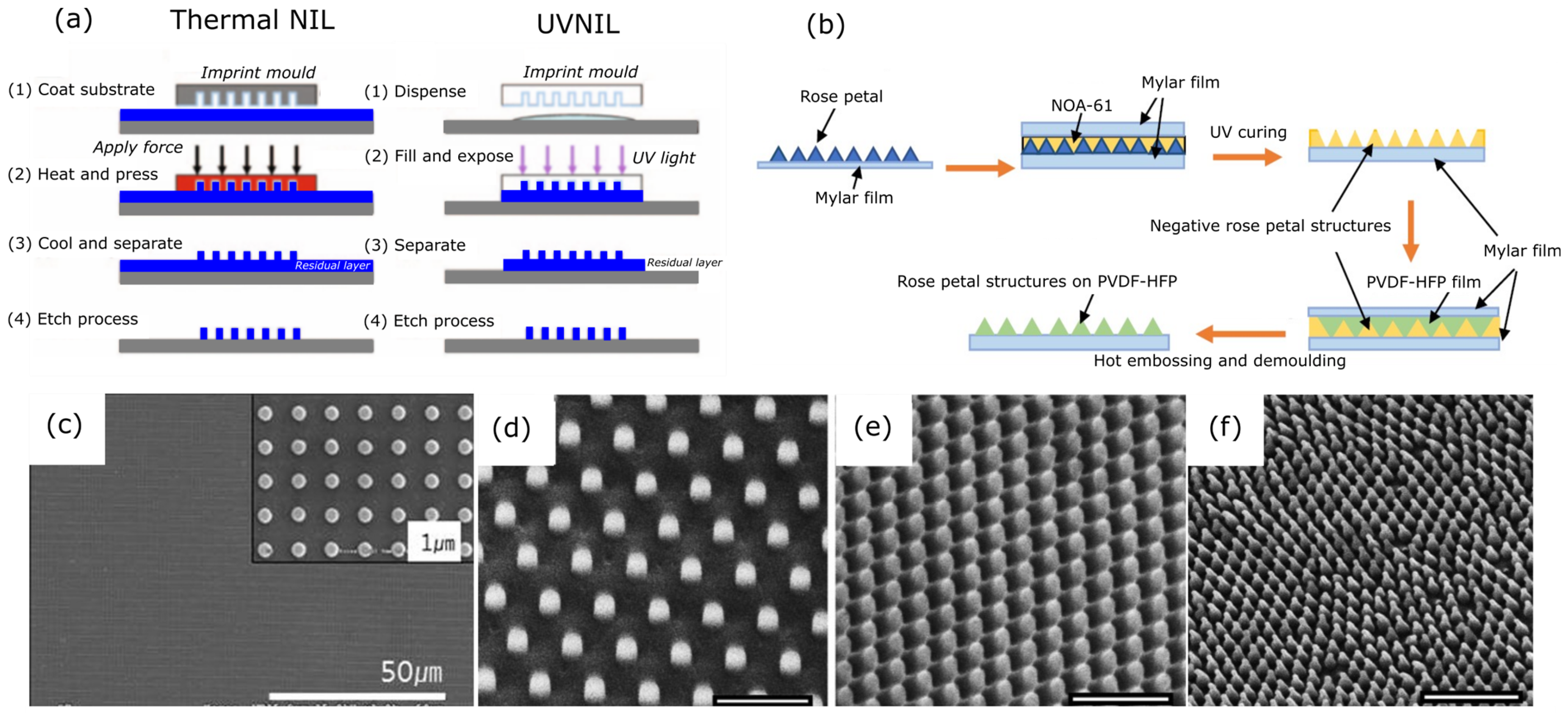
Nanoimprinting Lithography
Laser-Based Lithography Techniques

Anodic Aluminium Oxide Template
Electron Beam Lithography (EBL)
Hydrothermal Synthesis
| Method | Substrate Material | Supportive Processes | Feature Type | Nanofeature Parameters | Bactericidal Effect (Max. Efficiency %, Incubation Time in Hours) | Ref. | |
|---|---|---|---|---|---|---|---|
| NIL | PMMA | Silicon mould preparation | Nanopillars | H | 210 nm, 300 nm | Lethal to E. coli bacteria (~50%, 24 h) | [57] |
| Dt | 70 nm, 190 nm, 215 nm | ||||||
| Db | 100 nm, 130 nm, 380 nm | ||||||
| S | 170 nm, 320 nm, 595 nm | ||||||
| OrmoStamp (glass) | AAOT, deep RIE | Nanopillars | H | 200, 300, 400 nm | Lethal to S. aureus bacteria (~100%, 0.5 h) | [207] | |
| D | 70, 80 nm | ||||||
| PD | 40 µm2, 70 µm2 | ||||||
| PEGDMA | RIE | Nanoneedle | H | 300 nm | Lethal to E. coli bacteria (N/A) | [208] | |
| Dt | 50 nm | ||||||
| Db | 200 nm | ||||||
| P | 500 nm | ||||||
| PVDF-HFP | Hydrothermal method | Array of micropapillae with nanofold structures | D (papillae) | 20 µm | Lethal to Gram-positive S. agalactiae: Micropapillae (~97%, 12 h), Nanopillars (100%, 12 h); Gram-negative E. coli, Micropapillae (~100%, 12 h), Nanopillars (100%, 12 h) | [179] | |
| D (nanofold) | 300–400 nm | ||||||
| D | 300 nm | ||||||
| P | 600 nm | ||||||
| PC | RIE, EBL | Nanopillar | H | 25 nm | Lethal to E. coli (N/A) | [201] | |
| Db | 40 nm | ||||||
| S | 100, 200, 500 nm | ||||||
| Laser Lithography | PMMA | Thermal oxidation, RIE, chemical vapour deposition, TNIL | Nanopillars | H | 450 nm | Lethal to P. aeruginosa (~70%, 8 h) and E. coli (~70%, 8 h) | [176] |
| Db | 250 nm | ||||||
| AR | 3 | ||||||
| LIL | PS | None | Both pillar-like and lamella-shaped patterns | H | 2 µm | Lethal and anti-adhesion under flow conditions to S. aureus | [191] |
| W | 3 µm | ||||||
| S | 3 µm | ||||||
| Colloidal lithography | PET | RIE | Nanocones | H | 530–350 nm | Lethal to E. coli (~30%, 1 h) and K. pneumoniae (~30%, 1 h) | [161] |
| Dt | 20–300 nm | ||||||
| Db | 55–380 nm | ||||||
| AR | 1–7 | ||||||
| PEEK | RIE | Nanopillars, nanocones, micropillars, micro-cones | H | 350 nm | Lethal to E. coli: Nanopillars (>95%, 12 h), Micropillars (~90%, 12 h), Micro-cones (>75%, 12 h) | [162] | |
| P | 200, 250 nm | ||||||
| D | 100, 50 nm | ||||||
| AAOT | Polymer made from DGEBA and E230 | Wet etching, dip coating | Nanopillars | H | 500–600 nm | Lethal to P. aeruginosa (~99%, 3 h) and S. aureus (~99%, 3 h) | [196] |
| Dt | 100 nm | ||||||
| Db | 350 nm | ||||||
| PC | Hot embossing and wet etching | Nanopillars | H | 143–408 nm | Lethal to E. coli bacteria (>95%, 3 h) | [195] | |
| Dt | 26.7–33.4 nm | ||||||
| Db | 66.3–154.3 nm | ||||||
| S | 100–307.8 nm | ||||||
| RIE | PP | None | Nanofibrils | H | 0.5, 1 µm | Lethal to E. coli (~99.6%, 24 h) | [171] |
| D | 30, 40 nm | ||||||
| CPN | None | Nanopillars | H | 1–1.4 µm | Lethal to P. aeruginosa (~80%, 24 h) | [160] | |
| R | 0.92–1.42 µm | ||||||
| PET | None | Nanopillars | H | 128.67 nm | Lethal to P. aeruginosa (~99%, 24 h) | [172] | |
| D | 21.7 nm | ||||||
| Nanowires | H | 501.4 nm | |||||
| D | 29.54 nm | ||||||
5. Factors Affecting Bactericidal Activity of Mechano-Bactericidal Mechanism
6. Conclusions and Outlook
Author Contributions
Funding
Data Availability Statement
Acknowledgments
Conflicts of Interest
References
- Donlan, R.M. Biofilms: Microbial Life on Surfaces. Emerg. Infect. Dis. 2002, 8, 881–890. [Google Scholar] [CrossRef]
- Davies, D. Understanding Biofilm Resistance to Antibacterial Agents. Nat. Rev. Drug Discov. 2003, 2, 114–122. [Google Scholar] [CrossRef] [PubMed]
- Hernando-Amado, S.; Coque, T.M.; Baquero, F.; Martínez, J.L. Defining and Combating Antibiotic Resistance from One Health and Global Health Perspectives. Nat. Microbiol. 2019, 4, 1432–1442. [Google Scholar] [CrossRef] [PubMed]
- Li, B.; Webster, T.J. Bacteria Antibiotic Resistance: New Challenges and Opportunities for Implant-Associated Orthopedic Infections. J. Orthop. Res. 2017, 36, 22–32. [Google Scholar] [CrossRef] [PubMed]
- Varela, M.F.; Stephen, J.; Lekshmi, M.; Ojha, M.; Wenzel, N.; Sanford, L.M.; Hernandez, A.J.; Parvathi, A.; Kumar, S.H. Bacterial Resistance to Antimicrobial Agents. Antibiotics 2021, 10, 593. [Google Scholar] [CrossRef] [PubMed]
- Ashtiani, R.E.; Alam, M.; Tavakolizadeh, S.; Abbasi, K. The Role of Biomaterials and Biocompatible Materials in Implant-Supported Dental Prosthesis. Complement. Alternat. Med. 2021, 2021, 3349433. [Google Scholar]
- Santos, G.A. The Importance of Metallic Materials as Biomaterials. Adv. Tissue Eng. Regen. Med. Open Access 2017, 3, 200–302. [Google Scholar] [CrossRef]
- Klinke, T.; Daboul, A.; Maron, J.; Gredes, T.; Puls, R.; Jaghsi, A.; Biffar, R. Artifacts in Magnetic Resonance Imaging and Computed Tomography Caused by Dental Materials. PLoS ONE 2012, 7, e31766. [Google Scholar] [CrossRef]
- Prakasam, M.; Locs, J.; Salma-Ancane, K.; Loca, D.; Largeteau, A.; Berzina-Cimdina, L. Biodegradable Materials and Metallic Implants-A Review. J. Funct. Biomater. 2017, 8, 44. [Google Scholar] [CrossRef]
- Shekhawat, D.; Singh, A.; Bhardwaj, A.; Patnaik, A. A Short Review on Polymer, Metal and Ceramic Based Implant Materials. IOP Conf. Ser. Mater. Sci. Eng. 2021, 1017, 012038. [Google Scholar] [CrossRef]
- Mahanty, A.; Shikha, D. Changes in the Morphology, Mechanical Strength and Biocompatibility of Polymer and Metal/Polymer Fabricated Hydroxyapatite for Orthopaedic Implants: A Review. J. Polym. Eng. 2022, 42, 298–322. [Google Scholar] [CrossRef]
- Peschke, E.; Ulloa, P.; Jansen, O.; Hoevener, J.-B. Metallic Implants in MRI—Hazards and Imaging Artifacts. Rofo 2021, 193, 1285–1293. [Google Scholar] [CrossRef] [PubMed]
- Hargreaves, B.A.; Worters, P.W.; Pauly, K.B.; Pauly, J.M.; Koch, K.M.; Gold, G.E. Metal-Induced Artifacts in MRI. AJR Am. J. Roentgenol. 2011, 197, 547–555. [Google Scholar] [CrossRef] [PubMed]
- Ramakrishna, S.; Mayer, J.; Wintermantel, E.; Leong, K.W. Biomedical Applications of Polymer-Composite Materials: A Review. Compos. Sci. Technol. 2001, 61, 1189–1224. [Google Scholar] [CrossRef]
- Jaganathan, S.K.; Supriyanto, E.; Murugesan, S.; Balaji, A.; Asokan, M.K. Biomaterials in Cardiovascular Research: Applications and Clinical Implications. Biomed Res. Int. 2014, 2014, 459465. [Google Scholar] [CrossRef] [PubMed]
- Teo, A.J.T.; Mishra, A.; Park, I.; Kim, Y.-J.; Park, W.-T.; Yoon, Y.-J. Polymeric Biomaterials for Medical Implants and Devices. ACS Biomater. Sci. Eng. 2016, 2, 454–472. [Google Scholar] [CrossRef] [PubMed]
- Bozkurt, Y.; Karayel, E. 3D Printing Technology; Methods, Biomedical Applications, Future Opportunities and Trends. J. Mater. Res. Technol. 2021, 14, 1430–1450. [Google Scholar] [CrossRef]
- Gonzalez, G.; Roppolo, I.; Pirri, C.F.; Chiappone, A. Current and Emerging Trends in Polymeric 3D Printed Microfluidic Devices. Addit. Manuf. 2022, 55, 102867. [Google Scholar] [CrossRef]
- Kalirajan, C.; Dukle, A.; Nathanael, A.J.; Oh, T.-H.; Manivasagam, G. A Critical Review on Polymeric Biomaterials for Biomedical Applications. Polymers 2021, 13, 3015. [Google Scholar] [CrossRef]
- Senevirathne, S.W.M.A.I.; Hasan, J.; Mathew, A.; Woodruff, M.; Yarlagadda, P.K.D.V. Bactericidal Efficiency of Micro- and Nanostructured Surfaces: A Critical Perspective. RSC Adv. 2021, 11, 1883–1900. [Google Scholar] [CrossRef]
- Modaresifar, K.; Azizian, S.; Ganjian, M.; Fratila-Apachitei, L.E.; Zadpoor, A.A. Bactericidal Effects of Nanopatterns: A Systematic Review. Acta Biomater. 2019, 83, 29–36. [Google Scholar] [CrossRef] [PubMed]
- Slepicka, P.; Kasalkova, N.S.; Siegel, J.; Kolska, Z.; Bacakova, L.; Svorcik, V. Nano-Structured and Functionalized Surfaces for Cytocompatibility Improvement and Bactericidal Action. Biotechnol. Adv. 2015, 33, 1120–1129. [Google Scholar] [CrossRef] [PubMed]
- Patil, D.; Overland, M.; Stoller, M.; Chatterjee, K. Bioinspired Nanostructured Bactericidal Surfaces. Curr. Opin. Chem. Eng. 2021, 34, 100741. [Google Scholar] [CrossRef]
- Tripathy, A.; Sen, P.; Su, B.; Briscoe, W.H. Natural and Bioinspired Nanostructured Bactericidal Surfaces. Adv. Colloid Interface Sci. 2017, 248, 85–104. [Google Scholar] [CrossRef] [PubMed]
- Al-Shalawi, F.D.; Mohamed Ariff, A.H.; Jung, D.-W.; Mohd Ariffin, M.K.A.; Seng Kim, C.L.; Brabazon, D.; Al-Osaimi, M.O. Biomaterials as Implants in the Orthopedic Field for Regenerative Medicine: Metal versus Synthetic Polymers. Polymers 2023, 15, 2601. [Google Scholar] [CrossRef] [PubMed]
- Asghari, F.; Samiei, M.; Adibkia, K.; Akbarzadeh, A.; Davaran, S. Biodegradable and Biocompatible Polymers for Tissue Engineering Application: A Review. Artif. Cells Nanomed. Biotechnol. 2017, 45, 185–192. [Google Scholar] [CrossRef] [PubMed]
- Mi, H.-Y.; Salick, M.R.; Jing, X.; Jacques, B.R.; Crone, W.C.; Peng, X.-F.; Turng, L.-S. Characterization of Thermoplastic Polyurethane/Polylactic Acid (TPU/PLA) Tissue Engineering Scaffolds Fabricated by Microcellular Injection Molding. Mater. Sci. Eng. C Mater. Biol. Appl. 2013, 33, 4767–4776. [Google Scholar] [CrossRef] [PubMed]
- Qian, J.; Xu, W.; Yong, X.; Jin, X.; Zhang, W. Fabrication and in Vitro Biocompatibility of Biomorphic PLGA/NHA Composite Scaffolds for Bone Tissue Engineering. Mater. Sci. Eng. C Mater. Biol. Appl. 2014, 36, 95–101. [Google Scholar] [CrossRef]
- Badaraev, A.D.; Sidelev, D.V.; Yurjev, Y.N.; Bukal, V.R.; Tverdokhlebov, S.I. Modes Development of PLGA Scaffolds Modification by Magnetron Co-Sputtering of Cu and Ti Targets. J. Phys. Conf. Ser. 2021, 1799, 012001. [Google Scholar] [CrossRef]
- Sun, F.; Sun, X.; Wang, H.; Li, C.; Zhao, Y.; Tian, J.; Lin, Y. Application of 3D-Printed, PLGA-Based Scaffolds in Bone Tissue Engineering. Int. J. Mol. Sci. 2022, 23, 5831. [Google Scholar] [CrossRef]
- Maheshwari, U.; Samuel, S.; Nagiah, V.K. Fabrication and Evaluation of (PVA/HAp/PCL) Bilayer Composites as Potential Scaffolds for Bone Tissue Regeneration Application. Ceram Int. 2014, 40, 8469–8477. [Google Scholar] [CrossRef]
- Hajiali, F.; Tajbakhsh, S.; Shojaei, A. Fabrication and Properties of Polycaprolactone Composites Containing Calcium Phosphate-Based Ceramics and Bioactive Glasses in Bone Tissue Engineering: A Review. Polym. Rev. 2018, 58, 164–207. [Google Scholar] [CrossRef]
- Greene, J.P. Microstructures of Polymers. In Automotive Plastics and Composites; Elsevier: Amsterdam, The Netherlands, 2021; pp. 27–37. [Google Scholar]
- Gaaz, T.S.; Sulong, A.B.; Akhtar, M.N.; Kadhum, A.A.H.; Mohamad, A.B.; Al-Amiery, A.A. Properties and Applications of Polyvinyl Alcohol, Halloysite Nanotubes and Their Nanocomposites. Molecules 2015, 20, 22833–22847. [Google Scholar] [CrossRef] [PubMed]
- Jiang, S.; Liu, S.; Feng, W. PVA Hydrogel Properties for Biomedical Application. J. Mech. Behav. Biomed. Mater. 2011, 4, 1228–1233. [Google Scholar] [CrossRef] [PubMed]
- Nkhwa, S.; Lauriaga, K.F.; Kemal, E.; Deb, S. Poly(Vinyl Alcohol): Physical Approaches to Designing Biomaterials for Biomedical Applications. Conf. Pap. Sci. 2014, 2014, 403472. [Google Scholar] [CrossRef]
- Zare, M.; Ghomi, E.R.; Venkatraman, P.D.; Ramakrishna, S. Silicone-based Biomaterials for Biomedical Applications: Antimicrobial Strategies and 3D Printing Technologies. J. Appl. Polym. Sci. 2021, 138, 50969. [Google Scholar] [CrossRef]
- Ding, K.; Wang, Y.; Liu, S.; Wang, S.; Mi, J. Preparation of Medical Hydrophilic and Antibacterial Silicone Rubber via Surface Modification. RSC Adv. 2021, 11, 39950–39957. [Google Scholar] [CrossRef] [PubMed]
- Carvalho, J.R.G.; Conde, G.; Antonioli, M.L.; Santana, C.H.; Littiere, T.O.; Dias, P.P.; Chinelatto, M.A.; Canola, P.A.; Zara, F.J.; Ferraz, G.C. Long-Term Evaluation of Poly (Lactic Acid) (PLA) Implants in a Horse: An Experimental Pilot Study. Molecules 2021, 26, 7224. [Google Scholar] [CrossRef]
- Singhvi, M.S.; Zinjarde, S.S.; Gokhale, D.V. Polylactic Acid: Synthesis and Biomedical Applications. J. Appl. Microbiol. 2019, 127, 1612–1626. [Google Scholar] [CrossRef]
- Casalini, T.; Rossi, F.; Castrovinci, A.; Perale, G. A Perspective on Polylactic Acid-Based Polymers Use for Nanoparticles Synthesis and Applications. Front. Bioeng. Biotechnol. 2019, 7, 259. [Google Scholar] [CrossRef]
- Chen, T.; Zhao, X.; Weng, Y. Self-Assembled Polylactic Acid (PLA): Synthesis, Properties and Biomedical Applications. Front. Chem. 2022, 10, 1107620. [Google Scholar] [CrossRef]
- Ebrahimi, F.; Ramezani Dana, H. Poly Lactic Acid (PLA) Polymers: From Properties to Biomedical Applications. Int. J. Polym. Mater. 2022, 71, 1117–1130. [Google Scholar] [CrossRef]
- Pawar, R.P.; Tekale, S.U.; Shisodia, S.U.; Totre, J.T.; Domb, J.A. Biomedical Applications of Poly (Lactic Acid). Rec. Pat. Regen. Med. 2014, 4, 40–51. [Google Scholar] [CrossRef]
- Ludwicka, K.; Kolodziejczyk, M.; Gendaszewska-Darmach, E.; Chrzanowski, M.; Jedrzejczak-Krzepkowska, M.; Rytczak, P.; Bielecki, S. Stable Composite of Bacterial Nanocellulose and Perforated Polypropylene Mesh for Biomedical Applications. J. Biomed. Mater. Res. B Appl. Biomater. 2019, 107, 978–987. [Google Scholar] [CrossRef] [PubMed]
- Acik, G.; Altinkok, C.; Acik, B. Biodegradable and Antibacterial Chlorinated Polypropylene/Chitosan Based Composite Films for Biomedical Applications. Polym. Bull. 2022, 79, 9997–10011. [Google Scholar] [CrossRef]
- Ali, U.; Karim, K.J.B.A.; Buang, N.A. A Review of the Properties and Applications of Poly (Methyl methacrylate) (PMMA). Polym. Rev. 2015, 55, 678–705. [Google Scholar] [CrossRef]
- Frazer, R.Q.; Byron, R.T.; Osborne, P.B.; West, K.P. PMMA: An Essential Material in Medicine and Dentistry. J. Long Term Eff. Med. Implants 2005, 15, 629–639. [Google Scholar] [CrossRef] [PubMed]
- Díez-Pascual, A.M. PMMA-Based Nanocomposites for Odontology Applications: A State-of-the-Art. Int. J. Mol. Sci. 2022, 23, 10288. [Google Scholar] [CrossRef] [PubMed]
- Leigh, J.A. Use of PMMA in Expansion Dental Implants. J. Biomed. Mater. Res. 1975, 9, 233–242. [Google Scholar] [CrossRef]
- Wang, H.; Lu, T.; Meng, F.; Zhu, H.; Liu, X. Enhanced Osteoblast Responses to Poly Ether Ether Ketone Surface Modified by Water Plasma Immersion Ion Implantation. Colloids Surf. B Biointerfaces 2014, 117, 89–97. [Google Scholar] [CrossRef]
- Alqurashi, H.; Khurshid, Z.; Syed, A.U.Y.; Rashid Habib, S.; Rokaya, D.; Zafar, M.S. Polyetherketoneketone (PEKK): An Emerging Biomaterial for Oral Implants and Dental Prostheses. J. Adv. Res. 2021, 28, 87–95. [Google Scholar] [CrossRef] [PubMed]
- Pietrocola, G.; Campoccia, D.; Motta, C.; Montanaro, L.; Arciola, C.R.; Speziale, P. Colonization and Infection of Indwelling Medical Devices by Staphylococcus Aureus with an Emphasis on Orthopedic Implants. Int. J. Mol. Sci. 2022, 23, 5958. [Google Scholar] [CrossRef] [PubMed]
- Ardila, C.M.; Vivares-Builes, A.M. Antibiotic Resistance in Patients with Peri-Implantitis: A Systematic Scoping Review. Int. J. Environ. Res. Public Health 2022, 19, 15609. [Google Scholar] [CrossRef] [PubMed]
- Momand, P.; Becktor, J.P.; Naimi-Akbar, A.; Tobin, G.; Götrick, B. Effect of Antibiotic Prophylaxis in Dental Implant Surgery: A Multicenter Placebo-controlled Double-blinded Randomized Clinical Trial. Clin. Implant Dent. Relat. Res. 2022, 24, 116–124. [Google Scholar] [CrossRef] [PubMed]
- Rezić, I.; Majdak, M.; Ljoljić Bilić, V.; Pokrovac, I.; Martinaga, L.; Somogyi Škoc, M.; Kosalec, I. Development of Antibacterial Protective Coatings Active against MSSA and MRSA on Biodegradable Polymers. Polymers 2021, 13, 659. [Google Scholar] [CrossRef] [PubMed]
- Dickson, M.N.; Liang, E.I.; Rodriguez, L.A.; Vollereaux, N.; Yee, A.F. Nanopatterned Polymer Surfaces with Bactericidal Properties. Biointerphases 2015, 10, 021010. [Google Scholar] [CrossRef] [PubMed]
- Trott, A.T. Instruments, Suture Materials, and Closure Choices. In Wounds and Lacerations; Elsevier: Amsterdam, The Netherlands, 2012; pp. 82–94. [Google Scholar]
- Palispis, W.A.; Gupta, R. Biologic Augmentation in Peripheral Nerve Repair. In Biologics in Orthopaedic Surgery; Elsevier: Amsterdam, The Netherlands, 2019; pp. 141–163. [Google Scholar]
- Manoukian, O.S.; Sardashti, N.; Stedman, T.; Gailiunas, K.; Ojha, A.; Penalosa, A.; Mancuso, C.; Hobert, M.; Kumbar, S.G. Biomaterials for Tissue Engineering and Regenerative Medicine. In Encyclopedia of Biomedical Engineering; Elsevier: Amsterdam, The Netherlands, 2019; pp. 462–482. [Google Scholar]
- Houchin, M.L.; Topp, E.M. Physical Properties of PLGA Films during Polymer Degradation. J. Appl. Polym. Sci. 2009, 114, 2848–2854. [Google Scholar] [CrossRef]
- Vignali, A.; Iannace, S.; Falcone, G.; Utzeri, R.; Stagnaro, P.; Bertini, F. Lightweight Poly(ε-Caprolactone) Composites with Surface Modified Hollow Glass Microspheres for Use in Rotational Molding: Thermal, Rheological and Mechanical Properties. Polymers 2019, 11, 624. [Google Scholar] [CrossRef]
- Zhou, Z.-M.; Wang, K.; Lin, K.-W.; Wang, Y.-H.; Li, J.-Z. Influence of Characteristics of Thermoplastic Polyurethane on Graphene-Thermoplastic Polyurethane Composite Film. Micromachines 2021, 12, 129. [Google Scholar] [CrossRef]
- Wang, W.; Wang, C. Polyurethane for Biomedical Applications: A Review of Recent Developments. In The Design and Manufacture of Medical Devices; Elsevier: Amsterdam, The Netherlands, 2012; pp. 115–151. [Google Scholar]
- Vermette, P.; Griesser, H.J.; Laroche, G.; Guidoin, R. Biomedical Applications of Polyurethanes. In Tissue Engineering Intelligence Unit 6 Landes Bioscience; Landes Bioscience: Georgetown, TX, USA, 2001. [Google Scholar]
- Hoque, M.B.; Solaiman; Alam, A.B.M.H.; Mahmud, H.; Nobi, A. Mechanical, Degradation and Water Uptake Properties of Fabric Reinforced Polypropylene Based Composites: Effect of Alkali on Composites. Fibers 2018, 6, 94. [Google Scholar] [CrossRef]
- Islam, N.M.; Othman, N.; Ahmad, Z.; Ismail, H. Effect of Pro-Degradant Additives Concentration on Aging Properties of Polypropylene Films. Polym. Plast. Technol. Eng. 2010, 49, 272–278. [Google Scholar] [CrossRef]
- Premkumar, J.; SonicaSree, K.; Sudhakar, T. Polymers in Biomedical Use. In Handbook of Polymer and Ceramic Nanotechnology; Springer International Publishing: Cham, Switzerland, 2021; pp. 1–28. [Google Scholar]
- Paradossi, G.; Cavalieri, F.; Chiessi, E.; Spagnoli, C.; Cowman, M.K.J. Poly(vinyl alcohol) as versatile biomaterial for potential biomedical applications. Mater. Sci. Mater. Med. 2003, 14, 687–691. [Google Scholar] [CrossRef] [PubMed]
- Peng, C.; Chen, G. Preparation and Assessment of Heat-Treated α-Chitin Nanowhiskers Reinforced Poly(Viny alcohol) Film for Packaging Application. Materials 2018, 11, 1883. [Google Scholar] [CrossRef] [PubMed]
- Hashim, H.; El-Mekawey, F.; El-Kashef, H.; Ghazy, R. Determination of Scattering Parameters of Polyvinyl Alcohol by Static Laser Scattering. Beni-Suef Univ. J. Basic Appl. Sci. 2014, 3, 203–208. [Google Scholar] [CrossRef]
- Tubbs, R.K. Melting Point and Heat of Fusion of Poly(Vinyl Alcohol). J. Polym. Sci. A 1965, 3, 4181–4189. [Google Scholar] [CrossRef]
- Gundapaneni, D.; Goswami, T. Thermal Isotherms in PMMA and Cell Necrosis during Total Hip Arthroplasty. J. Appl. Biomater. Funct. Mater. 2014, 12, 193–202. [Google Scholar] [CrossRef] [PubMed]
- Clet, J.A.G.; Liou, N.-S.; Weng, C.-H.; Lin, Y.-S. A Parametric Study for Tensile Properties of Silicone Rubber Specimen Using the Bowden-Type Silicone Printer. Materials 2022, 15, 1729. [Google Scholar] [CrossRef] [PubMed]
- Zhao, J.; Jiang, N.; Zhang, D.; He, B.; Chen, X. Study on Optimization of Damping Performance and Damping Temperature Range of Silicone Rubber by Polyborosiloxane Gel. Polymers 2020, 12, 1196. [Google Scholar] [CrossRef]
- Verma, S.; Sharma, N.; Kango, S.; Sharma, S. Developments of PEEK (Polyetheretherketone) as a Biomedical Material: A Focused Review. Eur. Polym. J. 2021, 147, 110295. [Google Scholar] [CrossRef]
- Senra, M.R.; de Marques, M.F.V.; Monteiro, S.N. Poly (Ether-Ether-Ketone) for Biomedical Applications: From Enhancing Bioactivity to Reinforced-Bioactive Composites—An Overview. Polymers 2023, 15, 373. [Google Scholar] [CrossRef]
- Mbogori, M.; Vaish, A.; Vaishya, R.; Haleem, A.; Javaid, M. Poly-Ether-Ether-Ketone (PEEK) in Orthopaedic Practice- A Current Concept Review. J. Orthop. Rep. 2022, 1, 3–7. [Google Scholar] [CrossRef]
- Sakihara, M.; Taira, Y.; Sawase, T. Effects of Sulfuric and Vinyl Sulfonic Acid Etchants on Bond Strength of Resin Composite to Polyetherketoneketone. Odontology 2019, 107, 158–164. [Google Scholar] [CrossRef] [PubMed]
- Çaykara, T.; Sande, M.G.; Azoia, N.; Rodrigues, L.R.; Silva, C.J. Exploring the Potential of Polyethylene Terephthalate in the Design of Antibacterial Surfaces. Med. Microbiol. Immunol. 2020, 209, 363–372. [Google Scholar] [CrossRef] [PubMed]
- Panowicz, R.; Konarzewski, M.; Durejko, T.; Szala, M.; Łazińska, M.; Czerwińska, M.; Prasuła, P. Properties of Polyethylene Terephthalate (PET) after Thermo-Oxidative Aging. Materials 2021, 14, 3833. [Google Scholar] [CrossRef] [PubMed]
- Dhaka, V.; Singh, S.; Anil, A.G.; Sunil Kumar Naik, T.S.; Garg, S.; Samuel, J.; Kumar, M.; Ramamurthy, P.C.; Singh, J. Occurrence, Toxicity and Remediation of Polyethylene Terephthalate Plastics. A Review. Environ. Chem. Lett. 2022, 20, 1777–1800. [Google Scholar] [CrossRef] [PubMed]
- Aumsuwan, N.; Heinhorst, S.; Urban, M.W. Antibacterial Surfaces on Expanded Polytetrafluoroethylene; Penicillin Attachment. Biomacromolecules 2007, 8, 713–718. [Google Scholar] [CrossRef] [PubMed]
- Kalaycıoğlu, Z.; Torlak, E.; Akın-Evingür, G.; Özen, İ.; Erim, F.B. Antimicrobial and Physical Properties of Chitosan Films Incorporated with Turmeric Extract. Int. J. Biol. Macromol. 2017, 101, 882–888. [Google Scholar] [CrossRef] [PubMed]
- Shahidi, F.; Arachchi, J.K.V.; Jeon, Y.-J. Food Applications of Chitin and Chitosans. Trends Food Sci. Technol. 1999, 10, 37–51. [Google Scholar] [CrossRef]
- Shih, P.-Y.; Liao, Y.-T.; Tseng, Y.-K.; Deng, F.-S.; Lin, C.-H. A Potential Antifungal Effect of Chitosan against Candida Albicans Is Mediated via the Inhibition of SAGA Complex Component Expression and the Subsequent Alteration of Cell Surface Integrity. Front. Microbiol. 2019, 10, 602. [Google Scholar] [CrossRef]
- Azuma, K.; Osaki, T.; Minami, S.; Okamoto, Y. Anticancer and Anti-Inflammatory Properties of Chitin and Chitosan Oligosaccharides. J. Funct. Biomater. 2015, 6, 33–49. [Google Scholar] [CrossRef]
- Cho, Y.I.; No, H.K.; Meyers, S.P. Physicochemical Characteristics and Functional Properties of Various Commercial Chitin and Chitosan Products. J. Agric. Food Chem. 1998, 46, 3839–3843. [Google Scholar] [CrossRef]
- Dong, Y.; Ruan, Y.; Wang, H.; Zhao, Y.; Bi, D. Studies on Glass Transition Temperature of Chitosan with Four Techniques. J. Appl. Polym. Sci. 2004, 93, 1553–1558. [Google Scholar] [CrossRef]
- Jiang, S.; Qiao, C.; Wang, X.; Li, Z.; Yang, G. Structure and Properties of Chitosan/Sodium Dodecyl Sulfate Composite Films. RSC Adv. 2022, 12, 3969–3978. [Google Scholar] [CrossRef] [PubMed]
- Cabral, J.P.S. Water Microbiology. Bacterial Pathogens and Water. Int. J. Environ. Res. Public Health 2010, 7, 3657–3703. [Google Scholar] [CrossRef] [PubMed]
- Kreve, S.; Reis, A.C.D. Bacterial Adhesion to Biomaterials: What Regulates This Attachment? A Review. Jpn. Dent. Sci. Rev. 2021, 57, 85–96. [Google Scholar] [CrossRef]
- Renner, L.D.; Weibel, D.B. Physicochemical Regulation of Biofilm Formation. MRS Bull. 2011, 36, 347–355. [Google Scholar] [CrossRef] [PubMed]
- Zheng, S.; Bawazir, M.; Dhall, A.; Kim, H.-E.; He, L.; Heo, J.; Hwang, G. Implication of Surface Properties, Bacterial Motility, and Hydrodynamic Conditions on Bacterial Surface Sensing and Their Initial Adhesion. Front. Bioeng. Biotechnol. 2021, 9, 643722. [Google Scholar] [CrossRef] [PubMed]
- Dunne, W.M., Jr. Bacterial Adhesion: Seen Any Good Biofilms Lately? Clin. Microbiol. Rev. 2002, 15, 155–166. [Google Scholar] [CrossRef]
- Boks, N.P.; Busscher, H.J.; van der Mei, H.C.; Norde, W. Bond-Strengthening in Staphylococcal Adhesion to Hydrophilic and Hydrophobic Surfaces Using Atomic Force Microscopy. Langmuir 2008, 24, 12990–12994. [Google Scholar] [CrossRef]
- Wilson, W.W.; Wade, M.M.; Holman, S.C.; Champlin, F.R. Status of Methods for Assessing Bacterial Cell Surface Charge Properties Based on Zeta Potential Measurements. J. Microbiol. Methods 2001, 43, 153–164. [Google Scholar] [CrossRef]
- Hayashi, H.; Seiki, H.; Tsuneda, S.; Hirata, A.; Sasaki, H. Influence of Growth Phase on Bacterial Cell Electrokinetic Characteristics Examined by Soft Particle Electrophoresis Theory. J. Colloid Interface Sci. 2003, 264, 565–568. [Google Scholar] [CrossRef] [PubMed]
- Tuson, H.H.; Weibel, D.B. Bacteria–Surface Interactions. Soft Matter 2013, 9, 4368. [Google Scholar] [CrossRef] [PubMed]
- Limoli, D.H.; Jones, C.J.; Wozniak, D.J. Bacterial Extracellular Polysaccharides in Biofilm Formation and Function. Microbiol. Spectr. 2015, 3, 223–247. [Google Scholar] [CrossRef] [PubMed]
- Kelleher, S.M.; Habimana, O.; Lawler, J.; O’ Reilly, B.; Daniels, S.; Casey, E.; Cowley, A. Cicada Wing Surface Topography: An Investigation into the Bactericidal Properties of Nanostructural Features. ACS Appl. Mater. Interfaces 2016, 8, 14966–14974. [Google Scholar] [CrossRef] [PubMed]
- Watson, G.S.; Green, D.W.; Schwarzkopf, L.; Li, X.; Cribb, B.W.; Myhra, S.; Watson, J.A. A Gecko Skin Micro/Nano Structure—A Low Adhesion, Superhydrophobic, Anti-Wetting, Self-Cleaning, Biocompatible, Antibacterial Surface. Acta Biomater. 2015, 21, 109–122. [Google Scholar] [CrossRef] [PubMed]
- Campoccia, D.; Montanaro, L.; Arciola, C.R. The Significance of Infection Related to Orthopedic Devices and Issues of Antibiotic Resistance. Biomaterials 2006, 27, 2331–2339. [Google Scholar] [CrossRef] [PubMed]
- Ventola, C.L. The Antibiotic Resistance Crisis: Part 1: Causes and Threats. Pharm. Ther. 2015, 40, 277–283. [Google Scholar]
- Montanaro, L.; Speziale, P.; Campoccia, D.; Ravaioli, S.; Cangini, I.; Pietrocola, G.; Giannini, S.; Arciola, C.R. Scenery of Staphylococcus Implant Infections in Orthopedics. Future Microbiol. 2011, 6, 1329–1349. [Google Scholar] [CrossRef]
- Campoccia, D.; Montanaro, L.; Arciola, C.R. A Review of the Biomaterials Technologies for Infection-Resistant Surfaces. Biomaterials 2013, 34, 8533–8554. [Google Scholar] [CrossRef]
- Khatoon, Z.; McTiernan, C.D.; Suuronen, E.J.; Mah, T.-F.; Alarcon, E.I. Bacterial Biofilm Formation on Implantable Devices and Approaches to Its Treatment and Prevention. Heliyon 2018, 4, e01067. [Google Scholar] [CrossRef]
- Kandi, V.; Vadakedath, S. Implant-Associated Infections: A Review of the Safety of Cardiac Implants. Cureus 2020, 12, e12267. [Google Scholar] [CrossRef] [PubMed]
- Nicolle, L.E. Catheter Associated Urinary Tract Infections. Antimicrob. Resist. Infect. Control 2014, 3, 23. [Google Scholar] [CrossRef] [PubMed]
- Green, D.W.; Lee, K.K.-H.; Watson, J.A.; Kim, H.-Y.; Yoon, K.-S.; Kim, E.-J.; Lee, J.-M.; Watson, G.S.; Jung, H.-S. High Quality Bioreplication of Intricate Nanostructures from a Fragile Gecko Skin Surface with Bactericidal Properties. Sci. Rep. 2017, 7, 41023. [Google Scholar] [CrossRef] [PubMed]
- Ishihama, H.; Ishii, K.; Nagai, S.; Kakinuma, H.; Sasaki, A.; Yoshioka, K.; Kuramoto, T.; Shiono, Y.; Funao, H.; Isogai, N.; et al. An Antibacterial Coated Polymer Prevents Biofilm Formation and Implant-Associated Infection. Sci. Rep. 2021, 11, 3602. [Google Scholar] [CrossRef] [PubMed]
- Jahanmard, F.; Dijkmans, F.M.; Majed, A.; Vogely, H.C.; van der Wal, B.C.H.; Stapels, D.A.C.; Ahmadi, S.M.; Vermonden, T.; Amin Yavari, S. Toward Antibacterial Coatings for Personalized Implants. ACS Biomater. Sci. Eng. 2020, 6, 5486–5492. [Google Scholar] [CrossRef] [PubMed]
- Akshaya, S.; Rowlo, P.K.; Dukle, A.; Nathanael, A.J. Antibacterial Coatings for Titanium Implants: Recent Trends and Future Perspectives. Antibiotics 2022, 11, 1719. [Google Scholar] [CrossRef] [PubMed]
- Escobar, A.; Muzzio, N.; Moya, S.E. Antibacterial Layer-by-Layer Coatings for Medical Implants. Pharmaceutics 2020, 13, 16. [Google Scholar] [CrossRef] [PubMed]
- Chan, Y.; Wu, X.H.; Chieng, B.W.; Ibrahim, N.A.; Then, Y.Y. Superhydrophobic Nanocoatings as Intervention against Biofilm-Associated Bacterial Infections. Nanomaterials 2021, 11, 1046. [Google Scholar] [CrossRef]
- Steinerová, D.; Kalendová, A.; Machotová, J.; Knotek, P.; Humpolíček, P.; Vajdák, J.; Slang, S.; Krejčová, A.; Beneš, L.; Wolff-Fabris, F. Influence of Metal Oxide Nanoparticles as Antimicrobial Additives Embedded in Waterborne Coating Binders Based on Self-Crosslinking Acrylic Latex. Coatings 2022, 12, 1445. [Google Scholar] [CrossRef]
- Gallingani, T.; Resca, E.; Dominici, M.; Gavioli, G.; Laurita, R.; Liguori, A.; Mari, G.; Ortolani, L.; Pericolini, E.; Sala, A.; et al. A New Strategy to Prevent Biofilm and Clot Formation in Medical Devices: The Use of Atmospheric Non-Thermal Plasma Assisted Deposition of Silver-Based Nanostructured Coatings. PLoS ONE 2023, 18, e0282059. [Google Scholar] [CrossRef]
- Kara, F.; Aksoy, E.A.; Aksoy, S.; Hasirci, N. Coating of silver nanoparticles on polyurethane film surface by green chemistry approach and investigation of antibacterial activity against S. epidermidis. J. Bioact. Compat. Polym. 2022, 37, 205–219. [Google Scholar] [CrossRef]
- Zhang, J.; Gao, M.; Gao, W.; Yang, P.; Meng, F.; Liu, Q.; Chang, H. Functional Silver Nanoparticles as Broad-Spectrum Antimicrobial Agents. New J. Chem. 2022, 46, 16387–16393. [Google Scholar] [CrossRef]
- Gold, K.; Slay, B.; Knackstedt, M.; Gaharwar, A.K. Antimicrobial Activity of Metal and Metal-oxide Based Nanoparticles. Adv. Ther. 2018, 1, 1700033. [Google Scholar] [CrossRef]
- Azam, A.; Ahmed, A.S.; Oves, M.; Khan, M.S.; Habib, S.S.; Memic, A. Antimicrobial Activity of Metal Oxide Nanoparticles against Gram-Positive and Gram-Negative Bacteria: A Comparative Study. Int. J. Nano Med. 2012, 7, 6003–6009. [Google Scholar] [CrossRef] [PubMed]
- Mohammed, H.; Kumar, A.; Bekyarova, E.; Al-Hadeethi, Y.; Zhang, X.; Chen, M.; Ansari, M.S.; Cochis, A.; Rimondini, L. Antimicrobial Mechanisms and Effectiveness of Graphene and Graphene-Functionalized Biomaterials. A Scope Review. Front. Bioeng. Biotechnol. 2020, 8, 465. [Google Scholar] [CrossRef] [PubMed]
- Kumar, P.; Huo, P.; Zhang, R.; Liu, B. Antibacterial Properties of Graphene-Based Nanomaterials. Nanomaterials 2019, 9, 737. [Google Scholar] [CrossRef] [PubMed]
- Shang, L.; Qi, Y.; Lu, H.; Pei, H.; Li, Y.; Qu, L.; Wu, Z.; Zhang, W. Graphene and Graphene Oxide for Tissue Engineering and Regeneration. In Theranostic Bionanomaterials; Elsevier: Amsterdam, The Netherlands, 2019; pp. 165–185. [Google Scholar]
- Azizi-Lalabadi, M.; Hashemi, H.; Feng, J.; Jafari, S.M. Carbon Nanomaterials against Pathogens; the Antimicrobial Activity of Carbon Nanotubes, Graphene/Graphene Oxide, Fullerenes, and Their Nanocomposites. Adv. Colloid Interface Sci. 2020, 284, 102250. [Google Scholar] [CrossRef] [PubMed]
- Reynoso, E.; Durantini, A.M.; Solis, C.A.; Macor, L.P.; Otero, L.A.; Gervaldo, M.A.; Durantini, E.N.; Heredia, D.A. Photoactive Antimicrobial Coating Based on a PEDOT-Fullerene C60 Polymeric Dyad. RSC Adv. 2021, 11, 23519–23532. [Google Scholar] [CrossRef] [PubMed]
- Lyon, D.Y.; Adams, L.K.; Falkner, J.C.; Alvarezt, P.J.J. Antibacterial Activity of Fullerene Water Suspensions: Effects of Preparation Method and Particle Size. Environ. Sci. Technol. 2006, 40, 4360–4366. [Google Scholar] [CrossRef] [PubMed]
- Guo, L.; Yuan, W.; Lu, Z.; Li, C.M. Polymer/Nanosilver Composite Coatings for Antibacterial Applications. Colloids Surf. A Physicochem. Eng. Asp. 2013, 439, 69–83. [Google Scholar] [CrossRef]
- Ghazzy, A.; Naik, R.R.; Shakya, A.K. Metal–Polymer Nanocomposites: A Promising Approach to Antibacterial Materials. Polymers 2023, 15, 2167. [Google Scholar] [CrossRef] [PubMed]
- Patil, D.; Wasson, M.K.; Aravindan, S.; Perumal, V.; Rao, P.V. Fabrication of Silver Nanoparticles-Embedded Antibacterial Polymer Surface through Thermal Annealing and Soft Molding Technique. Mater. Res. Express 2019, 6, 045010. [Google Scholar] [CrossRef]
- Li, X.; Cheung, G.S.; Watson, G.S.; Watson, J.A.; Lin, S.; Schwarzkopf, L.; Green, D.W. The Nanotipped Hairs of Gecko Skin and Biotemplated Replicas Impair and/or Kill Pathogenic Bacteria with High Efficiency. Nanoscale 2016, 8, 18860–18869. [Google Scholar] [CrossRef] [PubMed]
- Linklater, D.P.; Saita, S.; Murata, T.; Yanagishita, T.; Dekiwadia, C.; Crawford, R.J.; Masuda, H.; Kusaka, H.; Ivanova, E.P. Nanopillar Polymer Films as Antibacterial Packaging Materials. ACS Appl. Nano Mater. 2022, 5, 2578–2591. [Google Scholar] [CrossRef]
- Sengul, A.B.; Asmatulu, E. Toxicity of Metal and Metal Oxide Nanoparticles: A Review. Environ. Chem. Lett. 2020, 18, 1659–1683. [Google Scholar] [CrossRef]
- Ng, C.T.; Yong, L.Q.; Hande, M.P.; Ong, C.N.; Yu, L.E.; Bay, B.H.; Baeg, G.H. Zinc Oxide Nanoparticles Exhibit Cytotoxicity and Genotoxicity through Oxidative Stress Responses in Human Lung Fibroblasts and Drosophila Melanogaster. Int. J. Nano Med. 2017, 12, 1621–1637. [Google Scholar] [CrossRef] [PubMed]
- Tan, K.X.; Barhoum, A.; Pan, S.; Danquah, M.K. Risks and Toxicity of Nanoparticles and Nanostructured Materials. In Emerging Applications of Nanoparticles and Architecture Nanostructures; Elsevier: Amsterdam, The Netherlands, 2018; pp. 121–139. [Google Scholar]
- De Berardis, B.; Civitelli, G.; Condello, M.; Lista, P.; Pozzi, R.; Arancia, G.; Meschini, S. Exposure to ZnO Nanoparticles Induces Oxidative Stress and Cytotoxicity in Human Colon Carcinoma Cells. Toxicol. Appl. Pharmacol. 2010, 246, 116–127. [Google Scholar] [CrossRef] [PubMed]
- Guan, R.; Kang, T.; Lu, F.; Zhang, Z.; Shen, H.; Liu, M. Cytotoxicity, Oxidative Stress, and Genotoxicity in Human Hepatocyte and Embryonic Kidney Cells Exposed to ZnO Nanoparticles. Nanoscale Res. Lett. 2012, 7, 602. [Google Scholar] [CrossRef]
- Haase, A.; Tentschert, J.; Jungnickel, H.; Graf, P.; Mantion, A.; Draude, F.; Plendl, J.; Goetz, M.E.; Galla, S.; Mašić, A.; et al. Toxicity of Silver Nanoparticles in Human Macrophages: Uptake, Intracellular Distribution and Cellular Responses. J. Phys. Conf. Ser. 2011, 304, 012030. [Google Scholar] [CrossRef]
- Mancuso, G.; Midiri, A.; Gerace, E.; Biondo, C. Bacterial Antibiotic Resistance: The Most Critical Pathogens. Pathogens 2021, 10, 1310. [Google Scholar] [CrossRef]
- Uddin, T.M.; Chakraborty, A.J.; Khusro, A.; Zidan, B.M.R.M.; Mitra, S.; Emran, T.B.; Dhama, K.; Ripon, M.K.H.; Gajdács, M.; Sahibzada, M.U.K.; et al. Antibiotic Resistance in Microbes: History, Mechanisms, Therapeutic Strategies and Future Prospects. J. Infect. Public Health 2021, 14, 1750–1766. [Google Scholar] [CrossRef]
- Jaggessar, A.; Shahali, H.; Mathew, A.; Yarlagadda, P.K.D.V. Bio-Mimicking Nano and Micro-Structured Surface Fabrication for Antibacterial Properties in Medical Implants. J. Nano Biotechnol. 2017, 15, 64. [Google Scholar] [CrossRef] [PubMed]
- Mainwaring, D.E.; Nguyen, S.H.; Webb, H.; Jakubov, T.; Tobin, M.; Lamb, R.N.; Wu, A.H.-F.; Marchant, R.; Crawford, R.J.; Ivanova, E.P. The Nature of Inherent Bactericidal Activity: Insights from the Nanotopology of Three Species of Dragonfly. Nanoscale 2016, 8, 6527–6534. [Google Scholar] [CrossRef] [PubMed]
- Nguyen, S.H.; Webb, H.K.; Mahon, P.J.; Crawford, R.J.; Ivanova, E.P. Natural Insect and Plant Micro-/Nanostructsured Surfaces: An Excellent Selection of Valuable Templates with Superhydrophobic and Self-Cleaning Properties. Molecules 2014, 19, 13614–13630. [Google Scholar] [CrossRef] [PubMed]
- Oopath, S.V.; Baji, A.; Abtahi, M.; Luu, T.Q.; Vasilev, K.; Truong, V.K. Nature-inspired Biomimetic Surfaces for Controlling Bacterial Attachment and Biofilm Development. Adv. Mater. Interfaces 2023, 10, 2201425. [Google Scholar] [CrossRef]
- Ivanova, E.P.; Hasan, J.; Webb, H.K.; Truong, V.K.; Watson, G.S.; Watson, J.A.; Baulin, V.A.; Pogodin, S.; Wang, J.Y.; Tobin, M.J.; et al. Natural Bactericidal Surfaces: Mechanical Rupture of Pseudomonas Aeruginosa Cells by Cicada Wings. Small 2012, 8, 2489–2494. [Google Scholar] [CrossRef] [PubMed]
- Ivanova, E.P.; Hasan, J.; Webb, H.K.; Gervinskas, G.; Juodkazis, S.; Truong, V.K.; Wu, A.H.F.; Lamb, R.N.; Baulin, V.A.; Watson, G.S.; et al. Bactericidal Activity of Black Silicon. Nat. Commun. 2013, 4, 2838. [Google Scholar] [CrossRef]
- Vassallo, E.; Pedroni, M.; Silvetti, T.; Morandi, S.; Toffolatti, S.; Angella, G.; Brasca, M. Bactericidal Performance of Nanostructured Surfaces by Fluorocarbon Plasma. Mater. Sci. Eng. C Mater. Biol. Appl. 2017, 80, 117–121. [Google Scholar] [CrossRef]
- Bandara, C.D.; Singh, S.; Afara, I.O.; Wolff, A.; Tesfamichael, T.; Ostrikov, K.; Oloyede, A. Bactericidal Effects of Natural Nanotopography of Dragonfly Wing on Escherichia Coli. ACS Appl. Mater. Interfaces 2017, 9, 6746–6760. [Google Scholar] [CrossRef]
- Truong, V.K.; Geeganagamage, N.M.; Baulin, V.A.; Vongsvivut, J.; Tobin, M.J.; Luque, P.; Crawford, R.J.; Ivanova, E.P. The Susceptibility of Staphylococcus Aureus CIP 65.8 and Pseudomonas Aeruginosa ATCC 9721 Cells to the Bactericidal Action of Nanostructured Calopteryx Haemorrhoidalis Damselfly Wing Surfaces. Appl. Microbiol. Biotechnol. 2017, 101, 4683–4690. [Google Scholar] [CrossRef]
- Linklater, D.P.; Baulin, V.A.; Juodkazis, S.; Crawford, R.J.; Stoodley, P.; Ivanova, E.P. Mechano-Bactericidal Actions of Nanostructured Surfaces. Nat. Rev. Microbiol. 2021, 19, 8–22. [Google Scholar] [CrossRef] [PubMed]
- Li, X. Bactericidal Mechanism of Nanopatterned Surfaces. Phys. Chem. Chem. Phys. 2016, 18, 1311–1316. [Google Scholar] [CrossRef] [PubMed]
- Jenkins, J.; Mantell, J.; Neal, C.; Gholinia, A.; Verkade, P.; Nobbs, A.H.; Su, B. Antibacterial Effects of Nanopillar Surfaces Are Mediated by Cell Impedance, Penetration and Induction of Oxidative Stress. Nat. Commun. 2020, 11, 1626. [Google Scholar] [CrossRef]
- Ivanova, E.P.; Linklater, D.P.; Werner, M.; Baulin, V.A.; Xu, X.; Vrancken, N.; Rubanov, S.; Hanssen, E.; Wandiyanto, J.; Truong, V.K.; et al. The Multi-Faceted Mechano-Bactericidal Mechanism of Nanostructured Surfaces. Proc. Natl. Acad. Sci. USA 2020, 117, 12598–12605. [Google Scholar] [CrossRef] [PubMed]
- Nowlin, K.; Boseman, A.; Covell, A.; LaJeunesse, D. Adhesion-Dependent Rupturing of Saccharomyces cerevisiae on Biological Antimicrobial Nanostructured Surfaces. J. R. Soc. Interface 2015, 12, 20140999. [Google Scholar] [CrossRef] [PubMed]
- Ivanova, E.P.; Linklater, D.P.; Aburto-Medina, A.; Le, P.; Baulin, V.A.; Khuong Duy Nguyen, H.; Curtain, R.; Hanssen, E.; Gervinskas, G.; Hock Ng, S.; et al. Antifungal versus Antibacterial Defence of Insect Wings. J. Colloid Interface Sci. 2021, 603, 886–897. [Google Scholar] [CrossRef] [PubMed]
- Yamamoto, M.; Nishikawa, N.; Mayama, H.; Nonomura, Y.; Yokojima, S.; Nakamura, S.; Uchida, K. Theoretical Explanation of the Lotus Effect: Superhydrophobic Property Changes by Removal of Nanostructures from the Surface of a Lotus Leaf. Langmuir 2015, 31, 7355–7363. [Google Scholar] [CrossRef]
- Jiang, R.; Hao, L.; Song, L.; Tian, L.; Fan, Y.; Zhao, J.; Liu, C.; Ming, W.; Ren, L. Lotus-Leaf-Inspired Hierarchical Structured Surface with Non-Fouling and Mechanical Bactericidal Performances. Chem. Eng. J. 2020, 398, 125609. [Google Scholar] [CrossRef]
- Chen, Y. Nanofabrication by Electron Beam Lithography and Its Applications: A Review. Microelectron. Eng. 2015, 135, 57–72. [Google Scholar] [CrossRef]
- Bhadra, C.M.; Truong, V.K.; Pham, V.T.H.; Kobaisi, M.; Seniutinas, G.; Wang, J.Y.; Juodkazis, S.; Crawford, R.J.; Ivanova, E.P. Antibacterial Titanium Nanopatterned Arrays Inspired by Dragonfly Wings. Sci. Rep. 2015, 5, 16817. [Google Scholar] [CrossRef]
- Patil, D.; Golia, V.; Overland, M.; Stoller, M.; Chatterjee, K. Mechanobactericidal nanotopography on nitrile surfaces toward antimicrobial protective gear. ACS Macro Lett. 2022, 12, 227–233. [Google Scholar] [CrossRef]
- Hazell, G.; Fisher, L.E.; Murray, W.A.; Nobbs, A.H.; Su, B. Bioinspired Bactericidal Surfaces with Polymer Nanocone Arrays. J. Colloid Interface Sci. 2018, 528, 389–399. [Google Scholar] [CrossRef] [PubMed]
- Mo, S.; Mehrjou, B.; Tang, K.; Wang, H.; Huo, K.; Qasim, A.M.; Wang, G.; Chu, P.K. Dimensional-Dependent Antibacterial Behavior on Bioactive Micro/Nano Polyetheretherketone (PEEK) Arrays. Chem. Eng. J. 2020, 392, 123736. [Google Scholar] [CrossRef]
- Kolodziej, C.M.; Maynard, H.D. Electron-Beam Lithography for Patterning Biomolecules at the Micron and Nanometer Scale. Chem. Mater. 2012, 24, 774–780. [Google Scholar] [CrossRef]
- Xu, R.; Zeng, Z.; Lei, Y. Well-Defined Nanostructuring with Designable Anodic Aluminum Oxide Template. Nat. Commun. 2022, 13, 2435. [Google Scholar] [CrossRef] [PubMed]
- Liu, W.-D.; Yang, B. Patterned Surfaces for Biological Applications: A New Platform Using Two Dimensional Structures as Biomaterials. Chin. Chem. Lett. 2017, 28, 675–690. [Google Scholar] [CrossRef]
- Zhu, K.; Hu, G. Supercritical Hydrothermal Synthesis of Titanium Dioxide Nanostructures with Controlled Phase and Morphology. J. Supercrit. Fluids 2014, 94, 165–173. [Google Scholar] [CrossRef]
- Zhang, C.; Yang, C.; Ding, D. Deep Reactive Ion Etching of PMMA. Appl. Surf. Sci. 2004, 227, 139–143. [Google Scholar] [CrossRef]
- Chekurov, N.; Grigoras, K.; Sainiemi, L.; Peltonen, A.; Tittonen, I.; Franssila, S. Dry Fabrication of Microdevices by the Combination of Focused Ion Beam and Cryogenic Deep Reactive Ion Etching. J. Micromech. Microeng. 2010, 20, 085009. [Google Scholar] [CrossRef]
- Roy, A.; Chatterjee, K. Bactericidal Anisotropic Nanostructures on Titanium Fabricated by Maskless Dry Etching. ACS Appl. Nano Mater. 2022, 5, 4447–4461. [Google Scholar] [CrossRef]
- Ganjian, M.; Modaresifar, K.; Zhang, H.; Hagedoorn, P.-L.; Fratila-Apachitei, L.E.; Zadpoor, A.A. Reactive Ion Etching for Fabrication of Biofunctional Titanium Nanostructures. Sci. Rep. 2019, 9, 18815. [Google Scholar] [CrossRef]
- Kayes, M.I. Micro/Nano Structured Materials for Enhanced Device Performance and Antibacterial Applications. Ph.D. Thesis, University of Pittsburgh, Pittsburgh, PA, USA, 2019. [Google Scholar]
- Roy, A.; Patil, D.; Yarlagadda, P.K.D.V.; Chatterjee, K. Cooperative Stiffening of Flexible High Aspect Ratio Nanostructures Impart Mechanobactericidal Activity to Soft Substrates. J. Colloid Interface Sci. 2023, 652, 2127–2138. [Google Scholar] [CrossRef]
- Liu, W.; Liu, X.; Fangteng, J.; Wang, S.; Fang, L.; Shen, H.; Xiang, S.; Sun, H.; Yang, B. Bioinspired Polyethylene Terephthalate Nanocone Arrays with Underwater Superoleophobicity and Anti-Bioadhesion Properties. Nanoscale 2014, 6, 13845–13853. [Google Scholar] [CrossRef]
- Guo, L.J. Nanoimprint Lithography: Methods and Material Requirements. Adv. Mater. 2007, 19, 495–513. [Google Scholar] [CrossRef]
- Stewart, M.D.; Willson, C.G. Imprint Materials for Nanoscale Devices. MRS Bull. 2005, 30, 947–951. [Google Scholar] [CrossRef]
- Kim, S.; Jung, U.T.; Kim, S.-K.; Lee, J.-H.; Choi, H.S.; Kim, C.-S.; Jeong, M.Y. Nanostructured Multifunctional Surface with Antireflective and Antimicrobial Characteristics. ACS Appl. Mater. Interfaces 2015, 7, 326–331. [Google Scholar] [CrossRef]
- Glinsner, T.; Kreindl, G.; Kast, M. Nanoimprint Lithography. Opt. Photonik 2010, 5, 42–45. [Google Scholar] [CrossRef]
- Kooy, N.; Mohamed, K.; Pin, L.T.; Guan, O.S. A Review of Roll-to-Roll Nanoimprint Lithography. Nanoscale Res. Lett. 2014, 9, 320. [Google Scholar] [CrossRef]
- Oopath, S.V.; Martins, J.; Kakarla, A.B.; Kong, I.; Petrovski, S.; Baji, A. Rose Petal Mimetic Surfaces with Antibacterial Properties Produced Using Nanoimprint Lithography. ACS Appl. Bio Mater. 2023, 6, 2690–2697. [Google Scholar] [CrossRef]
- Fruncillo, S.; Su, X.; Liu, H.; Wong, L.S. Lithographic Processes for the Scalable Fabrication of Micro- and Nanostructures for Biochips and Biosensors. ACS Sens. 2021, 6, 2002–2024. [Google Scholar] [CrossRef]
- Sung, Y.H.; Kim, Y.D.; Choi, H.-J.; Shin, R.; Kang, S.; Lee, H. Fabrication of Superhydrophobic Surfaces with Nano-in-Micro Structures Using UV-Nanoimprint Lithography and Thermal Shrinkage Films. Appl. Surf. Sci. 2015, 349, 169–173. [Google Scholar] [CrossRef]
- Lü, L.; Fuh, J.Y.H.; Wong, Y.S. Fundamentals of Laser-Lithography Processes. In Laser-Induced Materials and Processes for Rapid Prototyping; Springer: Boston, MA, USA, 2001; pp. 9–38. [Google Scholar]
- Rebollar, E.; Castillejo, M.; Ezquerra, T.A. Laser Induced Periodic Surface Structures on Polymer Films: From Fundamentals to Applications. Eur. Polym. J. 2015, 73, 162–174. [Google Scholar] [CrossRef]
- Tawfick, S.; De Volder, M.; Copic, D.; Park, S.J.; Oliver, C.R.; Polsen, E.S.; Roberts, M.J.; Hart, A.J. Engineering of Micro- and Nanostructured Surfaces with Anisotropic Geometries and Properties. Adv. Mater. 2012, 24, 1628–1674. [Google Scholar] [CrossRef] [PubMed]
- Shahali, H.; Hasan, J.; Cheng, H.-H.; Ramarishna, S.; Yarlagadda, P.K. A Systematic Approach towards Biomimicry of Nanopatterned Cicada Wings on Titanium Using Electron Beam Lithography. Nanotechnology 2021, 32, 065301. [Google Scholar] [CrossRef] [PubMed]
- Shimizu, Y. Laser Interference Lithography for Fabrication of Planar Scale Gratings for Optical Metrology. Nano Manuf. Metrol. 2021, 4, 3–27. [Google Scholar] [CrossRef]
- Du, K.; Jiang, Y.; Huang, P.-S.; Ding, J.; Gao, T.; Choi, C.-H. Self-Formation of Polymer Nanostructures in Plasma Etching: Mechanisms and Applications. J. Micromech. Microeng. 2018, 28, 014006. [Google Scholar] [CrossRef]
- Liu, R.; Cao, L.; Liu, D.; Wang, L.; Saeed, S.; Wang, Z. Laser Interference Lithography—A Method for the Fabrication of Controlled Periodic Structures. Nanomaterials 2023, 13, 1818. [Google Scholar] [CrossRef]
- Franssila, S.; Tuomikoski, S. MEMS Lithography. In Handbook of Silicon Based MEMS Materials and Technologies; Elsevier: Amsterdam, The Netherlands, 2015; pp. 427–443. [Google Scholar]
- Quilis, N.G.; Hageneder, S.; Fossati, S.; Auer, S.K.; Venugopalan, P.; Bozdogan, A.; Petri, C.; Moreno-Cencerrado, A.; Toca-Herrera, J.L.; Jonas, U.; et al. UV-Laser Interference Lithography for Local Functionalization of Plasmonic Nanostructures with Responsive Hydrogel. J. Phys. Chem. C Nanomater. Interfaces 2020, 124, 3297–3305. [Google Scholar] [CrossRef]
- Valle, J.; Burgui, S.; Langheinrich, D.; Gil, C.; Solano, C.; Toledo-Arana, A.; Helbig, R.; Lasagni, A.; Lasa, I. Evaluation of Surface Microtopography Engineered by Direct Laser Interference for Bacterial Anti-Biofouling. Macromol. Biosci. 2015, 15, 1060–1069. [Google Scholar] [CrossRef]
- Cheng, H.-C.; Huang, C.-F.; Lee, J.-L.; Lin, Y.; Shen, Y.-K. Morphological Characteristics of Nanostructured Surface Using Anodic Aluminum Template. Adv. Polym. Technol. 2016, 35, 152–161. [Google Scholar] [CrossRef]
- Sulka, G.D.; Parkoła, K.G. Temperature Influence on Well-Ordered Nanopore Structures Grown by Anodization of Aluminium in Sulphuric Acid. Electrochim. Acta 2007, 52, 1880–1888. [Google Scholar] [CrossRef]
- Zhou, F.; Baron-Wiecheć, A.; Garcia-Vergara, S.J.; Curioni, M.; Habazaki, H.; Skeldon, P.; Thompson, G.E. Effects of Current Density and Electrolyte Temperature on the Volume Expansion Factor of Anodic Alumina Formed in Oxalic Acid. Electrochim. Acta 2012, 59, 186–195. [Google Scholar] [CrossRef]
- Cui, Q.; Liu, T.; Li, X.; Song, K.; Ge, D. Nanopillared Polycarbonate Surfaces Having Variable Feature Parameters as Bactericidal Coatings. ACS Appl. Nano Mater. 2020, 3, 4599–4609. [Google Scholar] [CrossRef]
- Zhang, X.; Zhang, J.; Han, X.; Wang, S.; Hao, L.; Zhang, C.; Fan, Y.; Zhao, J.; Jiang, R.; Ren, L. A Photothermal Therapy Enhanced Mechano-Bactericidal Hybrid Nanostructured Surface. J. Colloid Interface Sci. 2023, 645, 380–390. [Google Scholar] [CrossRef]
- Chaliha, C.; Kalita, E. Nanosystems for Antimicrobial Interventions: Advanced Synthesis and Implementation Strategies. In Antimicrobial Nanosystems; Elsevier: Amsterdam, The Netherlands, 2023; pp. 3–22. [Google Scholar]
- Shahali, H.; Hasan, J.; Wang, H.; Tesfamichael, T.; Yan, C.; Yarlagadda, P.K.D.V. Evaluation of Particle Beam Lithography for Fabrication of Metallic Nano-Structures. Procedia Manuf. 2019, 30, 261–267. [Google Scholar] [CrossRef]
- Pala, N. Karabiyik, Electron Beam Lithography (EBL). In Encyclopedia of Nanotechnology; Springer: Dordrecht, The Netherlands, 2012; pp. 718–740. [Google Scholar]
- Prakash, S.; Yeom, J. Advanced Fabrication Methods and Techniques. In Nanofluidics and Microfluidics; Elsevier: Amsterdam, The Netherlands, 2014; pp. 87–170. [Google Scholar]
- Kallas, P.; Haugen, H.J.; Gadegaard, N.; Stormonth-Darling, J.; Hulander, M.; Andersson, M.; Valen, H. Adhesion of Escherichia Coli to Nanostructured Surfaces and the Role of Type 1 Fimbriae. Nanomaterials 2020, 10, 2247. [Google Scholar] [CrossRef]
- Chen, X.; Mao, S.S. Titanium Dioxide Nanomaterials: Synthesis, Properties, Modifications, and Applications. ChemInform 2007, 38, 2891–2959. [Google Scholar] [CrossRef]
- Zhao, L.; Liu, T.; Li, X.; Cui, Q.; Wu, Q.; Wang, X.; Song, K.; Ge, D. Low-Temperature Hydrothermal Synthesis of Novel 3D Hybrid Nanostructures on Titanium Surface with Mechano-Bactericidal Performance. ACS Biomater. Sci. Eng. 2021, 7, 2268–2278. [Google Scholar] [CrossRef]
- Diu, T.; Faruqui, N.; Sjöström, T.; Lamarre, B.; Jenkinson, H.F.; Su, B.; Ryadnov, M.G. Cicada-Inspired Cell-Instructive Nanopatterned Arrays. Sci. Rep. 2014, 4, 7122. [Google Scholar] [CrossRef]
- Jaggessar, A.; Senevirathne, S.W.M.A.I.; Velic, A.; Yarlagadda, P.K.D.V. Antibacterial Activity of 3D versus 2D TiO2 Nanostructured Surfaces to Investigate Curvature and Orientation Effects. Curr. Opin. Biomed. Eng. 2022, 23, 100404. [Google Scholar] [CrossRef]
- Yoo, K.; Lee, W.; Kang, K.; Kim, I.; Kang, D.; Oh, D.K.; Kim, M.C.; Choi, H.; Kim, K.; Kim, M.; et al. Low-Temperature Large-Area Fabrication of ZnO Nanowires on Flexible Plastic Substrates by Solution-Processible Metal-Seeded Hydrothermal Growth. Nano Converg. 2020, 7, 24. [Google Scholar] [CrossRef] [PubMed]
- Wu, S.; Zuber, F.; Maniura-Weber, K.; Brugger, J.; Ren, Q. Nanostructured Surface Topographies Have an Effect on Bactericidal Activity. J. Nanobiotechnology 2018, 16, 20. [Google Scholar] [CrossRef]
- Park, H.-H.; Sun, K.; Seong, M.; Kang, M.; Park, S.; Hong, S.; Jung, H.; Jang, J.; Kim, J.; Jeong, H.E. Lipid-Hydrogel-Nanostructure Hybrids as Robust Biofilm-Resistant Polymeric Materials. ACS Macro Lett. 2019, 8, 64–69. [Google Scholar] [CrossRef] [PubMed]
- Velic, A.; Jaggessar, A.; Tesfamichael, T.; Li, Z.; Yarlagadda, P.K.D.V. Effects of Nanopillar Size and Spacing on Mechanical Perturbation and Bactericidal Killing Efficiency. Nanomaterials 2021, 11, 2472. [Google Scholar] [CrossRef] [PubMed]
- Seng, P.; Bayle, S.; Alliez, A.; Romain, F.; Casanova, D.; Stein, A. The Microbial Epidemiology of Breast Implant Infections in a Regional Referral Centre for Plastic and Reconstructive Surgery in the South of France. Int. J. Infect. Dis. 2015, 35, 62–66. [Google Scholar] [CrossRef]
- Xie, Y.; Qu, X.; Li, J.; Li, D.; Wei, W.; Hui, D.; Zhang, Q.; Meng, F.; Yin, H.; Xu, X.; et al. Ultrafast Physical Bacterial Inactivation and Photocatalytic Self-Cleaning of ZnO Nanoarrays for Rapid and Sustainable Bactericidal Applications. Sci. Total Environ. 2020, 738, 139714. [Google Scholar] [CrossRef] [PubMed]
- Ishak, M.I.; Liu, X.; Jenkins, J.; Nobbs, A.H.; Su, B. Protruding Nanostructured Surfaces for Antimicrobial and Osteogenic Titanium Implants. Coatings 2020, 10, 756. [Google Scholar] [CrossRef]
- Fisher, L.E.; Yang, Y.; Yuen, M.-F.; Zhang, W.; Nobbs, A.H.; Su, B. Bactericidal Activity of Biomimetic Diamond Nanocone Surfaces. Biointerphases 2016, 11, 011014. [Google Scholar] [CrossRef]
- Velic, A.; Hasan, J.; Li, Z.; Yarlagadda, P.K.D.V. Mechanics of Bacterial Interaction and Death on Nanopatterned Surfaces. Biophys. J. 2021, 120, 217–231. [Google Scholar] [CrossRef]
- Valiei, A.; Lin, N.; McKay, G.; Nguyen, D.; Moraes, C.; Hill, R.J.; Tufenkji, N. Surface Wettability Is a Key Feature in the Mechano-Bactericidal Activity of Nanopillars. ACS Appl. Mater. Interfaces 2022, 14, 27564–27574. [Google Scholar] [CrossRef]
- Michalska, M.; Gambacorta, F.; Divan, R.; Aranson, I.S.; Sokolov, A.; Noirot, P.; Laible, P.D. Tuning Antimicrobial Properties of Biomimetic Nanopatterned Surfaces. Nanoscale 2018, 10, 6639–6650. [Google Scholar] [CrossRef]
- Nistor, C.L.; Mihaescu, C.I.; Bala, D.; Gifu, I.C.; Ninciuleanu, C.M.; Burlacu, S.G.; Petcu, C.; Vladu, M.-G.; Ghebaur, A.; Stroea, L.; et al. Novel Hydrophobic Nanostructured Antibacterial Coatings for Metallic Surface Protection. Coatings 2022, 12, 253. [Google Scholar] [CrossRef]
- Mandal, P.; Shishodia, A.; Ali, N.; Ghosh, S.; Arora, H.S.; Grewal, H.S.; Ghosh, S.K. Effect of Topography and Chemical Treatment on the Hydrophobicity and Antibacterial Activities of Micropatterned Aluminium Surfaces. Surf. Topogr. 2020, 8, 025017. [Google Scholar] [CrossRef]
- Jindai, K.; Nakade, K.; Masuda, K.; Sagawa, T.; Kojima, H.; Shimizu, T.; Shingubara, S.; Ito, T. Adhesion and Bactericidal Properties of Nanostructured Surfaces Dependent on Bacterial Motility. RSC Adv. 2020, 10, 5673–5680. [Google Scholar] [CrossRef]
- Hasan, J.; Jain, S.; Chatterjee, K. Nanoscale Topography on Black Titanium Imparts Multi-Biofunctional Properties for Orthopedic Applications. Sci. Rep. 2017, 7, 41118. [Google Scholar] [CrossRef] [PubMed]


Disclaimer/Publisher’s Note: The statements, opinions and data contained in all publications are solely those of the individual author(s) and contributor(s) and not of MDPI and/or the editor(s). MDPI and/or the editor(s) disclaim responsibility for any injury to people or property resulting from any ideas, methods, instructions or products referred to in the content. |
© 2023 by the authors. Licensee MDPI, Basel, Switzerland. This article is an open access article distributed under the terms and conditions of the Creative Commons Attribution (CC BY) license (https://creativecommons.org/licenses/by/4.0/).
Share and Cite
Kumara, S.P.S.N.B.S.; Senevirathne, S.W.M.A.I.; Mathew, A.; Bray, L.; Mirkhalaf, M.; Yarlagadda, P.K.D.V. Progress in Nanostructured Mechano-Bactericidal Polymeric Surfaces for Biomedical Applications. Nanomaterials 2023, 13, 2799. https://doi.org/10.3390/nano13202799
Kumara SPSNBS, Senevirathne SWMAI, Mathew A, Bray L, Mirkhalaf M, Yarlagadda PKDV. Progress in Nanostructured Mechano-Bactericidal Polymeric Surfaces for Biomedical Applications. Nanomaterials. 2023; 13(20):2799. https://doi.org/10.3390/nano13202799
Chicago/Turabian StyleKumara, S. P. S. N. Buddhika Sampath, S. W. M. Amal Ishantha Senevirathne, Asha Mathew, Laura Bray, Mohammad Mirkhalaf, and Prasad K. D. V. Yarlagadda. 2023. "Progress in Nanostructured Mechano-Bactericidal Polymeric Surfaces for Biomedical Applications" Nanomaterials 13, no. 20: 2799. https://doi.org/10.3390/nano13202799