Article

Bismuth Tungstate Nanoplates—Vis Responsive Photocatalyst for Water Oxidation

1
Institute for Catalysis, Hokkaido University, N21, W10, Sapporo 001-0021, Japan
2
Nanomaterials and Nanotechnology Department, Central Metallurgical Research and Development Institute (CMRDI), P.O. Box 87 Helwan, Cairo 11421, Egypt
3
Faculty of Chemistry, Jagiellonian University, Gronostajowa 2, 30-387 Krakow, Poland
*
Authors to whom correspondence should be addressed.
Nanomaterials 2023, 13(17), 2438; https://doi.org/10.3390/nano13172438
Submission received: 25 July 2023 / Revised: 21 August 2023 / Accepted: 25 August 2023 / Published: 28 August 2023
(This article belongs to the Topic New Materials and Advanced Applications in Photocatalysis)

Abstract

:
The development of visible-light-responsive (VLR) semiconductor materials for effective water oxidation is significant for a sustainable and better future. Among various candidates, bismuth tungstate (Bi2WO6; BWO) has attracted extensive attention because of many advantages, including efficient light-absorption ability, appropriate redox properties (for O2 generation), adjustable morphology, low cost, and profitable chemical and optical characteristics. Accordingly, a facile solvothermal method has been proposed in this study to synthesize two-dimensional (2D) BWO nanoplates after considering the optimal preparation conditions (solvothermal reaction time: 10–40 h). To find the key factors of photocatalytic performance, various methods and techniques were used for samples’ characterization, including XRD, FE-SEM, STEM, TEM, HRTEM, BET-specific surface area measurements, UV/vis DRS, and PL spectroscopy, and photocatalytic activity was examined for water oxidation under UV and/or visible-light (vis) irradiation. Famous commercial photocatalyst–P25 was used as a reference sample. It was found that BWO crystals grew anisotropically along the {001} basal plane to form nanoplates, and all properties were controlled simultaneously by tuning the synthesis time. Interestingly, the most active sample (under both UV and vis), prepared during the 30 h solvothermal reaction at 433 K (BWO–30), was characterized by the smallest specific surface area and the largest crystals. Accordingly, it is proposed that improved crystallinity (which hindered charge carriers’ recombination, as confirmed by PL), efficient photoabsorption (using the smallest bandgap), and 2D mesoporous structure are responsible for the best photocatalytic performance of the BWO–30 sample. This report shows for the first time that 2D mesoporous BWO nanoplates might be successfully prepared through a facile template-free solvothermal approach. All the above-mentioned advantages suggest that nanostructured BWO is a prospective candidate for photocatalytic applications under natural solar irradiation.

Graphical Abstract

1. Introduction

Environmental technology has received socioeconomic and scientific attention because of the global crises in energy, environment, water, and climate. Accordingly, photocatalytic water splitting (especially under natural solar radiation) has been suggested as a promising, sustainable, and eco-friendly “green” technology for hydrogen fuel production [1,2,3,4]. Regrettably, photocatalytic water oxidation (i.e., primary and key half-reaction of water splitting) is a major challenge because of the high energy barrier and the difficulty in both thermodynamics and kinetics [1,2,3]. Therefore, the development of novel photocatalysts for effective water oxidation is of high importance, and numerous studies focused on the development of effective photocatalysts, such as WO3 [5,6], BiVO4 [7,8], BiFeO3 [2], and Fe2O3 [9,10], have already been performed. For example, Djatoubai et al. prepared Ti–doped Bi2FeO3 nanoplates (with oxygen vacancies) by using a simple hydrothermal method that was followed by annealing in a hydrogen atmosphere, and created efficient photocatalytic water oxidation under vis irradiation [2]. It was proposed that improved activity could be attributed to the modulation of the electronic structure of Bi2FeO3 by incorporating titanium (doping) and forming oxygen vacancies, which resulted in enhanced light harvesting ability and charge carriers’ separation.
Bismuth tungstate (Bi2WO6; BWO), as one of the outstanding Aurivillius oxides, has received significant attention because of its tuned structures and compositions, as well as its outstanding performance, which makes it a promising VLR photocatalyst for different applications, specifically water oxidation [11,12]. Generally, russellite BWO (Aurivillius oxide with an orthorhombic structure) is built up by alternating (Bi2O2)2+ slabs and perovskite-like (WO42−) layers [12]. The layered structure is advantageous for boosting the electron conductivity and improving the vis-harvesting ability (bandgap of ca. 2.6–2.9 eV) [11,12]. Kudo et al. were the first to prepared BWO for oxygen evolution reaction [13], and since their work, significant interest has been dedicated to the synthesis, estimation of property-controlled activities, mechanism investigation, improvement of photocatalytic performance, and various potential applications of BWO photocatalysts. It should be pointed out that BWO is considered to be a significant candidate for water oxidation (i.e., oxygen generation) because of its redox properties (valence band position), cost-effectiveness, nontoxicity, and adjustable morphology [11,12]. However, its further applications are extremely restricted because of its inherent limitations, such as poor capability to absorb visible light and speedy recombination of charge carriers. To remove these drawbacks, various BWO modification strategies have already been proposed, such as the elemental doping [14,15,16,17,18,19,20,21], surface modification (e.g., with noble metals [19,22,23,24,25] and carbon-based materials [26,27,28,29,30,31,32]), the structural optimization [23,27,33,34,35,36,37,38,39,40], and the construction of heterojunctions with other materials, such as Bi2WO6/TiO2 [41], Bi2WO6/g-C3N4 [42], Bi2WO6/MoS2 [43], Bi2WO6/FeS2 [44], Bi2WO6/CoIn2S4 [45], Bi2WO6/AgIO3 [46], Bi2WO6/CNT/TiO2 [47], Bi2WO6/g–C3N4/TiO2 [48], and Bi2WO6/BiOI/g–C3N4 [49].
It is well recognized that one- and two-dimensional nanomaterials might efficiently improve photocatalytic performance because of their distinct structure and appealing characteristics, promoting the charge carriers’ transfer [11,50,51]. Accordingly, 2D nanostructures (nanosheets/nanoplates) of BWO have also been synthesized, e.g., by the cetyltrimethylammonium bromide (CTAB)-assisted solvo/hydrothermal method [35,52,53,54,55,56,57,58,59,60]. However, CTAB (organic surfactant) is expensive, and the removal of its residues requires organic solvents like chloroform. According to our knowledge, a template-free synthesis of 2D BWO photocatalysts has not been reported yet. Therefore, this is the first paper in which the facile template-free solvothermal route (at different durations) is proposed for the preparation of 2D mesoporous BWO nanoplates.

2. Materials and Methods

2.1. Materials

Bismuth(III) nitrate pentahydrate (Bi(NO3)3·5H2O, 99.9%), sodium tungstate(VI) dihydrate (Na2WO4·2H2O, 99.9%), acetic acid (CH3COOH, 99%), ethanol (EtOH, 99.5%), and silver fluoride (AgF, 99%) were purchased from Wako Pure Chemical Co., Ltd. (Osaka, Japan). No further purification was performed on any of the chemicals. All experiments were conducted with ultrapure water (UP–H2O) obtained from a Direct–Q Millipore system.

2.2. Synthesis

Two-dimensional mesoporous BWO nanoplates were prepared using a facile template-free solvothermal method, as follows: Bi(NO3)3·5H2O (0.002 mol) and Na2WO4·2H2O (0.001 mol) were separately dispersed in 40 mL of acetic acid and UP–H2O, respectively. Subsequently, Na2WO4·2H2O solution was slowly dropped to Bi(NO3)3·5H2O solution under continuous agitation, stirred for 2 h, and then sonicated for 30 min. The final mixture was placed into a 100–mL Teflon-lined stainless-steel autoclave and then heated at 433 K for different durations (10, 15, 20, 25, 30, and 40 h). After cooling down, the obtained suspensions were centrifuged. The resultant solid samples were washed four times with UP–H2O and EtOH and finally dried overnight at 333 K. The obtained samples were abbreviated according to the duration of the solvothermal reaction, e.g., BWO–10 indicates the BWO sample prepared during 10 h synthesis.

2.3. Characterization of Photocatalyst

The physicochemical characteristics of synthesized BWO photocatalysts were investigated via different methods, as described further below. The crystalline compositions were examined by X-ray powder diffraction (XRD with accelerating voltage: 40 kV, emission current: 30 mA; Rigaku intelligent XRD SmartLab with a Cu target, Rigaku, LTD., Tokyo, Japan). Samples were analyzed between 10° and 90° at 1°/min scan speed and scan step of 0.0081°. The crystal structure parameters were determined with Rigaku PDXL software (Version 2.6.1.2, Rigaku, LTD., Tokyo, Japan, 2007–2015). To determine the crystallinity of BWO samples, an internal standard method was applied using commercial NiO as a standard. The standard with the crystallinity of 96.6% was thoroughly mixed in an agate mortar with BWO powder (20/80 weight ratio of NiO to BWO), and the resultant mixture was analyzed with an XRD diffractometer; then, the crystallinity of BWO samples was calculated.
The morphology was characterized by field emission-scanning electron microscopy (FE–SEM) under a high vacuum (JSM–7400F, JEOL, Tokyo, Japan). The images were captured in a wide range of magnifications in secondary electron imaging mode (SEI). Furthermore, scanning transmission electron microscopy (STEM; HD–2000, Hitachi, Tokyo, Japan) was also conducted in three different modes: secondary electron image (SE), Z contrast image (ZC), and phase contrast image (TE). Additionally, transmission electron microscopy (TEM) with an accelerating voltage of up to 200 kV, magnification power of up to 600 kX and resolution power down to 0.2 nm (JEOL–JEM 1230, Tokyo, Japan), high-resolution TEM (HR-TEM), and selected area (electron) diffraction were also applied for the characterization of the most active sample. The specific surface area (SSA) and the pore size (PS) distribution were estimated by N2 adsorption-desorption isotherms at 77 K employing Brunauer–Emmett–Teller (BET), and Brunauer–Joyner–Hallenda (BJH) analysis, respectively (Quanta Chrome Instruments, NOVA 2000 series, Peterborough, UK).
The photoabsorption characteristics were investigated on a diffuse reflectance spectroscope (DRS; JASCO V-670) equipped with a PIN-757 integrating sphere (JASCO, LTD., Pfungstadt, Germany) and using BaSO4 as a reference. The diffuse reflectance mode (R) was transformed into the Kubelka–Munk function F(R) to distinguish light absorption from scattering. The energy gap (Eg) values were estimated by plotting the (F(R) hv)0.5) versus the light energy (hv), where F(R) × E0.5 = ((1 − R)2/2R × hv)0.5 [60]. The photoluminescence (PL) emission spectra were recorded with a Shimadzu spectrofluorophotometer (RF-5301PC; λex = 420 nm).

2.4. Photocatalytic Activity Tests
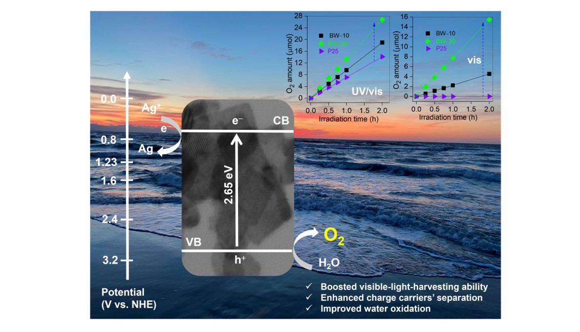
The photocatalytic water oxidation was carried out under UV and/or vis irradiation in the presence of in situ deposited silver as an electron scavenger. Typically, 0.05 g of BWO sample was dispersed in 5 mL of an aqueous suspension of AgF (0.05 mol L−1) in a 35 mL Pyrex test tube. The suspension (photocatalyst and AgF) was sonicated for ca. 10 min, deaerated with argon, and then the testing tube was sealed with a rubber septum and exposed to UV/vis (Hg lamp, λ > 290 nm) or vis (450 W–Xe lamp, λ > 400 nm; water IR filter, a cold mirror, and a cut–off filter: Y–42) irradiation. During the reaction, the suspension was constantly agitated in a thermostated water bath to keep the reaction temperature constant (about 298 K). The generated oxygen was quantified by employing a Shimadzu GC–8A chromatograph (Shimadzu Corporation, Kyoto, Japan), equipped with a thermal conductivity detector (TCD) and Poratak Q column (Agilent Technologies, Santa Clara, CA, USA). The recyclability tests were also performed (five repetitions) for the most active photocatalyst.

3. Results

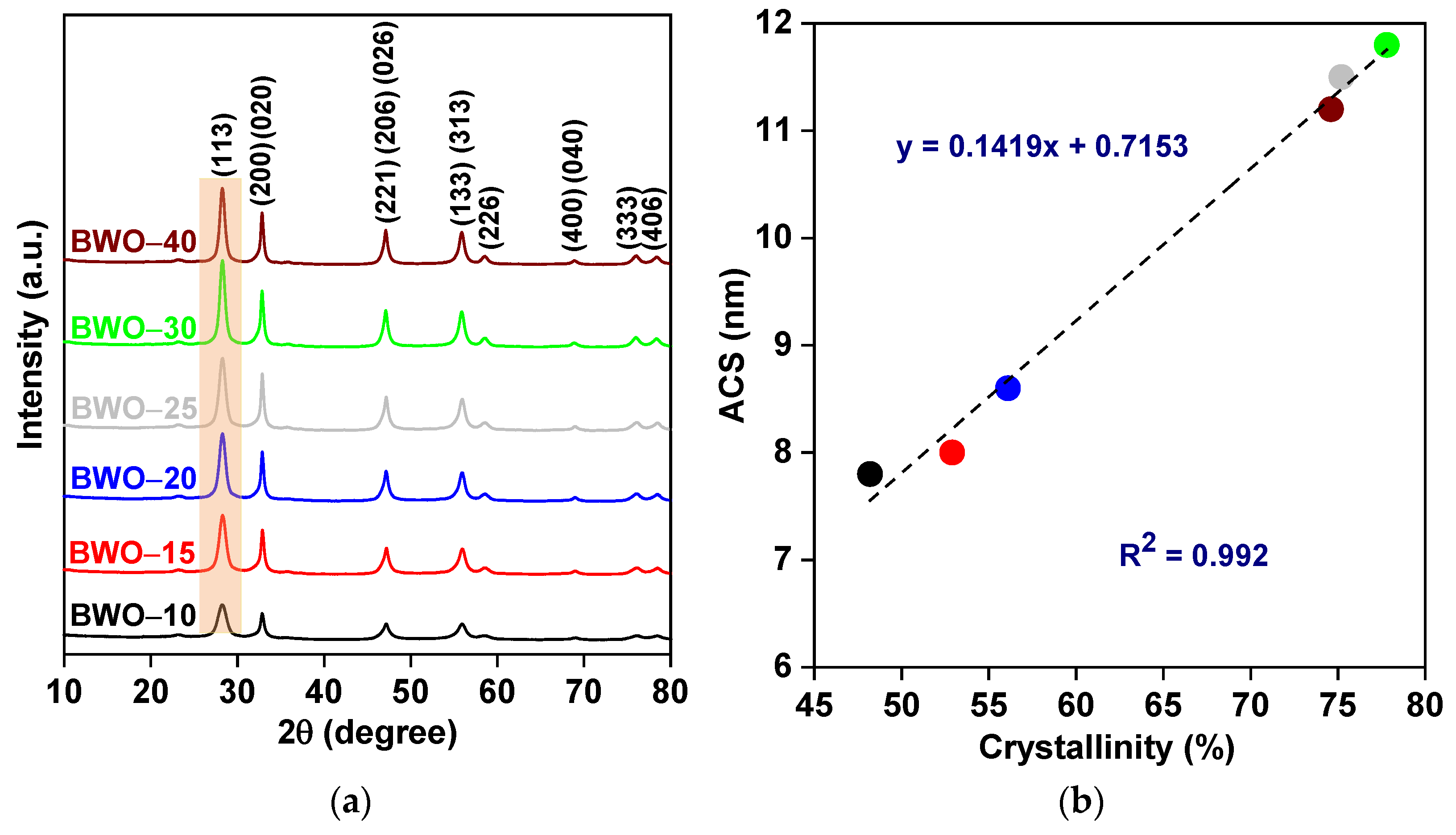
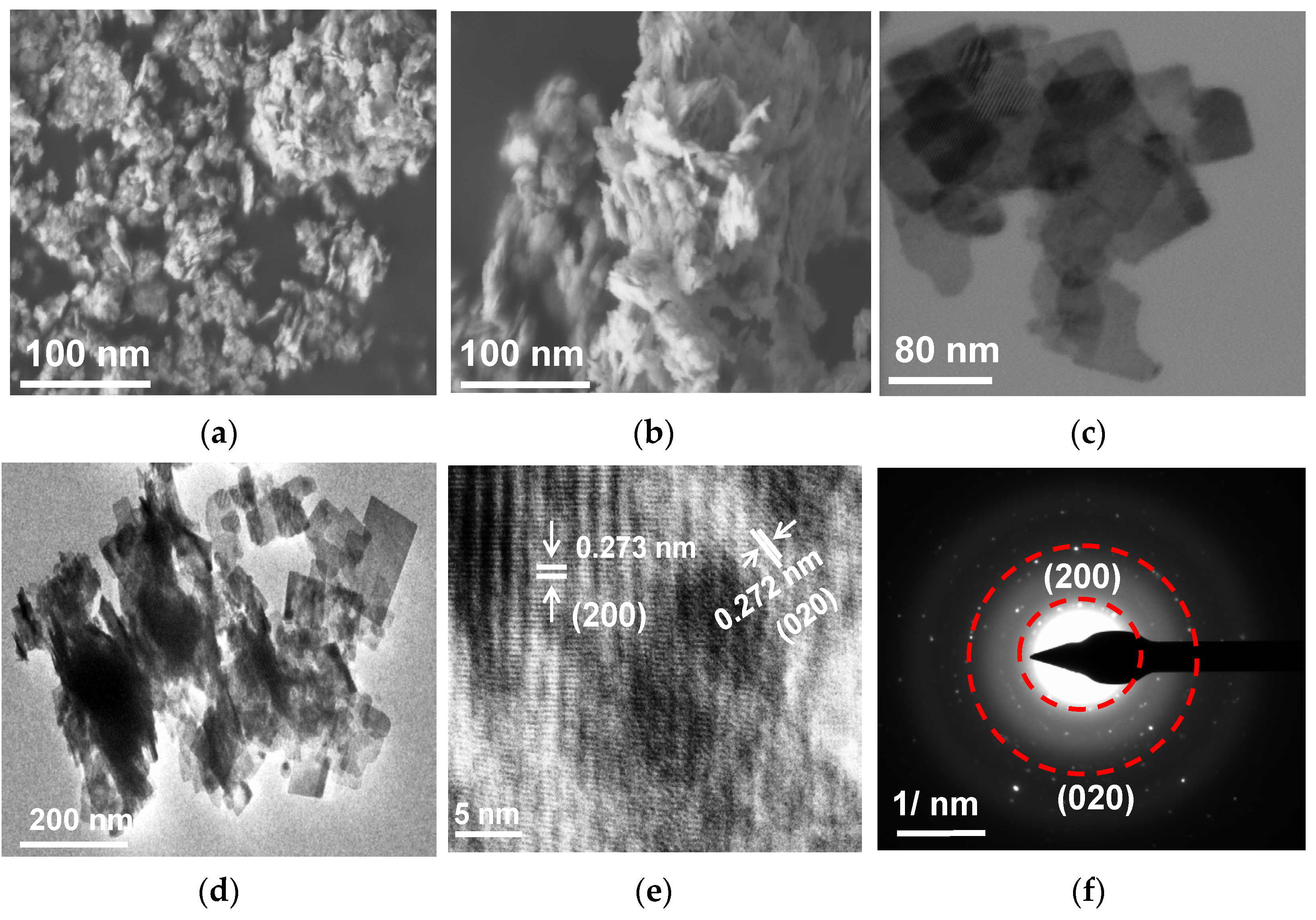
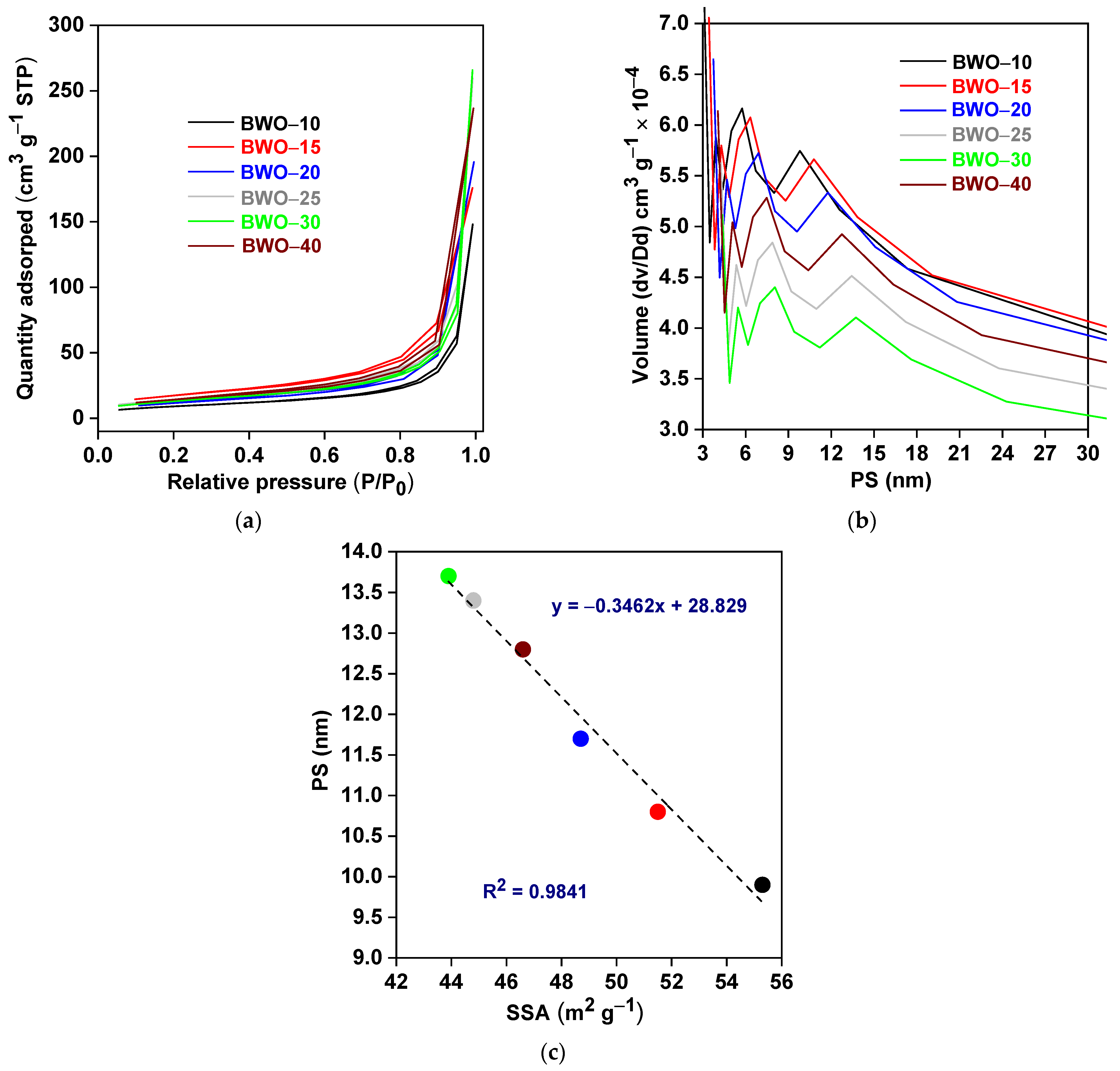
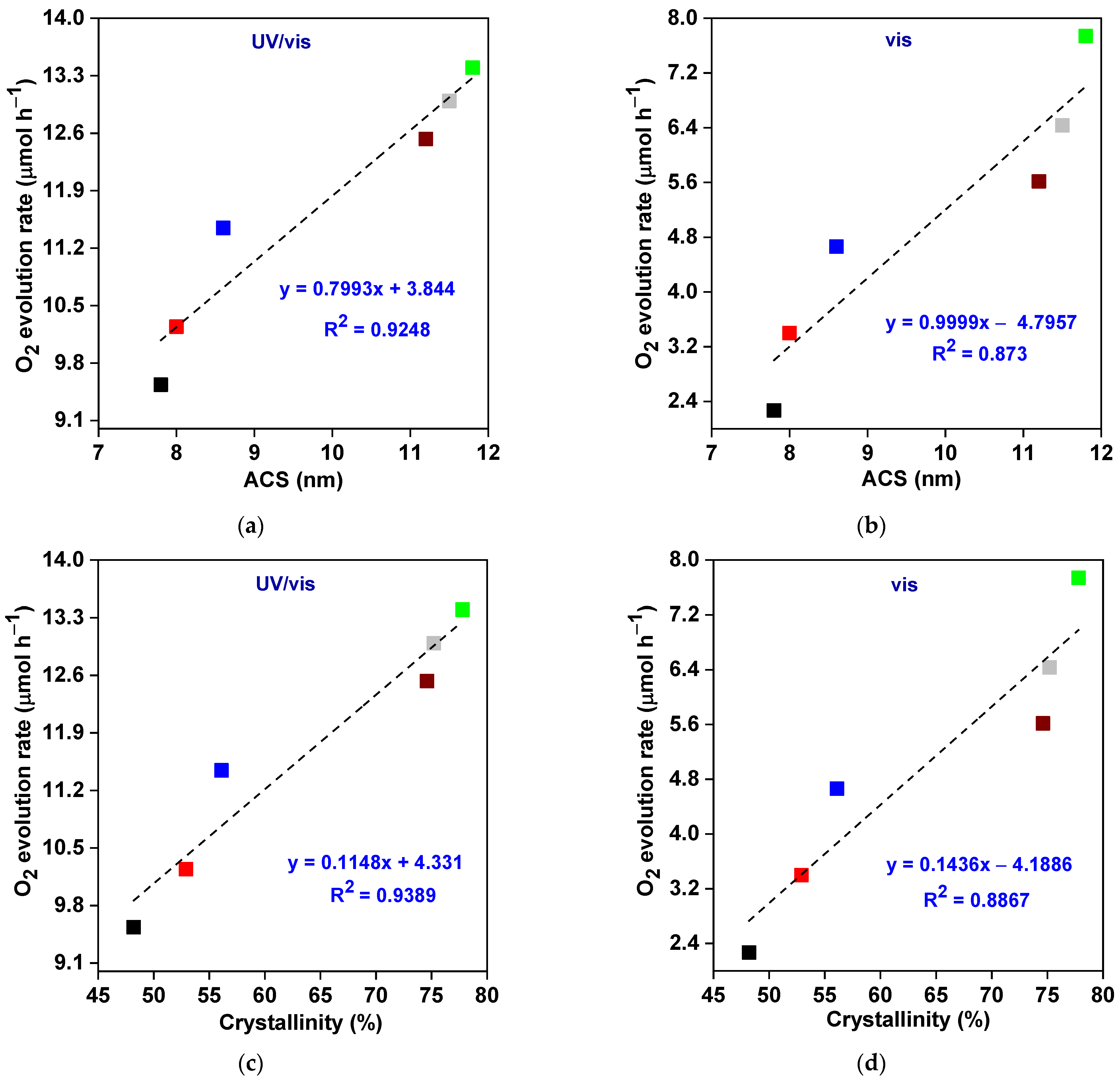
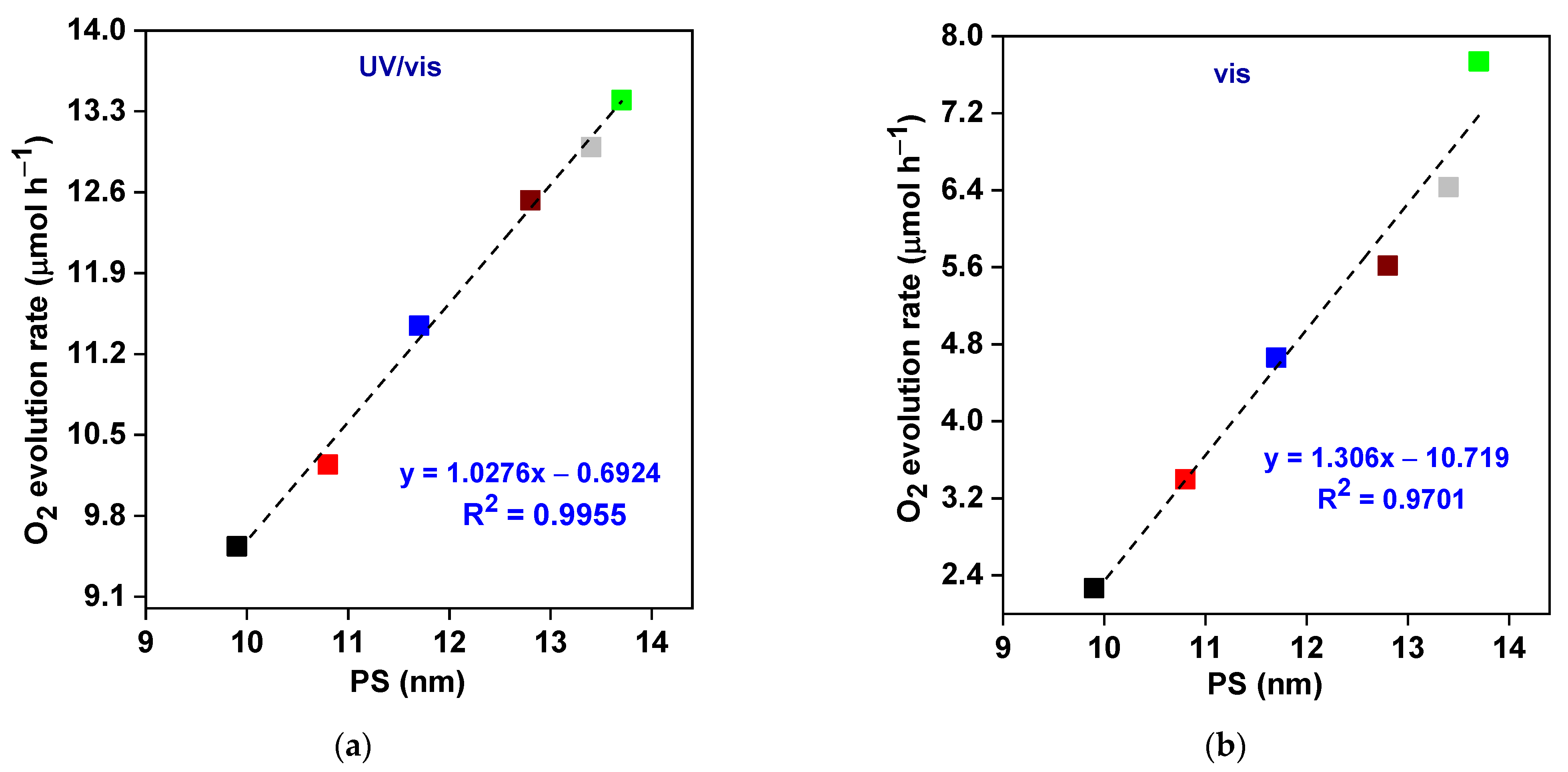
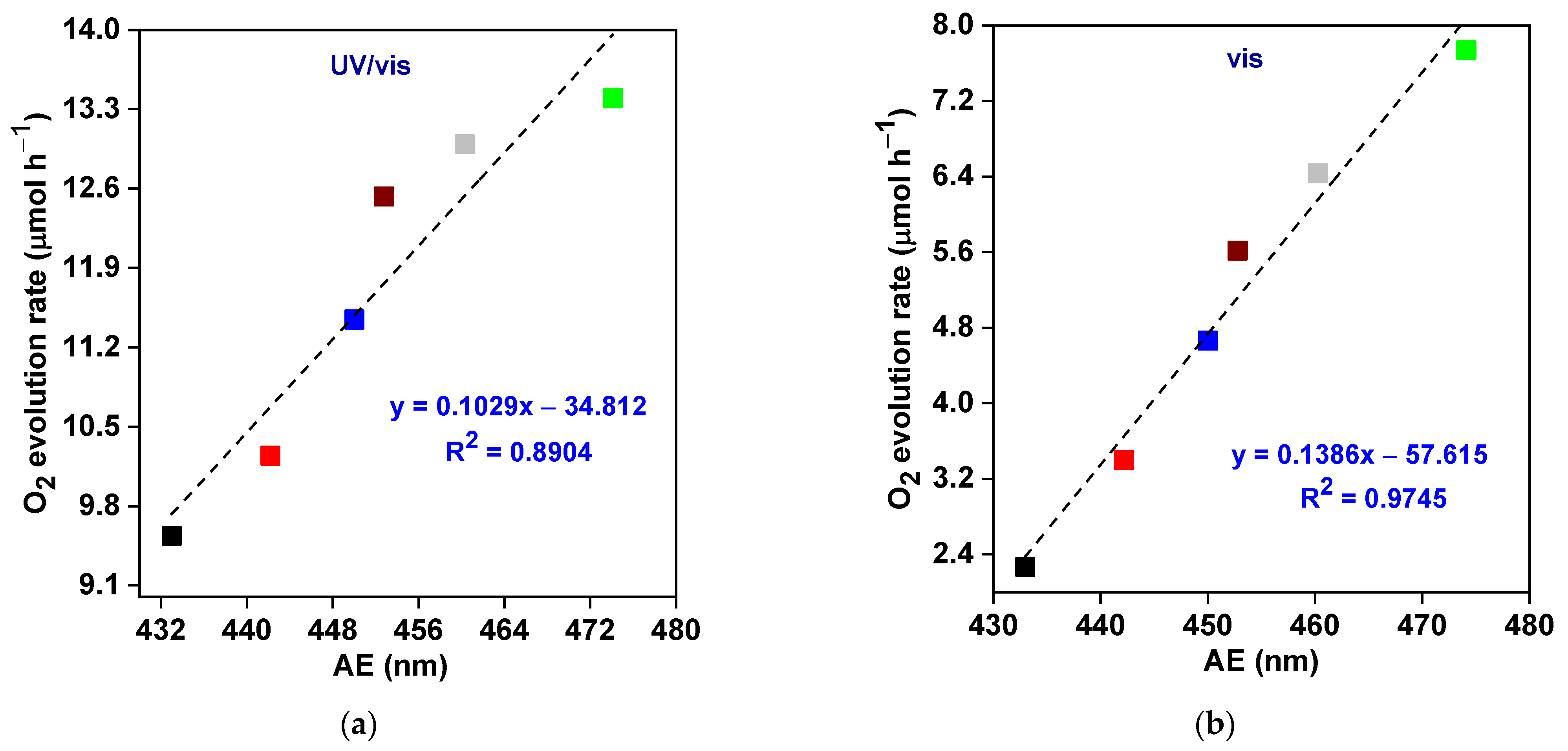
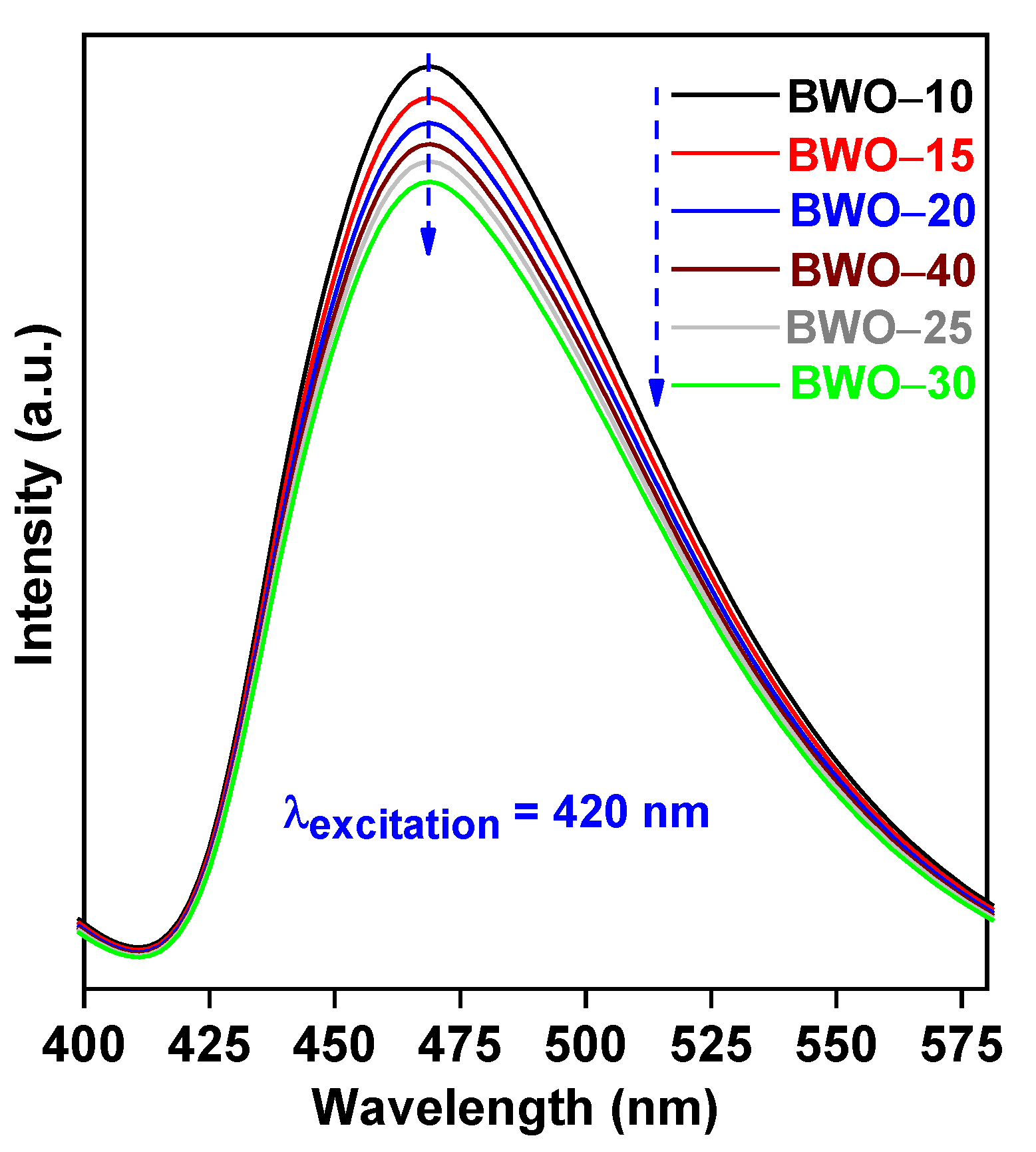
3.1. Characterization

The successful fabrication of BWO was confirmed by XRD analysis, and the obtained XRD patterns are shown in Figure 1a. The characteristic peaks at 2–theta of ca. 28.4°, 32.8°, 47.4°, 55.8°, 58.8°, 68.7°, 75.9°, and 78.5° could be assigned to (113), ((200) (020)), ((221) (206) (026)), ((313) (133)), (226), ((400) (040)), (333), and ((406) (046)) planes of BWO, respectively. These crystal planes are characteristic for the orthorhombic morphology of BWO (which resembles a perovskite-like structure with lattice parameters of a = 5.456 Å, b = 5.436 Å, and c = 16.428 Å, card no. 66579), and are consistent with previous findings [14,38,61,62,63]. Moreover, the lack of other diffraction peaks indicates the high crystal purity of the prepared samples. Although the duration of the solvothermal reaction shows no effect on the position of diffraction peaks, the increase in peaks’ intensity and peaks’ sharpening with the prolongation of reaction duration indicates the crystal growth and the improvement of crystallinity. Indeed, the crystallinity of BWO, estimated by an internal standard method, is improved by prolonging the reaction duration up to 30 h (Table 1). However, a further increase in the reaction time (40 h) causes a slight decrease in the crystallinity, which could be caused by the higher density of crystalline defects. Therefore, it might be concluded that the best crystalline properties are achieved during 30 h treatment (BWO–30), i.e., longer and shorter solvothermal time results in the formation of an imperfect crystalline structure. Similarly, Zhang et al. have also confirmed the influence of reaction duration (hydrothermal process) on the crystallinity of BWO [33].
Additionally, it has been found that the ratio of I(113)/I(200) (intensity of respective peaks) is lower than the standard value of 5, implying an anisotropic growth along the {001} basal plane, which suggests the formation of square-plate morphology (2D), as discussed later. It has already been suggested that large crystallites could be formed by an increase in solvo/hydrothermal time (as observed by intense and sharp XRD peaks) [33,62,64]. Accordingly, the average crystallite size (ACS) has been estimated, using the full-width half-maximum (FWHM) of (113) diffraction peak with the Debye–Scherrer equation [38], and obtained data are listed in Table 1. The ACS increases from 7.8 to 11.8 nm with an increase in the reaction duration till 30 h, and then slightly decreases to 11.2 nm for 40 h time. Therefore, it might be proposed that crystal growth is associated with crystallinity, as confirmed in Figure 1b. Accordingly, it has been found that both crystallinity and crystallite size are mainly controlled by the duration of the solvothermal reaction. Similarly, Li and coworkers found a correlation between the conditions of hydrothermal reaction and crystalline properties of BWO samples, i.e., an increase in crystallite sizes from 13.6 nm to 16.2 nm as the temperature of the hydrothermal reaction increased from 413 K to 433 K [25].
Summarizing, it might be concluded that the duration of the solvothermal reaction has a significant impact on the crystalline properties of BWO photocatalysts. Moreover, the estimation of optimal conditions for photocatalysts’ preparation is crucial for the preparation of perfect crystals. Similar findings have already been found for both BWO [33,62,65,66] and other photocatalysts, such as TiO2 [67,68,69,70,71], ZnO [72], MoS2 [73,74], and g–C3N4 [75].
The microscopic observations (Figure 2) have confirmed the self-assembly of nanoplate subunits during solvothermal growth, resulting in the formation of a layered microstructure. The obvious difference between BWO–10 and BWO–30 (a and b images, respectively) indicates that the small nanoparticles tend to aggregate and coalesce (forming larger particles with a clear–cut appearance), which is consistent with the XRD results (Table 1). The 2D structure of the BWO–30 sample is also clearly seen in STEM (Figure 2c) and TEM (Figure 2d) images. It should be noted that the BWO–30 sample consists of uniformly dispersed square nanoplates with smooth and transparent surfaces. To elucidate the growth orientation of the nanoplates, an HRTEM observation with SAED analysis was also carried out, and the obtained observations are displayed in Figure 2e,f. Accordingly, two sets of lattice fringes could be clearly observed, as seen by a marked interplanar spacing of ca. 0.273 nm and 0.272 nm, indexed to the (200) and (020) planes, respectively, of orthorhombic BWO (Figure 2e). Moreover, the SAED pattern exhibits a regular diffraction spot array with a spacing of 0.273 and 0.272 nm, corresponding to the (200) and (020) planes, respectively (Figure 2f). Therefore, it might be concluded that 2D BWO nanoplates preferably grow along the {001} facets, which is consistent with XRD results. This might be caused by a higher atom density on the {001} facets, leading to a slower growth along the {001} of BWO nanoplates [33,53,54,55,76,77].
To investigate the textural properties, BET/BJH measurements were performed at 77 K, and the obtained results are shown in Figure 3 and Table 2. All samples exhibit similar isotherms, categorized as type IV (according to IUPAC), with small hysteresis loops with a relative pressure of ca. 0.7–0.9, as shown in Figure 3a. This might be caused by irregular voids, resulting from large particles’ (plates) packing. Meanwhile, the surface characteristics were further estimated, and the obtained results, including the specific surface area (SSA), pore volume (PV), and pore size (PS), are presented in Figure 3b and Table 2. It has been found that the largest values of SSA (55.3 m2 g−1) and PV (5.8–6.2 × 10⁻⁴ cm3 g−1) are obtained for the BWO–10 sample, prepared during the shortest reaction (10 h). Clearly, the SSA and PV values decrease with increasing the reaction duration up to 30 h (43.9 m2 g−1 and 4.1–4.4 × 10⁻⁴ cm3 g−1, respectively). All obtained data correlate well with ACS, i.e., an increase in ACS (prolonged reaction) corresponds to a decrease in SSA. The longer reaction time might significantly affect the crystal growth since it allows smaller grains to grow, forming bigger crystallites. These large nanocrystallites might also be pushed into the channels of mesopores, decreasing the pore volume. It is clearly observed that all samples exhibit a mesoporous structure (PS = 3.9–13.7 nm). Figure 3c shows a clear correlation between SSA and pore size.
The photoabsorption features of BWO photocatalysts are displayed in Figure 4 and Table 2. Indeed, all photocatalysts could absorb a significant portion of visible light, i.e., the absorption edge (AE) ranges from 433.0 nm to 474.1 nm (Figure 4a). Therefore, prepared BWO samples are expected to act as VLR photocatalysts. Moreover, an obvious bathochromic shift in the AE (and the consequent bandgap narrowing, as displayed in Figure 4b) with an increase in the solvothermal time correlates well with an increase in particle/crystallite size (size-dependent light absorption), as the phonon frequency constant increases with an increase in crystal size [78,79,80,81]. The comparison of photoabsorption properties demonstrates that the absorption edge at the shortest wavelength (433 nm), and thus the sample with the largest energy bandgap (2.87 eV), was prepared during the shortest duration of solvothermal reaction (BWO–10), whereas BWO–30 exhibits the narrowest energy bandgap and the absorption edge at the longest wavelength (i.e., 2.65 eV and 474.1 nm, respectively), as presented in Figure 4 and Table 2.

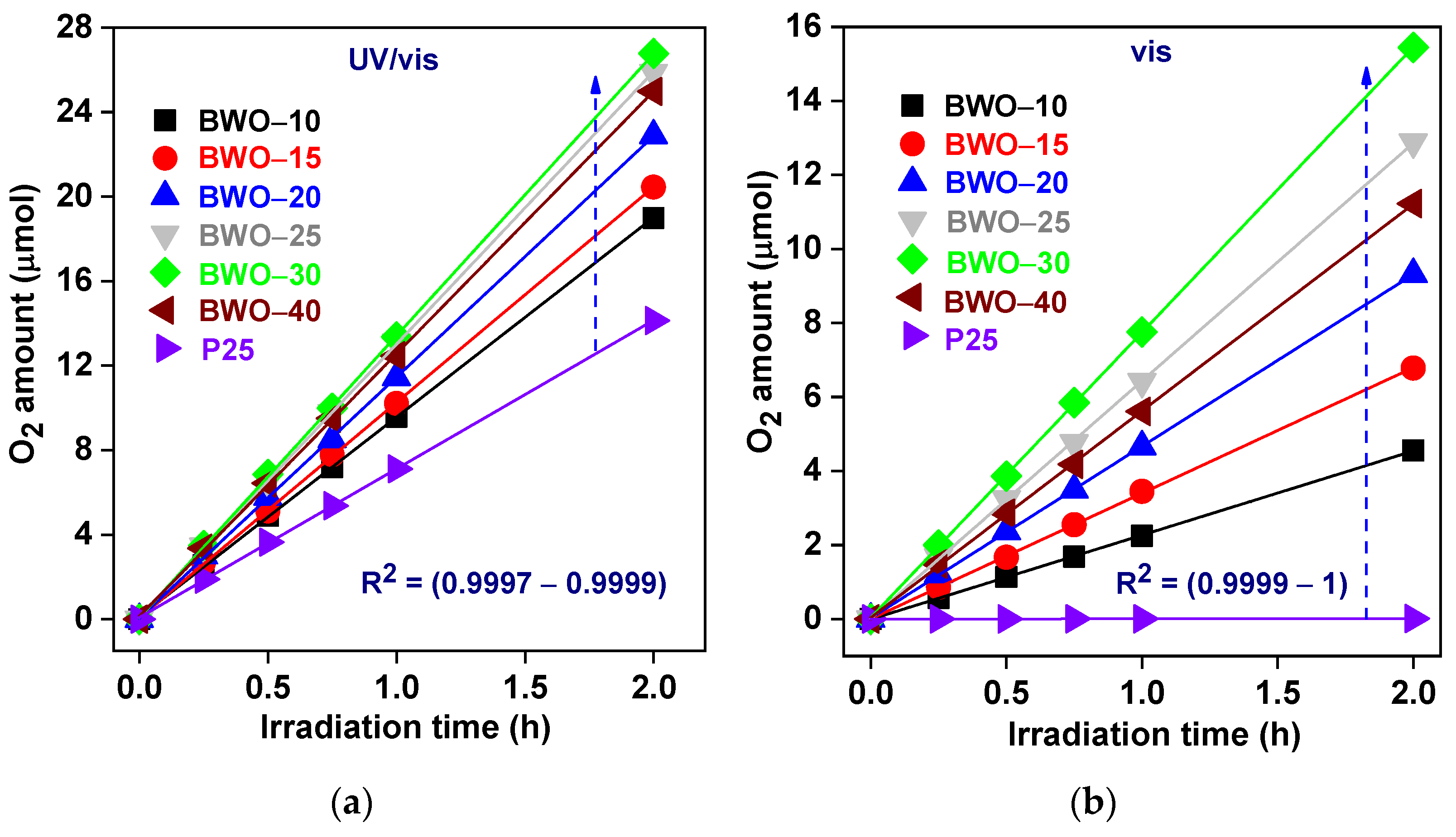
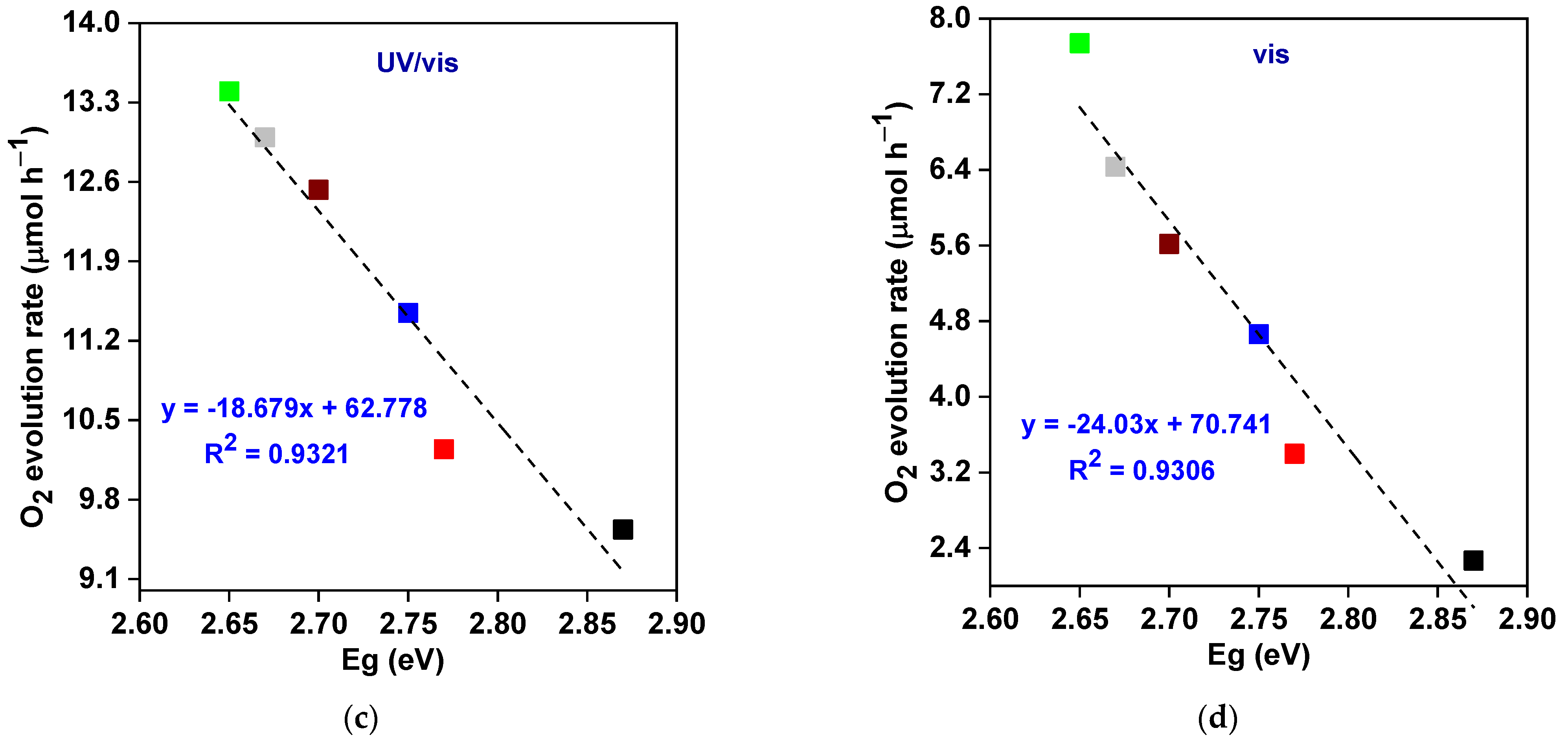
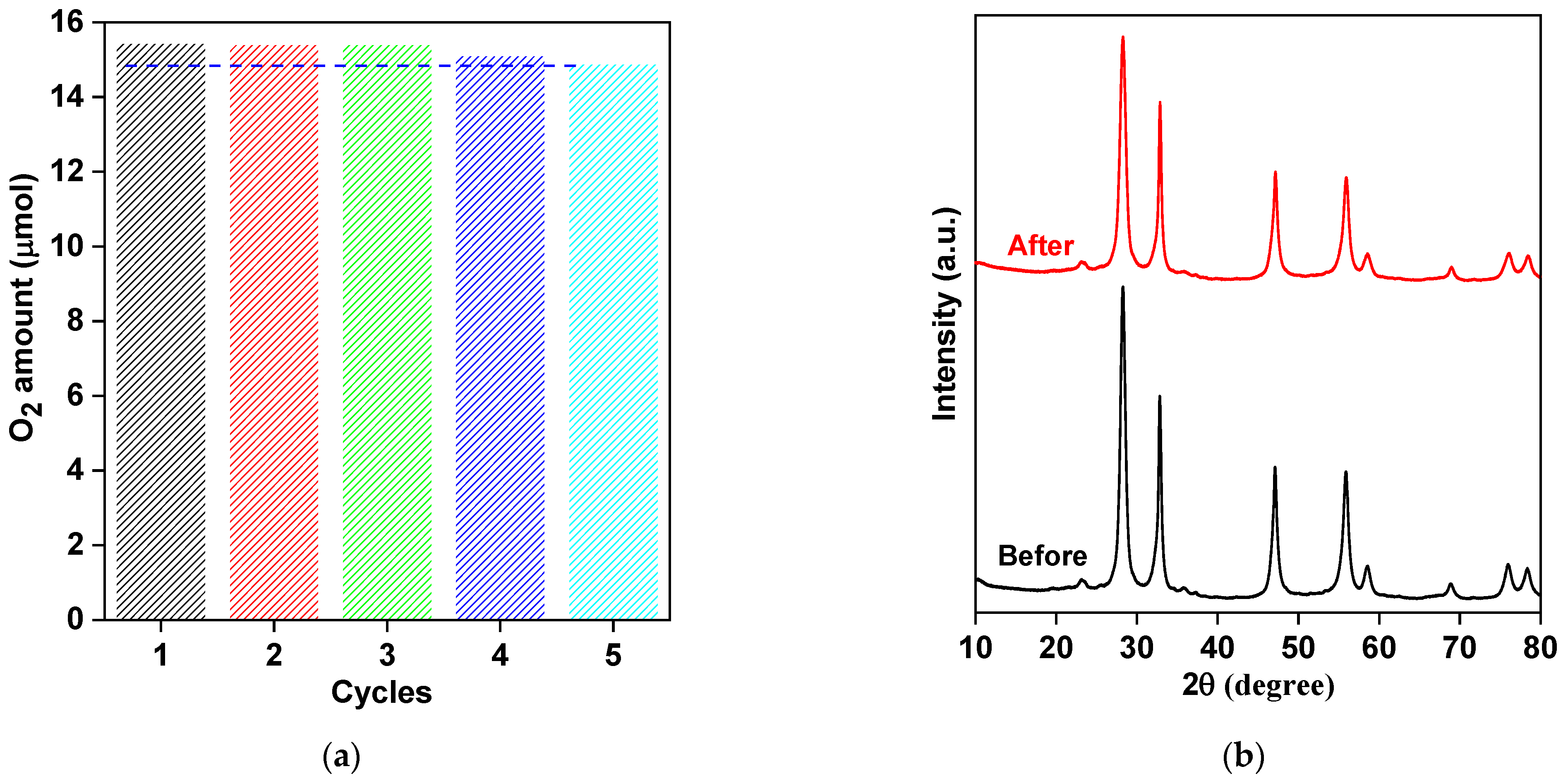
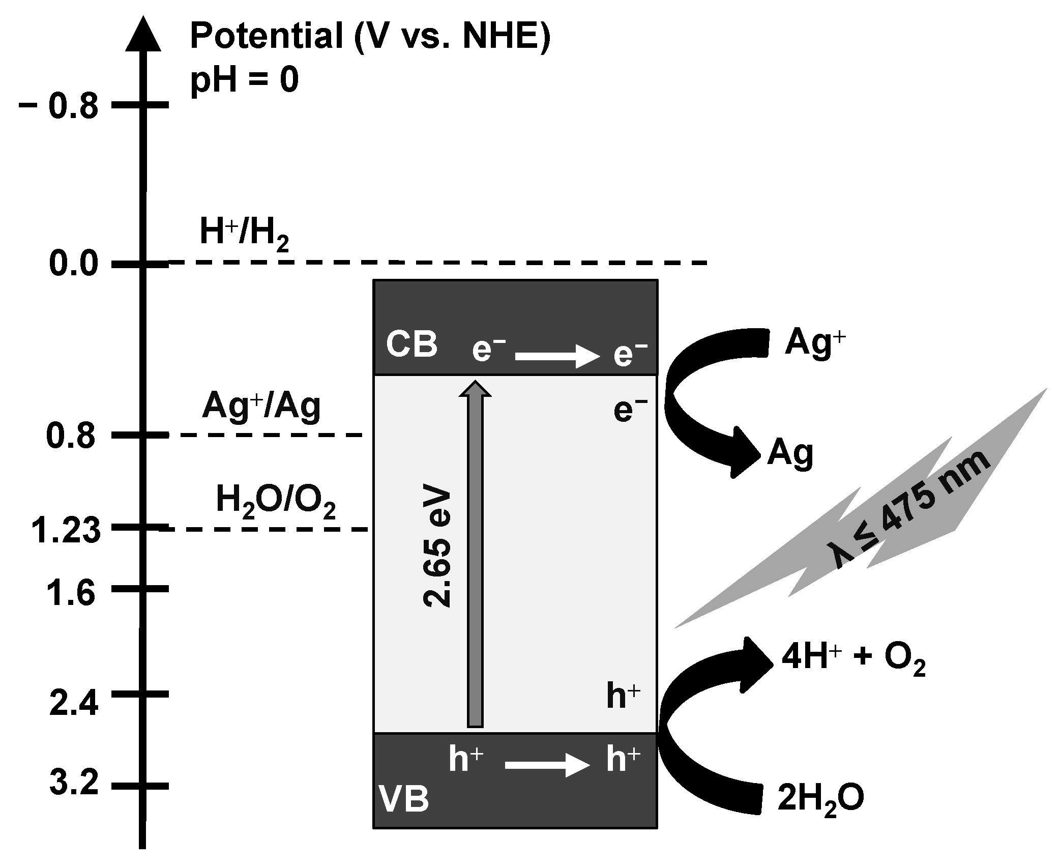
3.2. Photocatalytic O2 Evolution

The photocatalytic activity of BWO samples in comparison to that by famous P25 (common and standard photocatalyst [82,83]) was evaluated through photocatalytic O2 generation in an aqueous solution under UV and/or vis illumination in the presence of in situ deposited silver as an electron scavenger. It was found that O2 gas was not generated in the absence of either a photocatalyst (direct photolysis) or light (under dark conditions). Hence, it might be concluded that water oxidation must proceed via a photocatalytic mechanism, i.e., the formation of charge carriers under irradiation. Indeed, O2 evolution was observed in the presence of photocatalysts (BWO and P25) upon UV/vis and vis irradiation, as presented in Figure 5 (and Table 3). A linear evolution of oxygen for all samples during the whole duration of the reaction confirms the photocatalytic mechanism (not a “light-initiated” dark reaction), also revealing the high photostability of all materials.
Under UV/vis irradiation, all BWO samples exhibit much higher activity than that of one of the most active titania photocatalysts (P25) with similar surface properties (BET of ca. 50 m2 g−1 [84]) (Figure 5a). However, the crystallinity of BWO samples is much worse than that in P25 (>90% estimated by the same method [84]), but ACS of BWO is smaller than that in titania (25.3 and 39.6 nm for anatase and rutile, respectively) [84]). Accordingly, it might be concluded that a well-organized nanostructure (2D) might be responsible for the better performance of BWO. Moreover, as expected, P25–TiO2 is hardly active under vis irradiation due to a much wider bandgap (>3.0 eV) and, thus, a shorter wavelength edge (λ < 400 nm) (Figure 5b). The photocatalytic activity of BWO photocatalysts enhances with an increase in the synthesis time up to 30 h, reaching 13.40 μmol h−1 and 7.74 μmol h−1 of oxygen evolution rate under UV/vis and vis, respectively (BWO–30 sample), as shown in Figure 5 and Table 3.
Notably, the specific surface area is commonly considered one of the main factors controlling photocatalyst performance (similar to “dark” catalytic reactions). In this context, a large specific surface area could: (i) enhance the incident light-harvesting ability, (ii) create surface-active sites, (iii) promote the reactant molecules’ adsorption on the surface of the photocatalyst, (iv) increase the rate of reactants’ formation (e.g., reactive oxygen species), and thus significantly boost the photocatalytic performance for various applications (specifically, photocatalytic degradation of hazardous organic compounds) [85,86,87,88]. Surprisingly, the most active sample (BWO–30) is characterized by the smallest specific surface area (Table 2), implying that the specific surface area of the fabricated photocatalysts is not the reason behind increasing the photocatalytic activity in the current study. Similar findings have also been observed elsewhere [89,90,91,92]. It might be proposed that, in the case of direct redox reactions via photogenerated charges (here, silver reduction and water oxidation by photo-formed electrons and holes, respectively), instead of the involvement of intermediates (such as reactive oxygen species formed on the surface of photocatalyst; crucial for oxidative decomposing of organic compounds), the specific surface area does not govern photocatalytic performance. Therefore, other properties should be decisive for photocatalytic performance. Among them, crystallinity, nanoplate morphology, mesoporous structure (PS = 5.5–13.7 nm), and improved light absorption ability (especially under vis) must be considered. Indeed, the photocatalytic activity of the fabricated BWO materials correlates well with other properties (Figure 6, Figure 7 and Figure 8), i.e., crystallite size, crystallinity, pore size, absorption edge, and energy bandgap.
Intriguingly, crystallite size and crystallinity are two of the most crucial factors that significantly impact the photocatalytic activity of semiconductor photocatalysts [93,94]. However, contrary results can be found in the literature regarding the influence of crystallite/particle size on photocatalytic performance, i.e., the positive [93,95,96] and negative [94,97,98,99] impacts. In this study, the reaction rate under both UV/vis and vis irradiation increases upon an increase in the crystallite size, and the BWO–30 sample with the largest crystallites also exhibits the best photocatalytic activity, as depicted in Figure 6a,b. These findings could be attributed to enhancing the optical characteristics and promoting the charge carriers’ separation and mobility [93]. Moreover, it has been pointed out that the optimal value of crystallite size lies in the range of 7–15 nm for diverse photocatalytic applications [93,100,101]. In contrast, there is no disagreement about the impact of crystallinity on photocatalytic activity, as it is well-documented that higher crystallinity means better photocatalytic performance. The improved crystallinity correlates with lower content of crystalline defects, and thus a lower rate of charge carriers’ recombination (as confirmed also here, by PL data; discussed further). Similarly, the dependence of crystallinity on charge carriers’ separation efficiency (and thus photocatalytic activity) has already been proven for other photocatalysts, e.g., BiFeO3 [81] and TiO2 [102]. Therefore, the BWO–30 sample possessing the largest crystallinity shows the best activity towards O2 evolution under UV/vis and vis irradiation (Figure 6c,d). Here, during the formation a of 2D structure, it might be proposed that the large size of BWO–30 indicates the perfectly formed crystal, and thus the largest size corresponds to the best crystallinity.
Remarkably, the mesoporous structure introduces efficient paths for reactants’ transfer to reactive sites (mass transfer) and also improves multiple scattering of light, resulting in effective photon absorption and, hence, boosting photocatalytic activity [103,104,105]. As displayed in Figure 7a,b, the photocatalytic O2 evolution rate (under UV/vis and vis irradiation) improves upon an increase in the pore size of the synthesized BWO specimen, and the BWO–30 sample with the largest pore size exhibits the best photocatalytic activity. This might be caused by improved water adsorption and enhanced light penetration into the photocatalyst in the presence of larger pores, as already suggested in other reports [103,104,105].
Moreover, photocatalytic activity firmly relies on the optical properties of photocatalysts. In this sense, the photocatalytic O2 generation rate (under UV/vis and vis irradiation) increases upon an increase in the absorption edges (the efficient use of more photons) and, thus, a decrease in energy bandgaps (Eg) of fabricated BWO (the BWO–30 sample shows the superior photocatalytic performance) (Figure 8a–d). In addition, the nanoplate morphology is beneficial for charge carriers’ separation/transfer and efficient light harvesting ability, allowing multiple irradiation reflection (similar to photonic crystals [106]). Therefore, it should be concluded that the photocatalytic activity of BWO might be significantly enhanced by changing the physical properties via reaching the optimal conditions of solvothermal reaction, e.g., reaction time (here: 30 h). However, the prolonged reaction might also cause a decrease in photocatalytic activity because of a reduction in the quality of materials. For example, a 40 h solvothermal reaction causes a decrease in crystallinity (a higher density of crystalline defect).
To confirm that properties (mainly crystallinity) influence the h+/e recombination and, thus, the photocatalytic efficiency, PL spectroscopy was carried out, and the findings are presented in Figure 9. All BWO photocatalysts display photoluminescence at ca. 468.7 nm (after excitation at 420 nm), which correlates well with charge carries’ recombination [107]. However, the PL intensities between samples differ significantly. Indeed, the BW–30 sample with the highest crystallinity (and photocatalytic activities) possesses the weakest PL intensity (i.e., the best efficiency of photoinduced charge carriers’ separation), whereas the strongest PL signal (i.e., the fastest charge carries’ recombination rate) is obtained by the BWO–10 sample with the worst crystal properties. It has been confirmed that reaching optimal conditions of solvothermal reaction is important for getting the best photocatalytic performance, and thus, prolonged reaction (here > 30 h) might cause the preparation of samples with unfavorable crystalline defects.
To confirm the long-term photostability, the recyclability experiments were carried out for the most active sample (BW–30) during five cycles under vis irradiation, and the obtained data are displayed in Figure 10a. The high photocatalysis stability has been confirmed with only a slight change in oxygen generation between the first and fifth cycles (from 15.45 μmol to 14.89 μmol). Furthermore, XRD analysis of a recycled sample indicates that crystalline properties have not been changed (Figure 10b). Therefore, it could be proposed that 2D mesoporous BWO nanoplates are promising photocatalysts for vis applications, especially oxidation reactions). Indeed, 2D BWO photocatalysts exhibit superior photocatalytic activity toward O2 evolution, as compared to recently reported photocatalytic materials (Table 4) [2,108,109,110].
Finally, the mechanism of photocatalytic water oxidation on BWO photocatalyst (BWO–30) might be proposed (Figure 11). The edge potentials of the valence band (EVB) and conduction band (ECB) have been estimated using the Mulliken electronegativity theory (Equations (1) and (2)) [111]:
ECB = χ − Ee − 0.5Eg
EVB = ECB + Eg
Here, Eg is the energy bandgap of BWO–30 (2.65 eV); χ is the absolute electronegativity of Bi2WO6 (6.36 eV); and Ee is free electrons energy in the hydrogen scale (4.5 eV). Accordingly, estimated ECB and EVB are equal to 0.54 and 3.19 V, respectively. Therefore, it is possible to reduce silver cation (Ag+) into silver atom (Ag), as the ECB (0.54 V) of the photocatalyst is less positive than the standard redox potential of Ag+/Ag (0.7996 V). Moreover, water can be oxidized into O2, as the EVB (3.19 V) of the photocatalyst is more positive than the standard redox potential of H2O/O2 (1.23 V). Based on the above results, the mechanism of photocatalytic water oxidation over BW–30 (in the presence of AgF as an electron scavenger) has been proposed as the following: The h+/e pairs are generated under irradiation (both UV and vis could be used) since electrons (e) are transferred from the VB to the CB, leaving holes (h+) at VB of the photocatalyst (Equation (3)).
The photogenerated charge carriers (eCB and h+VB) migrate to the photocatalyst surface. Finally, the Ag+ cation (from AgF in the reaction suspension, Equation (4)) reacts with eCB to produce zero-valent silver (Equation (5)) and h+VB oxidizes water to form O2 (Equation (5)). Furthermore, the obtained Ag nanoparticles, loaded on the surface of BWO, might improve the photocatalytic activity and performance (working as a co-catalyst). It should be pointed out that the released proton (from water oxidation, Equation (6)) could not be reduced into H2, because the ECB of BWO–30 photocatalyst (0.54 V) is more positive than the standard redox potential of H+/H2 (0.0 V). Therefore, acidification of reaction suspension is proposed, e.g., as summarized in exemplary Equation (7).
Bi2WO6 + hν → eCB + h+VB
AgF → Ag+ + F
eCB + Ag+ → Ag
2h+VB + H2O → 2H+ + ½ O2
H+ + F → HF

4. Conclusions

The facile template-free solvothermal reaction has proven to be an efficient method for the preparation of a 2D porous BWO photocatalyst. BWO material shows significant activity in photocatalytic water oxidation under both UV and vis irradiation. The conditions of the solvothermal reaction are critical for the properties of BWO and, thus, the resultant photocatalytic performance. It has been found that a 30 h solvothermal reaction at 433 K results in the preparation of the most active photocatalyst with the best properties, i.e., nanoplate morphology (2D), mesoporous structure, high crystallinity (low content of defects) and efficient light harvesting ability. To the best of our knowledge, this is the first work displaying that 2D mesoporous BWO nanoplates could be successfully synthesized by a template-free solvothermal method and subsequently used for enhanced photocatalytic water oxidation under UV/vis and vis irradiation. Therefore, this study opens the door for other applications of BWO photocatalysts under natural solar radiation.

Author Contributions

T.M.K.: Conceptualization, Investigation, Data Analysis, Writing—original draft. E.K.: Data Analysis, Writing, and Editing, Reviewing, Supervision. S.M.E.-S.: Conceptualization, Investigation, Editing, Reviewing, Supervision. All authors have read and agreed to the published version of the manuscript.

Funding

This work was supported by the Egyptian Ministry of Higher Education, Egypt (Cultural Affairs and Missions Sector). The project is co-financed by the Polish National Agency for Academic Exchange within Polish Returns Program (BPN/PPO/2021/1/00037) and The National Science Centre (2022/01/1/ST4/00026).

Institutional Review Board Statement

Not applicable.

Informed Consent Statement

Not applicable.

Data Availability Statement

The data presented in this study are available on request from corresponding author (T.M.K.).

Acknowledgments

Tamer M. Khedr would like to acknowledge Bunsho Ohtani, Institute for Catalysis (ICAT), Hokkaido University for hosting him, as well as providing the access to the laboratory devices and research facilities at ICAT during this work; and Kunlei Wang for STEM measurements.

Conflicts of Interest

The authors declare no conflict of interest.

References

  1. Hayat, A.; Sohail, M.; Taha, T.A.; Kumar Baburao Mane, S.; Al-Sehemi, A.G.; Al-Ghamdi, A.A.; Nawawi, W.I.; Palamanit, A.; Amin, M.A.; Fallatah, A.M.; et al. Synergetic effect of bismuth vanadate over copolymerized carbon nitride composites for highly efficient photocatalytic H2 and O2 generation. J. Colloid Interface Sci. 2022, 627, 621–629. [Google Scholar] [CrossRef] [PubMed]
  2. Djatoubai, E.; Khan, M.S.; ul Haq, S.; Guo, P.; Shen, S. BiFeO3 bandgap engineering by dopants and defects control for efficient photocatalytic water oxidation. Appl. Catal. A-Gen. 2022, 643, 118737. [Google Scholar] [CrossRef]
  3. Ma, K.; Yang, B.; Su, X.; Fan, L. Two-phase solvothermal preparation of Co3O4/GO compound materials as catalysts for photocatalytic water oxidation. Mater. Lett. 2022, 324, 132615. [Google Scholar] [CrossRef]
  4. Zhang, Y.; Kong, L.; Konysheva, E.Y.; Xu, X. Expediting photocarrier separation in Ta3N5@CaTaO2N heterostructures with seamless interfaces for photocatalytic water oxidation under visible light. Appl. Catal. B 2022, 317, 121712. [Google Scholar] [CrossRef]
  5. Yang, M.; Li, J.; Ke, G.; Liu, B.; Dong, F.; Yang, L.; He, H.; Zhou, Y. WO3 homojunction photoanode: Integrating the advantages of WO3 different facets for efficient water oxidation. J. Energy Chem. 2021, 56, 37–45. [Google Scholar] [CrossRef]
  6. Chen, Z.; Geng, S.; Wang, Y.; Wang, Y.; Song, S. Boosting 2 e-Water oxidation reaction on WO3 by F-modification and revealing the mechanism by probing interfacial water structure. Appl. Catal. B 2022, 317, 121756. [Google Scholar] [CrossRef]
  7. Sun, W.; Dong, Y.; Zhai, X.; Zhang, M.; Li, K.; Wang, Q.; Ding, Y. Crystal facet engineering of BiVO4/CQDs/TPP with improved charge transfer efficiency for photocatalytic water oxidation. Chem. Eng. J. 2022, 430, 132872. [Google Scholar] [CrossRef]
  8. Ke, G.; Liu, B.; Duan, F.; Liu, X.; Wen, J.; Jia, B.; Liu, X.; He, H.; Zhou, Y. Resorcinol-formaldehyde resin nanoparticles as surface charge transfer and separation sites for the improvement of BiVO4 film photoanodes’ performance in solar water oxidation. Appl. Surf. Sci. 2022, 601, 154236. [Google Scholar] [CrossRef]
  9. Khan, N.A.; Rashid, N.; Ahmad, I.; Zahidullah; Zairov, R.; ur Rehman, H.; Nazar, M.F.; Jabeen, U. An efficient Fe2O3/FeS heterostructures water oxidation catalyst. Int. J. Hydrog. Energy 2022, 47, 22340–22347. [Google Scholar] [CrossRef]
  10. Duc Quang, N.; Cao Van, P.; Majumder, S.; Jeong, J.-R.; Kim, D.; Kim, C. Rational construction of S-doped FeOOH onto Fe2O3 nanorods for enhanced water oxidation. J. Colloid Interface Sci. 2022, 616, 749–758. [Google Scholar] [CrossRef]
  11. Zhu, Z.; Wan, S.; Zhao, Y.; Qin, Y.; Ge, X.; Zhong, Q.; Bu, Y. Recent progress in Bi2WO6-Based photocatalysts for clean energy and environmental remediation: Competitiveness, challenges, and future perspectives. Nano Sel. 2021, 2, 187–215. [Google Scholar] [CrossRef]
  12. Khedr, T.M.; Wang, K.; Kowalski, D.; El-Sheikh, S.M.; Abdeldayem, H.M.; Ohtani, B.; Kowalska, E. Bi2WO6-based Z-scheme photocatalysts: Principles, mechanisms and photocatalytic applications. J. Environ. Chem. Eng. 2022, 10, 107838. [Google Scholar] [CrossRef]
  13. Kudo, A.; Hijii, S. H2 or O2 evolution from aqueous solutions on layered oxide photocatalysts consisting of Bi3+ with 6s2 configuration and d0 transition metal ions. Chem. Lett. 1999, 28, 1103–1104. [Google Scholar] [CrossRef]
  14. Guo, W.; Jian, L.; Wang, X.; Zeng, W. Hydrothermal synthesis of Ni-doped hydrangea-like Bi2WO6 and the enhanced gas sensing property to n-butanol. Sens. Actuators B Chem. 2022, 357, 131396. [Google Scholar] [CrossRef]
  15. Yan, F.; Wang, Y.; Yi, C.; Xu, J.; Wang, B.; Ma, R.; Xu, M. Construction of carbon dots modified Cl-doped Bi2WO6 hollow microspheres for boosting photocatalytic degradation of tetracycline under visible light irradiation. Ceram. Int. 2022, 49, 7214–7222. [Google Scholar] [CrossRef]
  16. Bera, S.; Samajdar, S.; Pal, S.; Das, P.S.; Jones, L.A.H.; Finch, H.; Dhanak, V.R.; Ghosh, S. Effect of metal doping in Bi2WO6 micro-flowers for enhanced photoelectrochemical water splitting. Ceram. Int. 2022, 48, 35814–35824. [Google Scholar] [CrossRef]
  17. Su, H.; Li, S.; Xu, L.; Liu, C.; Zhang, R.; Tan, W. Hydrothermal preparation of flower-like Ni2+ doped Bi2WO6 for enhanced photocatalytic degradation. J. Phys. Chem. Solids 2022, 170, 110954. [Google Scholar] [CrossRef]
  18. Alhadi, A.; Ma, S. Synthesis of Sn doped-Bi2WO6 nanoslices for enhanced isopropanol sensing properties. Phys. B Condens. Matter 2022, 635, 413819. [Google Scholar] [CrossRef]
  19. Phuruangrat, A.; Buapoon, S.; Bunluesak, T.; Suebsom, P.; Wannapop, S.; Thongtem, T.; Thongtem, S. Hydrothermal preparation of Au-doped Bi2WO6 nanoplates for enhanced visible-light-driven photocatalytic degradation of rhodamine B. Solid State Sci. 2022, 128, 106881. [Google Scholar] [CrossRef]
  20. Ai, S.; Liu, Y.; Chai, Y.; Yuan, R.; Liu, H. Enhanced cathodic photocurrent derived from N-type S doped-Bi2WO6 nanoparticles through an antenna-like strategy for photoelectrochemical biosensor. Biosens. Bioelectron. 2022, 207, 114176. [Google Scholar] [CrossRef]
  21. Ning, J.; Zhang, J.; Dai, R.; Wu, Q.; Zhang, L.; Zhang, W.; Yan, J.; Zhang, F. Experiment and DFT study on the photocatalytic properties of La-doped Bi2WO6 nanoplate-like materials. Appl. Surf. Sci. 2022, 579, 152219. [Google Scholar] [CrossRef]
  22. Sun, D.; Le, Y.; Jiang, C.; Cheng, B. Ultrathin Bi2WO6 nanosheet decorated with Pt nanoparticles for efficient formaldehyde removal at room temperature. Appl. Surf. Sci. 2018, 441, 429–437. [Google Scholar] [CrossRef]
  23. Yuan, C.; Gao, H.; Xu, Q.; Song, X.; Zhai, C.; Zhu, M. Pt decorated 2D/3D heterostructure of Bi2WO6 nanosheet/Cu2S snowflake for improving electrocatalytic methanol oxidation with visible-light assistance. Appl. Surf. Sci. 2020, 521, 146431. [Google Scholar] [CrossRef]
  24. Jin, K.; Qin, M.; Li, X.; Wang, R.; Zhao, Y.; Li, Y.; Wang, H. A low-dosage silver-loaded flower-like Bi2WO6 nanosheets toward efficiently photocatalytic degradation of sulfamethoxazole. Mater. Sci. Semicond. Process. 2022, 139, 106338. [Google Scholar] [CrossRef]
  25. Li, Z.; Wang, K.; Zhang, J.; Chang, Y.; Kowalska, E.; Wei, Z. Enhanced Photocatalytic Activity of Hierarchical Bi2WO6 Microballs by Modification with Noble Metals. Catalysts 2022, 12, 130. [Google Scholar] [CrossRef]
  26. Di, J.; Xia, J.; Ge, Y.; Li, H.; Ji, H.; Xu, H.; Zhang, Q.; Li, H.; Li, M. Novel visible-light-driven CQDs/Bi2WO6 hybrid materials with enhanced photocatalytic activity toward organic pollutants degradation and mechanism insight. Appl. Catal. B 2015, 168–169, 51–61. [Google Scholar] [CrossRef]
  27. Wang, J.; Tang, L.; Zeng, G.; Deng, Y.; Dong, H.; Liu, Y.; Wang, L.; Peng, B.; Zhang, C.; Chen, F. 0D/2D interface engineering of carbon quantum dots modified Bi2WO6 ultrathin nanosheets with enhanced photoactivity for full spectrum light utilization and mechanism insight. Appl. Catal. B 2018, 222, 115–123. [Google Scholar] [CrossRef]
  28. Shad, N.A.; Sajid, M.M.; Afzal, A.M.; Amin, N.; Javed, Y.; Hassan, S.; Imran, Z.; Razaq, A.; Yousaf, M.I.; Munawar, A.; et al. Facile synthesis of Bi2WO6/rGO nanocomposites for photocatalytic and solar cell applications. Ceram. Int. 2021, 47, 16101–16110. [Google Scholar] [CrossRef]
  29. Jiang, H.; Sun, J.; Zang, S.; Gong, L.; Feng, L. Constructing broad spectrum response ROQDs/Bi2WO6/CQDs heterojunction nanoplates: Synergetic mechanism of boosting redox abilities for photocatalytic degradation pollutant. J. Environ. Chem. Eng. 2021, 9, 105674. [Google Scholar] [CrossRef]
  30. Madani, M.; Mansourian, M.; Almadari, S.; Mirzaee, O.; Tafreshi, M.J. Enhanced photosensitivity of heterostructure SiO2/Bi2WO6/GO composite nanoparticles. Phys. B Condens. Matter 2022, 645, 414241. [Google Scholar] [CrossRef]
  31. Riaz, A.; Saeed, M.; Munir, M.; Intisar, A.; Haider, S.; Tariq, S.; Hussain, N.; Kousar, R.; Bilal, M. Development of reduced graphene oxide-supported novel hybrid nanomaterials (Bi2WO6@rGO and Cu-WO4@rGO) for green and efficient oxidative desulfurization of model fuel oil for environmental depollution. Environ. Res. 2022, 212, 113160. [Google Scholar] [CrossRef] [PubMed]
  32. Ma, J.; Zhao, B.; Fan, X.; Wang, W.; Chen, X.; Shao, N.; Jiang, P. Ag/rGO/Bi2WO6 nanocomposite as a highly efficient and stable photocatalyst for rhodamine B degradation under visible light irradiation. Diam. Relat. Mater. 2022, 127, 109143. [Google Scholar] [CrossRef]
  33. Zhang, C.; Zhu, Y. Synthesis of Square Bi2WO6 Nanoplates as High-Activity Visible-Light-Driven Photocatalysts. Chem. Mater. 2005, 17, 3537–3545. [Google Scholar] [CrossRef]
  34. Li, Y.; Liu, J.; Huang, X. Synthesis and Visible-Light Photocatalytic Property of Bi2WO6 Hierarchical Octahedron-Like Structures. Nanoscale Res. Lett. 2008, 3, 365–371. [Google Scholar] [CrossRef]
  35. Yang, Z.; Huang, L.; Xie, Y.; Lin, Z.; Fan, Y.; Liu, D.; Chen, L.; Zhang, Z.; Wang, X. Controllable synthesis of Bi2WO6 nanoplate self-assembled hierarchical erythrocyte microspheres via a one-pot hydrothermal reaction with enhanced visible light photocatalytic activity. Appl. Surf. Sci. 2017, 403, 326–334. [Google Scholar] [CrossRef]
  36. Zhang, L.; Wang, W.; Zhou, L.; Xu, H. Bi2WO6 nano-and microstructures: Shape control and associated visible-light-driven photocatalytic activities. Small 2007, 3, 1618–1625. [Google Scholar] [CrossRef]
  37. Yang, P.; Chen, C.; Wang, D.; Ma, H.; Du, Y.; Cai, D.; Zhang, X.; Wu, Z. Kinetics, reaction pathways, and mechanism investigation for improved environmental remediation by 0D/3D CdTe/Bi2WO6 Z-scheme catalyst. Appl. Catal. B 2021, 285, 119877. [Google Scholar] [CrossRef]
  38. Chankhanittha, T.; Somaudon, V.; Photiwat, T.; Hemavibool, K.; Nanan, S. Preparation, characterization, and photocatalytic study of solvothermally grown CTAB-capped Bi2WO6 photocatalyst toward photodegradation of rhodamine B dye. Opt. Mater. 2021, 117, 111183. [Google Scholar] [CrossRef]
  39. Wu, L.; Zheng, S.; Lin, H.; Zhou, S.; Mahmoud Idris, A.; Wang, J.; Li, S.; Li, Z. In-situ assembling 0D/2D Z-scheme heterojunction of lead-free Cs2AgBiBr6/Bi2WO6 for enhanced photocatalytic CO2 reduction. J. Colloid Interface Sci. 2022, 629, 233–242. [Google Scholar] [CrossRef]
  40. Xiong, X.; Zhang, J.; Chen, C.; Yang, S.; Lin, J.; Xi, J.; Kong, Z. Novel 0D/2D Bi2WO6/MoSSe Z-scheme heterojunction for enhanced photocatalytic degradation and photoelectrochemical activity. Ceram. Int. 2022, 48, 31970–31983. [Google Scholar] [CrossRef]
  41. Jin, K.; Qin, M.; Li, X.; Wang, R.; Zhao, Y.; Wang, H. Z-scheme Au@TiO2/Bi2WO6 heterojunction as efficient visible-light photocatalyst for degradation of antibiotics. J. Mol. Liq. 2022, 364, 120017. [Google Scholar] [CrossRef]
  42. Zhang, R.; Jiang, J.; Zeng, K. Synthesis of Bi2WO6/g-C3N4 heterojunction on activated carbon fiber membrane as a thin-film photocatalyst for treating antibiotic wastewater. Inorg. Chem. Commun. 2022, 140, 109418. [Google Scholar] [CrossRef]
  43. Atla, R.; Oh, T.H. Novel fabrication of the recyclable MoS2/Bi2WO6 heterostructure and its effective photocatalytic degradation of tetracycline under visible light irradiation. Chemosphere 2022, 303, 134922. [Google Scholar] [CrossRef]
  44. Ye, J.; Zhang, Y.; Wang, J.; Liu, S.; Chang, Y.; Xu, X.; Feng, C.; Xu, J.; Guo, L.; Xu, J.; et al. Photo-Fenton and oxygen vacancies’ synergy for enhancing catalytic activity with S-scheme FeS2/Bi2WO6 heterostructure. Catal. Sci. Technol. 2022, 12, 4228–4242. [Google Scholar] [CrossRef]
  45. Li, Z.; Chen, S.; Li, Z.; Sun, J.; Yang, J.; Wei, J.; Wang, S.; Song, H.; Hou, Y. Visible light driven antibiotics degradation using S-scheme Bi2WO6/CoIn2S4 heterojunction: Mechanism, degradation pathways and toxicity assessment. Chemosphere 2022, 303, 135113. [Google Scholar] [CrossRef] [PubMed]
  46. Liu, Z.; Wang, H.; Duan, J.; Hou, B. An S-scheme heterojunction of Bi2WO6/AgIO3 nanocomposites that enhances photocatalytic degradation of rhodamine B and antifouling properties. Ceram. Int. 2022, 48, 24777–24787. [Google Scholar] [CrossRef]
  47. Rabanimehr, F.; Farhadian, M.; Nazar, A.R.S. A high-performance microreactor integrated with chitosan/Bi2WO6/CNT/TiO2 nanofibers for adsorptive/photocatalytic removal of cephalexin from aqueous solution. Int. J. Biol. Macromol. 2022, 208, 260–274. [Google Scholar] [CrossRef] [PubMed]
  48. Sattari, M.; Farhadian, M.; Reza Solaimany Nazar, A.; Moghadam, M. Enhancement of Phenol degradation, using of novel Z-scheme Bi2WO6/C3N4/TiO2 composite: Catalyst and operational parameters optimization. J. Photochem. Photobiol. A 2022, 431, 114065. [Google Scholar] [CrossRef]
  49. Chu, Y.; Fan, J.; Wang, R.; Liu, C.; Zheng, X. Preparation and immobilization of Bi2WO6/BiOI/g-C3N4 nanoparticles for the photocatalytic degradation of tetracycline and municipal waste transfer station leachate. Sep. Purif. Technol. 2022, 300, 121867. [Google Scholar] [CrossRef]
  50. Kowalski, D.; Kim, D.; Schmuki, P. TiO2 nanotubes, nanochannels and mesosponge: Self-organized formation and applications. Nano Today 2013, 8, 235–264. [Google Scholar] [CrossRef]
  51. Su, M.; Sun, H.; Tian, Z.; Zhao, Z.; Li, P. Z-scheme 2D/2D WS2/Bi2WO6 heterostructures with enhanced photocatalytic performance. Appl. Catal. A-Gen. 2022, 631, 118485. [Google Scholar] [CrossRef]
  52. Liang, L.; Lei, F.; Gao, S.; Sun, Y.; Jiao, X.; Wu, J.; Qamar, S.; Xie, Y. Single unit cell bismuth tungstate layers realizing robust solar CO2 reduction to methanol. Angew. Chemie-Int. Ed. 2015, 54, 13971–13974. [Google Scholar] [CrossRef]
  53. Zhou, Y.; Zhang, Y.; Lin, M.; Long, J.; Zhang, Z.; Lin, H.; Wu, J.C.-S.; Wang, X. Monolayered Bi2WO6 nanosheets mimicking heterojunction interface with open surfaces for photocatalysis. Nat. Commun. 2015, 6, 8340. [Google Scholar] [CrossRef] [PubMed]
  54. Zhang, P.; Hua, X.; Teng, X.; Liu, D.; Qin, Z.; Ding, S. CTAB assisted hydrothermal synthesis of lamellar Bi2WO6 with superior photocatalytic activity for rhodamine B degradation. Mater. Lett. 2016, 185, 275–277. [Google Scholar] [CrossRef]
  55. Zhou, Y.; Lv, P.; Meng, X.; Tang, Y.; Huang, P.; Chen, X.; Shen, X.; Zeng, X. CTAB-assisted fabrication of Bi2WO6 thin nanoplates with high adsorption and enhanced visible light-driven photocatalytic performance. Molecules 2017, 22, 859. [Google Scholar] [CrossRef]
  56. Jakhade, A.P.; Biware, M.V.; Chikate, R.C. Two-dimensional Bi2WO6 nanosheets as a robust catalyst toward photocyclization. ACS Omega 2017, 2, 7219–7229. [Google Scholar] [CrossRef]
  57. Wu, S.; Sun, J.; Li, Q.; Hood, Z.D.; Yang, S.; Su, T.; Peng, R.; Wu, Z.; Sun, W.; Kent, P.R.C.; et al. Effects of Surface Terminations of 2D Bi2WO6 on Photocatalytic Hydrogen Evolution from Water Splitting. ACS Appl. Mater. Interfaces 2020, 12, 20067–20074. [Google Scholar] [CrossRef]
  58. Saovakon, C.; Jansanthea, P. Inhibition of pathogenic bacteria by using cupric oxide nanostructures synthesized via CTAB-assisted hydrothermal route. J. Aust. Ceram. Soc. 2020, 56, 1385–1396. [Google Scholar] [CrossRef]
  59. Leandro, M.K.; Moura, J.V.; Freire, P.D.; Vega, M.L.; Lima, C.D.; Hidalgo, Á.A.; Araújo, A.C.; Freitas, P.R.; Paulo, C.L.; Sousa, A.K.; et al. Characterization and evaluation of layered Bi2WO6 nanosheets as a new antibacterial agent. Antibiotics 2021, 10, 1068. [Google Scholar] [CrossRef]
  60. Pang, B.; Liu, S.; Tu, Y.; Wang, X. Controllable synthesis and enhanced photoactivity of two-dimensional Bi2WO6 ultra-thin nanosheets. ChemistrySelect 2021, 6, 5381–5386. [Google Scholar] [CrossRef]
  61. Liu, Y.; Lv, H.; Hu, J.; Li, Z. Synthesis and characterization of Bi2WO6 nanoplates using egg white as a biotemplate through sol-gel method. Mater. Lett. 2015, 139, 401–404. [Google Scholar] [CrossRef]
  62. Cui, Z.; Yang, H.; Wang, B.; Li, R.; Wang, X. Effect of experimental parameters on the hydrothermal synthesis of Bi2WO6 nanostructures. Nanoscale. Res. Lett. 2016, 11, 190. [Google Scholar] [CrossRef]
  63. Zhou, Y.; Meng, X.; Tong, L.; Zeng, X.; Chen, X. Template-free fabrication of Bi2WO6 hierarchical hollow microspheres with visible-light-driven photocatalytic activity. Energies 2016, 9, 764. [Google Scholar] [CrossRef]
  64. Xie, H.; Shen, D.; Wang, X.; Shen, G. Microwave hydrothermal synthesis and visible-light photocatalytic activity of Bi2WO6 nanoplates. Mater. Chem. Phys. 2007, 103, 334–339. [Google Scholar] [CrossRef]
  65. Phuruangrat, A.; Dumrongrojthanath, P.; Ekthammathat, N.; Thongtem, S.; Thongtem, T. Hydrothermal synthesis, characterization, and visible light-driven photocatalytic properties of Bi2WO6 nanoplates. J. Nanomater. 2014, 2014, 138561. [Google Scholar] [CrossRef]
  66. Nagyné-Kovács, T.; Shahnazarova, G.; Lukács, I.E.; Szabó, A.; Hernadi, K.; Igricz, T.; László, K.; Szilágyi, I.M.; Pokol, G. Effect of pH in the hydrothermal preparation of Bi2WO6 nanostructures. Materials 2019, 12, 1728. [Google Scholar] [CrossRef] [PubMed]
  67. Wei, Z.; Kowalska, E.; Verrett, J.; Colbeau-Justin, C.; Remita, H.; Ohtani, B. Morphology-dependent photocatalytic activity of octahedral anatase particles prepared by ultrasonication–hydrothermal reaction of titanates. Nanoscale 2015, 7, 12392–12404. [Google Scholar] [CrossRef] [PubMed]
  68. Suwannaruang, T.; Rivera, K.K.P.; Neramittagapong, A.; Wantala, K. Effects of hydrothermal temperature and time on uncalcined TiO2 synthesis for reactive red 120 photocatalytic degradation. Surf. Coat. Technol. 2015, 271, 192–200. [Google Scholar] [CrossRef]
  69. Zhu, X.; Wen, G.; Liu, H.; Han, S.; Chen, S.; Kong, Q.; Feng, W. One-step hydrothermal synthesis and characterization of Cu-doped TiO2 nanoparticles/nanobucks/nanorods with enhanced photocatalytic performance under simulated solar light. J. Mater. Sci. Mater. Electron. 2019, 30, 13826–13834. [Google Scholar] [CrossRef]
  70. Tang, M.; Xia, Y.; Yang, D.; Liu, J.; Zhu, X.; Tang, R. Effects of hydrothermal time on structure and photocatalytic property of titanium dioxide for degradation of rhodamine B and tetracycline Hydrochloride. Materials 2021, 14, 5674. [Google Scholar] [CrossRef]
  71. Mezzourh, H.; Ben Moumen, S.; Amjoud, M.; Mezzane, D.; El Amraoui, Y.; Marbati, B.; Lahmar, A.; Jouiad, M.; El Marssi, M. Effect of growth time on structural and surface properties of TiO2 nanostructures deposited by single-step hydrothermal method. Mater. Today Proc. 2022, 51, 2053–2058. [Google Scholar] [CrossRef]
  72. Majid, F.; Bashir, M.; Bibi, I.; Raza, A.; Ezzine, S.; Alwadai, N.; Iqbal, M. ZnO nanofibers fabrication by hydrothermal route and effect of reaction time on dielectric, structural and optical properties. J. Mater. Res. Technol. 2022, 18, 4019–4029. [Google Scholar] [CrossRef]
  73. Li, M.; Wang, D.; Li, J.; Pan, Z.; Ma, H.; Jiang, Y.; Tian, Z. Facile hydrothermal synthesis of MoS2 nano-sheets with controllable structures and enhanced catalytic performance for anthracene hydrogenation. RSC Adv. 2016, 6, 71534–71542. [Google Scholar] [CrossRef]
  74. Luo, L.; Shi, M.; Zhao, S.; Tan, W.; Lin, X.; Wang, H.; Jiang, F. Hydrothermal synthesis of MoS2 with controllable morphologies and its adsorption properties for bisphenol A. J. Saudi Chem. Soc. 2019, 23, 762–773. [Google Scholar] [CrossRef]
  75. Zhang, J.; Huang, Y.; Nie, T.; Wang, R.; He, B.; Han, B.; Wang, H.; Tian, Y.; Gong, Y. Enhanced visible-light photocatalytic H2 production of hierarchical g-C3N4 hexagon by one-step self-assembly strategy. Appl. Surf. Sci. 2020, 499, 143942. [Google Scholar] [CrossRef]
  76. Sun, S.; Wang, W.; Zhang, L.; Gao, E.; Jiang, D.; Sun, Y.; Xie, Y. Ultrathin {001}-oriented bismuth tungsten oxide nanosheets as highly efficient photocatalysts. ChemSusChem 2013, 6, 1873–1877. [Google Scholar] [CrossRef]
  77. Zhang, M.; Zhang, Y.; Tang, L.; Zeng, G.; Wang, J.; Zhu, Y.; Feng, C.; Deng, Y.; He, W. Ultrathin Bi2WO6 nanosheets loaded g-C3N4 quantum dots: A direct Z-scheme photocatalyst with enhanced photocatalytic activity towards degradation of organic pollutants under wide spectrum light irradiation. J. Colloid Interface Sci. 2019, 539, 654–664. [Google Scholar] [CrossRef]
  78. Marotti, R.E.; Giorgi, P.; Machado, G.; Dalchiele, E.A. Crystallite size dependence of band gap energy for electrodeposited ZnO grown at different temperatures. Sol. Energy Mater. Sol. Cells 2006, 90, 2356–2361. [Google Scholar] [CrossRef]
  79. Singh, M.; Goyal, M.; Devlal, K. Size and shape effects on the band gap of semiconductor compound nanomaterials. J. Taibah Univ. Sci. 2018, 12, 470–475. [Google Scholar] [CrossRef]
  80. Singh, M.; Taele, B.M.; Goyal, M. Modeling of size and shape dependent band gap, dielectric constant and phonon frequency of semiconductor nanosolids. Chin. J. Phys. 2021, 70, 26–36. [Google Scholar] [CrossRef]
  81. Djatoubai, E.; Khan, M.S.; ul Haq, S.; Guo, P.; Shen, S. Rational design of BiFeO3 nanostructures for efficient charge carrier transfer and consumption for photocatalytic water oxidation. J. Alloys Compd. 2022, 911, 164920. [Google Scholar] [CrossRef]
  82. Ohtani, B.; Prieto-Mahaney, O.-O.; Li, D.; Abe, R. What is Degussa (Evonik) P25? Crystalline composition analysis, reconstruction from isolated pure particles and photocatalytic activity test. J. Photochem. Photobiol. A 2010, 216, 179–182. [Google Scholar] [CrossRef]
  83. Wang, K.; Wei, Z.; Ohtani, B.; Kowalska, E. Interparticle electron transfer in methanol dehydrogenation on platinum-loaded titania particles prepared from P25. Catal. Today 2018, 303, 327–333. [Google Scholar] [CrossRef]
  84. Wang, K.; Wei, Z.; Colbeau-Justin, C.; Nitta, A.; Kowalska, E. P25 and its components—Electronic properties and photocatalytic activities. Surf. Interfaces 2022, 31, 102057. [Google Scholar] [CrossRef]
  85. Amano, F.; Nogami, K.; Tanaka, M.; Ohtani, B. Correlation between surface area and photocatalytic activity for acetaldehyde decomposition over bismuth tungstate particles with a hierarchical structure. Langmuir 2010, 26, 7174–7180. [Google Scholar] [CrossRef]
  86. Liu, B.; Nakata, K.; Sakai, M.; Saito, H.; Ochiai, T.; Murakami, T.; Takagi, K.; Fujishima, A. Hierarchical TiO2 spherical nanostructures with tunable pore size, pore volume, and specific surface area: Facile preparation and high-photocatalytic performance. Catal. Sci. Technol. 2012, 2, 1933–1939. [Google Scholar] [CrossRef]
  87. Mohtar, S.S.; Aziz, F.; Ismail, A.F.; Sambudi, N.S.; Abdullah, H.; Rosli, A.N.; Ohtani, B. Impact of doping and additive applications on photocatalyst textural properties in removing organic pollutants: A review. Catalysts 2021, 11, 1160. [Google Scholar] [CrossRef]
  88. Khedr, T.M.; El-Sheikh, S.M.; Endo-Kimura, M.; Wang, K.; Ohtani, B.; Kowalska, E. Development of sulfur-doped graphitic carbon nitride for hydrogen evolution under visible-light irradiation. Nanomaterials 2023, 13, 62. [Google Scholar] [CrossRef] [PubMed]
  89. Faycal Atitar, M.; Ismail, A.A.; Al-Sayari, S.A.; Bahnemann, D.; Afanasev, D.; Emeline, A. V Mesoporous TiO2 nanocrystals as efficient photocatalysts: Impact of calcination temperature and phase transformation on photocatalytic performance. Chem. Eng. J. 2015, 264, 417–424. [Google Scholar] [CrossRef]
  90. Fao, G.D.; Catherine, H.N.; Huang, C.H.; Lee, Y.L.; Jiang, J.C.; Hu, C. Unraveling the effects of P and S doping over g-C3N4 in strengthening Lewis basicity for CO2/glycerol conversion: A theoretical and experimental study. Carbon 2023, 201, 129–140. [Google Scholar] [CrossRef]
  91. Alzahrani, K.A.; Ismail, A.A. α-Fe2O3/CeO2 S-scheme heterojunction photocatalyst for enhanced photocatalytic H2 evolution. Surf. Interfaces 2023, 39, 102935. [Google Scholar] [CrossRef]
  92. Alzahrani, K.A.; Ismail, A.A. Highly efficient AgVO3/WO3 photocatalyst n-n heterojunction toward visible-light induced degradation antibiotic. J. Ind. Eng. Chem. 2023, 124, 270–278. [Google Scholar] [CrossRef]
  93. Wang, X.; Sø, L.; Su, R.; Wendt, S.; Hald, P.; Mamakhel, A.; Yang, C.; Huang, Y.; Iversen, B.B.; Besenbacher, F. The influence of crystallite size and crystallinity of anatase nanoparticles on the photo-degradation of phenol. J. Catal. 2014, 310, 100–108. [Google Scholar] [CrossRef]
  94. Nandiyanto, A.B.D.; Zaen, R.; Oktiani, R. Correlation between Crystallite Size and Photocatalytic Performance of Micrometer-Sized Monoclinic WO3 Particles. Arab. J. Chem. 2020, 13, 1283–1296. [Google Scholar] [CrossRef]
  95. Tanaka, K.; Capule, M.F.V.; Hisanaga, T. Effect of crystallinity of TiO2 on its photocatalytic action. Chem. Phys. Lett. 1991, 187, 73–76. [Google Scholar] [CrossRef]
  96. Ohtani, B.; Ogawa, Y.; Nishimoto, S.I. Photocatalytic activity of amorphous-anatase mixture of titanium(iv) oxide particles suspended in aqueous solutions. J. Phys. Chem. B 1997, 101, 3746–3752. [Google Scholar] [CrossRef]
  97. Peng, T.; Zhao, D.; Dai, K.; Shi, W.; Hirao, K. Synthesis of titanium dioxide nanoparticles with mesoporous anatase wall and high photocatalytic activity. J. Phys. Chem. B 2005, 109, 4947–4952. [Google Scholar] [CrossRef]
  98. Arias, L.M.F.; Duran, A.A.; Cardona, D.; Camps, E.; Gómez, M.E.; Zambrano, G. Effect of annealing treatment on the photocatalytic activity of TiO2 thin films deposited by dc reactive magnetron sputtering. J. Phys. Conf. Ser. 2015, 614, 012008. [Google Scholar] [CrossRef]
  99. Vamvasakis, I.; Georgaki, I.; Vernardou, D.; Kenanakis, G.; Katsarakis, N. Synthesis of WO3 catalytic powders: Evaluation of photocatalytic activity under NUV/visible light irradiation and alkaline reaction pH. J. Sol-Gel Sci. Technol. 2015, 76, 120–128. [Google Scholar] [CrossRef]
  100. Zhang, Q.; Gao, L.; Guo, J. Effects of calcination on the photocatalytic properties of nanosized TiO2 powders prepared by TiCl4 hydrolysis. Appl. Catal. B Environ. 2000, 26, 207–215. [Google Scholar] [CrossRef]
  101. Liu, S.; Jaffrezic, N.; Guillard, C. Size effects in liquid-phase photo-oxidation of phenol using nanometer-sized TiO2 catalysts. Appl. Surf. Sci. 2008, 255, 2704–2709. [Google Scholar] [CrossRef]
  102. Murakami, N.; Kawakami, S.; Tsubota, T.; Ohno, T. Dependence of photocatalytic activity on particle size of a shape-controlled anatase titanium(IV) oxide nanocrystal. J. Mol. Catal. A Chem. 2012, 358, 106–111. [Google Scholar] [CrossRef]
  103. Rasalingam, S.; Wu, C.M.; Koodali, R.T. Modulation of pore sizes of titanium dioxide photocatalysts by a facile template free hydrothermal synthesis method: Implications for photocatalytic degradation of Rhodamine B. ACS Appl. Mater. Interfaces 2015, 7, 4368–4380. [Google Scholar] [CrossRef]
  104. Chandran, N.; Jayakrishnan, R.; Abraham, R. Role of pore size on the photocatalytic dilapidation of organic pollutant SRB in mesoporous In2S3. J. Chem. Sci. 2023, 135, 78. [Google Scholar] [CrossRef]
  105. Li, Z.; Liu, L.; Wang, Z.; Gao, P.; Li, G.K. Synthesis and application of mesoporous materials: Process status, technical problems, and development prospects: A mini-review. Energy Fuels 2023, 37, 3413–3427. [Google Scholar] [CrossRef]
  106. Raja-Mogan, T.; Ohtani, B.; Kowalska, E. Photonic crystals for plasmonic photocatalysis. Catalysts 2020, 10, 827. [Google Scholar] [CrossRef]
  107. Khedr, T.M.; El-Sheikh, S.M.; Kowalska, E.; Abdeldayem, H.M. The synergistic effect of anatase and brookite for photocatalytic generation of hydrogen and diclofenac degradation. J. Environ. Chem. Eng. 2021, 9, 106566. [Google Scholar] [CrossRef]
  108. Tian, L.; Xian, X.; Cui, X.; Tang, H.; Yang, X. Fabrication of modified g-C3N4 nanorod/Ag3PO4 nanocomposites for solar-driven photocatalytic oxygen evolution from water splitting. Appl. Surf. Sci. 2018, 430, 301–308. [Google Scholar] [CrossRef]
  109. Tian, L.; Yang, X.; Cui, X.; Liu, Q.; Tang, H. Fabrication of dual direct Z-scheme g-C3N4/MoS2/Ag3PO4 photocatalyst and its oxygen evolution performance. Appl. Surf. Sci. 2019, 463, 9–17. [Google Scholar] [CrossRef]
  110. Zhou, H.; Ke, J.; Xu, D.; Liu, J. MnWO4 nanorods embedded into amorphous MoSx microsheets in 2D/1D MoSx/MnWO4 S–scheme heterojunction for visible-light photocatalytic water oxidation. J. Mater. Sci. Technol. 2023, 136, 169–179. [Google Scholar] [CrossRef]
  111. Tang, Q.-Y.; Chen, W.-F.; Lv, Y.-R.; Yang, S.-Y.; Xu, Y.-H. Z-scheme hierarchical Cu2S/Bi2WO6 composites for improved photocatalytic activity of glyphosate degradation under visible light irradiation. Sep. Purif. Technol. 2020, 236, 116243. [Google Scholar] [CrossRef]
Figure 1. (a) XRD patterns; and (b) the correlation between crystallinity and average crystallite size (ACS) of BWO samples.
Nanomaterials 13 02438 g001
Figure 2. (a) FE-SEM of BWO–10 sample; (b) FE-SEM, (c) STEM, (d) TEM, (e) HRTEM, and (f) SAED images of BWO–30 sample.
Nanomaterials 13 02438 g002
Figure 3. (a) N2 adsorption-desorption isotherms; (b) pore size (PS) distribution; and (c) correlation between the specific surface area (SSA) and PS of the BWO samples.
Nanomaterials 13 02438 g003
Figure 4. (a) UV-vis diffuse reflectance spectra; (b) the corresponding Kubelka–Munk reflectance spectra for BWO samples.
Nanomaterials 13 02438 g004
Figure 5. The photocatalytic O2 generation: (a) under UU/vis; and (b) vis irradiation over BW-10, BWO-15, BWO-20, BWO-25, BWO-30, BWO-40, and P-25 photocatalysts.
Nanomaterials 13 02438 g005
Figure 6. The correlation between photocatalytic activity and average crystallite size (ACS) (a,b); and crystallinity% (c,d) of BWO samples.
Nanomaterials 13 02438 g006
Figure 7. The correlation between photocatalytic activity and pore size (PS) of BWO samples: under (a) UV/vis; and (b)vis irradiation.
Nanomaterials 13 02438 g007
Figure 8. The correlation between photocatalytic O2 evolution (under UV/vis and vis irradiation) and (a,b) absorption edge (AE); and (c,d) bandgap energy (Eg) of BWO samples.
Nanomaterials 13 02438 g008aNanomaterials 13 02438 g008b
Figure 9. PL spectra of BWO samples.
Nanomaterials 13 02438 g009
Figure 10. (a) Recyclability of the BW-30 in the photocatalytic O2 generation under vis irradiation; (b) XRD patterns of BWO-30 before and after the photocatalytic reaction.
Nanomaterials 13 02438 g010
Figure 11. Schematic diagram of the proposed mechanism of photocatalytic O2 generation over BWO (BWO-30) photocatalyst.
Nanomaterials 13 02438 g011
Table 1. The crystalline properties of BWO samples.
Sample IDHydrothermal Time
(h)
1 d (113)
(Ǻ)
2 FWHM
(Ǻ)
3 ACS
(nm)
Crystallinity
(%)
BWO–10103.1621.03717.848.2
BWO–15153.1601.0104852.9
BWO–20203.1590.94508.656.1
BWO–25253.1570.714911.575.2
BWO–30303.1560.684711.877.8
BWO–40403.1580.719111.274.6
1 d: d–spacing; 2 FWHM: full–width half–maximum; 3 ACS: average crystallite size.
Table 2. Textural and optical properties of BWO samples.
Catalyst ID1 SSA/m2 g−12 PV/cm3 g−1 × 10−43 PS/nm4 AE/nm5 Eg/eV
BWO–1055.35.8–6.23.9–9.9433.02.87
BWO–1551.55.7–6.04.3–10.8442.22.77
BWO–2048.75.3–5.74.7–11.7450.02.75
BWO–2544.84.5–4.85.4–13.4460.32.67
BWO–3043.94.1–4.45.5–13.7474.12.65
BWO–4046.64.9–5.45.1–12.8452.82.70
1 SSA: specific surface area; 2 PV: pore volume; 3 PS: pore size; 4 AE: absorption edge; 5 Eg: energy bandgap.
Table 3. Photocatalytic activity over P25–TiO2 and BWO photocatalysts.
Catalyst IDUnder UV-Vis IrradiationUnder Vis Irradiation
Evolved O2 Amount/
μ mol
Evolved O2 Rate/
μ mol h−1
R2Evolved O2 Amount/
μ mol
Evolved O2 Rate/
μ mol h−1
R2
BWO–1018.999.540.99984.552.270.9999
BWO–1520.4510.240.99996.783.400.9999
BWO–2022.8911.440.99979.314.660.9999
BWO–2525.8713.000.999911.225.620.9999
BWO–3026.7813.400.999915.457.741.0000
BWO–4024.9812.530.999812.896.430.9999
P2514.137.100.99980.0110.0060.9999
R2: regression coefficient.
Table 4. Comparison of photocatalytic O2 evolution efficiency over the prepared BWO photocatalyst and other photocatalysts in recent works.
Catalyst/
Dose (g)
Light SourceReactant SuspensionIrradiation Time (min)O2 Rate
(μmol h−1)
Ref.
g-C3N4/
Ag3PO4/
0.3
white LED light100 mL aq. solution
(1 g AgNO3)
603.30[108]
g-C3N4/
MoS2
Ag3PO4/
0.3
white LED light100 mL aq. solution
(1 g AgNO3)
406.99[109]
Ti/
BiFeO3/
0.01
300-W Xe lamp, λ > 420 nm; UV cut–off filter: Y–42)80 mL aq. solution
(0.14 g AgNO3 + 0.16 g La2O3)
3602.74[2]
MoS2/
MnWO4/
0.05
300-W Xe lamp, λ > 420 nm; UV cut–off filter: Y–42)200 mL aq. solution
(0.03 M AgNO3 + 0.2 g La2O3)
1805.19[111]
2D Bi2WO6/
0.05
Hg lamp, λ > 290 nm5 mL aq. solution
(0.05 M AgF)
12013.40This work
300-W Xe lamp, λ > 420 nm; UV cut–off filter: Y–42)7.74
Disclaimer/Publisher’s Note: The statements, opinions and data contained in all publications are solely those of the individual author(s) and contributor(s) and not of MDPI and/or the editor(s). MDPI and/or the editor(s) disclaim responsibility for any injury to people or property resulting from any ideas, methods, instructions or products referred to in the content.

