
Isotope Dilution Analysis for Particle Mass Determination Using Single-Particle Inductively Coupled Plasma Time-of-Flight Mass Spectrometry: Application to Size Determination of Silver Nanoparticles

 , , , and
Abstract
:1. Introduction
2. Materials and Methods
2.1. Reagents, Solutions and Materials
2.2. Instrumentation
2.3. Methodology for the Characterization of AgNPs by spICP-ToFMS and IDA
2.3.1. Determination of Sample Flow ()
2.3.2. Recalibration of Mass Spectra
2.3.3. Determination of the Mass Discrimination Factor (K Factor)
2.3.4. Determination of Transport Efficiency
2.3.5. Preparation and Analysis of AgNP Suspensions Spiked with 109Ag Isotopically Enriched Solutions
2.3.6. Data Acquisition and Treatment
3. Results and Discussion
3.1. Isotope Dilution Equations for spICP-MS
- Ms and Msp are the atomic weights of the element in the sample and the spike.
- and are the isotope abundances for the isotope i in the sample and the spike.
- is the isotope ratio a/b in the mixture, which varies with time as a result of the chromatographic separation.
3.2. Accurate and Precise Measurement of Isotope Ratios for Individual Nanoparticle Events (Rm)
3.2.1. Data Acquisition Mode in ICP-ToFMS for NP Detection
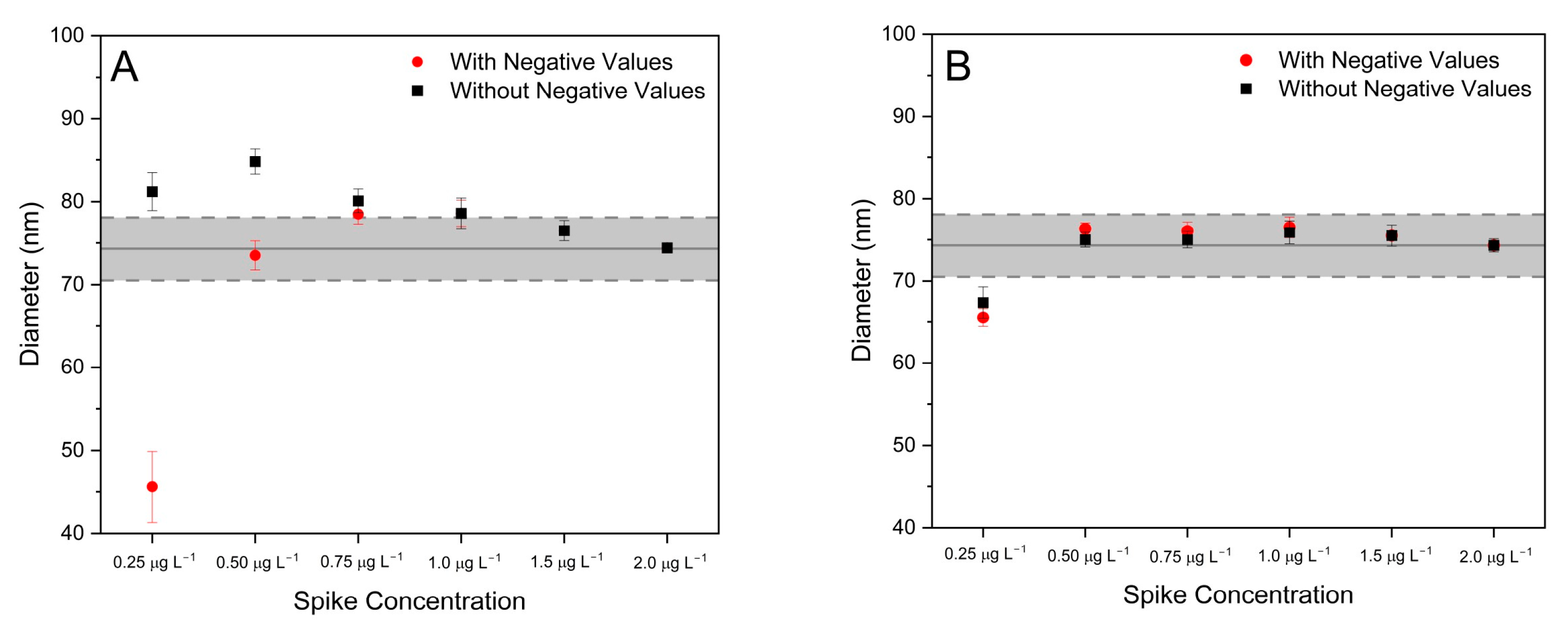
3.2.2. Effect of Integration Time (tint) and Composition/Concentration of the Spike
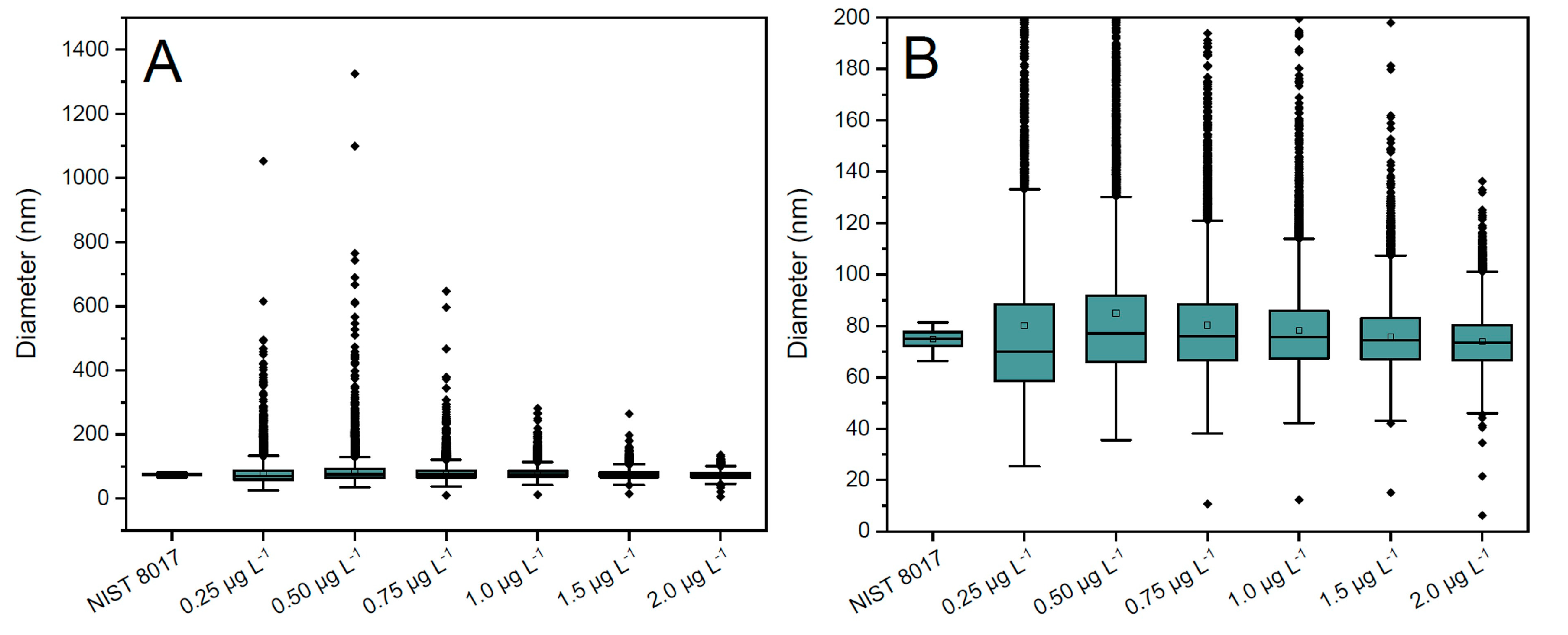
Selection of the Integration Time and Correction for Split Events
- Split events identification: split events are identified when two adjacent intense signals over a user-defined threshold are detected for the m/z = 107 trace (where contribution of the spike to the signal is low, as composition of the spike was selected to be ). This threshold was set as the average background for the m/z = 107 trace, as for the acquisition conditions used in this work, the probability for two adjacent signals coming from two different NPs instead of a split one is negligible [37].
- NP mass integration: the masses obtained for these adjacent signals after application of the IDA equation (Equation (3)) are integrated by the script.
- NP diameter calculation: the diameter of the NP is calculated with the integrated value obtained in step (2) (see Section 2.3. for more details).
Selection of the Isotopic Composition and Concentration of the Spike
3.2.3. Mass-Discrimination Correction
3.3. Application of the IDA Developed Method for Sizing AgNPs Using ICP-ToFMS
4. Conclusions
Author Contributions
Funding
Data Availability Statement
Acknowledgments
Conflicts of Interest
References
- Laborda, F.; Bolea, E.; Jiménez-Lamana, J. Single Particle Inductively Coupled Plasma Mass Spectrometry: A Powerful Tool for Nanoanalysis. Anal. Chem. 2014, 86, 2270–2278. [Google Scholar] [CrossRef]
- Krystek, P.; Ulrich, A.; Garcia, C.C.; Manohar, S.; Ritsema, R. Application of Plasma Spectrometry for the Analysis of Engineered Nanoparticles in Suspensions and Products. J. Anal. At. Spectrom. 2011, 26, 1701–1721. [Google Scholar] [CrossRef]
- Mitrano, D.M.; Ranville, J.F.; Bednar, A.; Kazor, K.; Hering, A.S.; Higgins, C.P. Tracking Dissolution of Silver Nanoparticles at Environmentally Relevant Concentrations in Laboratory, Natural, and Processed Waters Using Single Particle ICP-MS (SpICP-MS). Environ. Sci. Nano 2014, 1, 248–259. [Google Scholar] [CrossRef]
- Mozhayeva, D.; Engelhard, C. A Critical Review of Single Particle Inductively Coupled Plasma Mass Spectrometry—A Step towards an Ideal Method for Nanomaterial Characterization. J. Anal. At. Spectrom. 2020, 35, 1740–1783. [Google Scholar] [CrossRef]
- Bolea-Fernández, E.; Leite, D.; Rua-Ibarz, A.; Balcaen, L.; Aramendia, M.; Resano, M.; Vanhaecke, F. Characterization of SiO2 Nanoparticles by Single Particle-Inductively Coupled Plasma-Tandem Mass Spectrometry (SP-ICP-MS/MS). J. Anal. At. Spectrom. 2017, 32, 2140–2152. [Google Scholar] [CrossRef]
- Bolea-Fernandez, E.; Leite, D.; Rua-Ibarz, A.; Liu, T.; Woods, G.; Aramendia, M.; Resano, M.; Vanhaecke, F. On the Effect of Using Collision/Reaction Cell (CRC) Technology in Single-Particle ICP-Mass Spectrometry (SP-ICP-MS). Anal. Chim. Acta 2019, 1077, 95–106. [Google Scholar] [CrossRef]
- Pace, H.E.; Rogers, N.J.; Jarolimek, C.; Coleman, V.A.; Higgins, C.P.; Ranville, J.F. Determining Transport Efficiency for the Purpose of Counting and Sizing Nanoparticles via Single Particle Inductively Coupled Plasma Mass Spectrometry. Anal. Chem. 2011, 83, 9361–9369. [Google Scholar] [CrossRef]
- Montaño, M.D.; Olesik, J.W.; Barber, A.G.; Challis, K.; Ranville, J.F. Single Particle ICP-MS: Advances toward Routine Analysis of Nanomaterials. Anal. Bioanal. Chem. 2016, 408, 5053–5074. [Google Scholar] [CrossRef]
- Abad-Alvaro, I.; Leite, D.; Bartczak, D.; Cuello-Nunez, S.; Gomez-Gomez, B.; Madrid, Y.; Aramendia, M.; Resano, M.; Goenaga-Infante, H. An Insight into the Determination of Size and Number Concentration of Silver Nanoparticles in Blood Using Single Particle ICP-MS (SpICP-MS): Feasibility of Application to Samples Relevant to in Vivo Toxicology Studies. J. Anal. At. Spectrom. 2021, 36, 1180–1192. [Google Scholar] [CrossRef]
- Loula, M.; Kaňa, A.; Mestek, O. Non-Spectral Interferences in Single-Particle ICP-MS Analysis_ An Underestimated Phenomenon. Talanta 2019, 202, 565–571. [Google Scholar] [CrossRef]
- Aramendía, M.; García-Mesa, J.C.; Alonso, E.V.; Garde, R.; Bazo, A.; Resano, J.; Resano, M. A Novel Approach for Adapting the Standard Addition Method to Single Particle-ICP-MS for the Accurate Determination of NP Size and Number Concentration in Complex Matrices. Anal. Chim. Acta 2022, 1205, 339738. [Google Scholar] [CrossRef] [PubMed]
- Olesik, J.W.; Gray, P.J. Considerations for Measurement of Individual Nanoparticles or Microparticles by ICP-MS: Determination of the Number of Particles and the Analyte Mass in Each Particle. J. Anal. At. Spectrom. 2012, 27, 1143–1155. [Google Scholar] [CrossRef]
- Miyashita, S.; Mitsuhashi, H.; Fujii, S.; Takatsu, A.; Inagaki, K.; Fujimoto, T. High Transport Efficiency of Nanoparticles through a Total-Consumption Sample Introduction System and Its Beneficial Application for Particle Size Evaluation in Single-Particle ICP-MS. Anal. Bioanal. Chem. 2017, 409, 1531–1545. [Google Scholar] [CrossRef]
- Ramkorun-Schmidt, B.; Pergantis, S.A.; Esteban-Fernández, D.; Jakubowski, N.; Günther, D. Investigation of a Combined Microdroplet Generator and Pneumatic Nebulization System for Quantitative Determination of Metal-Containing Nanoparticles Using ICPMS. Anal. Chem. 2015, 87, 8687–8694. [Google Scholar] [CrossRef]
- Hendriks, L.; Ramkorun-Schmidt, B.; Gundlach-Graham, A.; Koch, J.; Grass, R.N.; Jakubowski, N.; Gunther, D. Single-Particle ICP-MS with Online Microdroplet Calibration: Toward Matrix Independent Nanoparticle Sizing. J. Anal. At. Spectrom. 2019, 34, 716–728. [Google Scholar] [CrossRef]
- Meermann, B.; Nischwitz, V. ICP-MS for the Analysis at the Nanoscale—A Tutorial Review. J. Anal. At. Spectrom. 2018, 33, 1432–1468. [Google Scholar] [CrossRef]
- Resano, M.; Aramendía, M.; García-Ruiz, E.; Bazo, A.; Bolea-Fernandez, E.; Vanhaecke, F. Living in a Transient World: ICP-MS Reinvented via Time-Resolved Analysis for Monitoring Single Events. Chem. Sci. 2022, 13, 4436–4473. [Google Scholar] [CrossRef]
- Bazo, A.; Aramendía, M.; Nakadi, F.V.; Resano, M. An Approach Based on an Increased Bandpass for Enabling the Use of Internal Standards in Single Particle ICP-MS: Application to AuNPs Characterization. Nanomaterials 2023, 13, 1838. [Google Scholar] [CrossRef]
- Agatemor, C.; Beauchemin, D. Matrix Effects in Inductively Coupled Plasma Mass Spectrometry: A Review. Anal. Chim. Acta 2011, 706, 66–83. [Google Scholar] [CrossRef]
- Rodríguez-González, P.; Marchante-Gayón, J.M.; García Alonso, J.I.; Sanz-Medel, A. Isotope Dilution Analysis for Elemental Speciation: A Tutorial Review. Spectrochim. Acta Part B At. Spectrosc. 2005, 60, 151–207. [Google Scholar] [CrossRef]
- Olesik, J.W. Investigating the Fate of Individual Sample Droplets in Inductively Coupled Plasmas. Appl. Spectrosc. 1997, 51, 158A–175A. [Google Scholar] [CrossRef]
- Garcia, C.C.; Murtazin, A.; Groh, S.; Horvatic, V.; Niemax, K. Characterization of Single Au and SiO2 Nano- and Microparticles by ICP-OES Using Monodisperse Droplets of Standard Solutions for Calibration. J. Anal. At. Spectrom. 2010, 25, 645–653. [Google Scholar] [CrossRef]
- Telgmann, L.; Metcalfe, C.D.; Hintelmann, H. Rapid Size Characterization of Silver Nanoparticles by Single Particle ICP-MS and Isotope Dilution. J. Anal. At. Spectrom. 2014, 29, 1265–1272. [Google Scholar] [CrossRef]
- Sötebier, C.A.; Kutscher, D.J.; Rottmann, L.; Jakubowski, N.; Panne, U.; Bettmer, J. Combination of Single Particle ICP-QMS and Isotope Dilution Analysis for the Determination of Size, Particle Number and Number Size Distribution of Silver Nanoparticles. J. Anal. At. Spectrom. 2016, 31, 2045–2052. [Google Scholar] [CrossRef]
- Rottmann, L.; Heumann, K.G. Development of an On-Line Isotope Dilution Technique with HPLC/ICP-MS for the Accurate Determination of Elemental Species. Fresenius J. Anal. Chem. 1994, 350, 221–227. [Google Scholar] [CrossRef]
- Rottmann, L.; Heumann, K.G. Determination of Heavy Metal Interactions with Dissolved Organic Materials in Natural Aquatic Systems by Coupling a High-Performance Liquid Chromatography System with an Inductively Coupled Plasma Mass Spectrometer. Anal. Chem. 1994, 66, 3709–3715. [Google Scholar] [CrossRef]
- Borovinskaya, O.; Hattendorf, B.; Tanner, M.; Gschwind, S.; Günther, D. A Prototype of a New Inductively Coupled Plasma Time-of-Flight Mass Spectrometer Providing Temporally Resolved, Multi-Element Detection of Short Signals Generated by Single Particles and Droplets. J. Anal. At. Spectrom. 2013, 28, 226–233. [Google Scholar] [CrossRef]
- Hendriks, L.; Gundlach-Graham, A.; Hattendorf, B.; Günther, D. Characterization of a New ICP-TOFMS Instrument with Continuous and Discrete Introduction of Solutions. J. Anal. At. Spectrom. 2017, 32, 548–561. [Google Scholar] [CrossRef]
- Von der Au, M.; Faßbender, S.; Chronakis, M.I.; Vogl, J.; Meermann, B. Size Determination of Nanoparticles by ICP-ToF-MS Using Isotope Dilution in Microdroplets. J. Anal. At. Spectrom. 2022, 37, 1203–1207. [Google Scholar] [CrossRef]
- Yongyang, S.; Wei, W.; Zhiming, L.; Hu, D.; Guoqing, Z.; Jiang, X.; Xiangjun, R. Direct Detection and Isotope Analysis of Individual Particles in Suspension by Single Particle Mode MC-ICP-MS for Nuclear Safety. J. Anal. At. Spectrom. 2015, 30, 1184–1190. [Google Scholar] [CrossRef]
- Kappel, S.; Boulyga, S.F.; Dorta, L.; Guenther, D.; Hattendorf, B.; Koffler, D.; Laaha, G.; Leisch, F.; Prohaska, T. Evaluation Strategies for Isotope Ratio Measurements of Single Particles by LA-MC-ICPMS. Anal. Bioanal. Chem. 2013, 405, 2943–2955. [Google Scholar] [CrossRef] [PubMed]
- Cuello-Nuñez, S.; Abad-Álvaro, I.; Bartczak, D.; del Castillo Busto, M.E.; Ramsay, D.A.; Pellegrino, F.; Goenaga-Infante, H. The Accurate Determination of Number Concentration of Inorganic Nanoparticles Using SpICP-MS with the Dynamic Mass Flow Approach. J. Anal. At. Spectrom. 2020, 35, 1832–1839. [Google Scholar] [CrossRef]
- Todolí, J.-L.; Mermet, J.-M. Acid Interferences in Atomic Spectrometry: Analyte Signal Effects and Subsequent Reduction. Spectrochim. Acta Part B At. Spectrosc. 1999, 54, 895–929. [Google Scholar] [CrossRef]
- Tuoriniemi, J.; Cornelis, G.; Hassellöv, M. Size Discrimination and Detection Capabilities of Single-Particle ICPMS for Environmental Analysis of Silver Nanoparticles. Anal. Chem. 2012, 84, 3965–3972. [Google Scholar] [CrossRef] [PubMed]
- Ulianov, A.; Müntener, O.; Schaltegger, U. The ICPMS Signal as a Poisson Process: A Review of Basic Concepts. J. Anal. At. Spectrom. 2015, 30, 1297–1321. [Google Scholar] [CrossRef]
- Peters, R.; Herrera-Rivera, Z.; Undas, A.; van der Lee, M.; Marvin, H.; Bouwmeester, H.; Weigel, S. Single Particle ICP-MS Combined with a Data Evaluation Tool as a Routine Technique for the Analysis of Nanoparticles in Complex Matrices. J. Anal. At. Spectrom. 2015, 30, 1274–1285. [Google Scholar] [CrossRef]
- Liu, J.; Murphy, K.E.; MacCuspie, R.I.; Winchester, M.R. Capabilities of Single Particle Inductively Coupled Plasma Mass Spectrometry for the Size Measurement of Nanoparticles: A Case Study on Gold Nanoparticles. Anal. Chem. 2014, 86, 3405–3414. [Google Scholar] [CrossRef]
- Petersen, E.J.; Montoro Bustos, A.R.; Toman, B.; Johnson, M.E.; Ellefson, M.; Caceres, G.C.; Neuer, A.L.; Chan, Q.; Kemling, J.W.; Mader, B.; et al. Determining What Really Counts: Modeling and Measuring Nanoparticle Number Concentrations. Environ. Sci. Nano 2019, 6, 2876–2896. [Google Scholar] [CrossRef]


| Component/Parameter | Type/Value/Mode |
|---|---|
| Plasma gas flow rate | 14.0 L min−1 |
| Auxiliary gas flow rate | 0.80 L min−1 |
| Nebulizer gas flow rate | 1.05 L min−1 |
| RF power | 1550 W |
| Integration time | 3 ms |
| Data acquisition mode | Continuous mode |
| Collision/reaction cell gas | Not used |
| Data acquisition isotopes | 107Ag, 109Ag |
| Detected mass range | 23Na-238U |
| Total acquisition time per sample | 1 min |
| Spike Concentration (µg L−1) | Negative Values (%) | Average Rm ± SD |
|---|---|---|
| 0.25 | 14.2 | 0.8633 ± 0.051 |
| 0.50 | 3.4 | 0.7167 ± 0.020 |
| 0.75 | 0.4 | 0.5969 ± 0.012 |
| 1.00 | 0.1 | 0.5217 ± 0.0069 |
| 1.50 | 0.0 | 0.4063 ± 0.0065 |
| 2.00 | 0.0 | 0.3217 ± 0.0019 |
| Spike Concentration (µg L−1) | Particle Mass Recovery (Median) (%) | Particle Mass Recovery (Average ± SD) (%) |
|---|---|---|
| 0.25 | 338 | 270 ± 96 |
| 0.50 | 293 | 373 ± 159 |
| 0.75 | 168 | 167 ± 36 |
| 1.00 | 142 | 138 ± 6.0 |
| 1.50 | 119 | 112 ± 10 |
| 2.00 | 98 | 98.0 ± 0.1 |
| Spike Concentration (µg L−1) | LODsize ± SD (nm) |
|---|---|
| 0.25 | 33.8 ± 1.7 |
| 0.50 | 33.1 ± 0.80 |
| 0.75 | 32.6 ± 0.57 |
| 1.00 | 32.7 ± 0.50 |
| 1.50 | 32.4 ± 0.82 |
| 2.00 | 32.3 ± 0.74 |
| Sample | K Factor ± SD |
|---|---|
| NPs | 1.0562 ± 0.0126 |
| Ionic solutions | 1.0554 ± 0.0033 |
| Batch 1 | Batch 2 | Batch 3 | Average of Batches (nm) ± Expanded Uncertainty (k = 2) | Reference Values (nm) ± Expanded Uncertainty (k = 2) 1 | ||||
|---|---|---|---|---|---|---|---|---|
| AFM | TEM | USAXS | DLS | |||||
| Average AgNP size (nm) | 69.6 | 69.3 | 70.3 | 69.8 ± 2.7 | 70.1 ± 6.0 | 74.6 ± 3.8 | 67.9 ± 0.8 | 105.6 ± 4.6 |
| RSD (%) | 0.31 | 0.48 | 0.84 | |||||
| Median AgNP size (nm) | 69.8 | 69.4 | 70.7 | 69.8 ± 2.7 | ||||
| RSD (%) | 0.28 | 0.67 | 1.40 | |||||
Disclaimer/Publisher’s Note: The statements, opinions and data contained in all publications are solely those of the individual author(s) and contributor(s) and not of MDPI and/or the editor(s). MDPI and/or the editor(s) disclaim responsibility for any injury to people or property resulting from any ideas, methods, instructions or products referred to in the content. |
© 2023 by the authors. Licensee MDPI, Basel, Switzerland. This article is an open access article distributed under the terms and conditions of the Creative Commons Attribution (CC BY) license (https://creativecommons.org/licenses/by/4.0/).
Share and Cite
Aramendía, M.; Leite, D.; Resano, J.; Resano, M.; Billimoria, K.; Goenaga-Infante, H. Isotope Dilution Analysis for Particle Mass Determination Using Single-Particle Inductively Coupled Plasma Time-of-Flight Mass Spectrometry: Application to Size Determination of Silver Nanoparticles. Nanomaterials 2023, 13, 2392. https://doi.org/10.3390/nano13172392
Aramendía M, Leite D, Resano J, Resano M, Billimoria K, Goenaga-Infante H. Isotope Dilution Analysis for Particle Mass Determination Using Single-Particle Inductively Coupled Plasma Time-of-Flight Mass Spectrometry: Application to Size Determination of Silver Nanoparticles. Nanomaterials. 2023; 13(17):2392. https://doi.org/10.3390/nano13172392
Chicago/Turabian StyleAramendía, Maite, Diego Leite, Javier Resano, Martín Resano, Kharmen Billimoria, and Heidi Goenaga-Infante. 2023. "Isotope Dilution Analysis for Particle Mass Determination Using Single-Particle Inductively Coupled Plasma Time-of-Flight Mass Spectrometry: Application to Size Determination of Silver Nanoparticles" Nanomaterials 13, no. 17: 2392. https://doi.org/10.3390/nano13172392