

Boron Nanoparticle-Enhanced Proton Therapy for Cancer Treatment

 , , , and
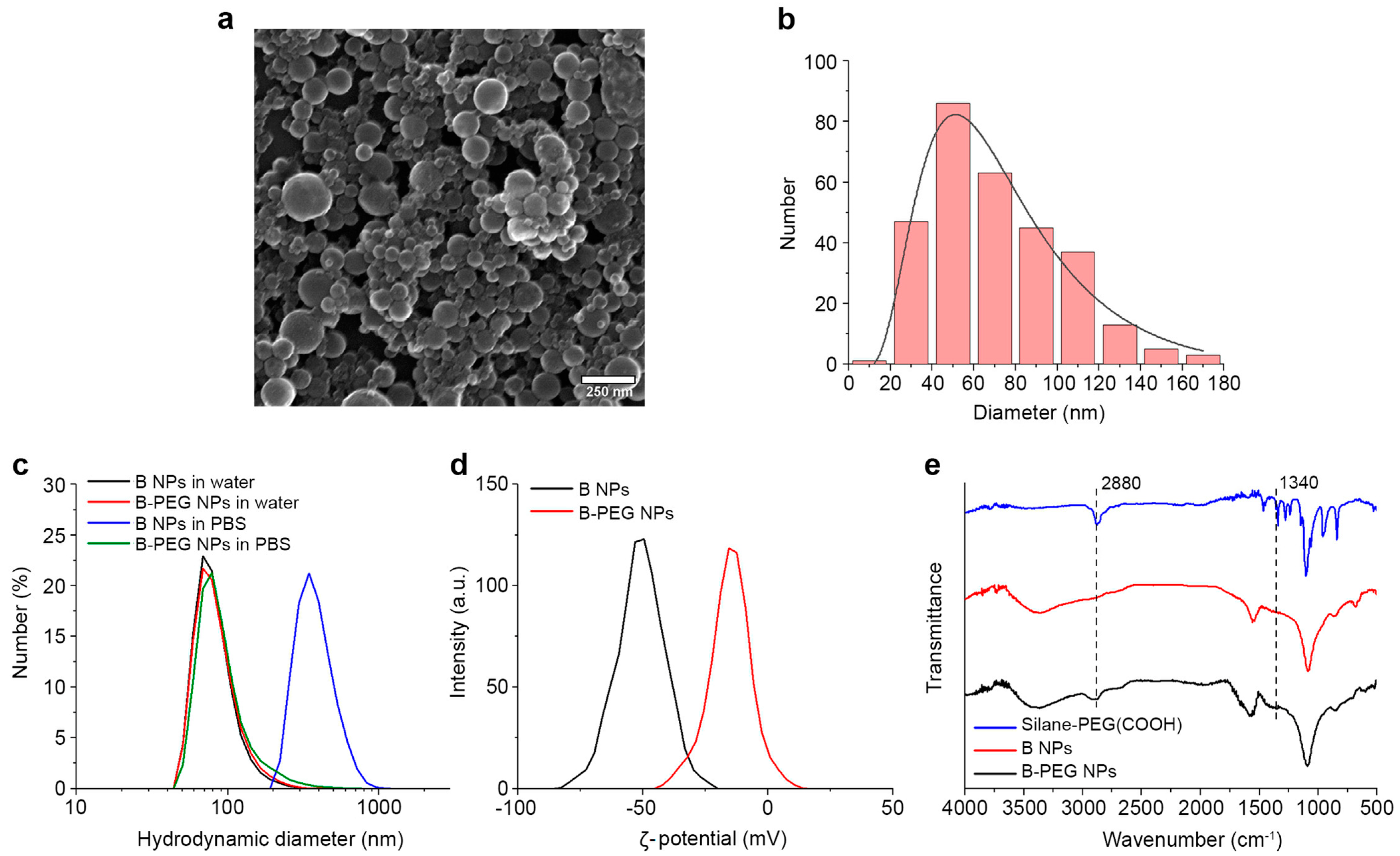
Abstract
:1. Introduction
2. Materials and Methods
2.1. Synthesis of Boron Nanoparticles
2.2. NP Characterization
2.3. Stabilization of Boron Nanoparticles
2.4. Proton Beam Irradiation
2.5. Cell Line
2.6. Clonogenic Assay
2.7. Apoptosis Analysis
2.8. Reactive Oxygen Species Detection
2.9. Analysis of Mitochondrial Membrane Potential (MMP)
2.10. Inductively Coupled Plasma Mass Spectrometry (ICP-MS)
2.11. Statistical Analysis
3. Results and Discussion
3.1. Synthesis and Characterization of Nanoparticles
3.2. Functionalization of Boron Nanoparticles
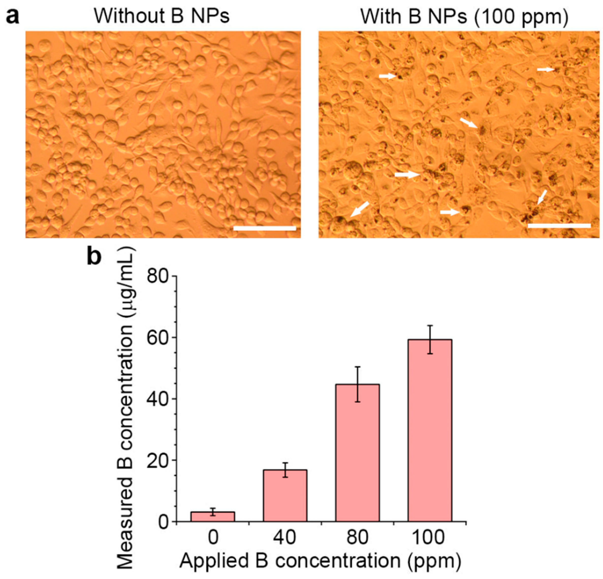
3.3. Cytotoxicity Studies
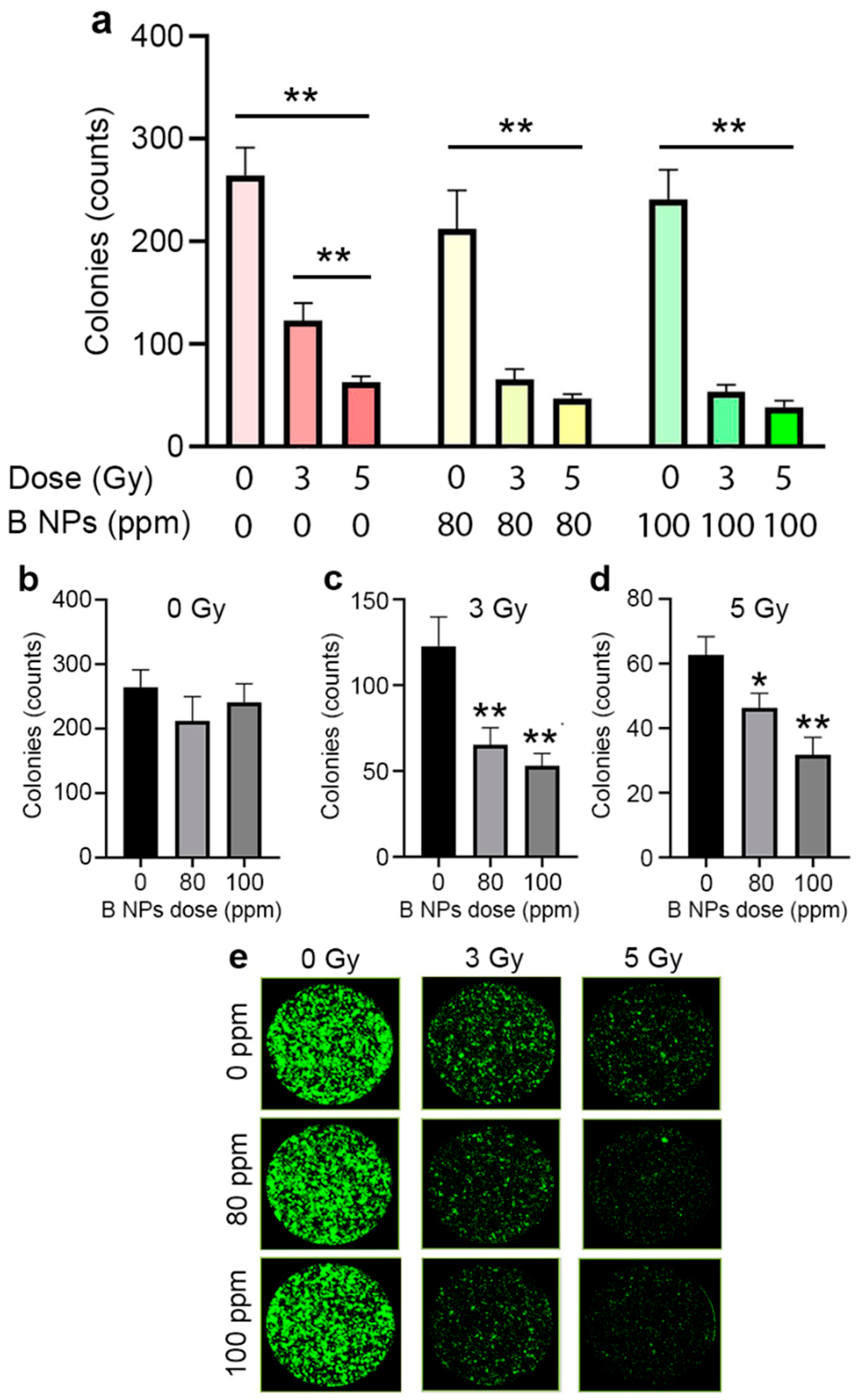
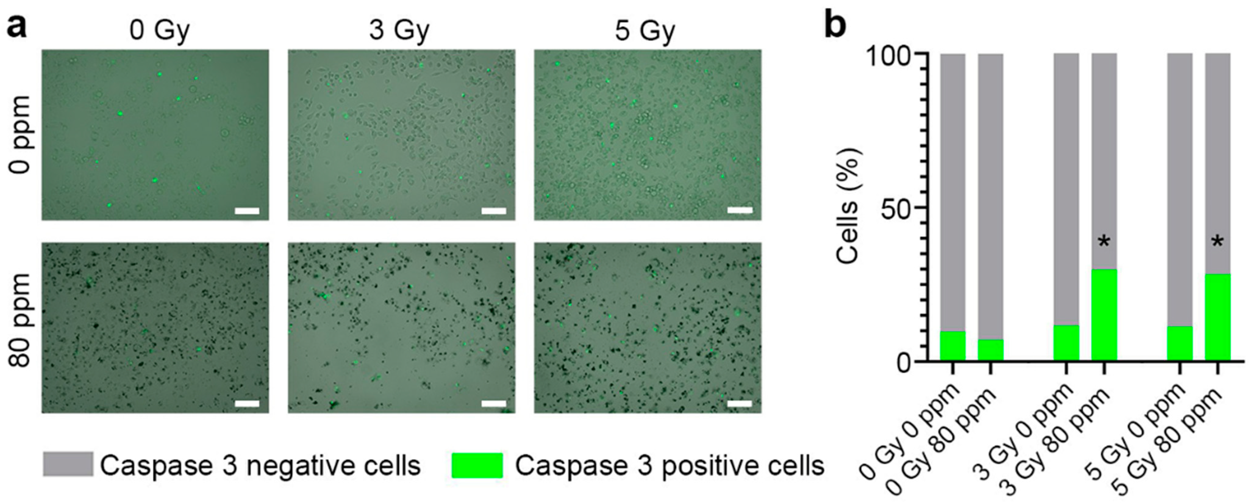
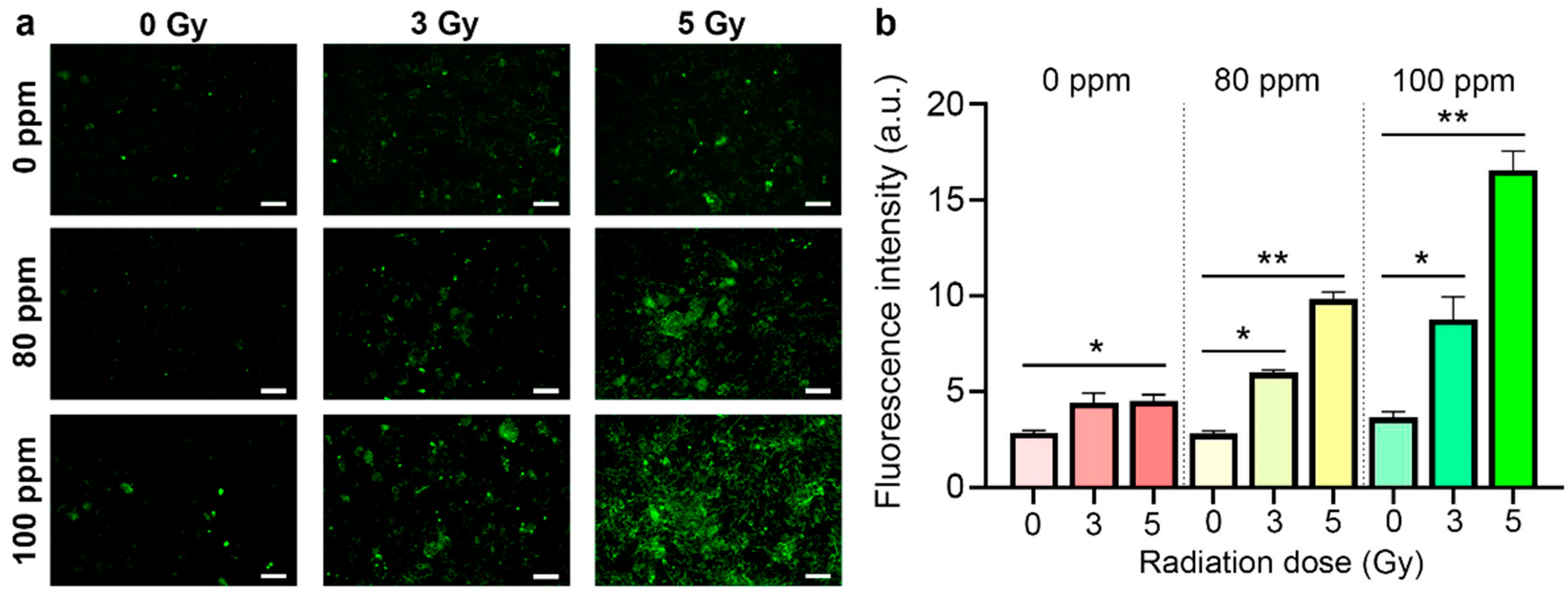
3.4. Proton Beam Irradiation Experiments
4. Discussion
5. Conclusions
Supplementary Materials
Author Contributions
Funding
Data Availability Statement
Conflicts of Interest
References
- Loeffler, J.S.; Durante, M. Charged particle therapy—Optimization, challenges and future directions. Nat. Rev. Clin. Oncol. 2013, 10, 411–424. [Google Scholar]
- Tommasino, F.; Durante, M. Proton radiobiology. Cancers 2015, 7, 353–381. [Google Scholar]
- Doyen, J.; Falk, A.T.; Floquet, V.; Hérault, J.; Hannoun-Lévi, J.-M. Proton beams in cancer treatments: Clinical outcomes and dosimetric comparisons with photon therapy. Cancer Treat. Rev. 2016, 43, 104–112. [Google Scholar]
- Bragg, W.H.; Kleeman, R. XXXIX. On the α particles of radium, and their loss of range in passing through various atoms and molecules. Lond. Edinb. Dublin Philos. Mag. J. Sci. 1905, 10, 318–340. [Google Scholar]
- Barth, R.F.; Soloway, A.H.; Fairchild, R.G. Boron neutron capture therapy for cancer. Sci. Am. 1990, 263, 100–107. [Google Scholar]
- Nedunchezhian, K.; Aswath, N.; Thiruppathy, M.; Thirugnanamurthy, S. Boron neutron capture therapy-a literature review. J. Clin. Diagn. Res. JCDR 2016, 10, ZE01. [Google Scholar]
- Malouff, T.D.; Seneviratne, D.S.; Ebner, D.K.; Stross, W.C.; Waddle, M.R.; Trifiletti, D.M.; Krishnan, S. Boron neutron capture therapy: A review of clinical applications. Front. Oncol. 2021, 11, 601820. [Google Scholar] [PubMed]
- Dee, P.I.; Gilbert, C. The disintegration of Boron into three α-particles. Proc. R. Soc. Lond. Ser. A-Math. Phys. Sci. 1936, 154, 279–296. [Google Scholar]
- Ruggiero, A.G. Nuclear Fusion of Protons with Boron; Brookhaven National Lab.: Upton, NY, USA, 1992. [Google Scholar]
- Yoon, D.-K.; Jung, J.-Y.; Suh, T.S. Application of proton boron fusion reaction to radiation therapy: A Monte Carlo simulation study. Appl. Phys. Lett. 2014, 105, 223507. [Google Scholar]
- Jung, J.-Y.; Yoon, D.-K.; Lee, H.C.; Lu, B.; Suh, T.S. The investigation of physical conditions of boron uptake region in proton boron fusion therapy (PBFT). AIP Adv. 2016, 6, 095119. [Google Scholar]
- Cirrone, G.; Manti, L.; Margarone, D.; Petringa, G.; Giuffrida, L.; Minopoli, A.; Picciotto, A.; Russo, G.; Cammarata, F.; Pisciotta, P. First experimental proof of Proton Boron Capture Therapy (PBCT) to enhance protontherapy effectiveness. Sci. Rep. 2018, 8, 1141. [Google Scholar]
- Ricciardi, V.; Bláha, P.; Buompane, R.; Crescente, G.; Cuttone, G.; Gialanella, L.; Michaličková, K.; Pacifico, S.; Porzio, G.; Manti, L. A New Low-Energy Proton Irradiation Facility to Unveil the Mechanistic Basis of the Proton-Boron Capture Therapy Approach. Appl. Sci. 2021, 11, 11986. [Google Scholar]
- Bláha, P.; Feoli, C.; Agosteo, S.; Calvaruso, M.; Cammarata, F.P.; Catalano, R.; Ciocca, M.; Cirrone, G.A.P.; Conte, V.; Cuttone, G. The proton-boron reaction increases the radiobiological effectiveness of clinical low-and high-energy proton beams: Novel experimental evidence and perspectives. Front. Oncol. 2021, 11, 682647. [Google Scholar]
- Cammarata, F.P.; Torrisi, F.; Vicario, N.; Bravatà, V.; Stefano, A.; Salvatorelli, L.; D’Aprile, S.; Giustetto, P.; Forte, G.I.; Minafra, L. Proton boron capture therapy (PBCT) induces cell death and mitophagy in a heterotopic glioblastoma model. Commun. Biol. 2023, 6, 388. [Google Scholar]
- Manandhar, M.; Bright, S.J.; Flint, D.B.; Martinus, D.K.; Kolachina, R.V.; Kacem, M.B.; Titt, U.; Martin, T.J.; Lee, C.L.; Morrison, K. Effect of boron compounds on the biological effectiveness of proton therapy. Med. Phys. 2022, 49, 6098–6109. [Google Scholar]
- Hosobuchi, M.; Kataoka, J.; Yokokawa, H.; Okazaki, Y.; Hirayama, R.; Inaniwa, T.; Ueda, M.; Kimura, M. Experimental verification of efficacy of pBCT in terms of physical and biological aspects. Nucl. Instrum. Methods Phys. Res. Sect. A Accel. Spectrometers Detect. Assoc. Equip. 2023, 1045, 167537. [Google Scholar]
- Shtam, T.; Burdakov, V.; Garina, A.; Garaeva, L.; Tran, N.H.; Volnitskiy, A.; Kuus, E.; Amerkanov, D.; Pack, F.; Andreev, G. Experimental validation of proton boron capture therapy for glioma cells. Sci. Rep. 2023, 13, 1341. [Google Scholar] [PubMed]
- Mazzone, A.; Finocchiaro, P.; Meo, S.L.; Colonna, N. On the (un) effectiveness of proton boron capture in proton therapy. Eur. Phys. J. Plus 2019, 134, 361. [Google Scholar]
- Kundrát, P.; Pachnerová Brabcová, K.; Jelínek Michaelidesová, A.; Zahradníček, O.; Danilová, I.; Štěpán, V.; Jamborová, Z.; Davídková, M. Boron-enhanced biological effectiveness of proton irradiation: Strategy to assess the underpinning mechanism. Radiat. Prot. Dosim. 2022, 198, 527–531. [Google Scholar]
- Song, G.; Cheng, L.; Chao, Y.; Yang, K.; Liu, Z. Emerging nanotechnology and advanced materials for cancer radiation therapy. Adv. Mater. 2017, 29, 1700996. [Google Scholar]
- Roy, I.; Krishnan, S.; Kabashin, A.V.; Zavestovskaya, I.N.; Prasad, P.N. Transforming nuclear medicine with nanoradiopharmaceuticals. ACS Nano 2022, 16, 5036–5061. [Google Scholar] [CrossRef] [PubMed]
- Matsumura, Y.; Maeda, H. A new concept for macromolecular therapeutics in cancer chemotherapy: Mechanism of tumoritropic accumulation of proteins and the antitumor agent smancs. Cancer Res. 1986, 46, 6387–6392. [Google Scholar]
- Chames, P.; Van Regenmortel, M.; Weiss, E.; Baty, D. Therapeutic antibodies: Successes, limitations and hopes for the future. Br. J. Pharmacol. 2009, 157, 220–233. [Google Scholar] [CrossRef] [Green Version]
- Tolmachev, V.M.; Chernov, V.I.; Deyev, S.M. Targeted nuclear medicine. Seek and destroy. Russ. Chem. Rev. 2022, 91, RCR5034. [Google Scholar] [CrossRef]
- Shipunova, V.O.; Komedchikova, E.N.; Kotelnikova, P.A.; Zelepukin, I.V.; Schulga, A.A.; Proshkina, G.M.; Shramova, E.I.; Kutscher, H.L.; Telegin, G.B.; Kabashin, A.V. Dual regioselective targeting the same receptor in nanoparticle-mediated combination immuno/chemotherapy for enhanced image-guided cancer treatment. ACS Nano 2020, 14, 12781–12795. [Google Scholar] [CrossRef]
- Anselmo, A.C.; Mitragotri, S. Nanoparticles in the clinic. Bioeng. Transl. Med. 2016, 1, 10–29. [Google Scholar] [CrossRef]
- Chen, G.; Roy, I.; Yang, C.; Prasad, P.N. Nanochemistry and nanomedicine for nanoparticle-based diagnostics and therapy. Chem. Rev. 2016, 116, 2826–2885. [Google Scholar] [CrossRef]
- Kabashin, A.V.; Singh, A.; Swihart, M.T.; Zavestovskaya, I.N.; Prasad, P.N. Laser-processed nanosilicon: A multifunctional nanomaterial for energy and healthcare. ACS Nano 2019, 13, 9841–9867. [Google Scholar] [CrossRef]
- Guo, T.; Tang, Q.; Guo, Y.; Qiu, H.; Dai, J.; Xing, C.; Zhuang, S.; Huang, G. Boron quantum dots for photoacoustic imaging-guided photothermal therapy. ACS Appl. Mater. Interfaces 2020, 13, 306–311. [Google Scholar] [CrossRef] [PubMed]
- Hao, J.; Tai, G.; Zhou, J.; Wang, R.; Hou, C.; Guo, W. Crystalline semiconductor boron quantum dots. ACS Appl. Mater. Interfaces 2020, 12, 17669–17675. [Google Scholar] [CrossRef]
- Rohani, P.; Kim, S.; Swihart, M.T. Boron nanoparticles for room-temperature hydrogen generation from water. Adv. Energy Mater. 2016, 6, 1502550. [Google Scholar] [CrossRef]
- Kögler, M.; Ryabchikov, Y.V.; Uusitalo, S.; Popov, A.; Popov, A.; Tselikov, G.; Välimaa, A.L.; Al-Kattan, A.; Hiltunen, J.; Laitinen, R. Bare laser-synthesized Au-based nanoparticles as nondisturbing surface-enhanced Raman scattering probes for bacteria identification. J. Biophotonics 2018, 11, e201700225. [Google Scholar] [CrossRef] [Green Version]
- Popov, A.A.; Tselikov, G.; Dumas, N.; Berard, C.; Metwally, K.; Jones, N.; Al-Kattan, A.; Larrat, B.; Braguer, D.; Mensah, S. Laser-synthesized TiN nanoparticles as promising plasmonic alternative for biomedical applications. Sci. Rep. 2019, 9, 1194. [Google Scholar] [CrossRef] [PubMed] [Green Version]
- Bulmahn, J.C.; Tikhonowski, G.; Popov, A.A.; Kuzmin, A.; Klimentov, S.M.; Kabashin, A.V.; Prasad, P.N. Laser-ablative synthesis of stable aqueous solutions of elemental bismuth nanoparticles for multimodal theranostic applications. Nanomaterials 2020, 10, 1463. [Google Scholar] [CrossRef]
- Kharin, A.Y.; Lysenko, V.V.; Rogov, A.; Ryabchikov, Y.V.; Geloen, A.; Tishchenko, I.; Marty, O.; Sennikov, P.G.; Kornev, R.A.; Zavestovskaya, I.N. Bi-Modal Nonlinear Optical Contrast from Si Nanoparticles for Cancer Theranostics. Adv. Opt. Mater. 2019, 7, 1801728. [Google Scholar] [CrossRef]
- Oleshchenko, V.; Kharin, A.Y.; Alykova, A.; Karpukhina, O.; Karpov, N.; Popov, A.; Bezotosnyi, V.; Klimentov, S.; Zavestovskaya, I.; Kabashin, A. Localized infrared radiation-induced hyperthermia sensitized by laser-ablated silicon nanoparticles for phototherapy applications. Appl. Surf. Sci. 2020, 516, 145661. [Google Scholar] [CrossRef]
- Tselikov, G.I.; Ermolaev, G.A.; Popov, A.A.; Tikhonowski, G.V.; Panova, D.A.; Taradin, A.S.; Vyshnevyy, A.A.; Syuy, A.V.; Klimentov, S.M.; Novikov, S.M. Transition metal dichalcogenide nanospheres for high-refractive-index nanophotonics and biomedical theranostics. Proc. Natl. Acad. Sci. USA 2022, 119, e2208830119. [Google Scholar] [CrossRef]
- Pastukhov, A.I.; Belyaev, I.B.; Bulmahn, J.C.; Zelepukin, I.V.; Popov, A.A.; Zavestovskaya, I.N.; Klimentov, S.M.; Deyev, S.M.; Prasad, P.N.; Kabashin, A.V. Laser-ablative aqueous synthesis and characterization of elemental boron nanoparticles for biomedical applications. Sci. Rep. 2022, 12, 9129. [Google Scholar] [CrossRef]
- Belyaev, I.; Zelepukin, I.; Pastukhov, A.; Shakhov, P.; Tikhonowski, G.; Popov, A.; Zakharkiv, A.Y.; Klimentov, S.; Garmash, A.; Zavestovskaya, I. Study of IR Photoheating of Aqueous Solutions of Boron Nanoparticles Synthesized by Pulsed Laser Ablation for Cancer Therapy. Bull. Lebedev Phys. Inst. 2022, 49, 185–189. [Google Scholar] [CrossRef]
- Kabashin, A.V.; Meunier, M. Synthesis of colloidal nanoparticles during femtosecond laser ablation of gold in water. J. Appl. Phys. 2003, 94, 7941–7943. [Google Scholar] [CrossRef]
- Shin, W.G.; Calder, S.; Ugurlu, O.; Girshick, S.L. Production and characterization of boron nanoparticles synthesized with a thermal plasma system. J. Nanopart. Res. 2011, 13, 7187–7191. [Google Scholar] [CrossRef]
- Zelepukin, I.V.; Griaznova, O.Y.; Shevchenko, K.G.; Ivanov, A.V.; Baidyuk, E.V.; Serejnikova, N.B.; Volovetskiy, A.B.; Deyev, S.M.; Zvyagin, A.V. Flash drug release from nanoparticles accumulated in the targeted blood vessels facilitates the tumour treatment. Nat. Commun. 2022, 13, 6910. [Google Scholar] [CrossRef] [PubMed]
- Lee, K.B.; Kim, K.-R.; Huh, T.-L.; Lee, Y.M. Proton induces apoptosis of hypoxic tumor cells by the p53-dependent and p38/JNK MAPK signaling pathways. Int. J. Oncol. 2008, 33, 1247–1256. [Google Scholar] [CrossRef] [PubMed]
- Alan Mitteer, R.; Wang, Y.; Shah, J.; Gordon, S.; Fager, M.; Butter, P.-P.; Jun Kim, H.; Guardiola-Salmeron, C.; Carabe-Fernandez, A.; Fan, Y. Proton beam radiation induces DNA damage and cell apoptosis in glioma stem cells through reactive oxygen species. Sci. Rep. 2015, 5, 13961. [Google Scholar] [CrossRef] [Green Version]
- Sia, J.; Szmyd, R.; Hau, E.; Gee, H. Molecular Mechanisms of Radiation-Induced Cancer Cell Death: A Primer. Front. Cell Dev. Biol. 2020, 8, 41. [Google Scholar] [CrossRef] [PubMed]
- Pinheiro, T.; Alves, L.C.; Corregidor, V.; Teixidor, F.; Viñas, C.; Marques, F. Metallacarboranes for proton therapy using research accelerators: A pilot study. EPJ Tech. Instrum. 2023, 10, 5. [Google Scholar] [CrossRef]
- Khaledi, N.; Wang, X.; Hosseinabadi, R.B.; Samiei, F. Is the proton–boron fusion therapy effective? J. Radiother. Pract. 2021, 20, 153–157. [Google Scholar] [CrossRef]
- Hideghéty, K.; Brunner, S.; Cheesman, A.; Szabo, E.R.; Polanek, R.; Margarone, D.; Tőkés, T.; Mogyorósi, K. 11Boron delivery agents for boron proton-capture enhanced proton therapy. Anticancer Res. 2019, 39, 2265–2276. [Google Scholar] [CrossRef] [Green Version]
- Mazzucconi, D.; Bortot, D.; Pola, A.; Fazzi, A.; Cazzola, L.; Conte, V.; Cirrone, G.; Petringa, G.; Cuttone, G.; Manti, L. Experimental investigation at CATANA facility of n-10B and p-11B reactions for the enhancement of proton therapy. Phys. Medica 2021, 89, 226–231. [Google Scholar] [CrossRef]
- Durante, M.; Orecchia, R.; Loeffler, J.S. Charged-particle therapy in cancer: Clinical uses and future perspectives. Nat. Rev. Clin. Oncol. 2017, 14, 483–495. [Google Scholar] [CrossRef]
- Mohan, R. A review of proton therapy—Current status and future directions. Precis. Radiat. Oncol. 2022, 6, 164–176. [Google Scholar] [CrossRef] [PubMed]
- Wang, S.; Zhou, Z.; Hu, R.; Dong, M.; Zhou, X.; Ren, S.; Zhang, Y.; Chen, C.; Huang, R.; Zhu, M.; et al. Metabolic Intervention Liposome Boosted Lung Cancer Radio-Immunotherapy via Hypoxia Amelioration and PD-L1 Restraint. Adv. Sci. 2023, 10, 2207608. [Google Scholar] [CrossRef] [PubMed]


Disclaimer/Publisher’s Note: The statements, opinions and data contained in all publications are solely those of the individual author(s) and contributor(s) and not of MDPI and/or the editor(s). MDPI and/or the editor(s) disclaim responsibility for any injury to people or property resulting from any ideas, methods, instructions or products referred to in the content. |
© 2023 by the authors. Licensee MDPI, Basel, Switzerland. This article is an open access article distributed under the terms and conditions of the Creative Commons Attribution (CC BY) license (https://creativecommons.org/licenses/by/4.0/).
Share and Cite
Zavestovskaya, I.N.; Popov, A.L.; Kolmanovich, D.D.; Tikhonowski, G.V.; Pastukhov, A.I.; Savinov, M.S.; Shakhov, P.V.; Babkova, J.S.; Popov, A.A.; Zelepukin, I.V.; et al. Boron Nanoparticle-Enhanced Proton Therapy for Cancer Treatment. Nanomaterials 2023, 13, 2167. https://doi.org/10.3390/nano13152167
Zavestovskaya IN, Popov AL, Kolmanovich DD, Tikhonowski GV, Pastukhov AI, Savinov MS, Shakhov PV, Babkova JS, Popov AA, Zelepukin IV, et al. Boron Nanoparticle-Enhanced Proton Therapy for Cancer Treatment. Nanomaterials. 2023; 13(15):2167. https://doi.org/10.3390/nano13152167
Chicago/Turabian StyleZavestovskaya, Irina N., Anton L. Popov, Danil D. Kolmanovich, Gleb V. Tikhonowski, Andrei I. Pastukhov, Maxim S. Savinov, Pavel V. Shakhov, Julia S. Babkova, Anton A. Popov, Ivan V. Zelepukin, and et al. 2023. "Boron Nanoparticle-Enhanced Proton Therapy for Cancer Treatment" Nanomaterials 13, no. 15: 2167. https://doi.org/10.3390/nano13152167