
Shear Bond Strength and Color Stability of Novel Antibacterial Nanofilled Dental Adhesive Resins
 ,
,  , ,
, ,  and
and
Abstract
:1. Introduction
2. Materials and Methods
2.1. Synthesis of Nanoparticles
2.2. Synthesis of Experimental Adhesives
2.3. Fabrication of Specimens
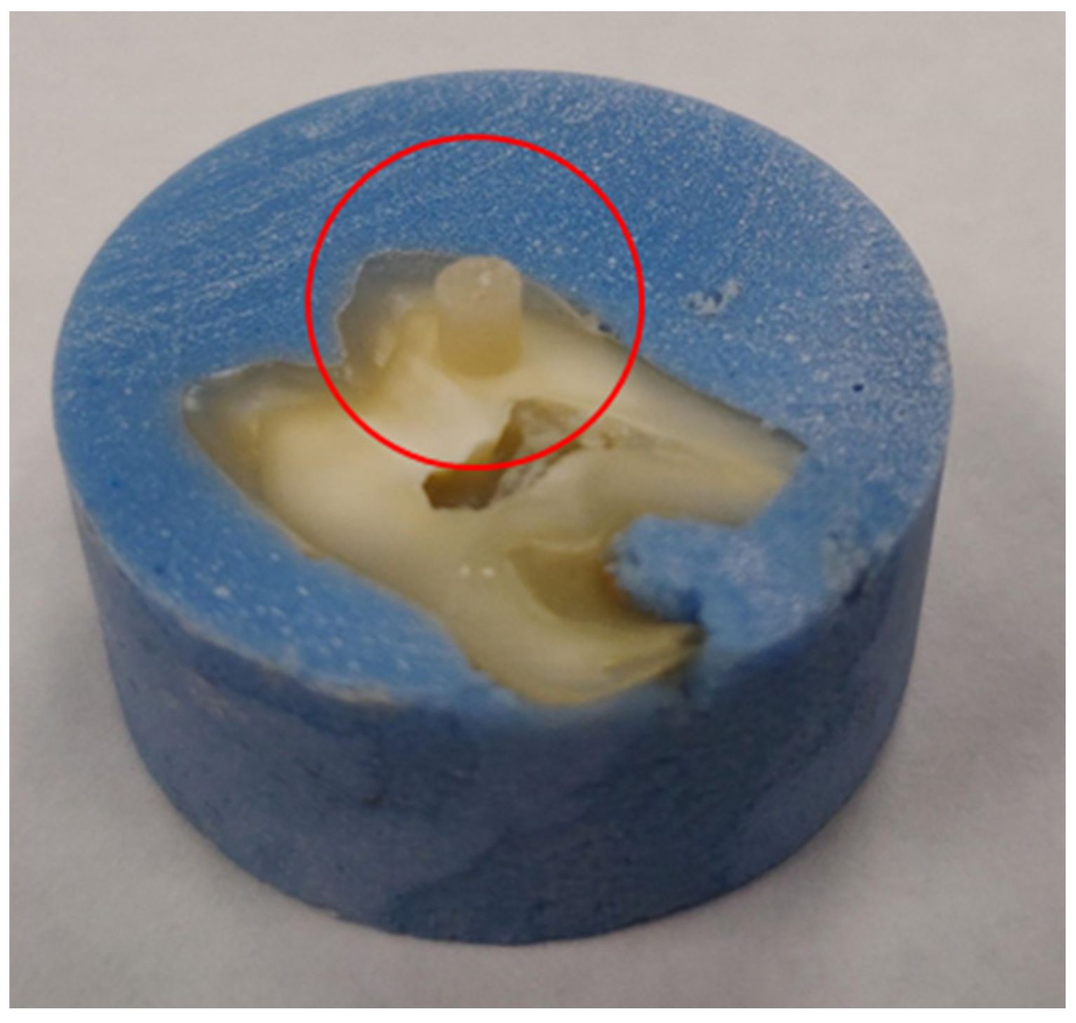
2.3.1. For Shear Bond Strength Test
2.3.2. For Color Stability Test
2.4. Shear Bond Strength Test
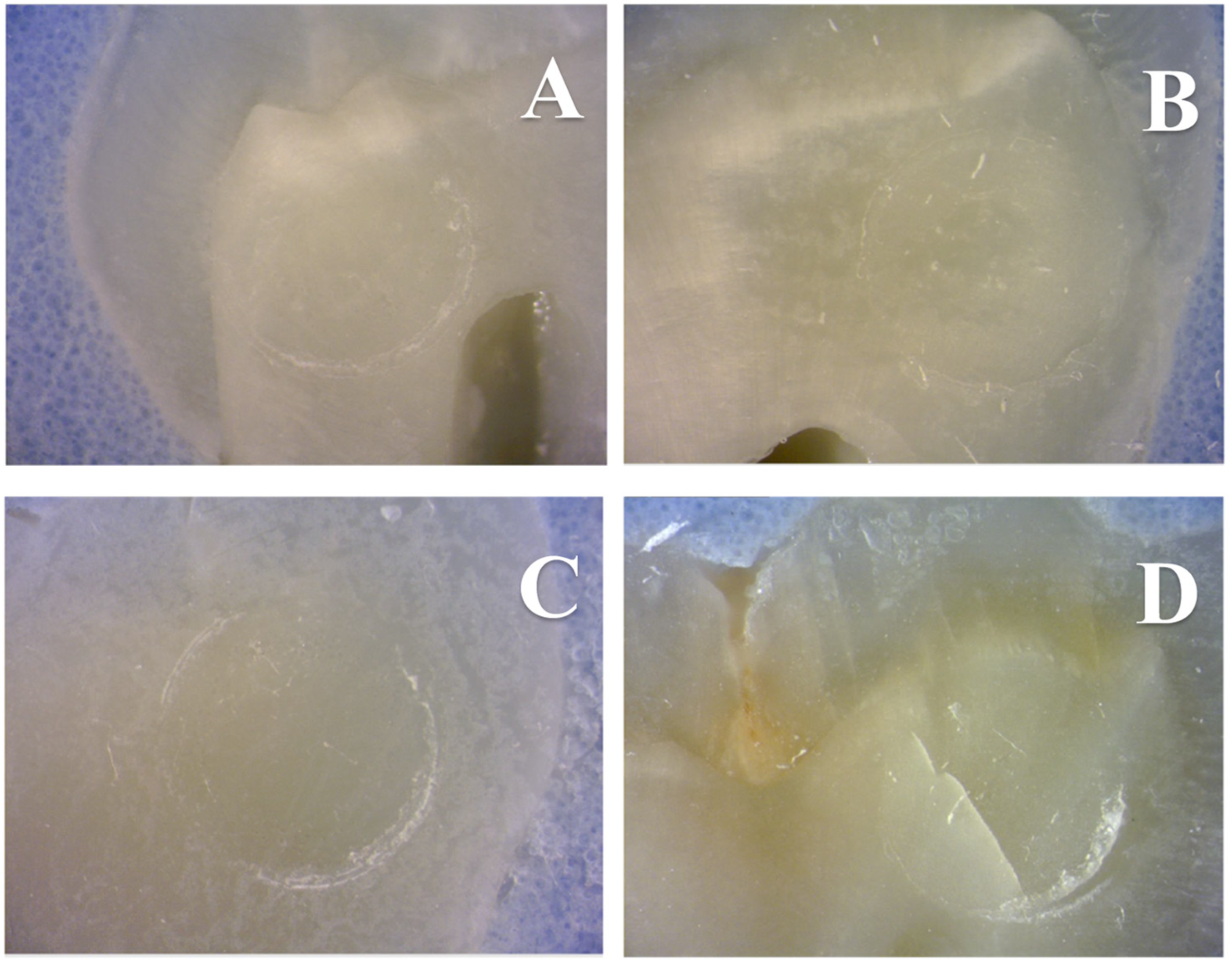
2.5. Scanning Electron Microscopy
2.6. Color Stability Test
2.7. Statistical Analysis
3. Results
4. Discussion
5. Conclusions
Author Contributions
Funding
Data Availability Statement
Conflicts of Interest
References
- Cho, K.; Rajan, G.; Farrar, P.; Prentice, L.; Prusty, B.G. Dental resin composites: A review on materials to product realizations. Compos. Part B Eng. 2022, 230, 109495. [Google Scholar] [CrossRef]
- Mai, S.; Zhang, Q.; Liao, M.; Ma, X.; Zhong, Y. Recent Advances in Direct Adhesive Restoration Resin-Based Dental Materials With Remineralizing Agents. Front. Dent. Med. 2022, 3, 22. [Google Scholar] [CrossRef]
- Pashley, D.H.; Tay, F.R.; Imazato, S. How to increase the Durability of Resin-Dentin Bonds. Compend. Contin. Educ. Dent. 2011, 32, 60–64. [Google Scholar]
- Burke, F.J.; Wilson, N.H.; Cheung, S.W.; Mjor, I.A. Influence of patient factors on age of restorations at failure and reasons for their placement and replacement. J. Dent. 2001, 29, 317–324. [Google Scholar] [CrossRef]
- Schricker, S.R. 9-Composite resin polymerization and relevant parameters. In Orthodontic Applications of Biomaterials; Eliades, T., Brantley, W.A., Eds.; Woodhead Publishing: Amsterdan, The Netherlands, 2017; pp. 153–170. [Google Scholar] [CrossRef]
- Gitalis, R.; Bae, J.H.; Preston, M.; Patel, M.; Liu, Z.; Sun, C.; Stewart, C.; Xiao, Y.; Siqueira, W.L.; Glogauer, M.; et al. Human neutrophils compromise the restoration-tooth interface. Acta Biomater. 2020, 117, 283–293. [Google Scholar] [CrossRef]
- Guo, X.; Yu, Y.; Gao, S.; Zhang, Z.; Zhao, H. Biodegradation of Dental Resin-Based Composite—A Potential Factor Affecting the Bonding Effect: A Narrative Review. Biomedicines 2022, 10, 2313. [Google Scholar]
- Bourbia, M.; Ma, D.; Cvitkovitch, D.G.; Santerre, J.P.; Finer, Y. Cariogenic bacteria degrade dental resin composites and adhesives. J. Dent. Res. 2013, 92, 989–994. [Google Scholar] [CrossRef]
- Sarikaya, R.; Song, L.; Yuca, E.; Xie, S.-X.; Boone, K.; Misra, A.; Spencer, P.; Tamerler, C. Bioinspired multifunctional adhesive system for next generation bio-additively designed dental restorations. J. Mech. Behav. Biomed. Mater. 2021, 113, 104135. [Google Scholar] [CrossRef]
- Spencer, P.; Ye, Q.; Misra, A.; Goncalves, S.E.P.; Laurence, J.S. Proteins, Pathogens, and Failure at the Composite-Tooth Interface. J. Dent. Res. 2014, 93, 1243–1249. [Google Scholar] [CrossRef] [Green Version]
- Spencer, P.; Ye, Q.; Park, J.; Topp, E.M.; Misra, A.; Marangos, O.; Wang, Y.; Bohaty, B.S.; Singh, V.; Sene, F.; et al. Adhesive/Dentin interface: The weak link in the composite restoration. Ann. Biomed. Eng. 2010, 38, 1989–2003. [Google Scholar] [CrossRef] [Green Version]
- Yuca, E.; Xie, S.-X.; Song, L.; Boone, K.; Kamathewatta, N.; Woolfolk, S.K.; Elrod, P.; Spencer, P.; Tamerler, C. Reconfigurable Dual Peptide Tethered Polymer System Offers a Synergistic Solution for Next Generation Dental Adhesives. Int. J. Mol. Sci. 2021, 22, 6552. [Google Scholar] [CrossRef]
- Sarikaya, R.; Song, L.; Ye, Q.; Misra, A.; Tamerler, C.; Spencer, P. Evolution of Network Structure and Mechanical Properties in Autonomous-Strengthening Dental Adhesive. Polymers 2020, 12, 2076. [Google Scholar] [CrossRef]
- Xie, S.X.; Song, L.; Yuca, E.; Boone, K.; Sarikaya, R.; VanOosten, S.K.; Misra, A.; Ye, Q.; Spencer, P.; Tamerler, C. Antimicrobial Peptide-Polymer Conjugates for Dentistry. ACS Appl. Polym. Mater. 2020, 2, 1134–1144. [Google Scholar] [CrossRef]
- Rai, M.; Yadav, A.; Gade, A. Silver nanoparticles as a new generation of antimicrobials. Biotechnol. Adv. 2009, 27, 76–83. [Google Scholar] [CrossRef]
- Cai, Y.; Strömme, M.; Zhang, P.; Engqvist, H.; Welch, K. Photocatalysis induces bioactivity of an organic polymer based material. RSC Adv. 2014, 4, 57715–57723. [Google Scholar] [CrossRef]
- Welch, K.; Cai, Y.; Engqvist, H.; Stromme, M. Dental adhesives with bioactive and on-demand bactericidal properties. Dent. Mater. 2010, 26, 491–499. [Google Scholar] [CrossRef]
- Pashley, D.H.; Tay, F.R.; Yiu, C.; Hashimoto, M.; Breschi, L.; Carvalho, R.M.; Ito, S. Collagen degradation by host-derived enzymes during aging. J. Dent. Res. 2004, 83, 216–221. [Google Scholar] [CrossRef]
- Hebling, J.; Pashley, D.H.; Tjaderhane, L.; Tay, F.R. Chlorhexidine arrests subclinical degradation of dentin hybrid layers in vivo. J. Dent. Res. 2005, 84, 741–746. [Google Scholar] [CrossRef]
- Carrilho, M.R.; Carvalho, R.M.; de Goes, M.F.; di Hipolito, V.; Geraldeli, S.; Tay, F.R.; Pashley, D.H.; Tjaderhane, L. Chlorhexidine preserves dentin bond in vitro. J. Dent. Res. 2007, 86, 90–94. [Google Scholar] [CrossRef] [Green Version]
- Makvandi, P.; Jamaledin, R.; Jabbari, M.; Nikfarjam, N.; Borzacchiello, A. Antibacterial quaternary ammonium compounds in dental materials: A systematic review. Dent. Mater. 2018, 34, 851–867. [Google Scholar] [CrossRef]
- Imazato, S.; Ebi, N.; Takahashi, Y.; Kaneko, T.; Ebisu, S.; Russell, R.R.B. Antibacterial activity of bactericide-immobilized filler for resin-based restoratives. Biomaterials 2003, 24, 3605–3609. [Google Scholar] [CrossRef]
- Zane, A.; Zuo, R.; Villamena, F.A.; Rockenbauer, A.; Digeorge Foushee, A.M.; Flores, K.; Dutta, P.K.; Nagy, A. Biocompatibility and antibacterial activity of nitrogen-doped titanium dioxide nanoparticles for use in dental resin formulations. Int. J. Nanomed. 2016, 11, 6459–6470. [Google Scholar] [CrossRef] [Green Version]
- Salehi, P.; Babanouri, N.; Roein-Peikar, M.; Zare, F. Long-term antimicrobial assessment of orthodontic brackets coated with nitrogen-doped titanium dioxide against Streptococcus mutans. Prog. Orthod. 2018, 19, 35. [Google Scholar] [CrossRef] [Green Version]
- Sodagar, A.; Akhoundi, M.S.A.; Bahador, A.; Jalali, Y.F.; Behzadi, Z.; Elhaminejad, F.; Mirhashemi, A.H. Effect of TiO2 nanoparticles incorporation on antibacterial properties and shear bond strength of dental composite used in Orthodontics. Dent. Press J. Orthod. 2017, 22, 67–74. [Google Scholar] [CrossRef] [Green Version]
- Chambers, C.; Stewart, S.B.; Su, B.; Jenkinson, H.F.; Sandy, J.R.; Ireland, A.J. Silver doped titanium dioxide nanoparticles as antimicrobial additives to dental polymers. Dent. Mater. 2017, 33, e115–e123. [Google Scholar] [CrossRef] [Green Version]
- Esteban Florez, F.L.; Hiers, R.D.; Larson, P.; Johnson, M.; O’Rear, E.; Rondinone, A.J.; Khajotia, S.S. Antibacterial dental adhesive resins containing nitrogen-doped titanium dioxide nanoparticles. Mater. Sci. Eng. C 2018, 93, 931–943. [Google Scholar] [CrossRef]
- Esteban Florez, F.L.; Kraemer, H.; Hiers, R.D.; Sacramento, C.M.; Rondinone, A.J.; Silvério, K.G.; Khajotia, S.S. Sorption, solubility and cytotoxicity of novel antibacterial nanofilled dental adhesive resins. Sci. Rep. 2020, 10, 13503. [Google Scholar] [CrossRef]
- Esteban Florez, F.L.; Trofimov, A.A.; Ievlev, A.; Qian, S.; Rondinone, A.J.; Khajotia, S.S. Advanced characterization of surface-modified nanoparticles and nanofilled antibacterial dental adhesive resins. Sci. Rep. 2020, 10, 9811. [Google Scholar] [CrossRef]
- Melo, M.A.; Cheng, L.; Zhang, K.; Weir, M.D.; Rodrigues, L.K.; Xu, H.H. Novel dental adhesives containing nanoparticles of silver and amorphous calcium phosphate. Dent. Mater. 2013, 29, 199–210. [Google Scholar] [CrossRef] [Green Version]
- Ashraf, M.A.; Peng, W.; Zare, Y.; Rhee, K.Y. Effects of Size and Aggregation/Agglomeration of Nanoparticles on the Interfacial/Interphase Properties and Tensile Strength of Polymer Nanocomposites. Nanoscale Res. Lett. 2018, 13, 214. [Google Scholar] [CrossRef]
- Meerbeek, V. Mechanisms of Resin Adhesion-Dentin and Enamel Bonding. Aegis Dent. Netw. 2008, 2, 2–8. [Google Scholar]
- Preethy, N.A.; Jeevanandan, G.; Govindaraju, L.; Subramanian, E. Comparison of Shear Bond Strength of Three Commercially Available Esthetic Restorative Composite Materials: An In Vitro Study. Int. J. Clin. Pediatr. Dent. 2020, 13, 635–639. [Google Scholar] [CrossRef]
- Nujella, B.P.S.; Choudary, M.T.; Reddy, S.P.; Kumar, M.K.; Gopal, T. Comparison of shear bond strength of aesthetic restorative materials. Contemp. Clin. Dent. 2012, 3, 22–26. [Google Scholar] [CrossRef]
- Sirisha, K.; Rambabu, T.; Shankar, Y.R.; Ravikumar, P. Validity of bond strength tests: A critical review: Part I. J. Conserv. Dent. 2014, 17, 305–311. [Google Scholar] [CrossRef] [Green Version]
- Sirisha, K.; Rambabu, T.; Ravishankar, Y.; Ravikumar, P. Validity of bond strength tests: A critical review-Part II. J. Conserv. Dent. 2014, 17, 420–426. [Google Scholar] [CrossRef] [Green Version]
- Van Meerbeek, B.; Peumans, M.; Poitevin, A.; Mine, A.; Van Ende, A.; Neves, A.; De Munck, J. Relationship between bond-strength tests and clinical outcomes. Dent. Mater. 2010, 26, e100–e121. [Google Scholar] [CrossRef]
- Hidari, T.; Takamizawa, T.; Imai, A.; Hirokane, E.; Ishii, R.; Tsujimoto, A.; Suzuki, T.; Miyazaki, M. Role of the functional monomer 10-methacryloyloxydecyl dihydrogen phosphate in dentin bond durability of universal adhesives in etch-&-rinse mode. Dent. Mater. J. 2020, 39, 616–623. [Google Scholar] [CrossRef] [Green Version]
- Takamizawa, T.; Barkmeier, W.W.; Tsujimoto, A.; Scheidel, D.D.; Watanabe, H.; Erickson, R.L.; Latta, M.A.; Miyazaki, M. Influence of water storage on fatigue strength of self-etch adhesives. J. Dent. 2015, 43, 1416–1427. [Google Scholar] [CrossRef]
- Sano, H.; Yoshikawa, T.; Pereira, P.N.; Kanemura, N.; Morigami, M.; Tagami, J.; Pashley, D.H. Long-term durability of dentin bonds made with a self-etching primer, in vivo. J. Dent. Res. 1999, 78, 906–911. [Google Scholar] [CrossRef]
- Abdalla, A.I. Effect of long-term water aging on microtensile bond strength of self-etch adhesives to dentin. Am. J. Dent. 2010, 23, 29. [Google Scholar]
- Osorio, R.; Pisani-Proenca, J.; Erhardt, M.C.G.; Osorio, E.; Aguilera, F.S.; Tay, F.R.; Toledano, M. Resistance of ten contemporary adhesives to resin–dentine bond degradation. J. Dent. 2008, 36, 163–169. [Google Scholar] [CrossRef]
- Dinh, C.-T.; Nguyen, T.-D.; Kleitz, F.; Do, T.-O. Shape-Controlled Synthesis of Highly Crystalline Titania Nanocrystals. ACS Nano 2009, 3, 3737–3743. [Google Scholar] [CrossRef]
- Huo, Y.; Bian, Z.; Zhang, X.; Jin, Y.; Zhu, J.; Li, H. Highly Active TiO2-xNx Visible Photocatalyst Prepared by N-Doping in Et3N/EtOH Fluid under Supercritical Conditions. J. Phys. Chem. C 2008, 112, 6546–6550. [Google Scholar] [CrossRef]
- Hiers, R.D.; Huebner, P.; Khajotia, S.S.; Florez, F.L.E. Characterization of Experimental Nanoparticulated Dental Adhesive Resins with Long-Term Antibacterial Properties. Nanomaterials 2022, 12, 3732. [Google Scholar] [CrossRef]
- Esteban Florez, F.L.; Hiers, R.D.; Smart, K.; Kreth, J.; Qi, F.; Merritt, J.; Khajotia, S.S. Real-time assessment of Streptococcus mutans biofilm metabolism on resin composite. Dent. Mater. 2016, 32, 1263–1269. [Google Scholar] [CrossRef] [Green Version]
- Esteban Florez, F.L.; Hiers, R.D.; Zhao, Y.; Merritt, J.; Rondinone, A.J.; Khajotia, S.S. Optimization of a real-time high-throughput assay for assessment of Streptococcus mutans metabolism and screening of antibacterial dental adhesives. Dent. Mater. 2020, 36, 353–365. [Google Scholar] [CrossRef]
- Sharma, G.; Wu, W.; Dalal, E.N. The CIEDE2000 color-difference formula: Implementation notes, supplementary test data, and mathematical observations. Color Res. Appl. 2005, 30, 21–30. [Google Scholar] [CrossRef]
- dos Santos Muniz Mota, G.M.; Kury, M.; Pereira da Silva Braga Tenório, C.; Lucisano Botelho do Amaral, F.; Turssi, C.P.; Cavalli, V. Effects of Artificial Staining and Bleaching Protocols on the Surface Roughness, Color, and Whiteness Changes of an Aged Nanofilled Composite. Front. Dent. Med. 2020, 1, 610586. [Google Scholar] [CrossRef]
- Štruncová, M.; Toma, S.H.; Araki, K.; Bresciani, E.; Rodrigues, F.P.; Medeiros, I.S.; Dutra-Correa, M. Silver nanoparticles added to a commercial adhesive primer: Colour change and resin colour stability with ageing. Int. J. Adhes. Adhes. 2020, 102, 102694. [Google Scholar] [CrossRef]
- Anjum, S.; Ishaque, S.; Fatima, H.; Farooq, W.; Hano, C.; Abbasi, B.H.; Anjum, I. Emerging Applications of Nanotechnology in Healthcare Systems: Grand Challenges and Perspectives. Pharmaceuticals 2021, 14, 707. [Google Scholar] [CrossRef]
- Yu, Z.; Li, Q.; Wang, J.; Yu, Y.; Wang, Y.; Zhou, Q.; Li, P. Reactive Oxygen Species-Related Nanoparticle Toxicity in the Biomedical Field. Nanoscale Res. Lett. 2020, 15, 115. [Google Scholar] [CrossRef]
- Sreenivasalu, P.K.P.; Dora, C.P.; Swami, R.; Jasthi, V.C.; Shiroorkar, P.N.; Nagaraja, S.; Asdaq, S.M.B.; Anwer, M.K. Nanomaterials in Dentistry: Current Applications and Future Scope. Nanomaterials 2022, 12, 1676. [Google Scholar] [CrossRef] [PubMed]
- Shcherbakov, A.B.; Reukov, V.V.; Yakimansky, A.V.; Krasnopeeva, E.L.; Ivanova, O.S.; Popov, A.L.; Ivanov, V.K. CeO2 Nanoparticle-Containing Polymers for Biomedical Applications: A Review. Polymers 2021, 13, 924. [Google Scholar] [CrossRef] [PubMed]
- Montanheiro, T.L.d.A.; Ribas, R.G.; Montagna, L.S.; Menezes, B.R.C.d.; Schatkoski, V.M.; Rodrigues, K.F.; Thim, G.P. A brief review concerning the latest advances in the influence of nanoparticle reinforcement into polymeric-matrix biomaterials. J. Biomater. Sci. Polym. Ed. 2020, 31, 1869–1893. [Google Scholar] [CrossRef] [PubMed]
- Al-Saleh, S.; Alateeq, A.; Alshaya, A.H.; Al-Qahtani, A.S.; Tulbah, H.I.; Binhasan, M.; Shabib, S.; Farooq, I.; Vohra, F.; Abduljabbar, T. Influence of TiO2 and ZrO2 Nanoparticles on Adhesive Bond Strength and Viscosity of Dentin Polymer: A Physical and Chemical Evaluation. Polymers 2021, 13, 3794. [Google Scholar] [CrossRef] [PubMed]
- Sun, J.; Forster, A.M.; Johnson, P.M.; Eidelman, N.; Quinn, G.; Schumacher, G.; Zhang, X.; Wu, W.-L. Improving performance of dental resins by adding titanium dioxide nanoparticles. Dent. Mater. 2011, 27, 972–982. [Google Scholar] [CrossRef]
- Sensi, L.G.; Lopes, G.C.; Monteiro, S., Jr.; Baratieri, L.N.; Vieira, L.C. Dentin bond strength of self-etching primers/adhesives. Oper. Dent. 2005, 30, 63–68. [Google Scholar]
- Šimunović, L.; Blagec, T.; Vrankić, A.; Meštrović, S. Color Stability of Orthodontic Brackets and Adhesives in Potentially Staining Beverages—In Vitro Study. Dent. J. 2022, 10, 115. [Google Scholar] [CrossRef]
- Porojan, L.; Toma, F.R.; Uțu, I.-D.; Vasiliu, R.D. Optical Behavior and Surface Analysis of Dental Resin Matrix Ceramics Related to Thermocycling and Finishing. Appl. Sci. 2022, 12, 4346. [Google Scholar] [CrossRef]
- Reddy, A.; Kumar, D.; Shivanna, V. Thermal cyclic changes on water sorption and solubility of composite restoratives—An in-vitro study. J. Conserv. Dent. 2006, 9, 63–71. [Google Scholar] [CrossRef]
- Gale, M.S.; Darvell, B.W. Thermal cycling procedures for laboratory testing of dental restorations. J. Dent. 1999, 27, 89–99. [Google Scholar] [CrossRef] [PubMed]
- El-Rashidy, A.A.; Abdelraouf, R.M.; Habib, N.A. Effect of two artificial aging protocols on color and gloss of single-shade versus multi-shade resin composites. BMC Oral Health 2022, 22, 321. [Google Scholar] [CrossRef] [PubMed]
- Peris, A.R.; Mitsui, F.H.; Lobo, M.M.; Bedran-russo, A.K.; Marchi, G.M. Adhesive systems and secondary caries formation: Assessment of dentin bond strength, caries lesions depth and fluoride release. Dent. Mater. 2007, 23, 308–316. [Google Scholar] [CrossRef] [PubMed]


| 24 h–3 Months | 3 Months–6 Months | |
|---|---|---|
| OPTB | −22.1% | −4.6% |
| 10% N_TiO2 | −15.4% | −16.0% |
| 20% N_TiO2 | −22.8% | −15.9% |
| 30% N_TiO2 | −20.7% | −12.5% |
| 24 h | 3 Months | 6 Months | |
|---|---|---|---|
| 10% N_TiO2 | 0.0% | 3.3% | 5.5% |
| 20% N_TiO2 | 4.5% | −1.4% | 0.8% |
| 30% N_TiO2 | −2.4% | −5.5% | 0.4% |
| ΔE00 | ||||||||||
|---|---|---|---|---|---|---|---|---|---|---|
| GROUP | NUMBER OF CYCLES | |||||||||
| 1000 | 2000 | 3000 | 4000 | 5000 | 6000 | 7000 | 8000 | 9000 | 10,000 | |
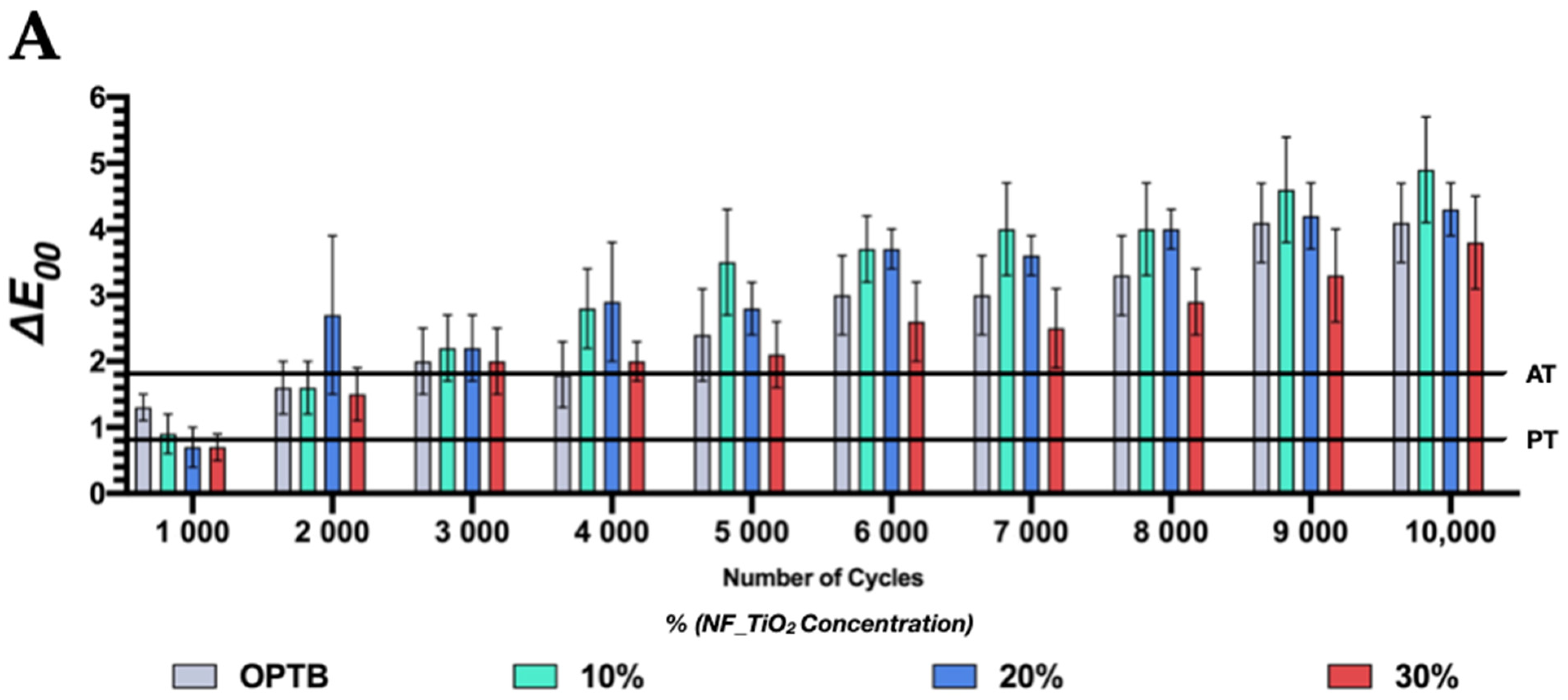
| OPTB | 1.3 (0.2) | 1.6 (0.4) | 2.0 (0.5) | 1.8 (0.5) | 2.4 (0.7) | 3.0 (0.6) | 3.0 (0.6) | 3.3 (0.6) | 4.1 (0.6) | 4.1 (0.6) |
| 10% NF_TiO2 | 0.9 (0.3) | 1.6 (0.4) | 2.2 (0.5) | 2.8 (0.6) | 3.5 (0.8) | 3.7 (0.5) | 4.0 (0.7) | 4.0 (0.7) | 4.6 (0.8) | 4.9 (0.8) |
| 20% NF_TiO2 | 0.7 (0.3) | 2.7 (1.2) | 2.2 (0.5) | 2.9 (0.9) | 2.8 (0.4) | 3.7 (0.3) | 3.6 (0.3) | 4.0 (0.3) | 4.2 (0.5) | 4.3 (0.4) |
| 30% NF_TiO2 | 0.7 (0.2) | 1.5 (0.4) | 2.0 (0.5) | 2.0 (0.3) | 2.1 (0.5) | 2.6 (0.6) | 2.5 (0.6) | 2.9 (0.5) | 3.3 (0.7) | 3.8 (0.7) |
| ΔEab | ||||||||||
|---|---|---|---|---|---|---|---|---|---|---|
| GROUP | NUMBER OF CYCLES | |||||||||
| 1000 | 2000 | 3000 | 4000 | 5000 | 6000 | 7000 | 8000 | 9000 | 10,000 | |
| OPTB | 1.0 (0.2) | 1.7 (0.6) | 2.3 (0.6) | 1.8 (0.6) | 2.5 (0.7) | 3.2 (0.7) | 3.2 (0.7) | 3.6 (0.8) | 4.5 (0.7) | 4.4 (1.1) |
| 10% NF_TiO2 | 1.1 (0.4) | 1.7 (0.6) | 2.5 (0.8) | 3.2 (0.9) | 3.9 (0.9) | 4.2 (0.6) | 4.6 (1.1) | 4.8 (1.1) | 5.1 (0.9) | 5.3 (1.0) |
| 20% NF_TiO2 | 0.9 (0.5) | 3.8 (1.8) | 2.5 (0.7) | 3.5 (1.4) | 2.8 (0.5) | 4.1 (0.7) | 3.9 (0.5) | 4.4 (0.5) | 4.7 (0.7) | 4.6 (0.5) |
| 30% NF_TiO2 | 0.9 (0.5) | 1.7 (0.5) | 2.2 (1.0) | 2.3 (0.5) | 2.2 (0.6) | 3.0 (0.7) | 2.6 (0.7) | 3.1 (0.7) | 3.5 (0.7) | 4.2 (0.6) |
Disclaimer/Publisher’s Note: The statements, opinions and data contained in all publications are solely those of the individual author(s) and contributor(s) and not of MDPI and/or the editor(s). MDPI and/or the editor(s) disclaim responsibility for any injury to people or property resulting from any ideas, methods, instructions or products referred to in the content. |
© 2022 by the authors. Licensee MDPI, Basel, Switzerland. This article is an open access article distributed under the terms and conditions of the Creative Commons Attribution (CC BY) license (https://creativecommons.org/licenses/by/4.0/).
Share and Cite
Hong, Q.; Pierre-Bez, A.C.; Kury, M.; Curtis, M.E.; Hiers, R.D.; Esteban Florez, F.L.; Mitchell, J.C. Shear Bond Strength and Color Stability of Novel Antibacterial Nanofilled Dental Adhesive Resins. Nanomaterials 2023, 13, 1. https://doi.org/10.3390/nano13010001
Hong Q, Pierre-Bez AC, Kury M, Curtis ME, Hiers RD, Esteban Florez FL, Mitchell JC. Shear Bond Strength and Color Stability of Novel Antibacterial Nanofilled Dental Adhesive Resins. Nanomaterials. 2023; 13(1):1. https://doi.org/10.3390/nano13010001
Chicago/Turabian StyleHong, Qing, Alexandra C. Pierre-Bez, Matheus Kury, Mark E. Curtis, Rochelle D. Hiers, Fernando L. Esteban Florez, and John C. Mitchell. 2023. "Shear Bond Strength and Color Stability of Novel Antibacterial Nanofilled Dental Adhesive Resins" Nanomaterials 13, no. 1: 1. https://doi.org/10.3390/nano13010001