
Advances in Electrochemical Detection Electrodes for As(III)


Abstract
:1. Introduction
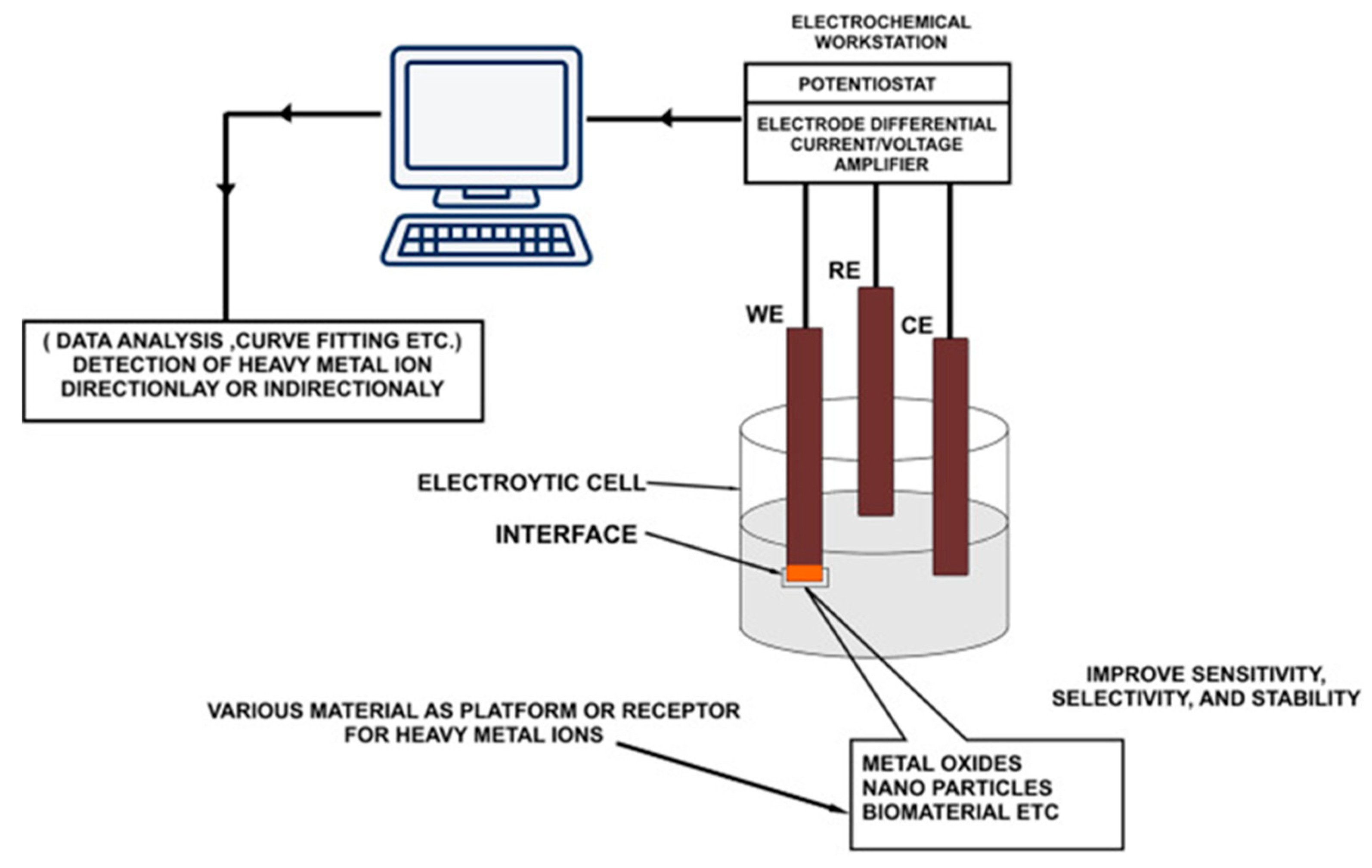
2. Principles of Electrochemical Detection of As(III)
3. Electrochemical Detection Electrodes for As(III)
3.1. Precious Metal Electrodes and Precious-Metal-Modified Electrodes
3.1.1. Gold Electrodes and Gold-Modified Electrodes
3.1.2. Platinum Electrodes and Platinum-Modified Electrodes
3.1.3. Silver Electrodes and Silver-Modified Electrodes
3.1.4. Other Precious-Metal-Modified Electrodes
3.2. Bimetallic Particle-Modified Electrodes
3.2.1. Gold–Platinum Bimetallic Modified Electrodes
3.2.2. Gold–Copper Bimetallic Modified Electrode
3.2.3. Silver–Gold Bimetallic Modified Electrode
3.3. Other Metals and Their Compound-Modified Electrodes
3.3.1. Fe and Its Compound-Modified Electrodes
3.3.2. Manganese and Cerium Oxide-Modified Electrodes
3.3.3. Cobalt Oxide-Modified Electrodes
3.3.4. Tin Oxide-Modified Electrodes
3.3.5. Strontium Compound-Modified Electrodes
3.3.6. Bismuth Compound-Modified Electrode
3.3.7. Zirconium Compound-Modified Electrodes
3.4. Carbon Nanomaterial-Modified Electrodes
3.4.1. Carbon Nanotube-Based Detection of Arsenic Ions
3.4.2. Graphene-Based Detection of Arsenic Ions
3.5. Biomolecule-Modified Electrodes
3.5.1. Arsenic Detection Based on DNA-Modified Electrodes
3.5.2. Aptamer Sensors for Arsenic Detection
3.5.3. Arsenic Detection Based on Other Biomolecules
3.6. Others
3.6.1. Silicon and Its Compound-Modified Electrodes
3.6.2. Novel Polymer-Modified Electrodes
4. Conclusions
Author Contributions
Funding
Institutional Review Board Statement
Informed Consent Statement
Data Availability Statement
Acknowledgments
Conflicts of Interest
References
- Mandal, B.K.; Suzuki, K.T. Arsenic round the world: A review. Talanta 2002, 58, 201–235. [Google Scholar] [CrossRef]
- Walsh, D.F.; Berger, B.L.; Bean, J.R. Mercury, arsenic, lead, cadmium, and selenium residues in fish, 1971–1973—National Pesticide Monitoring Program. Pestic. Monit. J. 1977, 11, 5–34. [Google Scholar] [PubMed]
- Waalkes, M.P.; Coogan, T.P.; Barter, R.A. Toxicological principles of metal carcinogenesis with special emphasis on cadmium. Crit. Rev. Toxicol. 1992, 22, 175–201. [Google Scholar] [CrossRef] [PubMed]
- Qiao, Y.L.; Taylor, P.R.; Yao, S.X.; Erozan, Y.S.; Luo, X.C.; Barrett, M.J.; Yan, Q.Y.; Giffen, C.A.; Huang, S.Q.; Maher, M.M.; et al. Risk factors and early detection of lung cancer in a cohort of Chinese tin miners. Ann. Epidemiol. 1997, 7, 533–541. [Google Scholar] [CrossRef]
- Lu, Y.J.; Sun, S.Q. Study of deposition and translocation of mineral dust in lungs of tin miners in Yunnan by X-ray microanalysis. Chin. J. Pathol. 1992, 21, 103–105. [Google Scholar]
- Kabir, R.; Sinha, P.; Mishra, S.; Ebenebe, O.V.; Taube, N.; Oeing, C.U.; Keceli, G.; Chen, R.; Paolocci, N.; Rule, A.; et al. Inorganic arsenic induces sex-dependent pathological hypertrophy in the heart. Am. J. Physiol. Heart Circ. Physiol. 2021, 320, H1321–H1336. [Google Scholar] [CrossRef]
- Ozturk, M.; Metin, M.; Altay, V.; Bhat, R.A.; Ejaz, M.; Gul, A.; Unal, B.T.; Hasanuzzaman, M.; Nibir, L.; Nahar, K.; et al. Arsenic and Human Health: Genotoxicity, Epigenomic Effects, and Cancer Signaling. Biol. Trace Elem. Res. 2022, 200, 988–1001. [Google Scholar] [CrossRef]
- Argos, M.; Kalra, T.; Rathouz, P.J.; Chen, Y.; Pierce, B.; Parvez, F.; Islam, T.; Ahmed, A.; Rakibuz-Zaman, M.; Hasan, R.; et al. Arsenic exposure from drinking water, and all-cause and chronic-disease mortalities in Bangladesh (HEALS): A prospective cohort study. Lancet 2010, 376, 252–258. [Google Scholar] [CrossRef] [Green Version]
- Martinez, V.D.; Vucic, E.A.; Lam, S.; Lam, W.L. Arsenic and Lung Cancer in Never-Smokers: Lessons from Chile. Am. J. Respir. Crit. Care Med. 2012, 185, 1131–1132. [Google Scholar] [CrossRef]
- Bostock, J. Observations on the Different Methods Recommended for Detecting Minute Portions of Arsenic. Edinb. Med. Surg. J. 1809, 5, 166–174. [Google Scholar]
- Delepine, S. The Detection of Arsenic in Beer and Brewing Material. Br. Med. J. 1901, 1, 81–84. [Google Scholar] [CrossRef] [PubMed] [Green Version]
- Scozzari, A. Electrochemical sensing methods: A brief review. In Proceedings of the Conference on NATO-Advanced-Study-Institute on Sensor Systems for Biological Threats—Algal Toxins Case, Pisa, Italy, 30 September–11 October 2008; pp. 335–351. [Google Scholar]
- Bansod, B.; Kumar, T.; Thakur, R.; Rana, S.; Singh, I. A review on various electrochemical techniques for heavy metal ions detection with different sensing platforms. Biosens. Bioelectron. 2017, 94, 443–455. [Google Scholar] [CrossRef] [PubMed]
- Aragay, G.; Pons, J.; Merkoci, A. Recent Trends in Macro-, Micro-, and Nanomaterial-Based Tools and Strategies for Heavy-Metal Detection. Chem. Rev. 2011, 111, 3433–3458. [Google Scholar] [CrossRef] [PubMed]
- Giacomino, A.; Abollino, O.; Lazzara, M.; Malandrino, M.; Mentasti, E. Determination of As(III) by anodic stripping voltammetry using a lateral gold electrode: Experimental conditions, electron transfer and monitoring of electrode surface. Talanta 2011, 83, 1428–1435. [Google Scholar] [CrossRef] [PubMed]
- Rahman, M.R.; Okajima, T.; Ohsaka, T. Selective Detection of As(III) at the Au(III)-like Polycrystalline Gold Electrode. Anal. Chem. 2010, 82, 9169–9176. [Google Scholar] [CrossRef] [PubMed]
- Diu, Y.; Zhao, W.; Xu, J.-J.; Chen, H.-Y. Electrochemical determination of arsenite in neutral media on reusable gold nanostructured films. Talanta 2009, 79, 243–248. [Google Scholar] [CrossRef]
- Dugo, G.; La Pera, L.; Lo Turco, V.; Di Bella, G. Speciation of inorganic arsenic in alimentary and environmental aqueous samples by using derivative anodic stripping chronopotentiometry (dASCP). Chemosphere 2005, 61, 1093–1101. [Google Scholar] [CrossRef]
- Viltchinskaia, E.A.; Zeigman, L.L.; Garcia, D.M.; Santos, P.F. Simultaneous determination of mercury and arsenic by anodic stripping voltammetry. Electroanalysis 1997, 9, 633–640. [Google Scholar] [CrossRef]
- HuiliangHuang, K.; Dasgupta, P. A field-deployable instrument for the measurement and speciation of arsenic in potable water. Anal. Chim. Acta 1999, 380, 27–37. [Google Scholar] [CrossRef]
- Alves, G.M.S.; Magalhaes, J.M.C.S.; Salauen, P.; van den Berg, C.M.G.; Soares, H.M.V.M. Simultaneous electrochemical determination of arsenic, copper, lead and mercury in unpolluted fresh waters using a vibrating gold microwire electrode. Anal. Chim. Acta 2011, 703, 1–7. [Google Scholar] [CrossRef]
- Bu, L.; Gu, T.; Ma, Y.; Chen, C.; Tan, Y.; Xie, Q.; Yao, S. Enhanced Cathodic Preconcentration of As(0) at Au and Pt Electrodes for Anodic Stripping Voltammetry Analysis of As(III) and As(V). J. Phys. Chem. C 2015, 119, 11400–11409. [Google Scholar] [CrossRef]
- Zhou, C.; Yang, M.; Li, S.-S.; Jiang, T.-J.; Liu, J.-H.; Huang, X.-J.; Chen, X. Electrochemically etched gold wire microelectrode for the determination of inorganic arsenic. Electrochim. Acta 2017, 231, 238–246. [Google Scholar] [CrossRef]
- Laschi, S.; Bagni, G.; Palchetti, I.; Mascini, M. As(III) voltammetric detection by means of disposable screen-printed gold electrochemical sensors. Anal. Lett. 2007, 40, 3002–3013. [Google Scholar] [CrossRef]
- Orellana Jaramillo, D.X.; Sukeri, A.; Saravia, L.P.H.; Espinoza-Montero, P.J.; Bertotti, M. Nanoporous Gold Microelectrode: A Novel Sensing Platform for Highly Sensitive and Selective Determination of Arsenic(III) using Anodic Stripping Voltammetry. Electroanalysis 2017, 29, 2316–2322. [Google Scholar] [CrossRef]
- Navratil, T.; Kopanica, M.; Krista, J. Anodic stripping voltammetry for arsenic determination on composite gold electrode. Chem. Anal. 2003, 48, 265–272. [Google Scholar]
- Prakash, R.; Srivastava, R.C.; Seth, P.K. Direct estimation of total arsenic using a novel metal side disk rotating electrode. Electroanalysis 2003, 15, 1410–1414. [Google Scholar] [CrossRef]
- Vandenhecke, J.; Waeles, M.; Riso, R.D.; Le Corre, P. A stripping chronopotentiometric (SCP) method with a gold film electrode for determining inorganic arsenic species in seawater. Anal. Bioanal. Chem. 2007, 388, 929–937. [Google Scholar] [CrossRef]
- Jena, B.K.; Raj, C.R. Gold nanoelectrode ensembles for the simultaneous electrochemical detection of ultratrace arsenic, mercury, and copper. Anal. Chem. 2008, 80, 4836–4844. [Google Scholar] [CrossRef]
- Li, D.; Li, J.; Jia, X.; Han, Y.; Wang, E. Electrochemical determination of arsenic(III) on mercaptoethylamine modified Au electrode in neutral media. Anal. Chim. Acta 2012, 733, 23–27. [Google Scholar] [CrossRef]
- Zakharova, E.A.; Noskova, G.N.; Antonova, S.G.; Kabakaev, A.S. Speciation of arsenic(III) and arsenic(V) by manganese-mediated stripping voltammetry at gold microelectrode ensemble in neutral and basic medium. Int. J. Environ. Anal. Chem. 2014, 94, 1478–1498. [Google Scholar] [CrossRef]
- Jiang, J.; Holm, N.; O’Brien, K. Improved Anodic Stripping Voltammetric Detection of Arsenic(III) Using Nanoporous Gold Microelectrode. ECS J. Solid State Sci. Technol. 2015, 4, S3024–S3029. [Google Scholar] [CrossRef]
- Korolczuk, M.; Ochab, M.; Rutyna, I. Determination of As(III) by anodic stripping voltammetry following double deposition and stripping steps at two gold working electrodes. Talanta 2015, 144, 517–521. [Google Scholar] [CrossRef] [PubMed]
- Jedryczko, D.; Pohl, P.; Welna, M. Inorganic arsenic speciation in natural mineral drinking waters by flow-through anodic stripping chronopotentiometry. Talanta 2016, 150, 265–271. [Google Scholar] [CrossRef] [PubMed]
- Kim, J.; Han, S.; Kim, Y. Electrochemical detection of arsenic(III) using porous gold via square wave voltammetry. Korean J. Chem. Eng. 2017, 34, 2096–2098. [Google Scholar] [CrossRef]
- Lestarini, D.T.; Ivandini, T.A. Electrochemical detection of As3+ and As5+ by anodic stripping voltammetry at a gold electrode. In Proceedings of the 2nd International Symposium on Current Progress in Functional Materials (ISCPFM)—Functional Materials for Energy and Environment Applications, Bali, Indonesia, 8–9 November 2019. [Google Scholar]
- Wang, L.; Lu, W.; Jing, G.; Cui, T. Trace Determination of Arsenite with an Ionophore-Coated Selective Micro Sensor. IEEE Sens. J. 2018, 18, 4364–4371. [Google Scholar] [CrossRef]
- Babar, N.-U.-A.; Joya, K.S.; Tayyab, M.A.; Ashiq, M.N.; Sohail, M. Highly Sensitive and Selective Detection of Arsenic Using Electrogenerated Nanotextured Gold Assemblage. ACS Omega 2019, 4, 13645–13657. [Google Scholar] [CrossRef] [Green Version]
- Dutta, S.; Strack, G.; Kurup, P. Gold nanostar electrodes for heavy metal detection. Sens. Actuators B Chem. 2019, 281, 383–391. [Google Scholar] [CrossRef]
- Majid, E.; Hrapovic, S.; Liu, Y.L.; Male, K.B.; Luong, J.H.T. Electrochemical determination of arsenite using a gold nanoparticle modified glassy carbon electrode and flow analysis. Anal. Chem. 2006, 78, 762–769. [Google Scholar] [CrossRef]
- Boonpeng, P.; Sooksamiti, P.; Lapanantnoppakhun, S.; Jakmunee, J. Determination of Inorganic Arsenic by Anodic Stripping Voltammetry with Gold Nanoparticles Modified Screen-printed Carbon Electrode and Anion Exchange Column Preconcentration. Chiang Mai J. Sci. 2019, 46, 106–117. [Google Scholar]
- Sakira, A.K.; Some, I.T.; Ziemons, E.; Dejaegher, B.; Mertens, D.; Hubert, P.; Kauffmann, J.-M. Determination of Arsenic(III) at a Nanogold Modified Solid Carbon Paste Electrode. Electroanalysis 2015, 27, 309–316. [Google Scholar] [CrossRef]
- Simm, A.O.; Banks, C.E.; Ward-Jones, S.; Davies, T.J.; Lawrence, N.S.; Jones, T.G.J.; Jiang, L.; Compton, R.G. Boron-doped diamond microdisc arrays: Electrochemical characterisation and their use as a substrate for the production of microelectrode arrays of diverse metals (Ag, Au, Cu) via electrodeposition. Analyst 2005, 130, 1303–1311. [Google Scholar] [CrossRef] [PubMed]
- Forsberg, G.; O’Laughlin, J.W.; Megargle, R.G.; Koirtyihann, S.R. Determination of arsenic by anodic stripping voltammetry and differential pulse anodic stripping voltammetry. Anal. Chem. 1975, 47, 1586–1592. [Google Scholar] [CrossRef]
- Duangthong, S.; Suwanin, A.; Chooto, P.; Innuphat, C. Flow injection-differential pulse anodic stripping voltammetry to measure As(III) and As(V) in natural water samples. ScienceAsia 2016, 42, 266–274. [Google Scholar] [CrossRef] [Green Version]
- Hassan, S.S.; Sirajuddin; Solangi, A.R.; Kazi, T.G.; Kalhoro, M.S.; Junejo, Y.; Tagar, Z.A.; Kalwar, N.H. Nafion stabilized ibuprofen-gold nanostructures modified screen printed electrode as arsenic(III) sensor. J. Electroanal. Chem. 2012, 682, 77–82. [Google Scholar] [CrossRef]
- Tran Ngoc, H.; Ganesh, T.; Kim, K.S.; Kim, S.; Han, S.-H.; Chung, H. A three-dimensional gold nanodendrite network porous structure and its application for an electrochemical sensing. Biosens. Bioelectron. 2011, 27, 183–186. [Google Scholar] [CrossRef]
- Anh Quang, D.; Do Mai, N.; Tran Thanh Tam, T. Determination of arsenic(III) in water using gold nanorods-modified electrode. J. Mater. Sci. Mater. Electron. 2021, 32, 27962–27974. [Google Scholar] [CrossRef]
- Lu, D.; Sullivan, C.; Brack, E.M.; Drew, C.P.; Kurup, P. Simultaneous voltammetric detection of cadmium(II), arsenic(III), and selenium(IV) using gold nanostar-modified screen-printed carbon electrodes and modified Britton-Robinson buffer. Anal. Bioanal. Chem. 2020, 412, 4113–4125. [Google Scholar] [CrossRef]
- Stankovic, A.; Kajinic, Z.; Turkalj, J.V.; Romic, Z.; Sikiric, M.D.; Asserghine, A.; Nagy, G.; Medvidovic-Kosanovic, M. Voltammetric Determination of Arsenic with Modified Glassy Carbon Electrode. Electroanalysis 2020, 32, 1043–1051. [Google Scholar] [CrossRef]
- Sullivan, C.; Lu, D.; Senecal, A.; Kurup, P. Voltammetric detection of Arsenic(III) using gold nanoparticles modified carbon screen printed electrodes: Application for facile and rapid analysis in commercial apple juice. Food Chem. 2021, 352, 129327. [Google Scholar] [CrossRef]
- Kamyabi, M.A.; Aghaei, A. Electromembrane extraction coupled to square wave anodic stripping voltammetry for selective preconcentration and determination of trace levels of As(III) in water samples. Electrochim. Acta 2016, 206, 192–198. [Google Scholar] [CrossRef]
- Rastogi, P.K.; Yadav, D.K.; Pandey, S.; Ganesan, V.; Sonkar, P.K.; Gupta, R. Synthesis and characterization of gold nanoparticles incorporated bentonite clay for electrocatalytic sensing of arsenic(III). J. Chem. Sci. 2016, 128, 349–356. [Google Scholar] [CrossRef]
- Tang, Q.; Zhu, G.; Ge, Y.; Yang, J.; Huang, M.; Liu, J. AuNPs-polyaniline nanosheet array on carbon nanofiber for the determination of As(III). J. Electroanal. Chem. 2020, 873, 114381. [Google Scholar] [CrossRef]
- Agir, S.K.; Kundu, M.; IEEE. Detection and Quantification of Arsenic in Water Using Electronic Tongue. In Proceedings of the 1st IEEE International Conference on Control, Measurement and Instrumentation (CMI), Jadavpur University, Kolkata, India, 8–10 January 2016; pp. 424–428. [Google Scholar]
- Chowdhury, A.-N.; Ferdousi, S.; Islam, M.M.; Okajima, T.; Ohsaka, T. Arsenic detection by nanogold/conducting-polymer-modified glassy carbon electrodes. J. Appl. Polym. Sci. 2007, 104, 1306–1311. [Google Scholar] [CrossRef]
- Song, Y.; Swain, G.M. Total inorganic arsenic detection in real water samples using anodic stripping voltammetry and a gold-coated diamond thin-film electrode. Anal. Chim. Acta 2007, 593, 7–12. [Google Scholar] [CrossRef] [PubMed]
- Xiao, L.; Wildgoose, G.G.; Compton, R.G. Sensitive electrochemical detection of Arsenic(III) using gold nanoparticle modified carbon nanotubes via anodic stripping voltammetry. Anal. Chim. Acta 2008, 620, 44–49. [Google Scholar] [CrossRef] [PubMed]
- Cui, H.; Yang, W.; Li, X.; Zhao, H.; Yuan, Z. An electrochemical sensor based on a magnetic Fe3O4 nanoparticles and gold nanoparticles modified electrode for sensitive determination of trace amounts of arsenic(III). Anal. Methods 2012, 4, 4176–4181. [Google Scholar] [CrossRef]
- Lan, Y.; Luo, H.; Ren, X.; Wang, Y.; Wang, L. Glassy Carbon Electrode Modified with Citrate Stabilized Gold Nanoparticles for Sensitive Arsenic(III) Detection. Anal. Lett. 2012, 45, 1184–1196. [Google Scholar] [CrossRef]
- Thotiyl, M.M.O.; Basit, H.; Sanchez, J.A.; Goyer, C.; Coche-Guerente, L.; Dumy, P.; Sampath, S.; Labbe, P.; Moutet, J.-C. Multilayer assemblies of polyelectrolyte-gold nanoparticles for the electrocatalytic oxidation and detection of arsenic(III). J. Colloid Interface Sci. 2012, 383, 130–139. [Google Scholar] [CrossRef]
- Huang, J.-F.; Chen, H.-H. Gold-nanoparticle-embedded nafion composite modified on glassy carbon electrode for highly selective detection of arsenic(III). Talanta 2013, 116, 852–859. [Google Scholar] [CrossRef]
- Liu, Y.; Huang, Z.; Xie, Q.; Sun, L.; Gu, T.; Li, Z.; Bu, L.; Yao, S.; Tu, X.; Luo, X.; et al. Electrodeposition of electroreduced graphene oxide-Au nanoparticles composite film at glassy carbon electrode for anodic stripping voltammetric analysis of trace arsenic(III). Sens. Actuators B Chem. 2013, 188, 894–901. [Google Scholar] [CrossRef]
- Pifferi, V.; Ardizzone, S.; Cappelletti, G.; Falciola, L.; Meroni, D. Ultra-Traces Detection by Gold-Based Electrodes in As(III) Novel Photoremediation. Electrocatalysis 2013, 4, 306–311. [Google Scholar] [CrossRef]
- Wang, D.; Zhao, Y.; Jin, H.; Zhuang, J.; Zhang, W.; Wang, S.; Wang, J. Synthesis of Au-Decorated Tripod-Shaped Te Hybrids for Applications in the Ultrasensitive Detection of Arsenic. ACS Appl. Mater. Interfaces 2013, 5, 5733–5740. [Google Scholar] [CrossRef] [PubMed]
- Wu, J.; Yang, M.; Xiao, J.; Fu, X.; Jin, J.; Li, L.; Chang, W.; Xie, C. Gold Nanoparticle Dropped Titania Microsphere Hybrids as an Enhanced Sensitive Material for Stripping Voltammetry Determination of As(III). J. Electrochem. Soc. 2013, 160, B225–B230. [Google Scholar] [CrossRef]
- Cinti, S.; Politi, S.; Moscone, D.; Palleschi, G.; Arduini, F. Stripping Analysis of As(III) by Means of Screen-Printed Electrodes Modified with Gold Nanoparticles and Carbon Black Nanocomposite. Electroanalysis 2014, 26, 931–939. [Google Scholar] [CrossRef]
- Wu, S.; Zhao, Q.; Zhou, L.; Zhang, Z. Stripping Analysis of Trace Arsenic Based on the MnOx/AuNPs Composite Film Modified Electrode in Alkaline Media. Electroanalysis 2014, 26, 1840–1849. [Google Scholar] [CrossRef]
- Sanghavi, B.J.; Gadhari, N.S.; Kalambate, P.K.; Karna, S.P.; Srivastava, A.K. Potentiometric stripping analysis of arsenic using a graphene paste electrode modified with a thiacrown ether and gold nanoparticles. Microchim. Acta 2015, 182, 1473–1481. [Google Scholar] [CrossRef]
- Kato, D.; Kamata, T.; Kato, D.; Yanagisawa, H.; Niwa, O. Au Nanoparticle-Embedded Carbon Films for Electrochemical As3+ Detection with High Sensitivity and Stability. Anal. Chem. 2016, 88, 2944–2951. [Google Scholar] [CrossRef]
- Mafa, J.P.; Mabuba, N.; Arotiba, O.A. An Exfoliated Graphite Based Electrochemical Sensor for As(III) in Water. Electroanalysis 2016, 28, 1462–1469. [Google Scholar] [CrossRef]
- Wei, J.; Li, S.-S.; Guo, Z.; Chen, X.; Liu, J.-H.; Huang, X.-J. Adsorbent Assisted in Situ Electrocatalysis: An Ultra-Sensitive Detection of As(III) in Water at Fe3O4 Nanosphere Densely Decorated with Au Nanoparticles. Anal. Chem. 2016, 88, 1154–1161. [Google Scholar] [CrossRef]
- Yang, M.; Chen, X.; Liu, J.-H.; Huang, X.-J. Enhanced anti-interference on electrochemical detection of arsenite with nanoporous gold in mild condition. Sens. Actuators B Chem. 2016, 234, 404–411. [Google Scholar] [CrossRef]
- Zhou, S.; Han, X.; Fan, H.; Liu, Y. Electrochemical Sensing toward Trace As(III) Based on Mesoporous MnFe2O4/Au Hybrid Nanospheres Modified Glass Carbon Electrode. Sensors 2016, 16, 935. [Google Scholar] [CrossRef] [Green Version]
- Carrera, P.; Espinoza-Montero, P.J.; Fernandez, L.; Romero, H.; Alvarado, J. Electrochemical determination of arsenic in natural waters using carbon fiber ultra-microelectrodes modified with gold nanoparticles. Talanta 2017, 166, 198–206. [Google Scholar] [CrossRef] [PubMed]
- Idris, A.O.; Mabuba, N.; Arotiba, O.A. Electrochemical co-detection of Arsenic and Selenium on a Glassy Carbon Electrode Modified with Gold Nanoparticles. Int. J. Electrochem. Sci. 2017, 12, 10–21. [Google Scholar] [CrossRef]
- Nunez-Bajo, E.; Carmen Blanco-Lopez, M.; Costa-Garcia, A.; Teresa Fernandez-Abedule, M. Electrogeneration of Gold Nanoparticles on Porous-Carbon Paper-Based Electrodes and Application to Inorganic Arsenic Analysis in White Wines by Chronoamperometric Stripping. Anal. Chem. 2017, 89, 6415–6423. [Google Scholar] [CrossRef] [PubMed] [Green Version]
- Sahoo, S.; Sahoo, P.K.; Satpati, A.K. Gold Nano Particle and Reduced Graphene Oxide Composite Modified Carbon Paste Electrode for the Ultra Trace Detection of Arsenic(III). Electroanalysis 2017, 29, 1400–1409. [Google Scholar] [CrossRef]
- Salunke, R.S.; Kasar, C.K.; Bangar, M.A.; Chavan, P.G.; Shirale, D.J. Electrodeposition of gold nanoparticles decorated single polypyrrole nanowire for arsenic detection in potable water: A chemiresistive sensor device. J. Mater. Sci. Mater. Electron. 2017, 28, 14672–14677. [Google Scholar] [CrossRef]
- Sun, F.; Fan, G. Electrochemical Detection of As(III) Ions in Hot Spring Water Based on a SiO2-Au Nanocomposite. Int. J. Electrochem. Sci. 2017, 12, 8982–8990. [Google Scholar] [CrossRef]
- Coneo Rodriguez, R.; Bruno, M.M.; Angelome, P.C. Au nanoparticles embedded in mesoporous ZrO2 films: Multifunctional materials for electrochemical detection. Sens. Actuators B Chem. 2018, 254, 603–612. [Google Scholar] [CrossRef]
- Ensafi, A.A.; Akbarian, F.; Heydari-Soureshjani, E.; Rezaei, B. A novel aptasensor based on 3D-reduced graphene oxide modified gold nanoparticles for determination of arsenite. Biosens. Bioelectron. 2018, 122, 25–31. [Google Scholar] [CrossRef]
- Nellaiappan, S.; Pillai, K.C.; Kumar, A.S. Flow-injection analysis coupled with electrochemical detection of poisonous inorganic arsenic(III) species using a gold nanoparticle/carbon nanofiber/chitosan chemically modified carbon screen printed electrode in neutral pH solution. Anal. Methods 2018, 10, 799–808. [Google Scholar] [CrossRef]
- Pungjunun, K.; Chaiyo, S.; Jantrahong, I.; Nantaphol, S.; Siangproh, W.; Chailapakul, O. Anodic stripping voltammetric determination of total arsenic using a gold nanoparticle-modified boron-doped diamond electrode on a paper-based device. Microchim. Acta 2018, 185, 324. [Google Scholar] [CrossRef] [PubMed]
- Yang, M.; Li, P.-H.; Xu, W.-H.; Wei, Y.; Li, L.-N.; Huang, Y.-Y.; Sun, Y.-F.; Chen, X.; Liu, J.-H.; Huang, X.-J. Reliable electrochemical sensing arsenic(III) in nearly groundwater pH based on efficient adsorption and excellent electrocatalytic ability of AuNPs/CeO2-ZrO2 nanocomposite. Sens. Actuators B Chem. 2018, 255, 226–234. [Google Scholar] [CrossRef]
- Buffa, A.; Mandler, D. Arsenic(III) detection in water by flow-through carbon nanotube membrane decorated by gold nanoparticles. Electrochim. Acta 2019, 318, 496–503. [Google Scholar] [CrossRef]
- Gimenez-Gomez, P.; Baldi, A.; Ayora, C.; Fernandez-Sanchez, C. Automated Determination of As(III) in Waters with an Electrochemical Sensor Integrated into a Modular Microfluidic System. ACS Sens. 2019, 4, 3156–3165. [Google Scholar] [CrossRef] [PubMed]
- Trachioti, M.G.; Karantzalis, A.E.; Hrbac, J.; Prodromidis, M.I. Low-cost screen-printed sensors on-demand: Instantly prepared sparked gold nanoparticles from eutectic Au/Si alloy for the determination of arsenic at the sub-ppb level. Sens. Actuators B Chem. 2019, 281, 273–280. [Google Scholar] [CrossRef]
- Wang, W.; Bao, N.; Yuan, W.; Si, N.; Bai, H.; Li, H.; Zhang, Q. Simultaneous determination of lead, arsenic, and mercury in cosmetics using a plastic based disposable electrochemical sensor. Microchem. J. 2019, 148, 240–247. [Google Scholar] [CrossRef]
- Zhao, G.; Liu, G. Electrochemical Deposition of Gold Nanoparticles on Reduced Graphene Oxide by Fast Scan Cyclic Voltammetry for the Sensitive Determination of As(III). Nanomaterials 2019, 9, 41. [Google Scholar] [CrossRef] [Green Version]
- Bu, L.; Xie, Q.; Ming, H. Gold nanoparticles decorated three-dimensional porous graphitic carbon nitrides for sensitive anodic stripping voltammetric analysis of trace arsenic(III). J. Alloys Compd. 2020, 823, 153723. [Google Scholar] [CrossRef]
- Chen, C.; Yu, S.; Jiang, S.; Liu, J.; Wang, Z.; Ye, B.-C. A novel and sensitive electrochemical sensor based on nanoporous gold for determination of As(III). Microchim. Acta 2020, 187, 395. [Google Scholar] [CrossRef]
- Ismail, S.; Yusof, N.A.; Abdullah, J.; Abd Rahman, S.F. Development of Electrochemical Sensor Based on Silica/Gold Nanoparticles Modified Electrode for Detection of Arsenite. IEEE Sens. J. 2020, 20, 3406–3414. [Google Scholar] [CrossRef]
- Kongor, A.; Panchal, M.; Athar, M.; Vora, M.; Verma, N.; Pandya, A.; Jha, P.C.; Bhadresha, K.; Rawal, R.; Jain, V. Colorimetric and electrochemical sensing of As(III) using calix 4 pyrrole capped gold nanoparticles and evaluation of its cytotoxic activity. J. Incl. Phenom. Macrocycl. Chem. 2020, 98, 29–41. [Google Scholar] [CrossRef]
- Sullivan, C.; Lu, D.; Brack, E.; Drew, C.; Kurup, P. Voltammetric codetection of arsenic(III) and copper(II) in alkaline buffering system with gold nanostar modified electrodes. Anal. Chim. Acta 2020, 1107, 63–73. [Google Scholar] [CrossRef] [PubMed]
- Udayan, A.P.M.; Kachwala, B.; Karthikeyan, K.G.; Gunasekaran, S. Ultrathin quasi-hexagonal gold nanostructures for sensing arsenic in tap water. RSC Adv. 2020, 10, 20211–20221. [Google Scholar] [CrossRef]
- Jijana, A.N.; Mphuthi, N.; Shumbula, P.; Vilakazi, S.; Sikhwivhilu, L. The Ultra-sensitive Electrochemical Detection of As(III) in Ground Water Using Disposable L-cysteine/Lipoic Acid Functionalised Gold Nanoparticle Modified Screen-Printed Electrodes. Electrocatalysis 2021, 12, 310–325. [Google Scholar] [CrossRef]
- Li, C.-Y.; Wei, Y.-Y.; Shen, W.; Dong, X.; Yang, M.; Wei, J. Ultrahigh sensitivity electroanalysis of trace As(III) in water and human serum via gold nanoparticles uniformly anchored to Co3O4 porous microsheets. Electrochim. Acta 2021, 368, 137605. [Google Scholar] [CrossRef]
- Nodehi, M.; Baghayeri, M.; Veisi, H. Preparation of GO/Fe3O4@PMDA/AuNPs nanocomposite for simultaneous determination of As3+ and Cu2+ by stripping voltammetry. Talanta 2021, 230, 122288. [Google Scholar] [CrossRef]
- Zhu, X.; Zhu, G.; Ge, Y.; Zhang, B.; Yang, J.; Hu, B.; Liu, J. AU(nano)/Fe-MOF hybrid electrode for highly sensitive determination of trace As(III). J. Electroanal. Chem. 2021, 899, 115642. [Google Scholar] [CrossRef]
- Tabibi, Z.; Massah, J.; Vakilian, K.A. A biosensor for the sensitive and specific measurement of arsenite using gold nanoparticles. Measurement 2022, 187, 110281. [Google Scholar] [CrossRef]
- Nguyen, T.S.V.; Huynh, T.M.; Nguyen, L.D.; Doan, T.C.D.; Baptist, R.; Dang, C.M. Sensitive electrochemical measurement of As(III) using Nafion modified platinum electrode via anode stripping voltammetry. Int. J. Nanotechnol. 2020, 17, 659–672. [Google Scholar] [CrossRef]
- Xu, H.; Zeng, L.; Xing, S.; Shi, G.; Chen, J.; Man, Y.; Jin, L. Highly ordered platinum-nanotube arrays for oxidative determination of trace arsenic(III). Electrochem. Commun. 2008, 10, 1893–1896. [Google Scholar] [CrossRef]
- Xu, H.; Zeng, L.; Xing, S.; Xian, Y.; Jin, L. Microwave-irradiated synthesized platinum nanoparticles/carbon nanotubes for oxidative determination of trace arsenic(III). Electrochem. Commun. 2008, 10, 551–554. [Google Scholar] [CrossRef]
- Sitorus, R.; Saefumillah, A.; Ivandini, T.A. Arsenic and arsenate detection with anodic stripping voltammetry technique using boron-doped diamond modified by platinum. IOP Conf. Ser. Mater. Sci. Eng. 2020, 763, 012013. [Google Scholar] [CrossRef]
- Kempegowda, R.; Antony, D.; Malingappa, P. Graphene-platinum nanocomposite as a sensitive and selective voltammetric sensor for trace level arsenic quantification. Int. J. Smart Nano Mater. 2014, 5, 17–32. [Google Scholar] [CrossRef] [Green Version]
- Hrapovic, S.; Liu, Y.; Luong, J.H.T. Reusable platinum nanoparticle modified boron doped diamond microelectrodes for oxidative determination of arsenite. Anal. Chem. 2007, 79, 500–507. [Google Scholar] [CrossRef]
- Dai, X.; Compton, R.G. Detection of As(III) via oxidation to As(V) using platinum nanoparticle modified glassy carbon electrodes: Arsenic detection without interference from copper. Analyst 2006, 131, 516–521. [Google Scholar] [CrossRef]
- Li, P.-H.; Yang, M.; Song, Z.-Y.; Chen, S.-H.; Xiao, X.-Y.; Lin, C.-H.; Huang, X.-J. Highly Sensitive and Stable Determination of As(III) under Near-Neutral Conditions: Benefit from the Synergetic Catalysis of Pt Single Atoms and Active S Atoms over Pt-1/MoS2. Anal. Chem. 2021, 93, 15115–15123. [Google Scholar] [CrossRef]
- Han, D.-D.; Liu, Z.-G.; Liu, J.-H.; Huang, X.-J. The size effect of Pt nanoparticles: A new route to improve sensitivity in electrochemical detection of As(III). RSC Adv. 2015, 5, 38290–38297. [Google Scholar] [CrossRef]
- Aguirre, M.d.C.; Rivas, B.L.; Basaez, L.; Pena-Farfal, C. Electrochemical Detection of Arsenite with Silver Electrodes in Inorganic Electrolyte and Natural System Mixtures. J. Braz. Chem. Soc. 2011, 22, 2362–2370. [Google Scholar] [CrossRef]
- Sonkoue, B.M.; Tchekwagep, P.M.S.; Nanseu-Njiki, C.P.; Ngameni, E. Electrochemical Determination of Arsenic Using Silver Nanoparticles. Electroanalysis 2018, 30, 2738–2743. [Google Scholar] [CrossRef]
- Prakash, S.; Chakrabarty, T.; Singh, A.K.; Shahi, V.K. Silver nanoparticles built-in chitosan modified glassy carbon electrode for anodic stripping analysis of As(III) and its removal from water. Electrochim. Acta 2012, 72, 157–164. [Google Scholar] [CrossRef]
- Dar, R.A.; Khare, N.G.; Cole, D.P.; Karna, S.P.; Srivastava, A.K. Green synthesis of a silver nanoparticle-graphene oxide composite and its application for As(III) detection. RSC Adv. 2014, 4, 14432–14440. [Google Scholar] [CrossRef]
- Wen, S.-H.; Liang, R.-P.; Zhang, L.; Qiu, J.-D. Multimodal Assay of Arsenite Contamination in Environmental Samples with Improved Sensitivity through Stimuli-Response of Multiligands Modified Silver Nanoparticles. ACS Sustain. Chem. Eng. 2018, 6, 6223–6232. [Google Scholar] [CrossRef]
- Agustiany, T.; Khalil, M.; Einaga, Y.; Jiwanti, P.K.; Ivandini, T.A. Stable iridium-modified boron-doped diamond electrode for the application in electrochemical detection of Arsenic(III). Mater. Chem. Phys. 2020, 244, 122723. [Google Scholar] [CrossRef]
- Mafakheri, E.; Salimi, A.; Hallaj, R.; Ramazani, A.; Kashi, M.A. Synthesis of Iridium Oxide Nanotubes by Electrodeposition into Polycarbonate Template: Fabrication of Chromium(III) and Arsenic(III) Electrochemical Sensor. Electroanalysis 2011, 23, 2429–2437. [Google Scholar] [CrossRef]
- Gupta, R.; Gamare, J.S.; Pandey, A.K.; Tyagi, D.; Kamat, J.V. Highly Sensitive Detection of Arsenite Based on Its Affinity Toward Ruthenium Nanoparticles Decorated on Glassy Carbon Electrode. Anal. Chem. 2016, 88, 2459–2465. [Google Scholar] [CrossRef]
- Dash, S.; Munichandraiah, N. Electroanalysis of As(III) at nanodendritic Pd on PEDOT. Analyst 2014, 139, 1789–1795. [Google Scholar] [CrossRef]
- Alam, M.M.; Rashed, M.A.; Rahman, M.M.; Rahman, M.M.; Nagao, Y.; Hasnat, M.A. Electrochemical oxidation of As(III) on Pd immobilized Pt surface: Kinetics and sensing performance. RSC Adv. 2018, 8, 8071–8079. [Google Scholar] [CrossRef] [Green Version]
- Murali Mohan, J.; Amreen, K.; Javed, A.; Dubey, S.K.; Goel, S. Electrochemical Mini-Platform with Thread-Based Electrodes for Interference Free Arsenic Detection. IEEE Trans. Nanobiosci. 1800, 21, 117–124. [Google Scholar] [CrossRef]
- Sanllorente-Mendez, S.; Dominguez-Renedo, O.; Arcos-Martinez, M.J. Determination of Arsenic(III) Using Platinum Nanoparticle-Modified Screen-Printed Carbon-Based Electrodes. Electroanalysis 2009, 21, 635–639. [Google Scholar] [CrossRef]
- Zhang, Y.; Li, D.; Compton, R.G. Arsenic(III) Detection with Underpotential Deposition and Anodic Stripping Voltammetry. ChemElectroChem 2021, 8, 3707–3715. [Google Scholar] [CrossRef]
- Mendez Cortes, S.P.; Andres Galan-Vidal, C.; Rodriguez Avila, J.A.; Alvarez Romero, G.A.; Paez-Hernandez, M.E. Square Wave Anodic Stripping Voltammetry Determination of Arsenic(III) onto Carbon Electrodes by Means of Co-deposition with Silver. J. Mex. Chem. Soc. 2018, 62, 314–322. [Google Scholar] [CrossRef]
- Salimi, A.; Hyde, M.E.; Banks, C.E.; Compton, R.G. Boron doped diamond electrode modified with iridium oxide for amperometic detection of ultra trace amounts of arsenic(III). Analyst 2004, 129, 9–14. [Google Scholar] [CrossRef]
- Ivandini, T.A.; Sato, R.; Makide, Y.; Fujishima, A.; Einaga, Y. Electrochemical detection of arsenic(III) using iridium-implanted boron-doped diamond electrodes. Anal. Chem. 2006, 78, 6291–6298. [Google Scholar] [CrossRef]
- Touilloux, R.; Tercier-Waeber, M.-L.; Bakker, E. Direct arsenic(III) sensing by a renewable gold plated Ir-based microelectrode. Analyst 2015, 140, 3526–3534. [Google Scholar] [CrossRef] [Green Version]
- Gumpu, M.B.; Veerapandian, M.; Krishnan, U.M.; Rayappan, J.B.B. Simultaneous electrochemical detection of Cd(II), Pb(II), As(III) and Hg(II) ions using ruthenium(II)-textured graphene oxide nanocomposite. Talanta 2017, 162, 574–582. [Google Scholar] [CrossRef] [PubMed]
- Gumpu, M.B.; Veerapandian, M.; Krishnan, U.M.; Rayappan, J.B.B. Amperometric determination of As(III) and Cd(II) using a platinum electrode modified with acetylcholinesterase, ruthenium(II)-tris(bipyridine) and graphene oxide. Microchim. Acta 2018, 185, 297. [Google Scholar] [CrossRef]
- Moghimi, N.; Mohapatra, M.; Leung, K.T. Bimetallic Nanoparticles for Arsenic Detection. Anal. Chem. 2015, 87, 5546–5552. [Google Scholar] [CrossRef] [PubMed]
- Lan, Y.; Luo, H.; Ren, X.; Wang, Y.; Liu, Y. Anodic stripping voltammetric determination of arsenic(III) using a glassy carbon electrode modified with gold-palladium bimetallic nanoparticles. Microchim. Acta 2012, 178, 153–161. [Google Scholar] [CrossRef]
- Zhang, Q.-X.; Yin, L.-B. Electrochemical performance of heterostructured Au-Pd bimetallic nanoparticles toward As(III) aqueous media. Electrochem. Commun. 2012, 22, 57–60. [Google Scholar] [CrossRef]
- Bu, L.; Liu, J.; Xie, Q.; Yao, S. Anodic stripping voltammetric analysis of trace arsenic(III) enhanced by mild hydrogen-evolution at a bimetallic Au-Pt nanoparticle modified glassy carbon electrode. Electrochem. Commun. 2015, 59, 28–31. [Google Scholar] [CrossRef]
- Yang, M.; Guo, Z.; Li, L.-N.; Huang, Y.-Y.; Liu, J.-H.; Zhou, Q.; Chen, X.; Huang, X.-J. Electrochemical determination of arsenic(III) with ultra-high anti-interference performance using Au-Cu bimetallic nanoparticles. Sens. Actuators B Chem. 2016, 231, 70–78. [Google Scholar] [CrossRef]
- Melinte, G.; Hosu, O.; Lettieri, M.; Cristea, C.; Marrazza, G. Electrochemical Fingerprint of Arsenic(III) by Using Hybrid Nanocomposite-Based Platforms. Sensors 2019, 19, 2279. [Google Scholar] [CrossRef] [PubMed] [Green Version]
- Mushiana, T.; Mabuba, N.; Idris, A.O.; Peleyeju, G.M.; Orimolade, B.O.; Nkosi, D.; Ajayi, R.F.; Arotiba, O.A. An aptasensor for arsenic on a carbon-gold bi-nanoparticle platform. Sens. Bio-Sens. Res. 2019, 24, 8–12. [Google Scholar] [CrossRef]
- Yadav, R.; Kushwah, V.; Gaur, M.S.; Bhadauria, S.; Berlina, A.N.; Zherdev, A.V.; Dzantiev, B.B. Electrochemical aptamer biosensor for As3+ based on apta deep trapped Ag-Au alloy nanoparticles-impregnated glassy carbon electrode. Int. J. Environ. Anal. Chem. 2020, 100, 623–634. [Google Scholar] [CrossRef]
- Sedki, M.; Zhao, G.; Ma, S.; Jassby, D.; Mulchandani, A. Linker-Free Magnetite-Decorated Gold Nanoparticles (Fe3O4-Au): Synthesis, Characterization, and Application for Electrochemical Detection of Arsenic(III). Sensors 2021, 21, 883. [Google Scholar] [CrossRef]
- Shin, S.-H.; Hong, H.-G. Anodic Stripping Voltammetric Detection of Arsenic(III) at Platinum-Iron(III) Nanoparticle Modified Carbon Nanotube on Glassy Carbon Electrode. Bull. Korean Chem. Soc. 2010, 31, 3077–3083. [Google Scholar] [CrossRef] [Green Version]
- Xu, W.-H.; Wang, L.; Wang, J.; Sheng, G.-P.; Liu, J.-H.; Yu, H.-Q.; Huang, X.-J. Superparamagnetic mesoporous ferrite nanocrystal clusters for efficient removal of arsenite from water. CrystEngComm 2013, 15, 7895–7903. [Google Scholar] [CrossRef]
- Devi, P.; Sharma, C.; Kumar, P.; Kumar, M.; Bansod, B.K.S.; Nayak, M.K.; Singla, M.L. Selective electrochemical sensing for arsenite using rGO/Fe3O4 nanocomposites. J. Hazard. Mater. 2017, 322, 85–94. [Google Scholar] [CrossRef]
- Hu, H.; Lu, W.; Liu, X.; Meng, F.; Zhu, J. A High-Response Electrochemical As(III) Sensor Using Fe3O4-rGO Nanocomposite Materials. Chemosensors 2021, 9, 150. [Google Scholar] [CrossRef]
- Chimezie, A.B.; Hajian, R.; Yusof, N.A.; Woi, P.M.; Shams, N. Fabrication of reduced graphene oxide-magnetic nanocomposite (rGO-Fe3O4) as an electrochemical sensor for trace determination of As(III) in water resources. J. Electroanal. Chem. 2017, 796, 33–42. [Google Scholar] [CrossRef]
- Gao, C.; Yu, X.-Y.; Xiong, S.-Q.; Liu, J.-H.; Huang, X.-J. Electrochemical Detection of Arsenic(III) Completely Free from Noble Metal: Fe3O4 Microspheres-Room Temperature Ionic Liquid Composite Showing Better Performance than Gold. Anal. Chem. 2013, 85, 2673–2680. [Google Scholar] [CrossRef]
- Huang, H.-Q.; Li, Y.-Y.; Chen, S.-H.; Liu, Z.-G.; Cui, Y.-M.; Li, H.-Q.; Guo, Z.; Huang, X.-J. Noble-metal-free Fe3O4/Co3S4 nanosheets with oxygen vacancies as an efficient electrocatalyst for highly sensitive electrochemical detection of As(III). Anal. Chim. Acta 2022, 1189, 339208. [Google Scholar] [CrossRef] [PubMed]
- Ren, B.; Sudarsanam, P.; Kandjani, A.E.; Hillary, B.; Amin, M.H.; Bhargava, S.K.; Jones, L.A. Electrochemical Detection of As(III) on a Manganese Oxide-Ceria (Mn2O3/CeO2) Nanocube Modified Au Electrode. Electroanalysis 2018, 30, 928–936. [Google Scholar] [CrossRef]
- Ponnaiah, S.K.; Periakaruppan, P.; Selvam, M.; Muthupandian, S.; Jeyaprabha, B.; Selvanathan, R. Clinically Pertinent Manganese Oxide/Polyoxytyramine/Reduced Graphene Oxide Nanocomposite for Voltammetric Detection of Salivary and Urinary Arsenic. J. Clust. Sci. 2020, 31, 877–885. [Google Scholar] [CrossRef]
- Salimi, A.; Manikhezri, H.; Hallaj, R.; Soltanian, S. Electrochemical detection of trace amount of arsenic(III) at glassy carbon electrode modified with cobalt oxide nanoparticles. Sens. Actuators B Chem. 2008, 129, 246–254. [Google Scholar] [CrossRef]
- Santhosh, C.; Velmurugan, V.; Jacob, G.; Jeong, S.K.; Grace, A.N.; Bhatnagar, A. Role of nanomaterials in water treatment applications: A review. Chem. Eng. J. 2016, 306, 1116–1137. [Google Scholar] [CrossRef]
- Jiang, T.-J.; Guo, Z.; Liu, J.-H.; Huang, X.-J. Gold electrode modified with ultrathin SnO2 nanosheets with high reactive exposed surface for electrochemical sensing of As(III). Electrochim. Acta 2016, 191, 142–148. [Google Scholar] [CrossRef]
- Bhanjana, G.; Mehta, N.; Chaudhary, G.R.; Dilbaghi, N.; Kim, K.-H.; Kumar, S. Novel electrochemical sensing of arsenic ions using a simple graphite pencil electrode modified with tin oxide nanoneedles. J. Mol. Liq. 2018, 264, 198–204. [Google Scholar] [CrossRef]
- Karthika, A.; Selvarajan, S.; Karuppasamy, P.; Suganthi, A.; Rajarajan, M. A novel highly efficient and accurate electrochemical detection of poisonous inorganic Arsenic(III) ions in water and human blood serum samples based on SrTiO3/beta-cyclodextrin composite. J. Phys. Chem. Solids 2019, 127, 11–18. [Google Scholar] [CrossRef]
- Ndlovu, T.; Mamba, B.B.; Sampath, S.; Krause, R.W.; Arotiba, O.A. Voltammetric detection of arsenic on a bismuth modified exfoliated graphite electrode. Electrochim. Acta 2014, 128, 48–53. [Google Scholar] [CrossRef]
- Mafa, P.J.; Idris, A.O.; Mabuba, N.; Arotiba, O.A. Electrochemical co-detection of As(III), Hg(II) and Pb(II) on a bismuth modified exfoliated graphite electrode. Talanta 2016, 153, 99–106. [Google Scholar] [CrossRef] [PubMed]
- Durai, L.; Badhulika, S. Ultra-selective, trace level detection of As3+ ions in blood samples using PANI coated BiVO4 modified SPCE via differential pulse anode stripping voltammetry. Mater. Sci. Eng. C Mater. Biol. Appl. 2020, 111, 110806. [Google Scholar] [CrossRef] [PubMed]
- Bhanjana, G.; Dilbaghi, N.; Chaudhary, S.; Kim, K.-H.; Kumar, S. Robust and direct electrochemical sensing of arsenic using zirconia nanocubes. Analyst 2016, 141, 4211–4218. [Google Scholar] [CrossRef]
- Noskova, G.N.; Zakharova, E.A.; Kolpakova, N.A.; Kabakaev, A.S. Electrodeposition and stripping voltammetry of arsenic(III) and arsenic(V) on a carbon black-polyethylene composite electrode in the presence of iron ions. J. Solid State Electrochem. 2012, 16, 2459–2472. [Google Scholar] [CrossRef]
- Saha, S.; Sarkar, P. Differential pulse anodic stripping voltammetry for detection of As(III) by Chitosan-Fe(OH)3 modified glassy carbon electrode: A new approach towards speciation of arsenic. Talanta 2016, 158, 235–245. [Google Scholar] [CrossRef]
- Xie, Z.; Xu, J.; Xie, F.; Xiong, S. Electrochemical Detection of As(III) by a rGO/Fe3O4-modified Screen-Printed Carbon Electrode. Anal. Sci. 2016, 32, 1053–1058. [Google Scholar] [CrossRef] [PubMed] [Green Version]
- Zhang, Z.; Ji, H.; Song, Y.; Zhang, S.; Wang, M.; Jia, C.; Tian, J.-Y.; He, L.; Zhang, X.; Liu, C.-S. Fe(III)-based metal-organic framework-derived core-shell nanostructure: Sensitive electrochemical platform for high trace determination of heavy metal ions. Biosens. Bioelectron. 2017, 94, 358–364. [Google Scholar] [CrossRef]
- Gounden, D.; Khene, S.; Nombona, N. Electroanalytical detection of heavy metals using metallophthalocyanine and silica-coated iron oxide composites. Chem. Pap. 2018, 72, 3043–3056. [Google Scholar] [CrossRef] [Green Version]
- Li, S.-S.; Zhou, W.-Y.; Jiang, M.; Guo, Z.; Liu, J.-H.; Zhang, L.; Huang, X.-J. Surface Fe(II)/Fe(III) Cycle Promoted Ultra-Highly Sensitive Electrochemical Sensing of Arsenic(III) with Dumbbell-Like Au/Fe3O4 Nanoparticles. Anal. Chem. 2018, 90, 4569–4577. [Google Scholar] [CrossRef]
- Zhang, R.; Hong, S.; Zhang, D.; Li, S.; Zhao, Z.; Bai, H. Nano alpha-FeOOH Modified Carbon Paste Electrode for Arsenic Determination in Natural Waters. Mater. Sci. Medzg. 2018, 24, 355–361. [Google Scholar] [CrossRef] [Green Version]
- Yogeeshwari, R.T.; Shreenivasa, L.; Krishna, R.H.; Prashanth, S.A.; Banuprakash, G.; Ashoka, S. Synthesis of acid resistant Fe2V4O13-polypyrrole nanocomposite: Its application towards the fabrication of disposable electrochemical sensor for the detection of As(III). Mater. Res. Express 2019, 6, 126448. [Google Scholar] [CrossRef]
- Zhao, Z.; Li, C.; Wu, H. Reduced graphene oxide nanosheets modified with plasmonic gold-based hybrid nanostructures and with magnetite (Fe3O4) nanoparticles for cyclic voltammetric determination of arsenic(III). Microchim. Acta 2019, 186, 226. [Google Scholar] [CrossRef] [PubMed]
- Saleemi, A.S.; Hafeez, M.; Munawar, A.; Akhtar, N.; Abbas, W.; Mazhar, M.E.; Shafiq, Z.; Davis, A.P.; Lee, S.-L. Synthesis and sensing efficiency of CN-wrapped ZnFe2O4 microsphere-ionic liquid composites towards ultra-high sensitive arsenic(iii) monitoring of ground drinking water. J. Mater. Chem. C 2020, 8, 12984–12992. [Google Scholar] [CrossRef]
- Zhou, S.-F.; Han, X.-J.; Fan, H.-L.; Zhang, Q.-X.; Liu, Y.-Q. Electrochemical detection of As(III) through mesoporous MnFe2O4 nanocrystal clusters by square wave stripping voltammetry. Electrochim. Acta 2015, 174, 1160–1166. [Google Scholar] [CrossRef]
- Devi, P.; Bansod, B.; Kaur, M.; Bagchi, S.; Nayak, M.K. Co-electrodeposited rGO/MnO2 nanohybrid for arsenite detection in water by stripping voltammetry. Sens. Actuators B Chem. 2016, 237, 652–659. [Google Scholar] [CrossRef]
- Yang, M.; Chen, X.; Jiang, T.-J.; Guo, Z.; Liu, J.-H.; Huang, X.-J. Electrochemical Detection of Trace Arsenic(III) by Nanocomposite of Nanorod-Like alpha-MnO2 Decorated with similar to 5 nm Au Nanoparticles: Considering the Change of Arsenic Speciation. Anal. Chem. 2016, 88, 9720–9728. [Google Scholar] [CrossRef] [PubMed]
- Jiang, T.; Qi, L.; Lu, X.; Hou, C.; Qin, W. Selective determination of Arsenic(III) using a Nafion/alpha-MnO2@polydopamine modified electrode. J. Electroanal. Chem. 2020, 878, 114562. [Google Scholar] [CrossRef]
- Gumpu, M.B.; Mani, G.K.; Nesakumar, N.; Kulandaisamy, A.J.; Babu, K.J.; Rayappan, J.B.B. Electrocatalytic nanocauliflower structured fluorine doped CdO thin film as a potential arsenic sensor. Sens. Actuators B Chem. 2016, 234, 426–434. [Google Scholar] [CrossRef]
- Jimana, A.; Peleyeju, M.G.; Tshwenya, L.; Pillay, K.; Arotiba, O.A. Voltammetric Analysis of As(III) at a Cobalt Nanoparticles/Reduced Graphene Oxide Modified Exfoliated Graphite Electrode. Int. J. Electrochem. Sci. 2018, 13, 10127–10140. [Google Scholar] [CrossRef]
- Kumar, P.; Devi, P.; Jain, R.; Saini, A.; Noetzel, R. Electrochemical Detection of Trace Arsenic(III) by functionalized In0.38Ga0.62N/Si(111) electrode. Mater. Lett. 2019, 236, 587–590. [Google Scholar] [CrossRef]
- Ramesha, G.K.; Sampath, S. In-situ formation of graphene-lead oxide composite and its use in trace arsenic detection. Sens. Actuators B Chem. 2011, 160, 306–311. [Google Scholar] [CrossRef]
- Dai, X.A.; Compton, R.G. Direct electrodeposition of gold nanoparticles onto indium tin oxide film coated glass: Application to the detection of arsenic(III). Anal. Sci. 2006, 22, 567–570. [Google Scholar] [CrossRef] [PubMed] [Green Version]
- El-Said, W.A.; Hajjar, D.; Makki, A.A.; Elshehy, E.A.; Choi, J.-W. A reusable Gemini surfactant-based electrochemical sensor for As(III) detection. Int. J. Environ. Anal. Chem. 2021, 101, 1–12. [Google Scholar] [CrossRef]
- Pereira, F.J.; Vazquez, M.D.; Deban, L.; Aller, A.J. Inorganic arsenic speciation by differential pulse anodic stripping voltammetry using thoria nanoparticles-carbon paste electrodes. Talanta 2016, 152, 211–218. [Google Scholar] [CrossRef]
- Zhang, X.; Zeng, T.; Hu, C.; Hu, S.; Tian, Q. Studies on fabrication and application of arsenic electrochemical sensors based on titanium dioxide nanoparticle modified gold strip electrodes. Anal. Methods 2016, 8, 1162–1169. [Google Scholar] [CrossRef]
- Pourbeyram, S.; Asadi, S. Time Resolved Direct Determination of Arsenate in the Presence of Arsenite on Pencil Graphite Electrode Modified by Graphene Oxide and Zirconium. Electroanalysis 2018, 30, 154–161. [Google Scholar] [CrossRef] [Green Version]
- Pandey, S.K.; Singh, P.; Singh, J.; Sachan, S.; Srivastava, S.; Singh, S.K. Nanocarbon-based Electrochemical Detection of Heavy Metals. Electroanalysis 2016, 28, 2472–2488. [Google Scholar] [CrossRef]
- Kumar, S.; Bhanjanaa, G.; Dilbaghi, N.; Kumar, R.; Umar, A. Fabrication and characterization of highly sensitive and selective arsenic sensor based on ultra-thin graphene oxide nanosheets. Sens. Actuators B Chem. 2016, 227, 29–34. [Google Scholar] [CrossRef]
- Yang, M.; Jiang, T.-J.; Wang, Y.; Liu, J.-H.; Li, L.-N.; Chen, X.; Huang, X.-J. Enhanced electrochemical sensing arsenic(III) with excellent anti-interference using amino-functionalized graphene oxide decorated gold microelectrode: XPS and XANES evidence. Sens. Actuators B Chem. 2017, 245, 230–237. [Google Scholar] [CrossRef]
- Gumpu, M.B.; Veerapandian, M.; Krishnan, U.M.; Rayappan, J.B.B. Electrochemical sensing platform for the determination of arsenite and arsenate using electroactive nanocomposite electrode. Chem. Eng. J. 2018, 351, 319–327. [Google Scholar] [CrossRef]
- Yuan, Y.-H.; Zhu, X.-H.; Wen, S.-H.; Liang, R.-P.; Zhang, L.; Qiu, J.-D. Electrochemical assay for As(III) by combination of highly thiol-rich trithiocyanuric acid and conductive reduced graphene oxide nanocomposites. J. Electroanal. Chem. 2018, 814, 97–103. [Google Scholar] [CrossRef]
- Baghayeri, M.; Ghanei-Motlagh, M.; Tayebee, R.; Fayazi, M.; Narenji, F. Application of graphene/zinc-based metal-organic framework nanocomposite for electrochemical sensing of As(III) in water resources. Anal. Chim. Acta 2020, 1099, 60–67. [Google Scholar] [CrossRef] [PubMed]
- Deshmukh, S.; Banerjee, D.; Bhattacharya, G.; Fishlock, S.J.; Barman, A.; McLaughlin, J.; Roy, S.S. Red Mud-Reduced Graphene Oxide Nanocomposites for the Electrochemical Sensing of Arsenic. ACS Appl. Nano Mater. 2020, 3, 4084–4090. [Google Scholar] [CrossRef]
- Nunez, C.; Jose Trivino, J.; Segura, R.; Arancibia, V. Development of a fast and sensitive method for the determination of As(III) at trace levels in urine by differential pulse anodic voltammetry using a simple graphene screen-printed electrode. Microchem. J. 2020, 159, 105393. [Google Scholar] [CrossRef]
- Liu, Y.; Wei, W. Jointly modified single-walled carbon nanotubes on low resistance monolayer modified electrode for arsenic(III) detection. J. Electroanal. Chem. 2008, 624, 299–304. [Google Scholar] [CrossRef]
- Liu, Y.; Wei, W. Layer-by-layer assembled DNA functionalized single-walled carbon nanotube hybrids for arsenic(III) detection. Electrochem. Commun. 2008, 10, 872–875. [Google Scholar] [CrossRef]
- Daud, N.; Yusof, N.A.; Tee, T.W.; Abdullah, A.H. Electrochemical Sensor for As(III) Utilizing CNTs/Leucine/Nafion Modified Electrode. Int. J. Electrochem. Sci. 2012, 7, 175–185. [Google Scholar]
- Yusof, N.A.; Daud, N.; Tee, T.W.; Abdullah, A.H. Electrocatalytic Characteristic of Carbon Nanotubes/Glutamine/Nafion Modified Platinum Electrode in Development of Sensor for Determination of As(III). Int. J. Electrochem. Sci. 2011, 6, 2385–2397. [Google Scholar]
- Gamboa, J.C.M.; Cornejo, L.; Squella, J.A. Vibrating screen printed electrode of gold nanoparticle-modified carbon nanotubes for the determination of arsenic(III). J. Appl. Electrochem. 2014, 44, 1255–1260. [Google Scholar] [CrossRef]
- Wang, Y.; Wang, P.; Wang, Y.; He, X.; Wang, K. Single strand DNA functionalized single wall carbon nanotubes as sensitive electrochemical labels for arsenite detection. Talanta 2015, 141, 122–127. [Google Scholar] [CrossRef]
- Roy, E.; Patra, S.; Madhuri, R.; Sharma, P.K. Europium doped magnetic graphene oxide-MWCNT nanohybrid for estimation and removal of arsenate and arsenite from real water samples. Chem. Eng. J. 2016, 299, 244–254. [Google Scholar] [CrossRef]
- RasulKhan, B.; Ponnaiah, S.K.; Periakaruppan, P.; Venkatachalam, G.; Balasubramanian, J. A new CQDs/f-MWCNTs/GO nanocomposite electrode for arsenic (10−12 M) quantification in bore-well water and industrial effluents. New J. Chem. 2020, 44, 18149–18156. [Google Scholar] [CrossRef]
- Baghayeri, M.; Amiri, A.; Karimabadi, F.; Di Masi, S.; Maleki, B.; Adibian, F.; Pourali, A.R.; Malitesta, C. Magnetic MWCNTs-dendrimer: A potential modifier for electrochemical evaluation of As(III) ions in real water samples. J. Electroanal. Chem. 2021, 888, 115059. [Google Scholar] [CrossRef]
- Zhang, W.; Chen, Z.; Guan, Y.; Liu, C.; Zheng, K.; Zou, X. Aptamer-functionalized screen-printed electrode coupled with graphene oxide and methylene blue nanocomposite as enhanced signal label for total arsenic determination in shellfish. Sens. Actuators B Chem. 2021, 335, 129383. [Google Scholar] [CrossRef]
- Arora, K.; Prabhakar, N.; Chand, S.; Malhotra, B.D. Immobilization of single stranded DNA probe onto polypyrrole-polyvinyl sulfonate for application to DNA hybridization biosensor. Sens. Actuators B Chem. 2007, 126, 655–663. [Google Scholar] [CrossRef]
- Labuda, J.; Bubnicova, K.; Kovalova, L.; Vanickova, M.; Mattusch, J.; Wennrich, R. Voltammetric detection of damage to DNA by arsenic compounds at a DNA biosensor. Sensors 2005, 5, 411–423. [Google Scholar] [CrossRef] [Green Version]
- Wen, S.; Zhang, C.; Liang, R.; Chi, B.; Yuan, Y.; Qiu, J. Highly sensitive voltammetric determination of arsenite by exploiting arsenite-induced conformational change of ssDNA and the electrochemical indicator Methylene Blue. Microchim. Acta 2017, 184, 4047–4054. [Google Scholar] [CrossRef]
- Mao, K.; Zhang, H.; Wang, Z.; Cao, H.; Zhang, K.; Li, X.; Yang, Z. Nanomaterial-based aptamer sensors for arsenic detection. Biosens. Bioelectron. 2020, 148, 111785. [Google Scholar] [CrossRef]
- Baghbaderani, S.S.; Noorbakhsh, A. Novel chitosan-Nafion composite for fabrication of highly sensitive impedimetric and colorimetric As(III) aptasensor. Biosens. Bioelectron. 2019, 131, 1–8. [Google Scholar] [CrossRef]
- Cui, L.; Wu, J.; Ju, H. Label-free signal-on aptasensor for sensitive electrochemical detection of arsenite. Biosens. Bioelectron. 2016, 79, 861–865. [Google Scholar] [CrossRef]
- Fuku, X.; Iftikar, F.; Hess, E.; Iwuoha, E.; Baker, P. Cytochrome c biosensor for determination of trace levels of cyanide and arsenic compounds. Anal. Chim. Acta 2012, 730, 49–59. [Google Scholar] [CrossRef]
- Hwang, J.-H.; Pathak, P.; Wang, X.; Rodriguez, K.L.; Park, J.; Cho, H.J.; Lee, W.H. A novel Fe-Chitosan-coated carbon electrode sensor for in situ As(III) detection in mining wastewater and soil leachate. Sens. Actuators B Chem. 2019, 294, 89–97. [Google Scholar] [CrossRef]
- Wen, S.-H.; Wang, Y.; Yuan, Y.-H.; Liang, R.-P.; Qiu, J.-D. Electrochemical sensor for arsenite detection using graphene oxide assisted generation of prussian blue nanoparticles as enhanced signal label. Anal. Chim. Acta 2018, 1002, 82–89. [Google Scholar] [CrossRef] [PubMed]
- Sawan, S.; Hamze, K.; Youssef, A.; Bouhadir, K.; Errachid, A.; Maalouf, R.; Jaffrezic-Renault, N. The Use of Voltammetry for Sorption Studies of Arsenic(III) Ions by Magnetic Beads Functionalized with Nucleobase Hydrazide Derivatives. Electroanalysis 2021, 33, 1789–1799. [Google Scholar] [CrossRef]
- Chen, L.; Zhou, N.; Li, J.; Chen, Z.; Liao, C.; Chen, J. Synergy of glutathione, dithiothreitol and N-acetyl-L-cysteine self-assembled monolayers for electrochemical assay: Sensitive determination of arsenic(III) in environmental and drinking water. Analyst 2011, 136, 4526–4532. [Google Scholar] [CrossRef] [PubMed]
- Shah, A. L-tryptophan modified glassy carbon electrode for the picomolar detection of As(III). J. Electrochem. Soc. 2020, 167, 117509. [Google Scholar] [CrossRef]
- Zaib, M.; Saeed, A.; Hussain, I.; Athar, M.M.; Iqbal, M. Voltammetric detection of As(III) with Porphyridium cruentum based modified carbon paste electrode biosensor. Biosens. Bioelectron. 2014, 62, 242–248. [Google Scholar] [CrossRef]
- Irvine, G.W.; Tan, S.N.; Stillman, M.J. A Simple Metallothionein-Based Biosensor for Enhanced Detection of Arsenic and Mercury. Biosensors 2017, 7, 14. [Google Scholar] [CrossRef] [Green Version]
- del Torno-de Roman, L.; Asuncion Alonso-Lomillo, M.; Dominguez-Renedo, O.; Julia Arcos-Martinez, M. Dual Biosensing Device for the Speciation of Arsenic. Electroanalysis 2015, 27, 302–308. [Google Scholar] [CrossRef]
- Gu, H.; Yang, Y.; Chen, F.; Liu, T.; Jin, J.; Pan, Y.; Miao, P. Electrochemical detection of arsenic contamination based on hybridization chain reaction and RecJ(f) exonuclease-mediated amplification. Chem. Eng. J. 2018, 353, 305–310. [Google Scholar] [CrossRef]
- Nunez, C.; Jose Trivino, J.; Arancibia, V. A electrochemical biosensor for As(III) detection based on the catalytic activity of Alcaligenes faecalis immobilized on a gold nanoparticle-modified screen-printed carbon electrode. Talanta 2021, 223, 121702. [Google Scholar] [CrossRef] [PubMed]
- Cortes-Salazar, F.; Beggah, S.; van der Meer, J.R.; Girault, H.H. Electrochemical As(III) whole-cell based biochip sensor. Biosens. Bioelectron. 2013, 47, 237–242. [Google Scholar] [CrossRef] [PubMed] [Green Version]
- Sanchez, S.; McDonald, M.; Silver, D.M.; de Bonnault, S.; Chen, C.; LeBlanc, K.; Hicks, E.C.; Mayall, R.M. The Integration of Whole-Cell Biosensors for the Field-Ready Electrochemical Detection of Arsenic. J. Electrochem. Soc. 2021, 168, 067508. [Google Scholar] [CrossRef]
- Sciuto, E.L.; Petralia, S.; van der Meer, J.R.; Conoci, S. Miniaturized electrochemical biosensor based on whole-cell for heavy metal ions detection in water. Biotechnol. Bioeng. 2021, 118, 1456–1465. [Google Scholar] [CrossRef] [PubMed]
- Vega-Figueroa, K.; Santillan, J.; Ortiz-Gomez, V.; Ortiz-Quiles, E.O.; Quinones-Colon, B.A.; Castilla-Casadiego, D.A.; Almodovar, J.; Bayro, M.J.; Rodriguez-Martinez, J.A.; Nicolau, E. Aptamer-Based Impedimetric Assay of Arsenite in Water: Interfacial Properties and Performance. ACS Omega 2018, 3, 1437–1444. [Google Scholar] [CrossRef]
- Ismail, S.; Yusof, N.A.; Abdullah, J.; Abd Rahman, S.F. Electrochemical Detection of Arsenite Using a Silica Nanoparticles-Modified Screen-Printed Carbon Electrode. Materials 2020, 13, 3168. [Google Scholar] [CrossRef]
- Rahman, M.M.; Hussein, M.A.; Aly, K.I.; Asiri, A.M. Thermally stable hybrid polyarylidene(azomethine-ether)s polymers (PAAP): An ultrasensitive arsenic(III) sensor approach. Des. Monomers Polym. 2018, 21, 82–98. [Google Scholar] [CrossRef] [Green Version]
- Ma, W.; Chang, Q.; Zhao, J.; Ye, B.-C. Novel electrochemical sensing platform based on ion imprinted polymer with nanoporous gold for ultrasensitive and selective determination of As3+. Microchim. Acta 2020, 187, 571. [Google Scholar] [CrossRef]


| Electrode | Method | Sensitivity (μA/ppb) | Linear Range(ppb) | LOD (ppb) | Reference |
|---|---|---|---|---|---|
| composite gold electrode | DPASV | 0.32 | [26] | ||
| gold side disk rotating electrode | DPASV | 5–80 | [27] | ||
| gold screen-printed electrode | SWASV | 0.03 | 0–200 | 2.5 | [24] |
| gold film | SCP 1 | 0.022 | [28] | ||
| gold nanoelectrode ensembles | SWASV | 3.14 | 0.02 | [29] | |
| gold nanofilm | LSV 2 | 0.2–375 | 0.04 | [17] | |
| Au(111)-like polycrystalline gold electrode | SWASV | 0.097 μA ppb−1 cm−2 | 0–1123.8 | 0.28 | [16] |
| vibrating gold microwire electrode | DPASV | 0.014 μA ppb−1 mm−1 V−1 | 0.07–3.0 | 0.07 | [21] |
| lateral gold electrode | ASV | 9.15 | 0.1–15 | 0.06 | [15] |
| MEA-modified Au electrode | DPASV | 0.0366 | 0.2–300 | 0.02 | [30] |
| Au-MEE | LSASV | 1–10 | 0.09 | [31] | |
| Au disc electrode | ASV | 18.69 | 0.75–299.68 | 0.075 | [22] |
| nanoporous gold microelectrode | LSV | 1.74 × 10⁻⁴ | 1.50–14,984 | 1.50 | [32] |
| two gold electrodes | ASV | 0.0374–0.7492 | 0.0097 | [33] | |
| Au-wire electrode | ASCP 3 | 0.42 | [34] | ||
| porous gold electrode | SWV | 0.1–14 | 0.1 | [35] | |
| gold electrode | ASV | 0−5000 | 850 | [36] | |
| nanoporous gold microelectrode | SWASV | 29.75 μA ppb−1 cm−2 | 2–30 and 10–200 | 0.62 | [25] |
| gold wire microelectrode | ASV | 6.8 μA ppb−1 cm−2 | 2.6 | [23] | |
| arsenite-selective ionophore film-Au | ASV | 10–100 | 1.10 | [37] | |
| gold nanotextured electrode | ASV | 39.54 μA ppb−1 cm−2 | 0.1–9 | 0.1 | [38] |
| gold nanostar | SWSV 4 | 2.5–764.2 | 0.8 | [39] |
| Electrode | Method | Sensitivity (μA/ppb) | Linear Range (ppb) | LOD (ppb) | Reference |
|---|---|---|---|---|---|
| Au-ITO | LSV | 5 | [55] | ||
| AuNPs | ASV | 0.400 μA·V ppb−1 | 0.5–15 | 0.25 | [40] |
| AuNPs-PANI | SWV | 0.4 | [56] | ||
| Au-coated boron-doped diamond thin-film | DPASV | 0.01–40 | 0.005 | [57] | |
| AuCNT | ASV | 26.49 | 0.1 | [58] | |
| 3DAu nanodendrite network porous structure | DPSV | 0.1–70 | 0.1 | [47] | |
| AuNPs/Fe3O4 | SWASV | 13.55 | 0.01–1 | 0.00097 | [59] |
| Nafion-Ibu-AuNSs | CV | 0.1–1800 | 0.018 | [46] | |
| citrate stabilized AuNPs | SWV | 0.05–1 and 1–15 | 0.025 | [60] | |
| PDDA-AuNPs | DPV | 0.017 | 0–7492 | 4.36 | [61] |
| NF (Au nano) | SWV | 0.32 | 0.1–12.0 | 0.047 | [62] |
| ERGO-AuNPs | ASV | 0.16 | 0.75–374.6 | 0.20 | [63] |
| AuNP-SPE | LSAdSV 1 | 0.014 | 0.75–749.2 | [64] | |
| Au/Te | SWASV | 6.35 | 0.0026 | [65] | |
| 3D porous Au /TiO2 | SWASV | 0.064 | 7.49–599.36 | 3.00 | [66] |
| CB-AuNPs/SPE | ASV | 0.63 | 2–30 | 0.4 | [67] |
| MnOx-AuNPs | LS-ASV | 2.73 µA ppb−1 cm−2 | 0.5–80 | 0.057 | [68] |
| AuNP | DPASV | 4–1498 | 0.9 | [42] | |
| TTCN-AuNPs | CV | 0.0019–2.55 | 0.0006 | [69] | |
| gold film | FI-DPASV | 1.0–30 | 0.81 | [45] | |
| AuNPs | EME 2-ASV | 0.5–10 and 10–600 | 0.18 | [52] | |
| AuNPs-C films | ASV | 0.026 | 1–100 | 0.55 | [70] |
| EG-AuNPs | SWASV | 0.58 | [71] | ||
| AuNPs-bt | CV | 74.92–127,364 | 7.49 | [53] | |
| Au@Fe3O4-RTIL | SWASV | 458.66 μA ppb−1 cm−2 /86.89 μA ppb−1 cm−2 | 0.1–1/1–10 | 0.0022 | [72] |
| np-Au | SWASV | 0.60 | 0.5–15 | 0.0315 | [73] |
| MnFe2O4/Au hybrid nanospheres | SWASV | 0.315 | 3.37 | [74] | |
| AuNpCµF | DPV | 0.9 | [75] | ||
| GCE-AuNPs | SWASV | 10–12,000 | 0.15 | [76] | |
| AuNPs-PCWEs | AS-chronoamperometry | 0.083 | 2.2 | [77] | |
| AuNPs-RGO | ASV | 0.092 | 1–20 | 0.13 | [78] |
| AuNPs-PpyNW | I-V | 0.0029 and 0.0585 | 7.49–599.36 and 749.21–5244.51 | 23.97 | [79] |
| Au/SiO2 | SWV | 0.1–40 | 0.07 | [80] | |
| ZrF-8CAu | CV | 5–700 | 1 | [81] | |
| 3D-rGO/AuNPs | EIS | 3.8 × 10⁻⁷–3.0 × 10⁻⁴ | 1.4 × 10⁻⁷ | [82] | |
| SPE/CNF-CHIT@Au nano | FIA-ECD | 0.2181 | 100–100,000 | 11.4 | [83] |
| AuNP/BDD | SWASV | 100–1500 | 20 | [84] | |
| AuNPs/CeO2-ZrO2 | SWASV | 0.976 | 0.5–15 | 0.137 | [85] |
| AuNPs-SPCE | ASV | 0.11 | 30–150 | 8.9 | [41] |
| Buckypaper modified by GNP | LSASV | 0.75–750 | 0.75 | [86] | |
| AuNP-Au film | ASLSV | 0.027 μA ppb−1 cm−2 | 1–150 | 0.42 | [87] |
| eAuNP-SPE | DPASV | 0.5–20 | 0.22 | [88] | |
| gold film–plastic | DPSV | 10–500 | 5 | [89] | |
| rGO-Au nano | SWASV | 1.0–50.0 | 0.08 | [90] | |
| AuNPs/gC3N4 | LSASV | 3.07 | 0.375–74.921 | 0.22 | [91] |
| 3D NPG-ITO | DPASV | 9.837 | 0.1–50 | 0.054 | [92] |
| SiNPs/AuNPs | LSASV | 10–100 | 5.6 | [93] | |
| MCPTH-AuNPs | CV | 1 | [94] | ||
| AuNS/SPCE | SWASV | 0.2213 | 0–100 | 0.8 | [49] |
| gold nanoparticles and crystal violet | DPV | 0.075 μA ppb−1 cm−2 | 149.84–1648.24 | 59.94 | [50] |
| gold nanostar | SWV | 0.101 | 0–100 | 2.9 | [95] |
| Au-PANI-Fe-CNF | SWASV | 0.04 | 5–400 | 0.5 | [54] |
| quasi-hexagonal gold nano | DPASV | 0.075–30 | 0.11 | [96] | |
| GNR | DPASV | 0.90–38.99 | 0.72 | [48] | |
| AuNP-rLA-Lcyst | SWV-ASV | 0.1 | 3–25 | 3 | [97] |
| AuNPs-Co3O4 | SWASV | 12.1/3.7 | 0.1–1/1–20 | 0.09/0.79 | [98] |
| GO/Fe3O4@ PMDA/AuNPs | SWASV | 5–500 | 0.15 | [99] | |
| AuNPs | SWSV | 0.1007 | 16.73 | [51] | |
| Au nano/Fe-MOF | SWASV | 4.708 | 2–30 | 0.0085 | [100] |
| GC-AuNP-ArOx | CV | 46.05 | 0.75–749.21 | 0.37 | [101] |
| Electrode | Method | Sensitivity (μA/ppb) | Linear Range (ppb) | LOD (ppb) | Reference |
|---|---|---|---|---|---|
| Pt nano | ASV | 2.94 × 10⁻³ | 74.92–3746.08 | 2.1 | [121] |
| Pt nano | SWV | 0–100 | 0.5 | [107] | |
| PtNTAEs | LSV | 0.011 | 749.21–14,984.32 | 0.1 | [103] |
| Pt nano/CNTs | LSV | 9.34 × 10⁻³ | 374.61–74,921.6 | 0.12 | [104] |
| Pt nano | CV | 5.68 | [122] | ||
| Gr-nPt | SWASV | 0.75–7.49 | 0.082 | [106] | |
| Pt nano (2.3 nm) | SWASV | 0.356 | 0–1000 | [110] | |
| Nafion/Pt | ASV | 0.036 | 0–40 | <10 | [102] |
| Pt nano | ASV | 0–100 | 16.50 | [105] | |
| Pt1 /MoS2 | SWASV | 3.31 | 0.5-8 | 0.05 | [109] |
| Pt nano | ASV | 6.3 × 10⁻⁷ C μM−1 | 3.75–74.92 | 4 | [123] |
| SWE | LSASV | 0.09 | [111] | ||
| AgNPs/CT | DPASV | 10–100 | 1.20 | [113] | |
| AgNPs-GO | ASV | 2.41 | 1–28.11 | 0.018 | [114] |
| Ag-GCE | SWASV | 0.98 | 10–60 | 4.2 | [124] |
| Ag-SPCE | SWASV | 0.6 | 10–80 | 8.4 | [124] |
| AgNPs | DPASV | 3.75–14.98 | 1.03 | [112] | |
| GSH/DTT/Asn-Ag NPs | DPV | 0.01–40 | 5.2 × 10⁻³ | [115] | |
| IrOx-BDD | chronoamperometry | 0.056 | 1.50–3746.08 | 0.15 | [125] |
| Ir-BDD | CV | 1.24 × 10⁻³ μA ppb−1 cm−2 | 1.5 | [126] | |
| IrO2 nanotubes | DPV | 0–5993.73 | 7.49 | [117] | |
| Au-IrM | SWASV | 3.19 × 10⁻⁴/2.64 × 10−3 | 0.75–3.75/0.07–0.75 | 0.037 | [127] |
| Ir-BDD | CV | 7.47 × 10⁻⁴ μA ppb−1 cm−2 | 74.92–7492.16 | 347.63 | [116] |
| Ru NPs | DPV | 2.38 × 10⁻³ | 0.1 | [118] | |
| [Ru(bpy)3]2+-GO | DPV | 0.32 | 7.49–14.98 | 0.015 | [128] |
| Ru(II)-tris(bipy)-GO | CV | 1.42 μA ppb−1 cm−2 | 3.75–59.93 | 2.25 | [129] |
| Pd-PEDOT | DPASV | 19.78 μA ppb−1 cm−2 | 0–749.21 | 0.52 | [119] |
| Pt–Pdsds | SWV | 74.92–16,857.36 | 0.2 | [120] |
| Electrode | Method | Sensitivity (μA/ppb) | Linear Range (ppb) | LOD (ppb) | Reference |
|---|---|---|---|---|---|
| Au-Pd NPs | SWV | 1–25 | 0.25 | [131] | |
| Au-Pd NPs | SWASV | 3.9 | 0.024 | [132] | |
| Au/Te crystals | SWASV | 6.35 | 0.1–10 | 0.0026 | [65] |
| Au-Pt NPs | LSASV | 0.37–224.76 | 0.28 | [133] | |
| Au-Cu | SWASV | 1.63 μA ppb−1 cm−2 | 2.09 | [134] | |
| Au-PtNPs/PANI | SWASV | 0.23 | 2.47–14.98 | 1.48 | [135] |
| C-AuNPs | SWV | 0.5–100 | 0.092 | [136] | |
| Ag-Au | CV/DPV | 0.01–10 | 0.003 × 10⁻³ | [137] | |
| Fe3O4-Au | SWASV | 122 | 1–100 | 0.22 | [138] |
| FePt | SWV | 0.42 | 1–5 | 0.8 | [130] |
| Pt-Fe | ASV | 0.064 | 0.75 | [139] |
| Electrode | Method | Sensitivity (μA/ppb) | Linear Range (ppb) | LOD (ppb) | Reference |
|---|---|---|---|---|---|
| Au NPs/Fe3O4 | SWV | 13.55 | 0.01–1 | 0.00097 | [59] |
| Fe-CB/PE | ASV | 0.4–20 | 0.16 | [157] | |
| Fe3O4-RTIL | SWASV | 4.91 | 1–10 | 0.0008 | [144] |
| Chitosan-Fe(OH)3 | ASV | 8.39 | 2–100 | 0.072 | [158] |
| Au @Fe3O4-RTIL | SWASV | 58.66 μA ppb−1 cm−2 | 0.1–1 | 0.0022 | [72] |
| rGO/Fe3O4 | 1.922 | 2–20 | 0.3 | [159] | |
| rGO-Fe3O4 | DPASV | 2–300 | 0.10 | [143] | |
| rGO/Fe3O4 | SWASV | 0.281 | 0.12 | [141] | |
| Fe-MOF @mFe3O4@mC | EIS | 7.49 × 10⁻⁴–0.75 | 5.04 × 10⁻⁴ | [160] | |
| Fe Pc/Si-NP | DPASV | 0.20 | 3.66 | [161] | |
| CoPc/Si-NP | DPASV | 0.18 | 4.39 | [161] | |
| Au/Fe3O4 | SWASV | 9.43 | 0.1–10 | 0.0215 | [162] |
| α-FeOOH | CV | 0.75–1498.43 | 0.37 | [163] | |
| Fe2V4O13–polypyrrole | DPASV | 0–500 | 0.3 | [164] | |
| Fe3O4-Ag/Au HNSs-rGONs | CV | 52 | 0.1–20 | 0.01 | [165] |
| CN-wrapped IL-modified ZF-Ms (CN@ZF-Ms-IL) | SWASV | 41.08 | 1–60 | 0.0006 | [166] |
| Fe3O4–rGO | SWV | 2.15 | 1–20 | 1.19 | [142] |
| GO/Fe3O4@PMDA/AuNPs | SWSV | 0.5–750 | 0.15 | [99] | |
| Fe3O4-Au-IL | SWASV | 122 | 1–100 | 0.22 | [138] |
| Fe3O4/Co3S4 | SWASV | 4.359 | 1.0–10.0 | 0.691 | [145] |
| MnOx/AuNPs | LS-ASV | 2.73 µA ppb−1 cm−2 | 0.5–80 | 0.057 | [68] |
| MnFe2O4 NCs | SWASV | 0.295 | 1.95 | [167] | |
| rGO/MnO2 | SWASV | 0.175 | 0.1–50 | 0.05 | [168] |
| AuNPs/α-MnO2 | SWASV | 0.828 | 1–10 | 0.019 | [169] |
| MnFe2O4/Au hybrid nanospheres | SWASV | 0.315 | 3.37 | [74] | |
| Mn2O3/CeO2 | SWASV | 0.0414 | 3.35 | [146] | |
| Nafion/α-MnO@PDA | SWASV | 0.13 | 10–150 | 3.2 | [170] |
| MnO2 /POT/rGO | DPV | 0.00163 | 0.01–0.9 | 0.042 | [147] |
| Bi-NPs | SWV | 5 | [153] | ||
| EG-Bi | SWASV | 0.014 | [154] | ||
| PANI@BiVO4 | DPASV | 6.06 μA ppb−1 cm−2 | 0.01–300 | 0.0072 | [155] |
| F-doped CdO thin films | CV | 5.747 × 10⁻³ | 4.55–41 | 0.00455 | [171] |
| CoOx | CV | 1.49 × 10⁻³ | 0.82 | [148] | |
| Co-rGO | ASV | 0.31 | [172] | ||
| AuNPs-Co3O4 | SWASV | 12.1/3.7 | 0.1–1/1–20 | 0.09/0.79 | [98] |
| Fe3O4 /Co3S4 | SWASV | 4.359 | 1.0–10.0 | 0.691 | [145] |
| In0.38Ga0.62N/Si(111) | SWV | 10–50 | 9.27 | [173] | |
| Pt1 /MoS 2 | SWASV | 3.31 | 0.5-8 | 0.05 | [109] |
| PbO2/rGO | 0.75 | [174] | |||
| Au-ITO | ASV | 5 | [175] | ||
| SnO2 nanosheets | SWASV | 0.058 | 5–300 | 4.6 | [150] |
| Nafion/SnO2 nanoneedles | CV | 28.13 μA ppb−1 cm−2 | 50–500 | 10 | [151] |
| 3D NPG-ITO | DPASV | 9.837 | 0.1–50 | 0.054 | [92] |
| Gemini-ITO | SWV | 1–100 | 0.88 | [176] | |
| SrTiO3 /β-CD | Amperometry | 0.0053 μA μM cm−2 | 749.21–10,489.02 | 1.50 | [152] |
| CP-ThO2 NP | DPASV | 0.54 | 3–180 | 0.1 | [177] |
| 3D porous Au/TiO2 | SWASV | 0.064 | 7.49–599.36 | 3.00 | [66] |
| TiO2-GSE | LSV | 1.10 | 10–80 | 10 | [178] |
| ZrO2-nanocubes | CV | 5–60 | 5 | [156] | |
| ZrF-8CAu | CV | 5–700 | 1 | [81] | |
| Zr-G-PGE (As(V)) | DPV | 1.36 | 0.10–40.0 | 0.12 | [179] |
| AuNPs/CeO2-ZrO2 | SWASV | 0.976 | 0.5–15 | 0.137 | [85] |
| Electrode | Method | Sensitivity (μA/ppb) | Linear Range (ppb) | LOD (ppb) | Reference |
|---|---|---|---|---|---|
| ERGO-AuNPs | ASV | 0.16 | 0.75–374.61 | 0.20 | [63] |
| Gr-nPt | SWASV | 0.75–7.5 | 0.082 | [106] | |
| Au/GO/Leucine/Nafion | CV | 0.03 μA ppb−1 cm−2 | 500 | [181] | |
| NH2-GO | SWASV | 130.631 μA ppb−1 cm−2 | 0.162 | [182] | |
| 3D-rGO/AuNPs | EIS | 3.8 × 10⁻⁷–3.0 × 10⁻⁴ | 1.4 × 10⁻⁷ | [82] | |
| [Ru(bpy)3]2+-GO | DPV | 6.00–1123.83 | 1.57 | [183] | |
| Ru(II)-tris(bipy)-GO | CV | 1.42 μA ppb−1 cm−2 | 3.75–59.94 | 2.25 | [129] |
| TTCA/rGO | SWASV | 0–10 | 0.054 | [184] | |
| Gr/MOF | DPASV | 0.2–25 | 0.06 | [185] | |
| RM-rGO | SWASV | 2.49 | 0.07 | [186] | |
| SPGE | DPAV | 0.0–5.0 | 0.28 | [187] | |
| Au-PANI-Fe-CNF | SWASV | 0.04 | 5–400 | 0.5 | [54] |
| SH-SWCNTs | LSV | 1.33 | 0.008 | [188] | |
| DNA–SWCNT | LSV | 0.17 | 0–33.6 | 0.05 | [189] |
| Pt nano/CNTs | LSV | 9.34 × 10⁻³ | 374.61–74,921.6 | 0.12 | [104] |
| CNTs/Leucine/Nafion | CV | 0.27 | 0.37–149.84 | 0.12 | [190] |
| CNTs/Nafion/Glutamine | 1.33 | 0.075–37.835 | 2.72 | [191] | |
| CNTs-GNPs | LSASV | 135 | 0.5 | [192] | |
| ssDNA/SWCNTs | DPV | 0.5–10 | 0.5 | [193] | |
| Eu-MGO/Au@MWCNT | SWSV | 0.99–100.0 | 0.27 | [194] | |
| Buckypaper modified by GNP | LSASV | 0.75–750 | 0.75 | [86] | |
| CQDs/f-MWCNTs/GO | DPV | 7.49 × 10⁻³–0.82 | 0.037 | [195] | |
| MMWCNTs-D-NH2 | SWASV | 0.5613 | 1.0–50.0 | 0.46 | [196] |
| Electrode | Method | Sensitivity (μA/ppb) | Linear Range (ppb) | LOD (ppb) | Reference |
|---|---|---|---|---|---|
| SBP DNA | DPV | 0.1–200 | 0.075 | [200] | |
| (GT)21-ssDNA and PB@GO | DPV | 0.2–500 | 0.058 | [206] | |
| GH-APTES-Fe3O4 NP | SWV | 1.92/0.12 | 13.3–65.8/117–241 | 1.6 | [207] |
| SAMs | ASV | 2–40 | 0.5 | [208] | |
| L-tryptophan | SWASV | 7.49 × 10⁻³–7.49 | 0.90 × 10⁻³ | [209] | |
| AuNP-rLA-Lcyst | SWV-ASV | 0.1 | 3–25 | 3 | [97] |
| P. cruentum | DPASV | 2.5–20 | 1.08 | [210] | |
| MTs | ASV | 5–1000 | 13 | [211] | |
| AgNPs/CT | DPASV | 10–100 | 1.20 | [113] | |
| Chitosan-Fe(OH)3 | ASV | 8.39 | 2–100 | 0.072 | [158] |
| SPE/CNF-CHIT@Au nano | FIA-ECD | 0.2181 | 100–100,000 | 11.4 | [83] |
| ACh-SPC | chronoamperometry | 2689.63 | [212] | ||
| HCR and RecJf exonuclease | EIS | 0.1–500 | 0.02 | [213] | |
| AF /AuNPs-SPCE | CV | 0.500–1999.61 | 137.85 | [214] | |
| GC-AuNP-ArOx | CV | 46.05 | 0.75–749.21 | 0.37 | [101] |
| Escherichia coli | CV | 0.94–3.75/3.75–30 | 0.8 | [215] | |
| Whole-Cell Biosensors | CV | 0–100 | [216] | ||
| E. coli | CV | 0.122 | 1.5 | [217] | |
| Ars-3 | DPV | 0.015–7.49 | 0.011 | [203] | |
| 3D-rGO/Au NPs/ssDNA | EIS | 3.8 × 10⁻⁷–3.0 × 10⁻⁴ | 1.4 × 10⁻⁷ | [82] | |
| ArsSApt | EIS | 50–10,000 | 59.94 | [218] | |
| C-AuNPs | SWV | 0.5–100 | 0.092 | [136] | |
| Ars-3/AuNPs-GO-MB | DPV | 0.4–10,000 | 0.2 | [197] |
Publisher’s Note: MDPI stays neutral with regard to jurisdictional claims in published maps and institutional affiliations. |
© 2022 by the authors. Licensee MDPI, Basel, Switzerland. This article is an open access article distributed under the terms and conditions of the Creative Commons Attribution (CC BY) license (https://creativecommons.org/licenses/by/4.0/).
Share and Cite
Hu, H.; Xie, B.; Lu, Y.; Zhu, J. Advances in Electrochemical Detection Electrodes for As(III). Nanomaterials 2022, 12, 781. https://doi.org/10.3390/nano12050781
Hu H, Xie B, Lu Y, Zhu J. Advances in Electrochemical Detection Electrodes for As(III). Nanomaterials. 2022; 12(5):781. https://doi.org/10.3390/nano12050781
Chicago/Turabian StyleHu, Haibing, Baozhu Xie, Yangtian Lu, and Jianxiong Zhu. 2022. "Advances in Electrochemical Detection Electrodes for As(III)" Nanomaterials 12, no. 5: 781. https://doi.org/10.3390/nano12050781