
Nanoparticle and Nanostructure Synthesis and Controlled Growth Methods
 , ,
, ,  , , and
Abstract
:1. Introduction
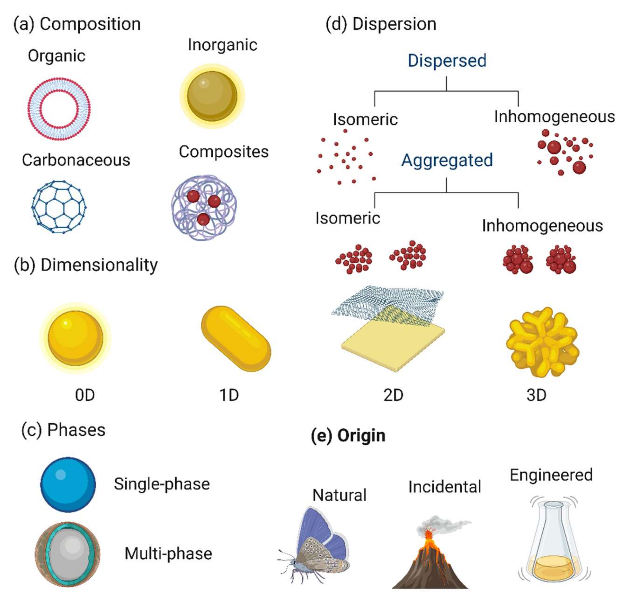
2. Classification of Nanomaterials
2.1. Classification Based on Dimensionality
- (1)
- Zero-dimensional (0D) NMs (all three dimensions in the nanoscale; i.e., up to 100 nm) include quantum dots (carbon, graphene, inorganic) and other spherical NMs (noble metals, fullerenes, polymers, metal organic framework, Up- and down-conversion nanoparticles) [22,23]. Due to their chemical inertness, biocompatibility, optical stability, cell permeability, and wavelength-dependent photoluminescence, they are interesting for biomedical and optoelectronic applications [24].
- (2)
- One-dimensional (1D) NMs (one dimension > 100 nm). In this class, nanotubes, nanorods, nanowires, and nanofibers [25] are made of polymer, carbon, metals, and metal oxides and are good electron emitters in a weak electric field. Other 1D NMs, such as veils, mats, and nonwovens, are made of polymer nanofibers [26,27]. Due to their important surface-to-volume ratio and small pores, they are used for filtration, decontamination, and catalysis and as scaffolds and super-absorbents for wound dressing and tissue engineering [28].
- (3)
- Two-dimensional (2D) NMs (two dimensions > 100 nm) include platelet-like forms, graphene (graphene oxide and re-reduced graphene oxide), transition metal dichalcogenides, metal oxides, silicates, graphitic carbon nitride, layered double hydroxides, black phosphorus, tin telluride nanosheets, antimonite, hexagonal boron nitride, boron nanosheets, and other sheet-like NMs [29,30]. Their physicochemical, biological, and optical properties explain their uniform shape, surface charge, and high surface-to-volume ratio [31].
- (4)
- Three-dimensional (3D) NMs (no dimension in the nanoscale range) include nanoporous powders, nanowire bundles, nanotube bundles, nanolayers, and nanostructured electrodes. Much research has been done on the development, fabrication, and evaluation of 3D NMs for storage devices (supercapacitors and batteries) for wastewater treatment and electrochemical conversion [32,33,34]. These complex NMs are important components of biomedical devices, solar cells, microelectromechanical systems, and robotic technology [35]. The use of 3D printing of NMs will allow the development of architectures with improved functional integration [21]. Figure 1 shows different NM classifications in the function of their properties.
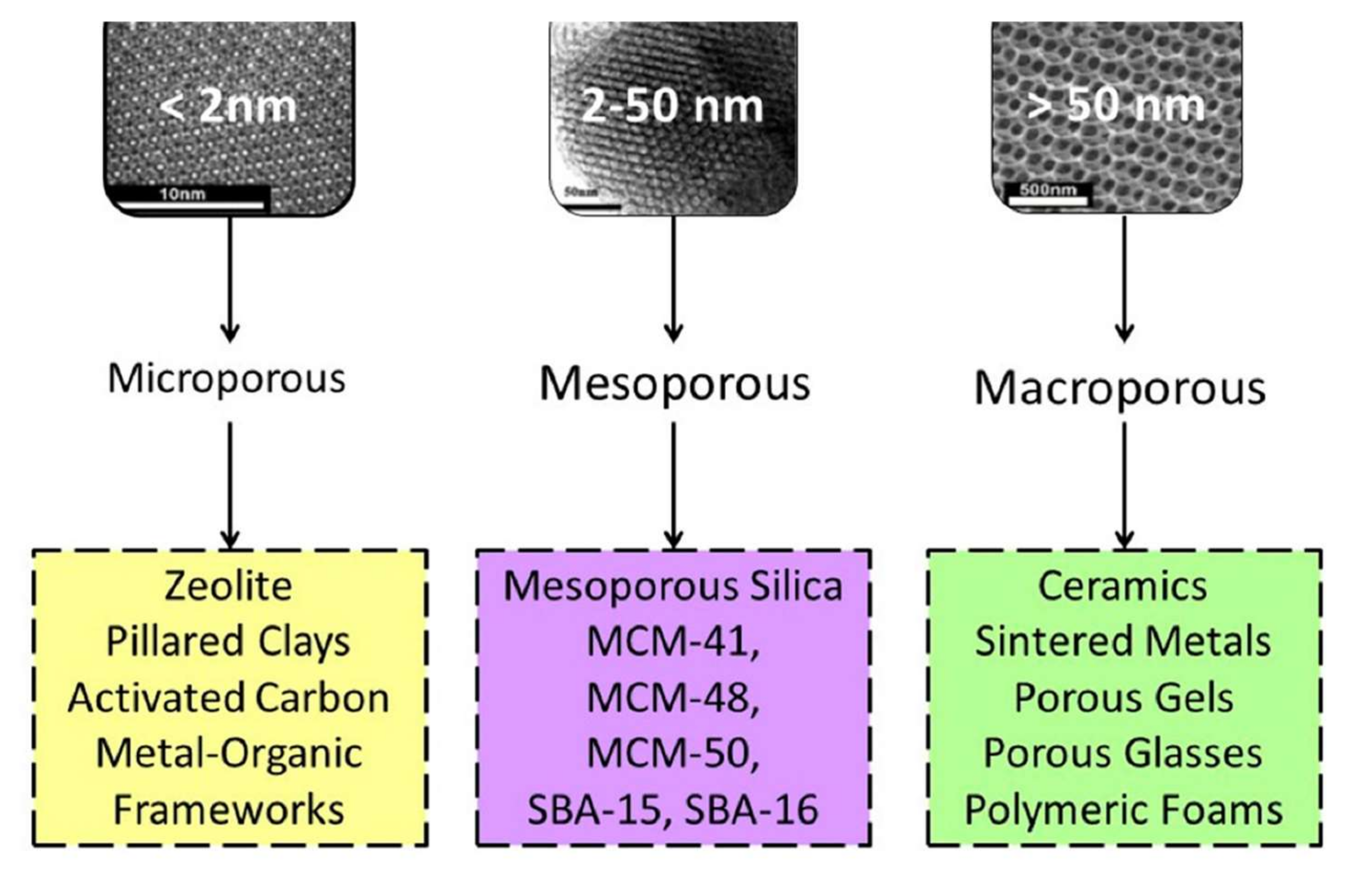
2.2. Classification Based on Porosity
- (1)
- Mesoporous NMs are highly porous compounds with pores of 2 to 50 nm in size. Moreover, according to IUPAC, highly permeable matter can be disorganized or organized in different units. Highly porous carbon is permeable in the microporous region, which significantly increases the insert surface area. Adsorbents are popular highly porous compounds that generally consist of a carbon structure with a permeable structure and a micropore volume, depending on how they were prepared [37].
- (2)
- Microporous NMs have pores < 2 nm in diameter and are commonly defined as nanopores (e.g., zeolites and metal-organic frameworks). Microporous materials are widely used for air filtration and gas separation to provide a contaminant-free gas exchange [38]. Mold spores, bacteria, and other air contaminants can be removed, while gas molecules can pass through the micropores. This allows obtaining a sterile environment within an enclosed area [39].
- (3)
- Macroporous NMs have pore sizes > 50 nm (e.g., macroporous arrays) and are particularly interesting due to their enhanced transport properties. Organized macroporous arrays should exhibit optimal fluxes, and diffusion should not be a limiting problem. This is a key issue for all processes where accessibility is crucial, for instance, delivery, sensing, catalysis, and sorption [40].
2.3. Classification Based on the Nanomaterial Source
- (1)
- Natural NMs are materials formed through natural (bio)geochemical or mechanical processes (e.g., combustion materials from forest fires, acid mine drainage, volcanic ash, sea spray, and radioactive radon gas waste) without any direct or indirect contribution by anthropogenic activities and processes [42]. Examples of natural NMs include the blue colors of tarantula, some butterfly wing scales, silk spiders and spider mites, foraminifera, viral structures such as capsids and proteins, wax crystal coating, lotus or nasturtium leaves, gecko foot spatula, natural colloids (milk and blood), human bone matrix, coral, nacre, and horn materials such as feathers, hair, skin, and claws [41,43]. Some inorganic NMs are formed naturally by crystal growth. For example, clays exhibit complex nanostructures due to their anisotropic crystal structure. Opals are probably formed by volcanic activity. Moreover, natural photonic crystals are considered NMs because of their nanoscale structure [44].
- (2)
- Incidental NMs are created unintentionally by direct or indirect human actions (e.g., vehicle engine exhaust, welding gasses, solid fuel combustion, and cooking). Incidental NMs unintentionally formed during an intentional process can increase air pollution. Many NMs (e.g., pigments, fumed silica, and cement) are formed during forest fires [41]. It is difficult to determine when incidental NMs started to be produced by humans. Usually, in incidental NMs, size and shape are not regular. They strongly affect the environment and should be compared to engineered NMs [45].
- (3)
- Engineered NMs are manufactured to fulfill specific needs (e.g., nanostructured medical implants) [46]. These nanoparticles have regular shapes and sizes (rings, fullerenes, carbon nanotubes, spheres, and graphene), whereas natural and incidental nanoparticles have irregular shapes and sizes, such as carbon black [41,47]. In the 1940s, the first commercialized NMs were prepared from fumed silica, and in the 1960s, the first silica nanospheres were fabricated from aqueous solutions [45].
- (4)
- Bioinspired NMs are fabricated to obtain specific nanostructures, features, or functions to mimic natural materials or living organisms. In many bioinspired NMs, advanced nanofabrication techniques are used to modulate their structures and obtain specific functions. For example, the rapid color change observed in chameleons when fighting or during courtship is mainly explained by the lattice adjustment of guanine nanocrystals in iridophore cells [48]. The photonic structure of chameleon iridophores can be mimicked by incorporating silica nanocrystals into mechanochromic elastomer sensors as non-dense packed crystals. These sensors change color when stretched (from red to blue) and when compressed (from red to green). This effect is reversible, as observed in chameleons. Such sensors may be used in wallpaper, signs, and optical records [41,49].
2.4. Nanomaterial Classification in the Function of Their Chemical Composition
- (1)
- Carbon-based NMs can be produced from sp2 carbon (e.g., fullerenes, graphene, carbon nanotubes, nanohorns, nano-onions, nanographite, nanodiamonds, carbon nanofibers) using various techniques, such as laser ablation, arc discharge, and chemical vapor deposition (CVD) [50,51]. Carbon-based nanoparticles are a special NM type due to their wide range of allotropies and can be considered to be organic NMs due to the presence of C-C bonds. Nanodiamonds, carbon black, and activated carbon (made of non-sp2 hybridized carbon atoms) also belong to this category. Milling or seeding can be used to reduce the size of most NMs present in the environment (e.g., CVD for nanodiamonds) [52]. Carbon-based NMs have been playing an important part in human activities (e.g., composites, pigments, reinforcing materials, fuels). In the field of renewable energy, graphite blocks are used as reflectors and moderators in nuclear reactors [14]. Moreover, carbon nanostructures serve as electrodes in electrochemical sensors, rechargeable batteries, and supercapacitors, [53,54].
- (2)
- Organic NMs are mainly made of carbon and hydrogen, with which other elements are chemically associated to obtain NMs with specific functionalities (e.g., dendrimers, micelles, liposomes, and ferritin). Organic NMs also include lipid and polymer nanoparticles that usually have a nano-encapsulated form (10–1000 nm in size) [55]. The polar lipid assemblies at the cell membranes of some bacteria and viruses are called lipid bilayers. These bilayers are mimicked by Langmuir–Blodgett films made of amphiphilic organic compounds in which one polar nanoblock interacts with another polar nanoblock. The head is on the polar side while the tail is on the polar side, and both have the same size [10]. In these fabricated films, the hydrophilic “head” and the hydrophobic “tail” allow the formation of micelles, liposomes, and single or bilayer films. Micelles and liposomes have a hollow core [56,57].
- (3)
- Inorganic NMs are composed of or include non-carbon elements (e.g., metals, metal oxides, and metal salts). Such NMs have many shapes (e.g., cylinders, wafers, ellipses, cubes, spheres, stars) in the function of the atom packing while maintaining the crystalline nature of metal-based compounds [58,59]. In addition, there are amorphous inorganic nanoparticles. Due to the pendulous bonds of atoms, the surface of inorganic NMs is very reactive and sensitive. This drawback can be overcome through functionalization. Some inorganic NMs have remarkable features, particularly metal-based quantum dots (1-10 nm) due to the transition stage between mass and few atoms, and magnetic nanoparticles [e.g., iron (Fe), magnetite (Fe3O4), and γ-Fe2O3 [60,61] due to their strong coercive forces and paramagnetic properties [62]. Nanoclays (1nm-thick 2D silicates) are biocompatible and have low toxicity [63]. The main applications of nanoclays are membrane coatings, polymer reinforcement, barriers, toxin adsorption, and sterilizing materials. Zeolite is a non-toxic, nanoporous, hydrated crystalline aluminosilicate with ion exchange properties for the removal of hazardous pollutants from wastewater [13].
- (4)
- Hybrid NMs are multiphase solid materials in which one of the phases has dimensions less than 100 nm [64]. In polymeric nanohybrids, polymers serve as a matrix for organic or inorganic nanoparticles in various forms [65,66]. This class also includes porous media, colloids, gels, and copolymers. Inorganic nanocomposites combine two or more metals in metal nanocomposites, such as intermetallic compounds, alloys with nanometals, core-shell nanoparticles, and banded components [67]. One of the most important nanohybrids is the carbon nanotube-metal matrix composite, an emerging new material being developed to take advantage of high tensile strength and electrical conductivity. Nanohybrids occur in nature, for example in the structure of abalone shells and bones [68,69].
2.5. Other Classifications
3. Mechanisms of Nanoparticle Formation
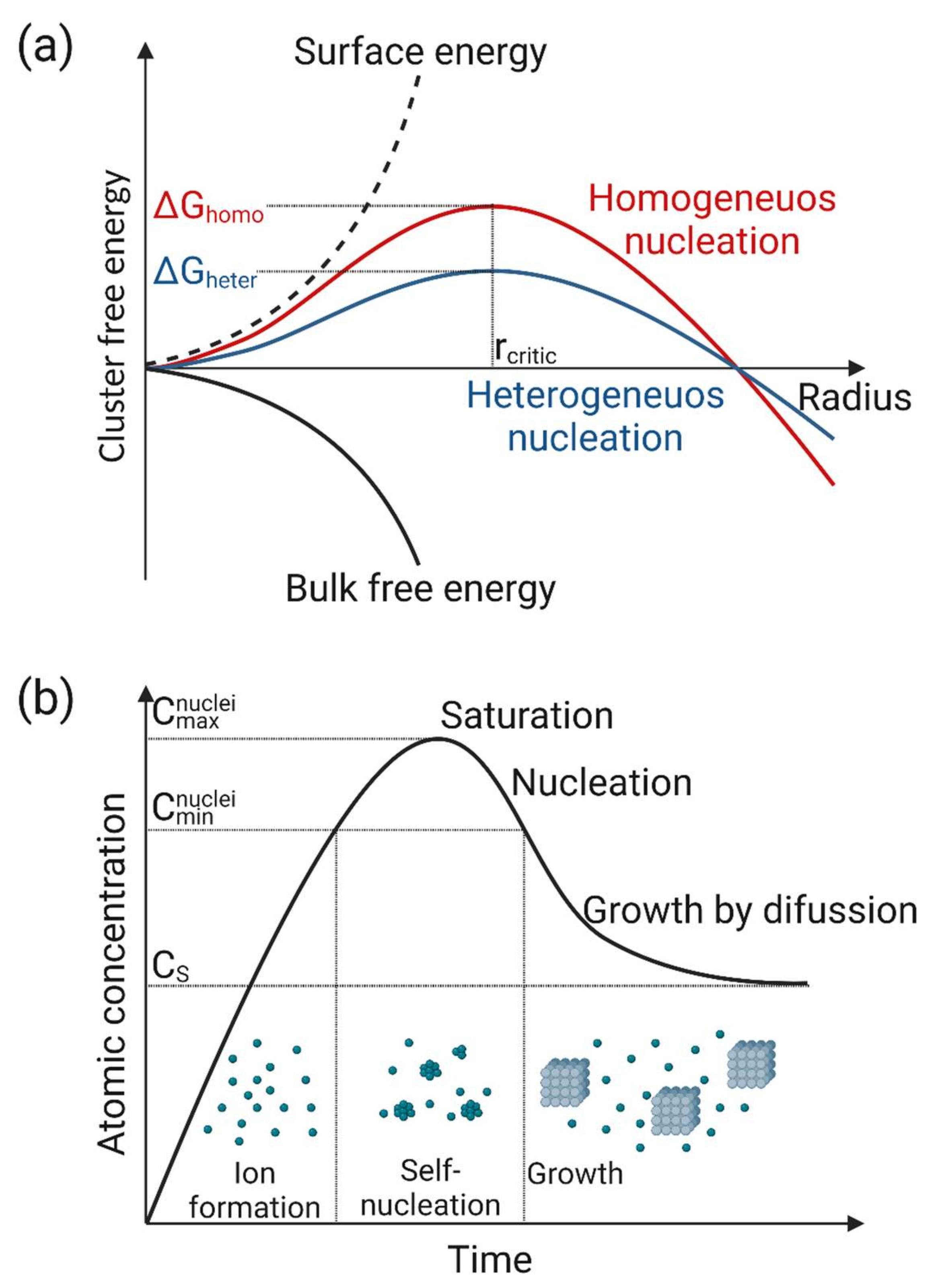
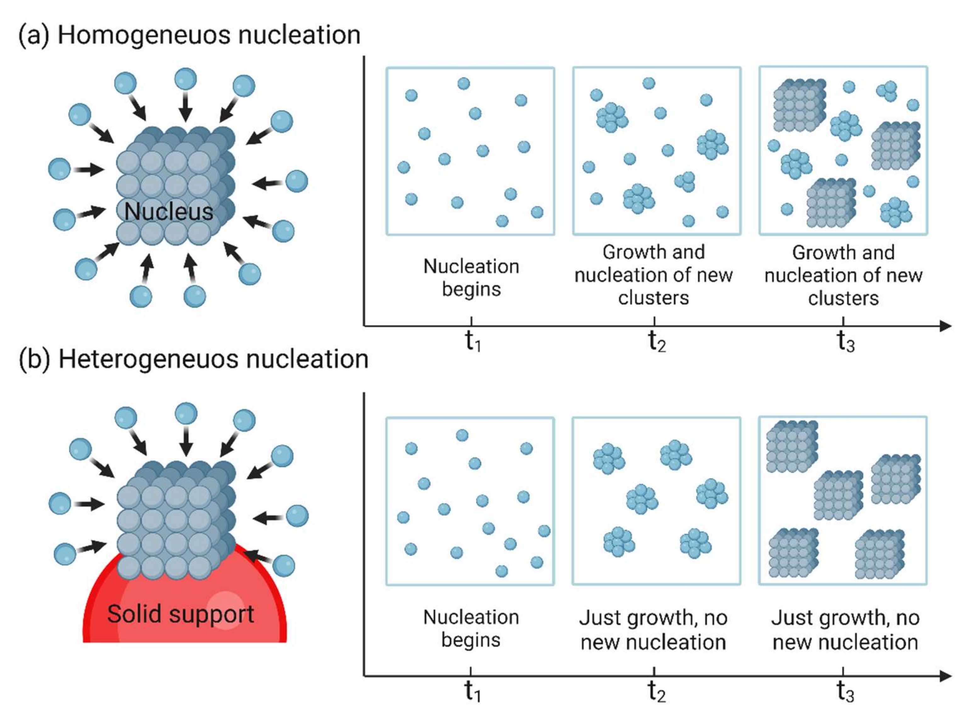
3.1. Nucleation
3.2. Growth
4. Classification of Nanomaterial Synthesis Methods in Function of the Starting Materials
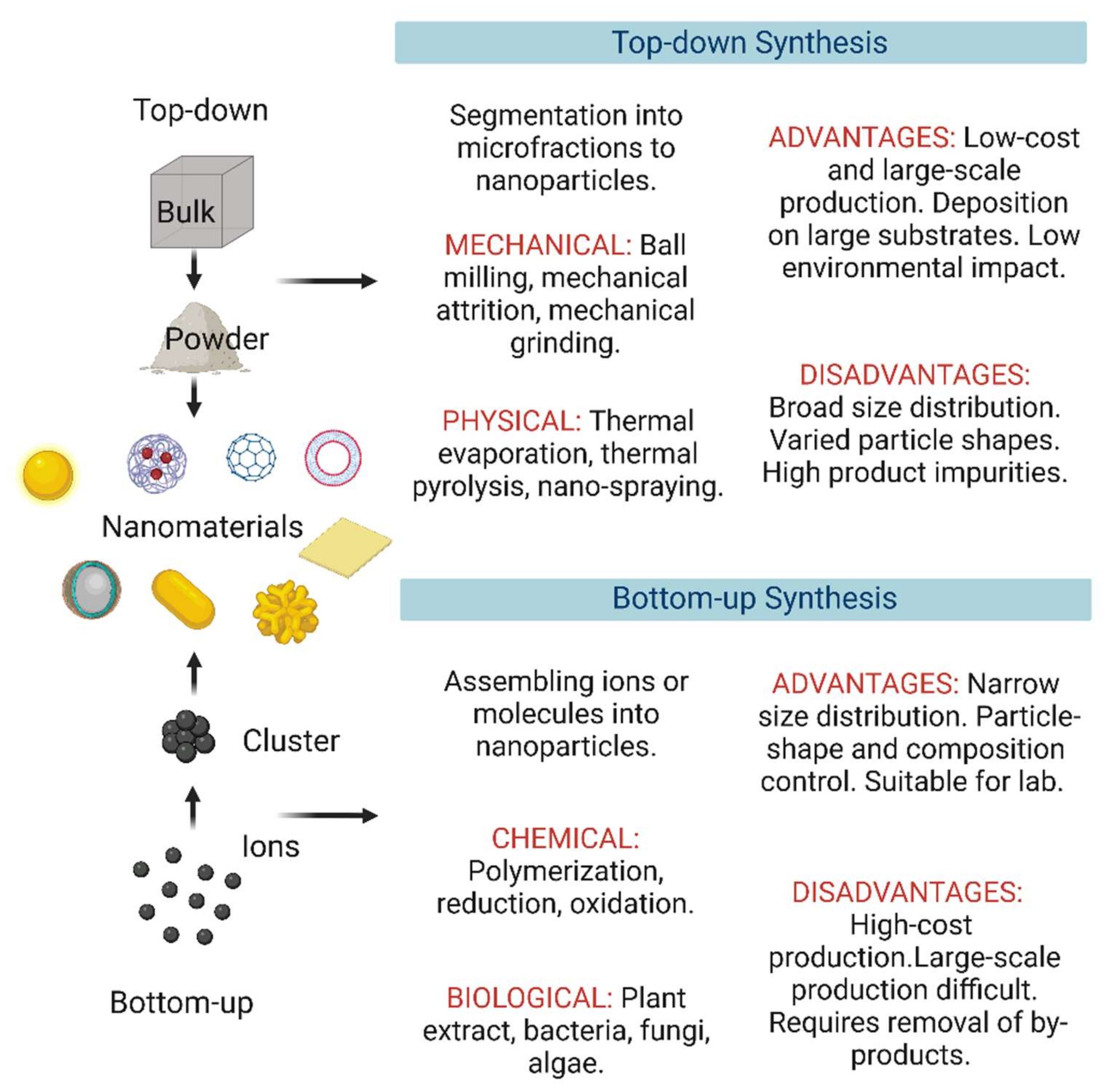
4.1. Top-Down Approaches
4.2. Bottom-Up Approaches
4.3. Hybrid Approaches
5. Classification of Nanomaterial Synthesis Techniques in Function of the Deriving Forces
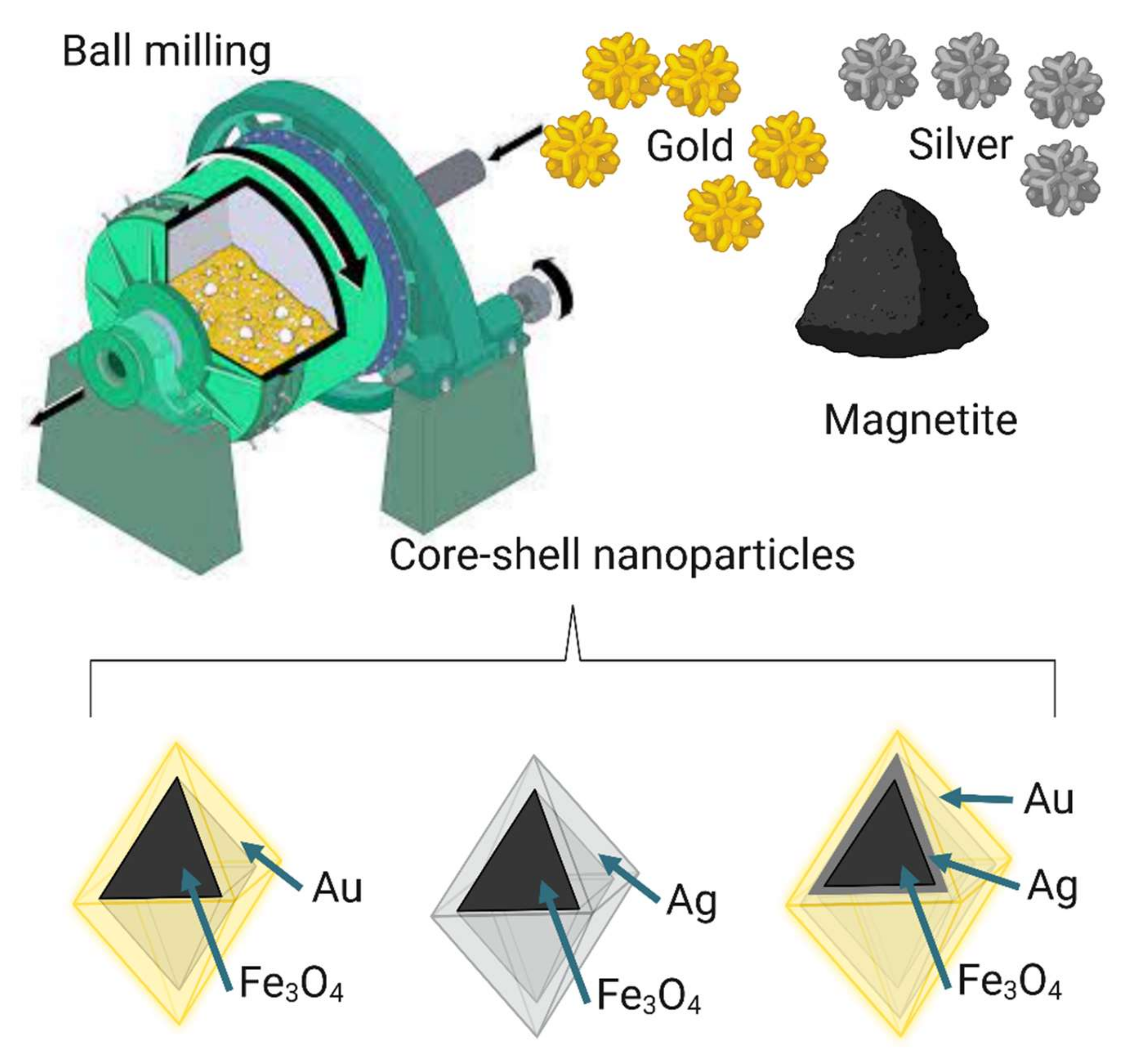
5.1. Mechanical Methods
5.2. Physical Synthesis Methods
5.3. Chemical and Physicochemical Synthesis Methods
5.4. Biological or Green Synthesis Methods
6. Nanosynthesis Method Classification in the Function of the Reaction Phase
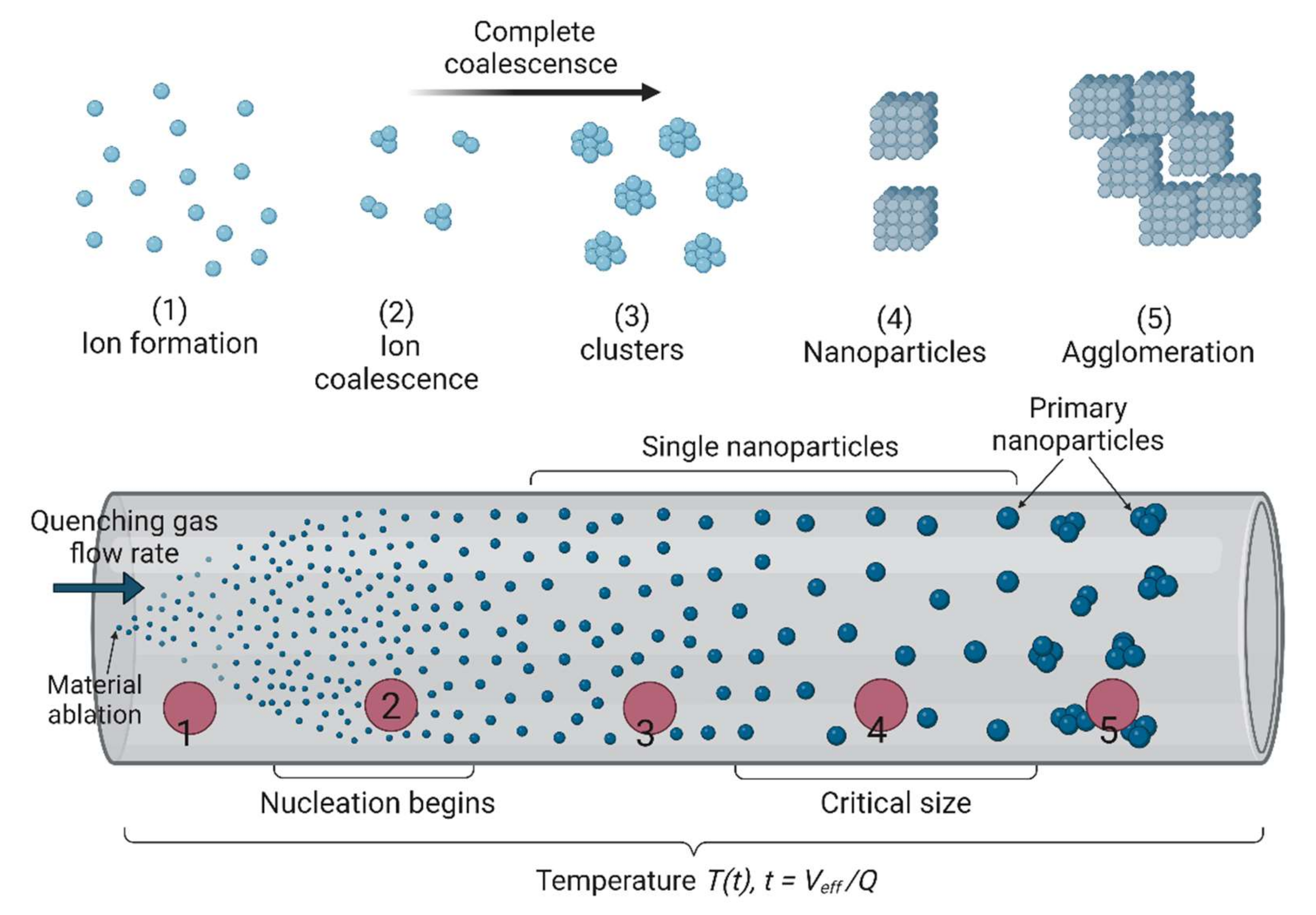
6.1. Gas Phase Synthesis
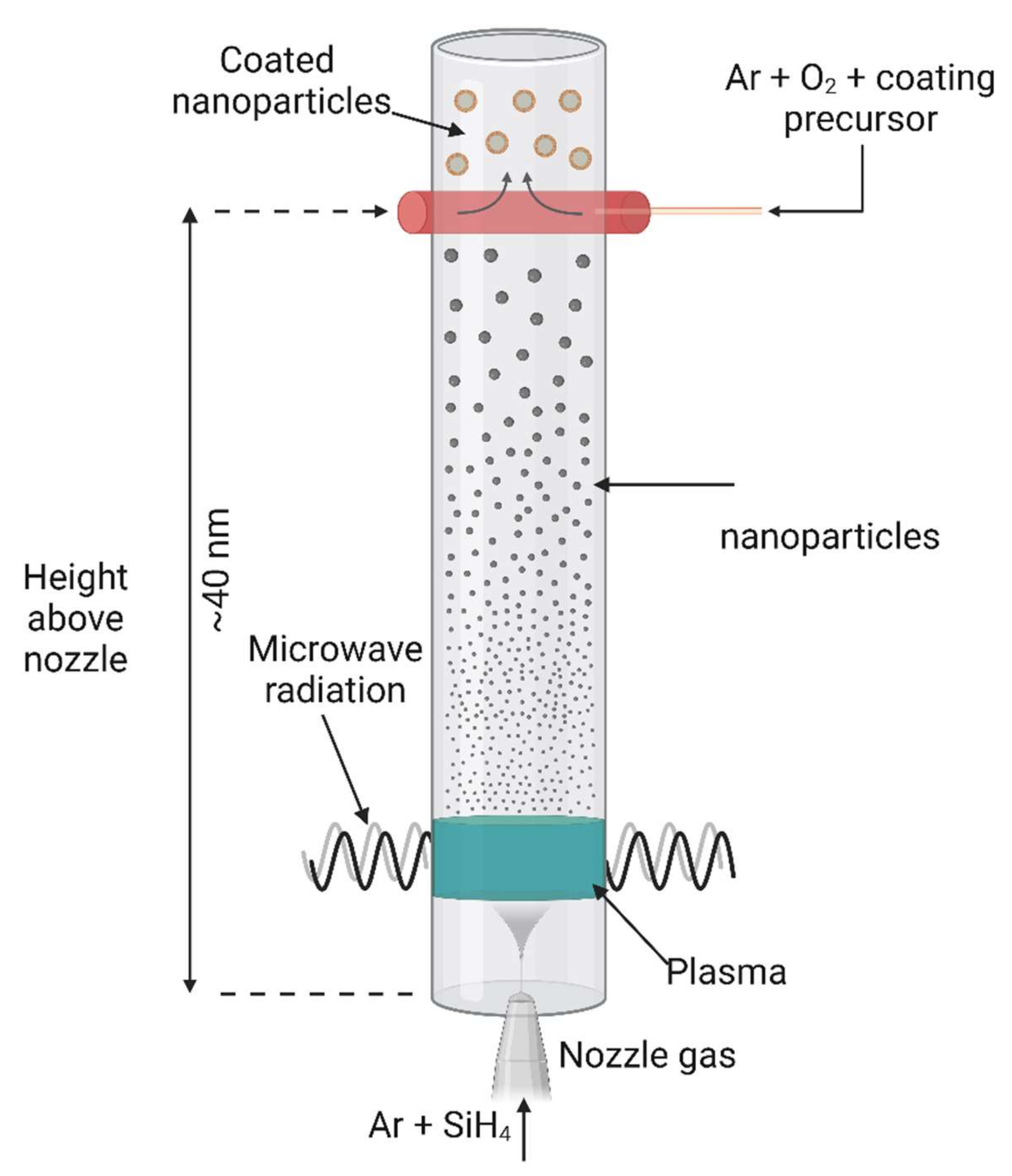
6.2. Plasma Synthesis
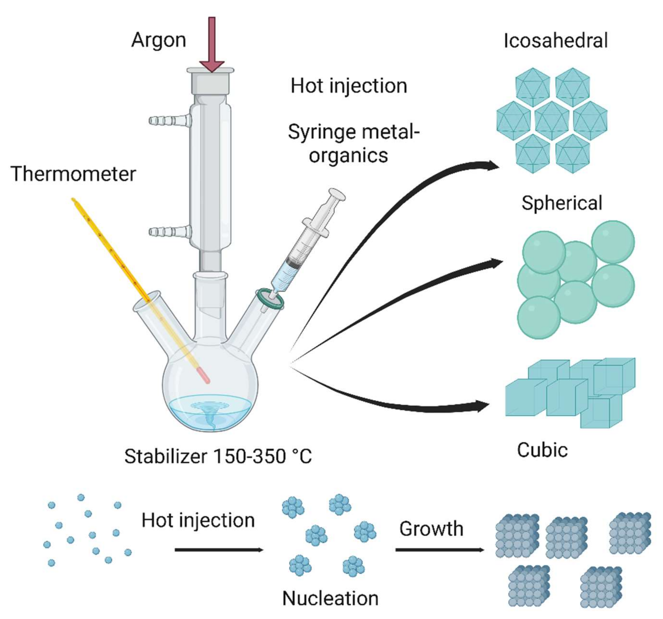
6.3. Liquid Phase Synthesis
6.4. Supercritical Fluid Synthesis
6.5. Solid Phase Synthesis
7. Concluding Remarks
Author Contributions
Funding
Conflicts of Interest
References
- Lal, S.; Jana, U.; Manna, P.K.; Mohanta, G.P.; Manavalan, R.; Pal, S.L. Nanoparticle: An Overview of Preparation and Characterization. J. Appl. Pharm. Sci. 2011, 1, 228–234. [Google Scholar]
- Jeevanandam, J.; Ling, J.K.U.; Tiong, M.; Barhoum, A.; Chan, Y.S.; Acquah, C.; Danquah, M.K. Nanocelluloses: Sources, Types, Unique Properties, Market, and Regulations. In Handbook of Nanocelluloses; Springer: Cham, Switzerland, 2022; pp. 3–34. [Google Scholar] [CrossRef]
- Khan, I.; Saeed, K.; Khan, I. Nanoparticles: Properties, Applications and Toxicities. Arab. J. Chem. 2019, 12, 908–931. [Google Scholar] [CrossRef]
- Hasan, S. A Review on Nanoparticles: Their Synthesis and Types. Res. J. Recent Sci. 2015, 4, 1–3. [Google Scholar]
- Meftahi, A.; Momeni Heravi, M.E.; Barhoum, A.; Samyn, P.; Najarzadeh, H.; Alibakhshi, S. Cellulose Nanofibers. Handb. Nanocelluloses 2022, 233–262. [Google Scholar] [CrossRef]
- Ijaz, I.; Gilani, E.; Nazir, A.; Bukhari, A. Detail review on chemical, physical and green synthesis, classification, characterizations and applications of nanoparticles. Taylor Fr. 2020, 13, 59–81. [Google Scholar] [CrossRef]
- Harish, V.; Tewari, D.; Gaur, M.; Yadav, A.B.; Swaroop, S.; Bechelany, M.; Barhoum, A. Review on Nanoparticles and Nanostructured Materials: Bioimaging, Biosensing, Drug Delivery, Tissue Engineering, Antimicrobial, and Agro-Food Applications. Nanomater 2022, 12, 457. [Google Scholar] [CrossRef]
- Barhoum, A.; Bechelany, M.; Makhlouf, A.S.H. Handbook of Nanofibers; Springer International Publishing: Cham, Switzerland, 2019; ISBN 978-3-319-53654-5. [Google Scholar]
- Barhoum, A.; García-Betancourt, M.L.; Jeevanandam, J.; Hussien, E.A.; Mekkawy, S.A.; Mostafa, M.; Omran, M.M.; Abdalla, M.S.; Bechelany, M. Review on Natural, Incidental, Bioinspired, and Engineered Nanomaterials: History, Definitions, Classifications, Synthesis, Properties, Market, Toxicities, Risks, and Regulations. Nanomater 2022, 12, 177. [Google Scholar] [CrossRef] [PubMed]
- Gaur, M.; Misra, C.; Yadav, A.B.; Swaroop, S.; Maolmhuaidh, F.Ó.; Bechelany, M.; Barhoum, A. Biomedical Applications of Carbon Nanomaterials: Fullerenes, Quantum Dots, Nanotubes, Nanofibers, and Graphene. Materials 2021, 14, 5978. [Google Scholar] [CrossRef]
- Bayda, S.; Adeel, M.; Tuccinardi, T.; Cordani, M.; Molecules, F.R. The History of Nanoscience and Nanotechnology: From Chemical–Physical Applications to Nanomedicine. Molecules 2019, 25, 112. [Google Scholar] [CrossRef]
- Jeevanandam, J.; Barhoum, A.; Chan, Y.S.; Dufresne, A.; Danquah, M.K. Review on Nanoparticles and Nanostructured Materials: History, Sources, Toxicity and Regulations. Beilstein J. Nanotechnol. 2018, 9, 1050–1074. [Google Scholar] [CrossRef]
- Salama, A.; Abouzeid, R.; Leong, W.S.; Jeevanandam, J.; Samyn, P.; Dufresne, A.; Bechelany, M.; Barhoum, A. Nanocellulose-Based Materials for Water Treatment: Adsorption, Photocatalytic Degradation, Disinfection, Antifouling, and Nanofiltration. Nanomaterials 2021, 11, 3008. [Google Scholar] [CrossRef] [PubMed]
- Barhoum, A.; El-Maghrabi, H.H.; Nada, A.A.; Sayegh, S.; Roualdes, S.; Renard, A.; Iatsunskyi, I.; Coy, E.; Bechelany, M. Simultaneous Hydrogen and Oxygen Evolution Reactions Using Free-Standing Nitrogen-Doped-Carbon–Co/CoOx Nanofiber Electrodes Decorated with Palladium Nanoparticles. J. Mater. Chem. A 2021, 9, 17724–17739. [Google Scholar] [CrossRef]
- Haichao, L.; Li, H.; Bubakir, M.M.; Yang, W.; Barhoum, A. Engineering Nanofibers as Electrode and Membrane Materials for Batteries, Supercapacitors, and Fuel Cells. In Handbook of Nanofibers; Springer: Cham, Switzerland, 2019; pp. 1105–1130. [Google Scholar] [CrossRef]
- Abdel-Haleem, F.M.; Saad, M.; Barhoum, A.; Bechelany, M.; Rizk, M.S. PVC Membrane, Coated-Wire, and Carbon-Paste Ion-Selective Electrodes for Potentiometric Determination of Galantamine Hydrobromide in Physiological Fluids. Mater. Sci. Eng. C. Mater. Biol. Appl. 2018, 89, 140–148. [Google Scholar] [CrossRef] [PubMed]
- Barhoum, A.; Forster, R.J. Label-Free Electrochemical Immunosensor for Picomolar Detection of the Cervical Cancer Biomarker MCM5. Anal. Chim. Acta 2022, 1225, 340226. [Google Scholar] [CrossRef]
- Mostafa, M.; Barhoum, A.; Sehit, E.; Gewaid, H.; Mostafa, E.; Omran, M.M.; Abdalla, M.S.; Abdel-Haleem, F.M.; Altintas, Z.; Forster, R.J. Current Trends in COVID-19 Diagnosis and Its New Variants in Physiological Fluids: Surface Antigens, Antibodies, Nucleic Acids, and RNA Sequencing. Trends Analyt. Chem. 2022, 116750, in press. [Google Scholar] [CrossRef] [PubMed]
- Rabie, A.M.I.; Ali, A.S.M.; Al-Zeer, M.A.; Barhoum, A.; El-Hallouty, S.; Shousha, W.G.; Berg, J.; Kurreck, J.; Khalil, A.S.G. Spontaneous Formation of 3D Breast Cancer Tissues on Electrospun Chitosan/Poly(Ethylene Oxide) Nanofibrous Scaffolds. ACS Omega 2022, 7, 2114–2126. [Google Scholar] [CrossRef]
- Hamimed, S.; Abdeljelil, N.; Landoulsi, A.; Chatti, A.; Aljabali, A.A.A.; Barhoum, A. Bacterial Cellulose Nanofibers. In Handbook of Nanocelluloses; Barhoum, A., Ed.; Springer: Cham, Switzerland, 2022; pp. 1–38. [Google Scholar] [CrossRef]
- Paramasivam, G.; Palem, V.V.; Sundaram, T.; Sundaram, V.; Kishore, S.C.; Bellucci, S. Nanomaterials: Synthesis and Applications in Theranostics. Nanomaterials 2021, 11, 3228. [Google Scholar] [CrossRef]
- Barhoum, A.; García-Betancourt, M.L.; Rahier, H.; Van Assche, G. Physicochemical Characterization of Nanomaterials: Polymorph, Composition, Wettability, and Thermal Stability. In Emerging Applications of Nanoparticles and Architectural Nanostructures: Current Prospects and Future Trends; Elsevier: Amsterdam, The Netherlands, 2018; pp. 255–278. ISBN 9780128135167. [Google Scholar]
- Djamila, B.; Eddine, L.S.; Abderrhmane, B.; Nassiba, A.; Barhoum, A. In Vitro Antioxidant Activities of Copper Mixed Oxide (CuO/Cu2O) Nanoparticles Produced from the Leaves of Phoenix Dactylifera L. Biomass Convers. Biorefinery 2022, 1, 1–14. [Google Scholar] [CrossRef]
- Sannino, D. Types and Classification of Nanomaterials. In Nanotechnology; Springer: Singapore, 2021; pp. 15–38. [Google Scholar] [CrossRef]
- Gugulothu, D.; Barhoum, A.; Afzal, S.M.; Venkateshwarlu, B.; Uludag, H. Structural Multifunctional Nanofibers and Their Emerging Applications. In Handbook of Nanofibers; Springer: Cham, Switzerland, 2018; pp. 1–41. [Google Scholar] [CrossRef]
- Ganjali, M.; Ganjali, M.; Sereshki, M.M.A.; Ahmadinasab, N.; Ghalandarzadeh, A.; Aljabali, A.A.A.; Barhoum, A. Bionanomaterials for cancer therapy. In Bionanotechnology: Emerging Applications of Bionanomaterials; Elsevier: Amsterdam, The Netherlands, 2022; pp. 443–468. [Google Scholar] [CrossRef]
- Barhoum, A.; Favre, T.; Sayegh, S.; Tanos, F.; Coy, E.; Iatsunskyi, I.; Razzouk, A.; Cretin, M.; Bechelany, M. 3D Self-Supported Nitrogen-Doped Carbon Nanofiber Electrodes Incorporated Co/CoOx Nanoparticles: Application to Dyes Degradation by Electro-Fenton-Based Process. Nanomater 2021, 11, 2686. [Google Scholar] [CrossRef]
- Quan, L.N.; Kang, J.; Ning, C.Z.; Yang, P.D.; Deng, J.; Su, Y.; Liu, D.; Liu, B.; Liu, C. Introduction: 1D Nanomaterials/Nanowires. Chem. Rev. 2019, 119, 8955–8957. [Google Scholar] [CrossRef]
- Youssef, A.M.; Moustafa, H.A.; Barhoum, A.; Hakim, A.E.-F.A.A.; Dufresne, A. Evaluation of the Morphological, Electrical and Antibacterial Properties of Polyaniline Nanocomposite Based on Zn/Al-Layered Double Hydroxides. ChemistrySelect 2017, 2, 8553–8566. [Google Scholar] [CrossRef]
- Abdel-Haleem, F.M.; Gamal, E.; Rizk, M.S.; El Nashar, R.M.; Anis, B.; Elnabawy, H.M.; Khalil, A.S.G.; Barhoum, A. t-Butyl calixarene/Fe2O3@MWCNTs composite-based potentiometric sensor for determination of ivabradine hydrochloride in pharmaceutical formulations. Mat. Sci.Eng. C 2020, 116, 111110. [Google Scholar] [CrossRef] [PubMed]
- Zhang, H. Ultrathin Two-Dimensional Nanomaterials. ACS Nano 2015, 9, 9451–9469. [Google Scholar] [CrossRef] [PubMed]
- Moodley, K.G.; Arumugam, V.; Barhoum, A. Nanocellulose-Based Materials for Wastewater Treatment. In Handbook of Nanocelluloses: Classification, Properties, Fabrication, and Emerging Applications; Springer International Publishing: Cham, Switzerland, 2021; pp. 1–33. [Google Scholar] [CrossRef]
- Hamimed, S.; Jebli, N.; Othmani, A.; Hamimed, R.; Barhoum, A.; Chatti, A. Nanocelluloses for Removal of Heavy Metals From Wastewater. In Handbook of Nanocelluloses: Classification, Properties, Fabrication, and Emerging Applications; Springer: Cham, Switzerland, 2022; pp. 1–42. [Google Scholar]
- Meftahi, A.; Samyn, P.; Geravand, S.A.; Khajavi, R.; Alibkhshi, S.; Bechelany, M.; Barhoum, A. Nanocelluloses as Skin Biocompatible Materials for Skincare, Cosmetics, and Healthcare: Formulations, Regulations, and Emerging Applications. Carbohydr. Polym. 2022, 278, 118956. [Google Scholar] [CrossRef]
- El-Beshlawy, M.; Abdel-Haleem, F.; Barhoum, A. Molecularly Imprinted Polymer-Based Potentiometric Biosensor for Nanomolar Determination of Pioglitazone Hydrochloride in Pharmaceutical Formulations. Electroanalysis 2021, 33, 1244–1254. [Google Scholar] [CrossRef]
- Barhoum, A.; Melcher, J.; Van Assche, G.; Rahier, H.; Bechelany, M.; Fleisch, M.; Bahnemann, D. Synthesis, Growth Mechanism, and Photocatalytic Activity of Zinc Oxide Nanostructures: Porous Microparticles versus Nonporous Nanoparticles. J. Mater. Sci. 2017, 52, 2746–2762. [Google Scholar] [CrossRef]
- Szczesniak, B.; Borysiuk, S.; Choma, J.; Jaroniec, M. Mechanochemical Synthesis of Highly Porous Materials. Mater. Horizons 2020, 7, 1457–1473. [Google Scholar] [CrossRef]
- Barhoum, A.; Van Assche, G.; Rahier, H.; Fleisch, M.; Bals, S.; Delplancked, M.P.; Leroux, F.; Bahnemann, D. Sol-Gel Hot Injection Synthesis of ZnO Nanoparticles into a Porous Silica Matrix and Reaction Mechanism. Mater. Des. 2017, 119, 270–276. [Google Scholar] [CrossRef]
- Kang, H.J.; Rafiqul Bari, G.A.K.M.; Lee, T.G.; Khan, T.T.; Park, J.W.; Hwang, H.J.; Cho, S.Y.; Jun, Y.S. Microporous Carbon Nanoparticles for Lithium–Sulfur Batteries. Nanomaterials 2020, 10, 2012. [Google Scholar] [CrossRef]
- Karunagaran, R.; Tran, D.; Tung, T.T.; Shearer, C.; Losic, D. A Unique Synthesis of Macroporous N-Doped Carbon Composite Catalyst for Oxygen Reduction Reaction. Nanomaterials 2021, 11, 43. [Google Scholar] [CrossRef]
- Hochella, M.F.; Mogk, D.W.; Ranville, J.; Allen, I.C.; Luther, G.W.; Marr, L.C.; McGrail, B.P.; Murayama, M.; Qafoku, N.P.; Rosso, K.M.; et al. Natural, Incidental, and Engineered Nanomaterials and Their Impacts on the Earth System. Science 2019, 363, eaau8299. [Google Scholar] [CrossRef] [PubMed]
- Griffin, S.; Masood, M.I.; Nasim, M.J.; Sarfraz, M.; Ebokaiwe, A.P.; Schäfer, K.H.; Keck, C.M.; Jacob, C. Natural Nanoparticles: A Particular Matter Inspired by Nature. Antioxidants 2017, 7, 3. [Google Scholar] [CrossRef] [PubMed]
- Shalan, A.E.; Barhoum, A.; Elseman, A.M.; Rashad, M.M.; Lira-Cantú, M. Nanofibers as Promising Materials for New Generations of Solar Cells. In Handbook of Nanofibers; Springer International Publishing: Cham, Switzerland, 2018; pp. 1–33. [Google Scholar] [CrossRef]
- Lespes, G.; Faucher, S.; Slaveykova, V.I. Natural Nanoparticles, Anthropogenic Nanoparticles, Where Is the Frontier? Front. Environ. Sci. 2020, 8, 71. [Google Scholar] [CrossRef]
- Westerhoff, P.; Atkinson, A.; Fortner, J.; Wong, M.S.; Zimmerman, J.; Gardea-Torresdey, J.; Ranville, J.; Herckes, P. Low Risk Posed by Engineered and Incidental Nanoparticles in Drinking Water. Nat. Nanotechnol. 2018, 13, 661–669. [Google Scholar] [CrossRef]
- Prasad, S.; Kumar, V.; Kirubanandam, S.; Barhoum, A. Engineered Nanomaterials: Nanofabrication and Surface Functionalization. In Emerging Applications of Nanoparticles and Architectural Nanostructures: Current Prospects and Future Trends; Elsevier Inc.: Amsterdam, The Netherlands, 2018; pp. 305–340. ISBN 9780128135167. [Google Scholar]
- Nnaji, C.O.; Jeevanandam, J.; Chan, Y.S.; Danquah, M.K.; Pan, S.; Barhoum, A. Engineered Nanomaterials for Wastewater Treatment: Current and Future Trends. In Fundamentals of Nanoparticles; Elsevier: Amsterdam, The Netherlands, 2018; pp. 129–168. [Google Scholar]
- Aljabali, A.A.A.; Obeid, M.A.; Awadeen, S.A.; Migdadi, E.M.; Barhoum, A.; Al Zoubi, M.S.; Kumar Chellappan, D.; Mishra, V.; Charbe, N.B.; Dureja, H.; et al. Nature Bioinspired and Engineered Nanomaterials. In Fundamentals of Bionanomaterials; Elsevier: Amsterdam, The Netherlands, 2022; pp. 31–58. [Google Scholar] [CrossRef]
- Bhattacharya, P.; Du, D.; Lin, Y. Bioinspired Nanoscale Materials for Biomedical and Energy. Artic. J. R. Soc. Interface 2014, 11, 20131067. [Google Scholar] [CrossRef]
- Kumar, S.; Bhushan, P.; Bhattacharya, S. Fabrication of Nanostructures with Bottom-up Approach and Their Utility in Diagnostics, Therapeutics, and Others. In Environmental, Chemical and Medical Sensors; Springer: Singapore, 2018; pp. 167–198. [Google Scholar] [CrossRef]
- Butler, J.E.; Sumant, A.V. The CVD of Nanodiamond Materials. Chem. Vap. Depos. 2008, 14, 145–160. [Google Scholar] [CrossRef]
- Barhoum, A.; El-Maghrabi, H.H.; Iatsunskyi, I.; Coy, E.; Renard, A.; Salameh, C.; Weber, M.; Sayegh, S.; Nada, A.A.; Roualdes, S.; et al. Atomic Layer Deposition of Pd Nanoparticles on Self-Supported Carbon-Ni/NiO-Pd Nanofiber Electrodes for Electrochemical Hydrogen and Oxygen Evolution Reactions. J. Colloid Interface Sci. 2020, 569, 286–297. [Google Scholar] [CrossRef]
- Abdel-Haleem, F.M.; Mahmoud, S.; Abdel-Ghani, N.E.T.; El Nashar, R.M.; Bechelany, M.; Barhoum, A. Polyvinyl Chloride Modified Carbon Paste Electrodes for Sensitive Determination of Levofloxacin Drug in Serum, Urine, and Pharmaceutical Formulations. Sensors 2021, 21, 3150. [Google Scholar] [CrossRef]
- Oschatz, M.; Borchardt, L.; Hippauf, F.; Nickel, W.; Kaskel, S.; Brunner, E. Interactions between Electrolytes and Carbon-Based Materials—NMR Studies on Electrical Double-Layer Capacitors, Lithium-Ion Batteries, and Fuel Cells. In Annual reports on NMR Spectroscopy; Academic Press: Cambridge, MA, USA, 2016. [Google Scholar]
- Ramsden, J.J. The Impact of Nanotechnology. In Nanotechnology: An Introduction, 2nd ed.; Ramsden, J.J., Ed.; William Andrew Publishing: Norwich, NY, USA, 2016; pp. 279–304. [Google Scholar] [CrossRef]
- Wagner, A.M.; Knipe, J.M.; Orive, G.; Peppas, N.A. Quantum Dots in Biomedical Applications. Acta Biomater. 2019, 94, 44–63. [Google Scholar] [CrossRef]
- Azam, N.; Najabat Ali, M.; Javaid Khan, T. Carbon Quantum Dots for Biomedical Applications: Review and Analysis. Front. Mater. 2021, 8, 700403. [Google Scholar] [CrossRef]
- Wang, X.; Zhong, X.; Li, J.; Liu, Z.; Cheng, L. Inorganic Nanomaterials with Rapid Clearance for Biomedical Applications. Chem. Soc. Rev. 2021, 50, 8669–8742. [Google Scholar] [CrossRef] [PubMed]
- Aljabali, A.A.A.; Obeid, M.A.; Al Zoubi, M.S.; Charbe, N.B.; Chellappan, D.K.; Mishra, V.; Dureja, H.; Gupta, G.; Prasher, P.; Dua, K.; et al. Nanocelluloses in Sensing Technology. In Handbook of Nanocelluloses; Springer: Cham, Switzerland, 2021. [Google Scholar] [CrossRef]
- Fernandes, N.; Rodrigues, C.F.; Moreira, A.F.; Correia, I.J. Overview of the Application of Inorganic Nanomaterials in Cancer Photothermal Therapy. Biomater. Sci. 2020, 8, 2990–3020. [Google Scholar] [CrossRef] [PubMed]
- Gul, S.; Khan, S.B.; Rehman, I.U.; Khan, M.A.; Khan, M.I. A Comprehensive Review of Magnetic Nanomaterials Modern Day Theranostics. Front. Mater. 2019, 6, 179. [Google Scholar] [CrossRef]
- Chen, X.; Cheng, L.; Li, H.; Barhoum, A.; Zhang, Y.; He, X.; Yang, W.; Bubakir, M.M.; Chen, H. Magnetic Nanofibers: Unique Properties, Fabrication Techniques, and Emerging Applications. ChemistrySelect 2018, 3, 9127–9143. [Google Scholar] [CrossRef]
- Esaifan, M.; Rahier, H.; Barhoum, A.; Khoury, H.; Hourani, M.; Wastiels, J. Development of Inorganic Polymer by Alkali-Activation of Untreated Kaolinitic Clay: Reaction Stoichiometry, Strength and Dimensional Stability. Constr. Build. Mater. 2015, 91, 251–259. [Google Scholar] [CrossRef]
- Ma, D. Hybrid Nanoparticles: An Introduction. In Noble Metal-Metal Oxide Hybrid Nanoparticles: Fundamentals and Applications; Woodhead Publishing: Sawston, UK, 2019; pp. 3–6. [Google Scholar] [CrossRef]
- Abdel-Haleem, F.M.; Gamal, E.; Rizk, M.S.; Madbouly, A.; El Nashar, R.M.; Anis, B.; Elnabawy, H.M.; Khalil, A.S.G.; Barhoum, A. Molecularly Imprinted Electrochemical Sensor-Based Fe2O3@MWCNTs for Ivabradine Drug Determination in Pharmaceutical Formulation, Serum, and Urine Samples. Front. Bioeng. Biotechnol. 2021, 9, 648704. [Google Scholar] [CrossRef]
- Barhoum, A.; Van Lokeren, L.; Rahier, H.; Dufresne, A.; Van Assche, G. Roles of in Situ Surface Modification in Controlling the Growth and Crystallization of CaCO3; Nanoparticles, and Their Dispersion in Polymeric Materials. J. Mater. Sci. 2015, 50, 7908–7918. [Google Scholar] [CrossRef]
- Ahmed, E.M. Hydrogel: Preparation, Characterization, and Applications: A Review. J. Adv. Res. 2015, 6, 105–121. [Google Scholar] [CrossRef]
- Saboori, A.; Dadkhah, M.; Fino, P.; Pavese, M. An Overview of Metal Matrix Nanocomposites Reinforced with Graphene Nanoplatelets; Mechanical, Electrical and Thermophysical Properties. Metals 2018, 8, 423. [Google Scholar] [CrossRef] [Green Version]
- El-Sheikh, S.M.; Barhoum, A.; El-Sherbiny, S.; Morsy, F.; El-Midany, A.A.-H.; Rahier, H. Preparation of superhydrophobic nanocalcite crystals using Box–Behnken design. Arabian J. Chem. 2019, 12, 1479–1486. [Google Scholar] [CrossRef]
- Morsy, F.A.; El-Sheikh, S.M.; Barhoum, A. Nano-Silica and SiO2/CaCO3 Nanocomposite Prepared from Semi-Burned Rice Straw Ash as Modified Papermaking Fillers. Arab. J. Chem. 2019, 12, 1186–1196. [Google Scholar] [CrossRef]
- Barhoum, A.; Van Assche, G.; Makhlouf, A.S.H.; Terryn, H.; Baert, K.; Delplancke, M.-P.; El-Sheikh, S.M.; Rahier, H. A Green, Simple Chemical Route for the Synthesis of Pure Nanocalcite Crystals. Cryst. Growth Des. 2015, 15, 573–580. [Google Scholar] [CrossRef]
- Polte, J. Fundamental Growth Principles of Colloidal Metal Nanoparticles—a New Perspective. CrystEngComm 2015, 17, 6809–6830. [Google Scholar] [CrossRef]
- Barberio, M.; Antici, P. In Situ Study of Nucleation and Aggregation Phases for Nanoparticles Grown by Laser-Driven Methods. Sci. Reports 2017 71 2017, 7, srep41372. [Google Scholar] [CrossRef]
- Karatutlu, A.; Barhoum, A.; Sapelkin, A. Liquid-Phase Synthesis of Nanoparticles and Nanostructured Materials. In Emerging Applications of Nanoparticles and Architecture Nanostructures; Elsevier: Amsterdam, The Netherlands, 2018. [Google Scholar] [CrossRef]
- McGinty, J.; Yazdanpanah, N.; Price, C.; Ter Horst, J.H.; Sefcik, J. Nucleation and Crystal Growth in Continuous Crystallization. In The Handbook of Continuous Crystallization; The Royal Society of Chemistry: London, UK, 2020; pp. 1–50. [Google Scholar] [CrossRef]
- Karatutlu, A.; Barhoum, A.; Sapelkin, A. Theories of Nanoparticle and Nanostructure Formation in Liquid Phase. In Emerging Applications of Nanoparticles and Architectural Nanostructures: Current Prospects and Future Trends; Elsevier Inc.: Amsterdam, The Netherlands, 2018; pp. 597–619. ISBN 9780128135167. [Google Scholar]
- Finney, E.E.; Finke, R.G. Nanocluster Nucleation and Growth Kinetic and Mechanistic Studies: A Review Emphasizing Transition-Metal Nanoclusters. J. Colloid Interface Sci. 2008, 317, 351–374. [Google Scholar] [CrossRef]
- Hebié, S.; Holade, Y.; Servat, K.; Kokoh, B.K.; Napporn, T.W. Electrochemical Reactivity at Free and Supported Gold Nanocatalysts Surface. In Catalytic Application of Nano-Gold Catalysts; Intech: Rijeka, Croatia, 2016. [Google Scholar]
- Simchi, A.; Ahmadi, R.; Reihani, S.S.; Mahdavi, A. Kinetics and Mechanisms of Nanoparticle Formation and Growth in Vapor Phase Condensation Process. Mater. Des. 2007, 28, 850–856. [Google Scholar] [CrossRef]
- Thanh, N.T.K.; Maclean, N.; Mahiddine, S. Mechanisms of Nucleation and Growth of Nanoparticles in Solution. Chem. Rev. 2014, 114, 7610–7630. [Google Scholar] [CrossRef]
- Velusamy, P.; Kumar, G.V.; Jeyanthi, V.; Das, J.; Pachaiappan, R. Bio-Inspired Green Nanoparticles: Synthesis, Mechanism, and Antibacterial Application. Toxicol. Res. 2016, 32, 95–102. [Google Scholar] [CrossRef]
- Jun, Y.-S.; Kim, D.; Neil, C. undefined Heterogeneous Nucleation and Growth of Nanoparticles at Environmental Interfaces. ACS Publ. 2016, 49, 30. [Google Scholar] [CrossRef]
- Jung, H.; Jun, Y.S. Ionic Strength-Controlled Mn (Hydr)Oxide Nanoparticle Nucleation on Quartz: Effect of Aqueous Mn(OH)2. Environ. Sci. Technol. 2016, 50, 105–113. [Google Scholar] [CrossRef]
- Winkler, P.M.; Wagner, P.E. Characterization Techniques for Heterogeneous Nucleation from the Gas Phase. J. Aerosol Sci. 2022, 159, 105875. [Google Scholar] [CrossRef]
- Mozaffari, S.; Li, W.; Thompson, C.; Ivanov, S.; Seifert, S.; Lee, B.; Kovarik, L.; Karim, A.M. Colloidal Nanoparticle Size Control: Experimental and Kinetic Modeling Investigation of the Ligand–Metal Binding Role in Controlling the Nucleation and Growth Kinetics. Nanoscale 2017, 9, 13772–13785. [Google Scholar] [CrossRef]
- Whitehead, C.B.; Özkar, S.; Finke, R.G. LaMer’s 1950 model of particle formation: A review and critical analysis of its classical nucleation and fluctuation theory basis, of competing models and mechanisms for phase-changes and particle formation, and then of its application to silver halide, semiconductor, metal, and metal-oxide nanoparticles. Mater. Adv. 2021, 2, 186–235. [Google Scholar]
- Barhoum, A.; Rehan, M.; Rahier, H.; Bechelany, M.; Van Assche, G. Seed-Mediated Hot-Injection Synthesis of Tiny Ag Nanocrystals on Nanoscale Solid Supports and Reaction Mechanism. ACS Appl. Mater. Interfaces 2016, 8, 10551–10561. [Google Scholar] [CrossRef] [PubMed]
- Rehan, M.; Khattab, T.A.; Barohum, A.; Gätjen, L.; Wilken, R. Development of Ag/AgX (X = Cl, I) nanoparticles toward antimicrobial, UV-protected and self-cleanable viscose fibers. Carbohydr. Polym. 2018, 197, 227–236. [Google Scholar] [CrossRef] [PubMed]
- Qian, Y.; Da Silva, A.; Yu, E.; Anderson, C.L.; Liu, Y.; Theis, W.; Ercius, P.; Xu, T. Crystallization of Nanoparticles Induced by Precipitation of Trace Polymeric Additives. Nat. Commun. 2021, 12, 2767. [Google Scholar]
- Madkour, L.H. Introduction to Nanotechnology (NT) and Nanomaterials (NMs). Adv. Struct. Mater. 2019, 116, 1–47. [Google Scholar] [CrossRef]
- Xie, P.; Cao, X.; Lin, Z.; Javanmard, M. Top-down Fabrication Meets Bottom-up Synthesis for Nanoelectronic Barcoding of Microparticles. Lab A Chip 2017, 17, 1939–1947. [Google Scholar] [CrossRef]
- Isaacoff, B.P.; Brown, K.A. Progress in Top-Down Control of Bottom-Up Assembly. Nano Lett. 2017, 17, 6508–6510. [Google Scholar] [CrossRef]
- Ranjan, S.; Dasgupta, N.; Rajendran, B.; Avadhani, G.S.; Ramalingam, C.; Kumar, A. Microwave-Irradiation-Assisted Hybrid Chemical Approach for Titanium Dioxide Nanoparticle Synthesis: Microbial and Cytotoxicological Evaluation. Environ. Sci. Pollut. Res. 2016, 23, 12287–12302. [Google Scholar] [CrossRef]
- Gherbi, B.; Laouini, S.E.; Meneceur, S.; Bouafia, A.; Hemmami, H.; Tedjani, M.L.; Thiripuranathar, G.; Barhoum, A.; Menaa, F. Effect of pH Value on the Bandgap Energy and Particles Size for Biosynthesis of ZnO Nanoparticles: Efficiency for Photocatalytic Adsorption of Methyl Orange. Sustainability 2022, 14, 11300. [Google Scholar] [CrossRef]
- Iqbal, P.; Preece, J.A.; Mendes, P.M. Nanotechnology: The “Top-Down” and “Bottom-Up” Approaches. In Supramolecular Chemistry; Gale, P.A., Steed, J.W., Eds.; John Wiley & Sons: Hoboken, NJ, USA, 2012. [Google Scholar] [CrossRef]
- Marcelo, G.A.; Lodeiro, C.; Capelo, J.L.; Lorenzo, J.; Oliveira, E. Magnetic, Fluorescent and Hybrid Nanoparticles: From Synthesis to Application in Biosystems; Elsevier: Amsterdam, The Netherlands, 2020. [Google Scholar]
- Hoang, V.V. Atomic Mechanism of Vitrification Process in Simple Monatomic Nanoparticles. Springer 2011, 61, 627–635. [Google Scholar] [CrossRef]
- Barhoum, A.; Jeevanandam, J.; Rastogi, A.; Samyn, P.; Boluk, Y.; Dufresne, A.; Danquah, M.K.; Bechelany, M. Plant Celluloses, Hemicelluloses, Lignins, and Volatile Oils for the Synthesis of Nanoparticles and Nanostructured Materials. Nanoscale 2020, 12, 22845–22890. [Google Scholar] [CrossRef]
- Prasad Yadav, T.; Manohar Yadav, R.; Pratap Singh, D. Mechanical Milling: A Top Down Approach for the Synthesis of Nanomaterials and Nanocomposites. Nanosci. Nanotechnol. 2012, 2, 22–48. [Google Scholar] [CrossRef]
- Liu, Y.; Chabane, D.; Elkedim, O. Intermetallic Compounds Synthesized by Mechanical Alloying for Solid-State Hydrogen Storage: A Review. Energies 2021, 14, 5758. [Google Scholar] [CrossRef]
- Alghanimi, S.M.K.; Hadi, S.S. The Environmental Effects of Nano Powder against Some Microbes That Isolated from Oral Cavity. IOP Conf. Series: Earth Environ. Sci. 2021, 790, 012067. [Google Scholar] [CrossRef]
- Singh, A.K.; Kumar, A.; Sharma, V.; Kala, P. Sustainable Techniques in Grinding: State of the Art Review. J. Clean. Prod. 2020, 269, 121876. [Google Scholar] [CrossRef]
- Feng, Y.; Li, Y.; Cui, L.; Yan, L.; Zhao, C.; Dong, Y. Cold Condensing Scrubbing Method for Fine Particle Reduction from Saturated Flue Gas. Energy 2019, 171, 1193–1205. [Google Scholar] [CrossRef]
- Jiang, A.; Wang, F.; Xia, D.; Li, M.; Qiang, L.; Zhu, Z.; Wang, P.; Fan, R.; Lin, K.; Yang, Y. Aluminum Nanoparticles Manufactured Using a Ball-Milling Method with Ammonium Chloride as a Grinding Aid: Achieving Energy Release at Low Temperature. New J. Chem. 2019, 43, 1851–1856. [Google Scholar] [CrossRef]
- Guo, M.; Ye, L.; Zhao, L. Solid-State-Grinding Method to Synthesize NiCoFe Alloy/NiCoFe–OH Nanosheets for Asymmetric Supercapacitor. J. Alloys Compd. 2021, 850, 156787. [Google Scholar] [CrossRef]
- Li, X.; Chen, G.; Le, Z.; Li, X.; Nie, P.; Liu, X.; Xu, P.; Wu, H.B.; Liu, Z.; Lu, Y. Well-Dispersed Phosphorus Nanocrystals within Carbon via High-Energy Mechanical Milling for High Performance Lithium Storage. Nano Energy 2019, 59, 464–471. [Google Scholar] [CrossRef]
- Mattli, M.R.; Shakoor, A.; Matli, P.R.; Mohamed, A.M.A. Microstructure and Compressive Behavior of Al–Y2O3 Nanocomposites Prepared by Microwave-Assisted Mechanical Alloying. Metals 2019, 9, 414. [Google Scholar] [CrossRef]
- Baric, V.; Ciacchi, L.C.; Mädler, L. Compaction-Induced Restructuring of Aggregated Nanoparticle Films Using the Discrete Element Method. Powder Technol. 2019, 342, 773–779. [Google Scholar] [CrossRef]
- Kmis, F.E.; Fissan, H.; Rellinghaus, B. Sintering and Evaporation Characteristics of Gas-Phase Synthesis of Size-Selected PbS Nanoparticles. Mater. Sci. Eng. B 2000, 69–70, 329–334. [Google Scholar] [CrossRef]
- Magnusson, M.H.; Deppert, K.; Malm, J.O.; Bovin, J.O.; Samuelson, L. Gold Nanoparticles: Production, Reshaping, and Thermal Charging. J. Nanoparticle Res. 1999, 1, 243–251. [Google Scholar] [CrossRef]
- Jung, J.H.; Cheol Oh, H.; Soo Noh, H.; Ji, J.H.; Soo Kim, S. Metal Nanoparticle Generation Using a Small Ceramic Heater with a Local Heating Area. J. Aerosol Sci. 2006, 37, 1662–1670. [Google Scholar] [CrossRef]
- Baig, N.; Kammakakam, I.; Falath, W. Nanomaterials: A Review of Synthesis Methods, Properties, Recent Progress, and Challenges. Mater. Adv. 2021, 2, 1821–1871. [Google Scholar] [CrossRef]
- De Luna, M.M.; Karandikar, P.; Gupta, M. Synthesis of Inorganic/Organic Hybrid Materials via Vapor Deposition onto Liquid Surfaces. ACS Appl. Nano Mater. 2018, 1, 6575–6579. [Google Scholar] [CrossRef]
- Devasia, R.; Painuly, A.; Devapal, D.; Sreejith, K.J. Continuous Fiber Reinforced Ceramic Matrix Composites. In Fiber Reinforced Composites; Woodhead Publishing: Sawston, UK, 2021; pp. 669–751. [Google Scholar] [CrossRef]
- Krishnia, L.; Thakur, P.; Thakur, A. Synthesis of Nanoparticles by Physical Route. In Synthesis and Applications of Nanoparticles; Springer: Singapore, 2022; pp. 45–59. [Google Scholar] [CrossRef]
- Chavali, M.S.; Nikolova, M.P. Metal Oxide Nanoparticles and Their Applications in Nanotechnology. SN Appl. Sci. 2019, 1, 607. [Google Scholar] [CrossRef]
- Farooq, S.A.; Raina, A.; Mohan, S.; Singh, R.A.; Jayalakshmi, S.; Haq, M.I.U. Nanostructured Coatings: Review on Processing Techniques, Corrosion Behaviour and Tribological Performance. Nanomater 2022, 12, 1323. [Google Scholar] [CrossRef]
- Krella, A. Resistance of PVD Coatings to Erosive and Wear Processes: A Review. Coatings 2020, 10, 921. [Google Scholar] [CrossRef]
- Eslamian, M.; Shekarriz, M. Recent Advances in Nanoparticle Preparation by Spray and Microemulsion Methods. In Recent Patents on Nanotechnology; Bentham Science Publishers: Sharjah, UAE, 2009. [Google Scholar]
- Malamatari, M.; Charisi, A.; Malamataris, S.; Kachrimanis, K.; Nikolakakis, I. Spray Drying for the Preparation of Nanoparticle-Based Drug Formulations as Dry Powders for Inhalation. Process 2020, 8, 788. [Google Scholar] [CrossRef]
- Sriram, S.R.; Parne, S.R.; Pothukanuri, N.; Edla, D.R. Prospects of Spray Pyrolysis Technique for Gas Sensor Applications—A Comprehensive Review. J. Anal. Appl. Pyrolysis 2022, 164, 105527. [Google Scholar] [CrossRef]
- Mueller, R.; Mädler, L.; Pratsinis, S.E. Nanoparticle Synthesis at High Production Rates by Flame Spray Pyrolysis. Chem. Eng. Sci. 2003, 58, 1969–1976. [Google Scholar] [CrossRef]
- Abdelwahed, W.; Degobert, G.; Stainmesse, S.; Fessi, H. Freeze-Drying of Nanoparticles: Formulation, Process and Storage Considerations. Adv. Drug Deliv. Rev. 2006, 58, 1688–1713. [Google Scholar] [CrossRef] [PubMed]
- Ennab, R.M.; Aljabali, A.A.A.; Charbe, N.B.; Barhoum, A.; Alqudah, A.; Tambuwala, M.M. Nanocelluloses in Wound Healing Applications. In Handbook of Nanocelluloses: Classification, Properties, Fabrication, and Emerging Applications; Springer International Publishing: Cham, Switzerland, 2021; pp. 1–28. [Google Scholar] [CrossRef]
- Aljabali, A.A.A.; Obeid, M.A.; Rezigue, M.M.; Alqudah, A.; Charbe, N.B.; Chellappan, K.; Mishra, V.; Pardhi, D.M.; Dureja, H.; Gupta, G.; et al. Nanocelluloses as a Novel Vehicle for Controlled Drug Delivery. In Handbook of Nanocelluloses: Classification, Properties, Fabrication, and Emerging Applications; Springer International Publishing: Cham, Switzerland, 2021; pp. 1–37. [Google Scholar] [CrossRef]
- Su, W.; Zhang, H.; Xing, Y.; Li, X.; Wang, J.; Cai, C. A Bibliometric Analysis and Review of Supercritical Fluids for the Synthesis of Nanomaterials. Nanomater 2021, 11, 336. [Google Scholar] [CrossRef]
- Rasouli, R.; Barhoum, A.; Bechelany, M.; Dufresne, A. Nanofibers for Biomedical and Healthcare Applications. Macromol. Biosci. 2019, 19, 1800256. [Google Scholar] [CrossRef]
- Zhou, H.; Shi, Z.; Wan, X.; Fang, H.; Yu, D.G.; Chen, X.; Liu, P. The Relationships between Process Parameters and Polymeric Nanofibers Fabricated Using a Modified Coaxial Electrospinning. Nanomater 2019, 9, 843. [Google Scholar] [CrossRef]
- Barhoum, A.; Pal, K.; Rahier, H.; Uludag, H.; Kim, I.S.; Bechelany, M. Nanofibers as New-Generation Materials: From Spinning and Nano-Spinning Fabrication Techniques to Emerging Applications. Appl. Mater. Today 2019, 17, 1–35. [Google Scholar] [CrossRef]
- Kausar, A. Polymeric Nanocomposite via Electrospinning: Assessment of Morphology, Physical Properties and Applications. J. Plast. Film Sheeting 2020, 37, 70–92. [Google Scholar] [CrossRef]
- Pang, Y.; Liu, Y.; Gao, M.; Ouyang, L.; Liu, J.; Wang, H.; Zhu, M.; Pan, H. A Mechanical-Force-Driven Physical Vapour Deposition Approach to Fabricating Complex Hydride Nanostructures. Nat. Commun. 2014, 5, 3519. [Google Scholar] [CrossRef] [PubMed]
- Ahmed, M.K.; El-Naggar, M.E.; Aldalbahi, A.; El-Newehy, M.H.; Menazea, A.A. Methylene Blue Degradation under Visible Light of Metallic Nanoparticles Scattered into Graphene Oxide Using Laser Ablation Technique in Aqueous Solutions. J. Mol. Liq. 2020, 315, 113794. [Google Scholar] [CrossRef]
- Mishra, A.; Pandey, V.K.; Shankar, B.S.; Melo, J.S. Spray Drying as an Efficient Route for Synthesis of Silica Nanoparticles-Sodium Alginate Biohybrid Drug Carrier of Doxorubicin. Colloids Surfaces B Biointerfaces 2021, 197, 111445. [Google Scholar] [CrossRef] [PubMed]
- Kianfar, P.; Vitale, A.; Dalle Vacche, S.; Bongiovanni, R. Photo-Crosslinking of Chitosan/Poly(Ethylene Oxide) Electrospun Nanofibers. Carbohydr. Polym. 2019, 217, 144–151. [Google Scholar] [CrossRef]
- Chen, S.; Yang, J.; Jia, Y.G.; Lu, B.; Ren, L. A Study of 3D-Printable Reinforced Composite Resin: PMMA Modified with Silver Nanoparticles Loaded Cellulose Nanocrystal. Materials 2018, 11, 2444. [Google Scholar] [CrossRef]
- Khan, F.A. Synthesis of Nanomaterials: Methods & Technology. In Applications of Nanomaterials in Human Health; Springer: Singapore, 2020; pp. 15–21. [Google Scholar] [CrossRef]
- Tudose, I.V.; Comanescu, F.; Pascariu, P.; Bucur, S.; Rusen, L.; Iacomi, F.; Koudoumas, E.; Suchea, M.P. Chemical and Physical Methods for Multifunctional Nanostructured Interface Fabrication. In Functional Nanostructured Interfaces for Environmental and Biomedical Applications; Elsevier: Amsterdam, The Netherlands, 2019; pp. 15–26. [Google Scholar] [CrossRef]
- Jin, H.; Guo, C.; Liu, X.; Liu, J.; Vasileff, A.; Jiao, Y.; Zheng, Y.; Qiao, S.Z. Emerging Two-Dimensional Nanomaterials for Electrocatalysis. Chem. Rev. 2018, 118, 6337–6408. [Google Scholar] [CrossRef] [PubMed]
- Barhoum, A.; Samyn, P.; Öhlund, T.; Dufresne, A. Review of Recent Research on Flexible Multifunctional Nanopapers. Nanoscale 2017, 9, 15181–15205. [Google Scholar] [CrossRef]
- Sawy, A.M.; Barhoum, A.; Abdel Gaber, S.A.; El-Hallouty, S.M.; Shousha, W.G.; Maarouf, A.A.; Khalil, A.S.G. Insights of Doxorubicin Loaded Graphene Quantum Dots: Synthesis, DFT Drug Interactions, and Cytotoxicity. Mater. Sci. Eng. C 2021, 122, 111921. [Google Scholar] [CrossRef] [PubMed]
- Jalali-Moghadam, E.; Shariatinia, Z. Quantum Dot Sensitized Solar Cells Fabricated by Means of a Novel Inorganic Spinel Nanoparticle. Appl. Surf. Sci. 2018, 441, 1–11. [Google Scholar] [CrossRef]
- Drewelow, G.; Wook Song, H.; Jiang, Z.T.; Lee, S. Factors Controlling Conductivity of PEDOT Deposited Using Oxidative Chemical Vapor Deposition. Appl. Surf. Sci. 2020, 501, 144105. [Google Scholar] [CrossRef]
- Petrov, S.; Rafailov, P.M.; Marinova, V.; Lin, S.H.; Lai, Y.C.; Yu, P.; Chi, G.C.; Dimitrov, D.Z.; Karashanova, D.; Gospodinov, M. Chemical Vapor Deposition Growth of Bilayer Graphene via Altering Gas Flux Geometry. Thin Solid Films 2019, 690, 137521. [Google Scholar] [CrossRef]
- Behera, A.; Mallick, P.; Mohapatra, S.S. Nanocoatings for Anticorrosion: An Introduction. In Corrosion Protection at the Nanoscale; Elsevier: Amsterdam, The Netherlands, 2020; pp. 227–243. [Google Scholar] [CrossRef]
- Lebedeva, O.; Kultin, D.; Kustov, L. Electrochemical Synthesis of Unique Nanomaterials in Ionic Liquids. Nanomater 2021, 11, 3270. [Google Scholar] [CrossRef] [PubMed]
- Ledo-Suárez, A.; Rodríguez-Sánchez, L.; Blanco, M.C.; López-Quintela, M.A. Electrochemical Synthesis and Stabilization of Cobalt Nanoparticles. Phys. Status Solidi 2006, 203, 1234–1240. [Google Scholar] [CrossRef]
- Younis, A.; Li, S.; Chu, D. Recent Advances In Resistive Switching Nanocrystalline Thin Films Derived Via Solution Processed Techniques. In Functional Materials and Electronics; Apple Academic Press: Palm Bay, FL, USA, 2018; pp. 163–193. [Google Scholar] [CrossRef]
- El Nashar, R.M.; Abdel Ghani, N.T.; El Gohary, N.A.; Barhoum, A.; Madbouly, A. Molecularly Imprinted Polymers Based Biomimetic Sensors for Mosapride Citrate Detection in Biological Fluids. Mater. Sci. Eng. C. Mater. Biol. Appl. 2017, 76, 123–129. [Google Scholar] [CrossRef] [PubMed]
- El-hoshoudy, A.N.M.B. Emulsion Polymerization Mechanism. In Recent Research in Polymerization; IntechOpen: London, UK, 2018. [Google Scholar] [CrossRef]
- El-Maghrabi, H.H.; Barhoum, A.; Nada, A.A.; Moustafa, Y.M.; Seliman, S.M.; Youssef, A.M.; Bechelany, M. Synthesis of Mesoporous Core-Shell CdS@TiO2 (0D and 1D) Photocatalysts for Solar-Driven Hydrogen Fuel Production. J. Photochem. Photobiol. A Chem. 2018, 351, 261–270. [Google Scholar] [CrossRef]
- Madkour, L.H. Nanoparticle and Polymeric Nanoparticle-Based Targeted Drug Delivery Systems. In Nucleic Acids as Gene Anticancer Drug Delivery Therapy; Academic Press: Cambridge, MA, USA, 2019; pp. 191–240. [Google Scholar] [CrossRef]
- Bhat, G.; Kandagor, V. Synthetic Polymer Fibers and Their Processing Requirements. In Advances in Filament Yarn Spinning of Textiles and Polymers; Woodhead Publishing: Sawston, UK, 2014; pp. 3–30. [Google Scholar] [CrossRef]
- Cho, Y.-S.; Ji, S.; Kim, Y.S. Synthesis of Polymeric Nanoparticles by Emulsion Polymerization for Particle Self-Assembly Applications. J. Nanosci. Nanotechnol. 2019, 19, 6398–6407. [Google Scholar] [CrossRef] [PubMed]
- Lu, L.; Sevonkaev, I.; Kumar, A.; Goia, D.V. Strategies for Tailoring the Properties of Chemically Precipitated Metal Powders. Powder Technol. 2014, 261, 87–97. [Google Scholar] [CrossRef] [Green Version]
- Guo, J.K.; Li, J.; Kou, H.M. Advanced Ceramic Materials. In Modern Inorganic Synthetic Chemistry; Elsevier: Amsterdam, The Netherlands, 2017; pp. 463–492. [Google Scholar] [CrossRef]
- Dorozhkin, S.V. Surface Modification of Magnesium and Its Biodegradable Alloys by Calcium Orthophosphate Coatings to Improve Corrosion Resistance and Biocompatibility. Surf. Modif. Magnes. Its Alloy. Biomed. Appl. 2015, 2, 151–191. [Google Scholar] [CrossRef]
- Guo, K.; Baidak, A.; Yu, Z. Recent Advances in Green Synthesis and Modification of Inorganic Nanomaterials by Ionizing and Non-Ionizing Radiation. J. Mater. Chem. A 2020, 8, 23029–23058. [Google Scholar] [CrossRef]
- Flores-Rojas, G.G.; López-Saucedo, F.; Bucio, E. Gamma-Irradiation Applied in the Synthesis of Metallic and Organic Nanoparticles: A Short Review. Radiat. Phys. Chem. 2020, 169, 107962. [Google Scholar] [CrossRef]
- Ashfaq, A.; Clochard, M.C.; Coqueret, X.; Dispenza, C.; Driscoll, M.S.; Ulański, P.; Al-Sheikhly, M. Polymerization Reactions and Modifications of Polymers by Ionizing Radiation. Polym. 2020, 12, 2877. [Google Scholar] [CrossRef] [PubMed]
- Lumen, D.; Wang, S.; Mäkilä, E.; Imlimthan, S.; Sarparanta, M.; Correia, A.; Westerveld Haug, C.; Hirvonen, J.; Santos, H.A.; Airaksinen, A.J.; et al. Investigation of Silicon Nanoparticles Produced by Centrifuge Chemical Vapor Deposition for Applications in Therapy and Diagnostics. Eur. J. Pharm. Biopharm. 2021, 158, 254–265. [Google Scholar] [CrossRef] [PubMed]
- Anandgaonker, P.; Kulkarni, G.; Gaikwad, S.; Rajbhoj, A. Synthesis of TiO2 Nanoparticles by Electrochemical Method and Their Antibacterial Application. Arab. J. Chem. 2019, 12, 1815–1822. [Google Scholar] [CrossRef]
- Mhlongo, G.H.; Motaung, D.E.; Cummings, F.R.; Swart, H.C.; Ray, S.S. A Highly Responsive NH3 Sensor Based on Pd-Loaded ZnO Nanoparticles Prepared via a Chemical Precipitation Approach. Sci. Reports 2019, 9, 9881. [Google Scholar] [CrossRef] [PubMed]
- Khudiar, S.S.; Mutlak, F.A.H.; Nayef, U.M. Synthesis of ZnO Nanostructures by Hydrothermal Method Deposited on Porous Silicon for Photo-Conversion Application. Optik 2021, 247, 167903. [Google Scholar] [CrossRef]
- Yang, Y.; Johansson, M.; Wiorek, A.; Tarakina, N.V.; Sayed, F.; Mathieu, R.; Jonsson, M.; Soroka, I.L. Gamma-Radiation Induced Synthesis of Freestanding Nickel Nanoparticles. Dalt. Trans. 2021, 50, 376–383. [Google Scholar] [CrossRef]
- Willner, I.; Baron, R.; Willner, B. Growing Metal Nanoparticles by Enzymes. Adv. Mater. 2006, 18, 1109–1120. [Google Scholar] [CrossRef]
- Jeevanandam, J.; Chan, Y.S.; Danquah, M.K. Biosynthesis of Metal and Metal Oxide Nanoparticles. ChemBioEng Rev. 2016, 3, 55–67. [Google Scholar] [CrossRef]
- Narayanan, K.B.; Sakthivel, N. Biological Synthesis of Metal Nanoparticles by Microbes. Adv. Colloid Interface Sci. 2010, 156, 1–13. [Google Scholar] [CrossRef]
- Parsons, J.G.; Peralta-Videa, J.R.; Gardea-Torresdey, J.L. Chapter 21 Use of Plants in Biotechnology: Synthesis of Metal Nanoparticles by Inactivated Plant Tissues, Plant Extracts, and Living Plants. Dev. Environ. Sci. 2007, 5, 463–485. [Google Scholar]
- Mittal, A.K.; Chisti, Y.; Banerjee, U.C. Synthesis of Metallic Nanoparticles Using Plant Extracts. Biotechnol. Adv. 2013, 31, 346–356. [Google Scholar] [CrossRef] [PubMed]
- Rai, M.; Yadav, A. Plants as Potential Synthesiser of Precious Metal Nanoparticles: Progress and Prospects. IET Nanobiotechnology 2013, 7, 117–124. [Google Scholar] [CrossRef]
- Thakkar, K.N.; Mhatre, S.S.; Parikh, R.Y. Biological Synthesis of Metallic Nanoparticles. Nanomed. Nanotechnol. Biol. Med. 2010, 6, 257–262. [Google Scholar] [CrossRef]
- Salem, S.S.; Fouda, A. Green Synthesis of Metallic Nanoparticles and Their Prospective Biotechnological Applications: An Overview. Biol. Trace Elem. Res. 2021, 199, 344–370. [Google Scholar] [CrossRef] [PubMed]
- Ovais, M.; Khalil, A.T.; Ayaz, M.; Ahmad, I.; Nethi, S.K.; Mukherjee, S. Biosynthesis of Metal Nanoparticles via Microbial Enzymes: A Mechanistic Approach. Int. J. Mol. Sci. 2018, 19, 4100. [Google Scholar] [CrossRef] [PubMed]
- Priyadarshini, E.; Priyadarshini, S.S.; Cousins, B.G.; Pradhan, N. Metal-Fungus Interaction: Review on Cellular Processes Underlying Heavy Metal Detoxification and Synthesis of Metal Nanoparticles. Chemosphere 2021, 274, 129976. [Google Scholar] [CrossRef]
- Li, J.; Ma, G.; Liu, H.; Liu, H. Yeast Cells Carrying Metal Nanoparticles. Mater. Chem. Phys. 2018, 207, 373–379. [Google Scholar] [CrossRef]
- Iravani, S.; Zolfaghari, B. Plant Viruses and Bacteriophages for Eco-Friendly Synthesis of Nanoparticles: Recent Trends and Important Challenges. Comments Inorg. Chem. 2021, 42, 226–248. [Google Scholar] [CrossRef]
- Jayachandran, A.; Aswathy, T.R.; Nair, A.S. Green Synthesis and Characterization of Zinc Oxide Nanoparticles Using Cayratia Pedata Leaf Extract. Biochem. Biophys. Rep. 2021, 26, 100995. [Google Scholar] [CrossRef]
- Shah, M.Z.; Guan, Z.H.; Din, A.U.; Ali, A.; Rehman, A.U.; Jan, K.; Faisal, S.; Saud, S.; Adnan, M.; Wahid, F.; et al. Synthesis of Silver Nanoparticles Using Plantago Lanceolata Extract and Assessing Their Antibacterial and Antioxidant Activities. Sci. Rep. 2021, 11, 20754. [Google Scholar] [CrossRef]
- Saifuddin, N.; Wong, C.W.; Yasumira, A.A. Rapid Biosynthesis of Silver Nanoparticles Using Culture Supernatant of Bacteria with Microwave Irradiation. E-J. Chem. 2009, 6, 61–70. [Google Scholar] [CrossRef]
- Gupta, K.; Chundawat, T.S. Bio-Inspired Synthesis of Platinum Nanoparticles from Fungus Fusarium Oxysporum: Its Characteristics, Potential Antimicrobial, Antioxidant and Photocatalytic Activities. Mater. Res. Express 2019, 6, 1050d6. [Google Scholar] [CrossRef]
- Olobayotan, I.; Akin-Osanaiye, B. Biosynthesis of Silver Nanoparticles Using Baker’s Yeast, Saccharomyces Cerevisiae and Its Antibacterial Activities. Access Microbiol. 2019, 1, 526. [Google Scholar] [CrossRef]
- Huttel, Y. Gas-Phase Synthesis of Nanoparticles; Wiley-VCH: Weinheim, Germany, 2017. [Google Scholar] [CrossRef]
- Popok, V.N.; Kylián, O. Gas-Phase Synthesis of Functional Nanomaterials. Appl. Nano 2020, 1, 25–58. [Google Scholar] [CrossRef]
- Barhoum, A.; Rahier, H.; Benelmekki, M.; Van Assche, G. Recent Trends in Nanostructured Particles: Synthesis, Functionalization, and Applications. In Fundamentals of Nanoparticles; Elsevier: Amsterdam, The Netherlands, 2018. [Google Scholar]
- Sudha, P.N.; Sangeetha, K.; Vijayalakshmi, K.; Barhoum, A. Nanomaterials History, Classification, Unique Properties, Production and Market. In Emerging Applications of Nanoparticles and Architecture Nanostructures; Elsevier: Amsterdam, The Netherlands, 2018; pp. 341–384. [Google Scholar] [CrossRef]
- Feng, J.; Biskos, G.; Schmidt-Ott, A. Toward Industrial Scale Synthesis of Ultrapure Singlet Nanoparticles with Controllable Sizes in a Continuous Gas-Phase Process. Sci. Rep. 2015, 5, 15788. [Google Scholar] [CrossRef] [PubMed] [Green Version]
- Vollath, D. Plasma Synthesis of Nanoparticles. KONA Powder Part. J. 2007, 25, 39–55. [Google Scholar] [CrossRef]
- Pohl, P. Plasma-Based Synthesis and Modification of Nanomaterials. Nanomaterials 2019, 9, 278. [Google Scholar] [CrossRef]
- Michael, P.; Lam, Y.T.; Filipe, E.C.; Tan, R.P.; Chan, A.H.; Lee, B.S.; Feng, N.; Hung, J.; Cox, T.R.; Santos, M.; et al. Plasma Polymerized Nanoparticles Effectively Deliver Dual SiRNA and Drug Therapy in Vivo. Sci. Rep. 2020, 10, 12836. [Google Scholar] [CrossRef]
- Kumar Kaushik, N.; Kaushik, N.; Linh, N.N.; Ghimire, B.; Pengkit, A.; Sornsakdanuphap, J.; Lee, S.-J.; Choi, E.H. Plasma and Nanomaterials: Fabrication and Biomedical Applications. Nanomaterials 2019, 9, 98. [Google Scholar] [CrossRef]
- Saito, G.; Akiyama, T. Nanomaterial Synthesis Using Plasma Generation in Liquid. J. Nanomater. 2015, 2015, 123696. [Google Scholar] [CrossRef]
- Dasgupta, M.; Fortugno, P.; Wiggers, H. Plasma-assisted Gas-phase Synthesis and In-line Coating of Silicon Nanoparticles. Plasma Process. Polym. 2020, 17, 1900245. [Google Scholar] [CrossRef]
- Jeevanandam, J.; Ung Ling, J.K.; Barhoum, A.; Chan, Y.S.; Danquah, M.K. Bionanomaterials: Definitions, sources, types, properties, toxicity, and regulations. In Fundamentals of Bionanomaterials; Elsevier: Amsterdam, The Netherlands, 2022; pp. 1–29. [Google Scholar] [CrossRef]
- Niraula, G.; Shrivastava, N.; Akhtar, K.; Javed, Y.; Coaquira, J.A.H.; Sharma, S.K. Liquid-Phase Synthesis of Multifunctional Nanomaterials: A Recent Update. In Magnetic Nanoheterostructures; Springer: Cham, Switzerland, 2020; pp. 1–56. [Google Scholar] [CrossRef]
- Lim, S.-C.; Chan, C.-Y.; Chen, K.-T.; Tuan, H.-Y. The Shape-Controlled Synthesis of Gallium–Palladium (GaPd2) Nanomaterials as High-Performance Electrocatalysts for the Hydrogen Evolution Reaction. Nanoscale 2019, 11, 8518–8527. [Google Scholar] [CrossRef] [PubMed]
- Xu, Y.; Musumeci, V.; Aymonier, C. Chemistry in Supercritical Fluids for the Synthesis of Metal Nanomaterials. React. Chem. Eng. 2019, 4, 2030–2054. [Google Scholar] [CrossRef]
- Yang, G.; Li, Z.; Wu, F.; Chen, M.; Wang, R.; Zhu, H.; Li, Q.; Yuan, Y. Improving Solubility and Bioavailability of Breviscapine with Mesoporous Silica Nanoparticles Prepared Using Ultrasound-Assisted Solution-Enhanced Dispersion by Supercritical Fluids Method. Int. J. Nanomedicine 2020, 15, 1661–1675. [Google Scholar] [CrossRef]
- Peng, H.H.; Hong, D.X.; Guan, Y.X.; Yao, S.J. Preparation of PH-Responsive DOX-Loaded Chitosan Nanoparticles Using Supercritical Assisted Atomization with an Enhanced Mixer. Int. J. Pharm. 2019, 558, 82–90. [Google Scholar] [CrossRef] [PubMed]
- Paliwal, R.; Babu, R.J.; Palakurthi, S. Nanomedicine Scale-up Technologies: Feasibilities and Challenges. Ageing Int. 2014, 15, 1527–1534. [Google Scholar] [CrossRef]
- Ciaglia, E.; Montella, F.; Trucillo, P.; Ciardulli, M.C.; Di Pietro, P.; Amodio, G.; Remondelli, P.; Vecchione, C.; Reverchon, E.; Maffulli, N.; et al. A Bioavailability Study on Microbeads and Nanoliposomes Fabricated by Dense Carbon Dioxide Technologies Using Human-Primary Monocytes and Flow Cytometry Assay. Int. J. Pharm. 2019, 570, 118686. [Google Scholar] [CrossRef]
- Shi, C.; Huang, H.; Xia, Y.; Yu, J.; Fang, R.; Liang, C.; Zhang, J.; Gan, Y.; Zhang, W. Importing Tin Nanoparticles into Biomass-Derived Silicon Oxycarbides with High-Rate Cycling Capability Based on Supercritical Fluid Technology. Chem. A Eur. J. 2019, 25, 7719–7725. [Google Scholar] [CrossRef]
- Yang, K.M.; Shin, I.C.; Park, J.W.; Kim, K.-S.; Kim, K.; Park, K.; Kim, K.; Kim, D.K. Nanoparticulation Improves Bioavailability of Erlotinib. Taylor Fr. 2017, 43, 1557–1565. [Google Scholar] [CrossRef]
- Chen, B.-Q.; Kankala, R.K.; Chen, A.-Z.; Yang, D.-Z.; Cheng, X.-X.; Jiang, N.-N.; Zhu, K.; Wang, S.-B. Investigation of Silk Fibroin Nanoparticle-Decorated Poly(l-Lactic Acid) Composite Scaffolds for Osteoblast Growth and Differentiation. Int. J. Nanomed. 2017, 12, 1877–1890. [Google Scholar] [CrossRef]
- Ha, E.-S.; Kim, J.-S.; Baek, I.-H.; Yoo, J.-W.; Jung, Y.; Moon, H.R.; Kim, M.-S. Development of Megestrol Acetate Solid Dispersion Nanoparticles for Enhanced Oral Delivery by Using a Supercritical Antisolvent Process. Drug Des. Devel. Ther. 2015, 9, 4269–4277. [Google Scholar] [CrossRef] [PubMed]
- Chen, A.-Z.; Chen, L.-Q.; Wang, S.-B.; Wang, Y.-Q.; Zha, J.-Z. Study of Magnetic Silk Fibroin Nanoparticles for Massage-like Transdermal Drug Delivery. Int. J. Nanomed. 2015, 10, 4639–4651. [Google Scholar] [CrossRef] [PubMed]
- Cho, W.; Kim, M.-S.; Jung, M.-S.; Park, J.; Cha, K.-H.; Kim, J.-S.; Park, H.J.; Alhalaweh, A.; Velaga, S.P.; Hwang, S.-J. Design of Salmon Calcitonin Particles for Nasal Delivery Using Spray-Drying and Novel Supercritical Fluid-Assisted Spray-Drying Processes. Int. J. Pharm. 2015, 478, 288–296. [Google Scholar] [CrossRef] [PubMed]
- Kim, M.-S.; Baek, I.-H. Fabrication and Evaluation of Valsartan-Polymer- Surfactant Composite Nanoparticles by Using the Supercritical Antisolvent Process. Int. J. Nanomed. 2014, 9, 5167–5176. [Google Scholar] [CrossRef]
- Gómez-Caballero, A.; Elejaga-Jimeno, A.; García del Caño, G.; Unceta, N.; Guerreiro, A.; Saumell-Esnaola, M.; Sallés, J.; Goicolea, M.A.; Barrio, R.J. Solid-Phase Synthesis of Imprinted Nanoparticles as Artificial Antibodies against the C-Terminus of the Cannabinoid CB1 Receptor: Exploring a Viable Alternative for Bioanalysis. Microchim. Acta 2021, 188, 368. [Google Scholar] [CrossRef]
- Eisa, W.H.; Abdelgawad, A.; Rojas, O.J. Solid-State Synthesis of Metal Nanoparticles Supported on Cellulose Nanocrystals and Their Catalytic Activity. ACS Publ. 2018, 6, 3974–3983. [Google Scholar] [CrossRef]
- Nalluri, S.R.; Nagarjuna, R.; Patra, D.; Ganesan, R.; Balaji, G. Large Scale Solid-State Synthesis of Catalytically Active Fe3O4@M (M = Au, Ag and Au-Ag Alloy) Core-Shell Nanostructures. Sci. Rep. 2019, 9, 6603. [Google Scholar] [CrossRef] [Green Version]


| Method | Nanoparticles | Size and Shape | Features/Applications | Refs. |
|---|---|---|---|---|
| Ball milling | Al nanoparticles | ~30 nm, spherical | Quick combustion at a flame temperature of over 1100 °C | [104] |
| Grinding | NiCoFe–OH Nanoparticles | ~15 nm, Nanosheets | Improved electrical conductivity for asymmetric supercapacitor | [105] |
| High-energy ball milling | P nanoparticles in a carbon matrix | 100–300 nm, Carbon fringe | Suitable for large-scale production and better performance in the phosphorus-based anode material | [106] |
| Mechanical alloying | Al–Y2O3 Nanocomposites | 50–70 nm, spherical Y2O3 | Improved mechanical properties suitable for automotive industries | [107] |
| Mechanical compaction | TiO2 Nanoparticles | ~10 nm, Spherical | Improved blending and film forming properties for gas sensing, energy storage, and production | [108] |
| Method | Nanoparticles | Size and Shape | Features/Applications | Refs. |
|---|---|---|---|---|
| Physical vapor deposition | Magnesium alanate and lithium borohydride | 20–40 nm, nanorods 10–40 nm, nanobelts | Improved hydrogen storage | [131] |
| Pulsed laser ablation | CuOx/GrO nanosheets | ~60 nm, Spherical (CuOx)/nanosheets (GrO) | Enhanced dye removal than graphene oxide | [132] |
| Spray drying | SiO2 nanoparticles modified with alginate | 890 nm, smooth doughnut | Drug carrier for cancer treatment | [133] |
| Solution electrospinning | Chitosan/poly(ethylene oxide) | 270 nm, nanofibers | Good mechanical properties with improved properties for drug delivery | [134] |
| 3D printing | Polymethyl methacrylate modified with cellulose nanocrystal-coated Ag nanoparticles | 80 nm in width, elongated rods | Antimicrobial biomaterials for functional dental restoration and other biomedical applications | [135] |
| Methods | Nanoparticles | Size and Shape | Applications | Ref. |
|---|---|---|---|---|
| Chemical vapor deposition | Si nanoparticles | 210 nm | Promising for therapeutic and diagnostic applications | [160] |
| Electrochemical | TiO2, nanoparticles | 25–30 nm, tetragonal | Provide antibacterial activity against human pathogens | [161] |
| Chemical precipitation | Pd-loaded on ZnO nanoparticles | 40 nm, nanograins | NH3 sensing in dry and humid environments | [162] |
| Hydrothermal | ZnO nanoparticles on porous silicon | 20 nm, hexagonal | Improved photo-conversion characteristics | [163] |
| Radiation | Ni Nanoparticles | ~4 nm, Aggregated | Good candidate for energy storage devices as catalysts | [164] |
| Method | Nanoparticles | Size and Shape | Applications/Features | Refs. |
|---|---|---|---|---|
| Plant extract-based synthesis | ZnO nanoparticles from Cayratia pedata leaf extract | 52 nm, Spherical | immobilization of the enzyme and bioactive compounds | [177] |
| Plant extract-based synthesis | Ag nanoparticles from Plantago lanceolate | 30 nm, Spherical | Antibacterial and antioxidant attributes | [178] |
| Bacterial synthesis | Au nanoparticles from Bacillus subtilis | 50 nm, triangular | Drug delivery and biomedical applications | [179] |
| Fungal synthesis | Pt nanoparticles from Fusarium oxysporum | 25 nm, Spherical | Potent antimicrobial, antioxidant and photocatalytic activity | [180] |
| Yeast synthesis | Silver nanoparticles from Saccharomyces cerevisiae | 16 nm, Oval | Antibacterial applications | [181] |
| Nanomaterials | Size | Morphology | Applications | Refs. |
|---|---|---|---|---|
| Breviscapine-loaded mesoporus silica nanoparticles | 177 nm | Spherical shape | Drug delivery for cardiovascular diseases | [197] |
| Microbeads and nanoliposomes | 1 µm (microbeads), 0.2 µm (nanoliposomes) | Spherical shape | Transporters for the delivery of a variety of drugs | [200] |
| Silicon oxycarbides (SiOC) | 2–200 nm | Irregular surface | Improved electrochemical properties for Lithium storage materials | [201] |
| pH-responsive doxycycline-loaded chitosan nanoparticles | 120–250 nm | Spherical shape | Improved adsorption properties for Chemotherapy | [198] |
| NUFS™-erlotinib nanoparticles | 220–250 nm | Round shape | NUFS™-erlotinib more effectively prevents epidermal growth factor receptor (EGFR) signaling and inhibits the proliferation of the non-small cell lung cancer A549 cell line. | [202] |
| Silk fibroin nanoparticle-decorated poly(l-lactic acid) composite scaffolds | 296 nm | Spherical shape | Bone tissue engineering | [203] |
| Megestrol acetate solid dispersion nanoparticles | 500 nm | Spherical shape | Enhance megestrol acetate bioavailability | [204] |
| Magnetic silk fibroin nanoparticles | 75 nm | Spherical shape | Enhance skin permeation (massage-like transdermal drug delivery) | [205] |
| Salmon calcitonin particles | 737 nm | Irregularly shape | Nasal delivery of peptides and proteins | [206] |
| Valsartan polymer-surfactant composite nanoparticles | 400 nm | Spherical | Improve the absorption and bioavailability of weakly water-soluble drugs | [207] |
Publisher’s Note: MDPI stays neutral with regard to jurisdictional claims in published maps and institutional affiliations. |
© 2022 by the authors. Licensee MDPI, Basel, Switzerland. This article is an open access article distributed under the terms and conditions of the Creative Commons Attribution (CC BY) license (https://creativecommons.org/licenses/by/4.0/).
Share and Cite
Harish, V.; Ansari, M.M.; Tewari, D.; Gaur, M.; Yadav, A.B.; García-Betancourt, M.-L.; Abdel-Haleem, F.M.; Bechelany, M.; Barhoum, A. Nanoparticle and Nanostructure Synthesis and Controlled Growth Methods. Nanomaterials 2022, 12, 3226. https://doi.org/10.3390/nano12183226
Harish V, Ansari MM, Tewari D, Gaur M, Yadav AB, García-Betancourt M-L, Abdel-Haleem FM, Bechelany M, Barhoum A. Nanoparticle and Nanostructure Synthesis and Controlled Growth Methods. Nanomaterials. 2022; 12(18):3226. https://doi.org/10.3390/nano12183226
Chicago/Turabian StyleHarish, Vancha, Md Mustafiz Ansari, Devesh Tewari, Manish Gaur, Awadh Bihari Yadav, María-Luisa García-Betancourt, Fatehy M. Abdel-Haleem, Mikhael Bechelany, and Ahmed Barhoum. 2022. "Nanoparticle and Nanostructure Synthesis and Controlled Growth Methods" Nanomaterials 12, no. 18: 3226. https://doi.org/10.3390/nano12183226