Article

Tuning Multicolor Emission of Manganese-Activated Gallogermanate Nanophosphors by Regulating Mn Ions Occupying Sites for Multiple Anti-Counterfeiting Application

1
College of Science, Xi’an University of Architecture and Technology, Xi’an 710055, China
2
Department of Physics, The University of Texas at Arlington, Arlington, TX 76019-0059, USA
*
Authors to whom correspondence should be addressed.
Nanomaterials 2022, 12(12), 2029; https://doi.org/10.3390/nano12122029
Submission received: 10 May 2022 / Revised: 4 June 2022 / Accepted: 9 June 2022 / Published: 13 June 2022
(This article belongs to the Special Issue Luminescence Nanomaterials and Applications)

Abstract

:
The ability to manipulate the luminescent color, intensity and long lifetime of nanophosphors is important for anti-counterfeiting applications. Unfortunately, persistent luminescence materials with multimode luminescent features have rarely been reported, even though they are expected to be highly desirable in sophisticated anti-counterfeiting. Here, the luminescence properties of Zn3Ga2GeO8:Mn phosphors were tuned by using different preparation approaches, including a hydrothermal method and solid-state reaction approach combining with non-equivalent ion doping strategy. As a result, Mn-activated Zn3Ga2GeO8 phosphors synthesized by a hydrothermal method demonstrate an enhanced red photoluminescence at 701 nm and a strong green luminescence with persistent luminescence and photostimulated luminescence at 540 nm. While Mn-activated Zn3Ga2GeO8 phosphors synthesized by solid-state reactions combined with a hetero-valent doping approach only exhibit an enhanced single-band red emission. Keeping the synthetic method unchanged, the substitution of hetero-valent dopant ion Li+ into different sites is valid for spectral fine-tuning. A spectral tuning mechanism is also proposed. Mn-activated Zn3Ga2GeO8 phosphors synthesized by a hydrothermal approach with multimodal luminescence is especially suitable for multiple anti-counterfeiting, multicolor display and other potential applications.

Graphical Abstract

1. Introduction

The ability to tune the luminescent color of materials is essential for various applications, such as anti-counterfeiting, three-dimensional displays, information coding, bio-imaging, optoelectronic devices and luminescent labeling [1,2,3]. Conventional approaches involve utilizing specially designed organic dyes or quantum dots upon ultraviolet or blue light excitation, where the emission color is modulated by tuning the wavelengths or the power density of excitation light [4,5], thus posing limitations in the resolution of imaging due to auto-fluorescence. Persistent luminescence (PersL) phosphors, which can emit luminescence lasting for hours after the stoppage of the excitation light [6], are particularly suitable for such imaging applications as they emit no background fluorescence [7,8].
The transition metal Mn with the multiple oxidation states, e.g., +2, +3 and +4, provides an opportunity for multi-color emission [9]. In recent years, Mn-doped ZnGa2O4 microcrystals with unique luminescence features have attracted much attention because a ZnGa2O4 host has two kinds of stable chemical coordination structure, including Ga3+ sites with octahedral coordination and Zn2+ sites with tetrahedral coordination [10]. Generally, a Mn2+ activation center occupying the tetrahedral-coordinated sites shows a green emission with long PersL, while a Mn4+ activation center with an octahedral-coordinated structure demonstrates a luminescence emission from red to deep red. Apparently, the Mn activator shows a green to deep red emission, and the emission color is determined by the coordination environment of Mn ions in the crystal structure.
In our work, Mn-activated Zn3Ga2GeO8 phosphors are successfully prepared using a hydrothermal method and solid-state reaction approach. Interestingly, it was found that changing the preparation route was a more efficient method for the spectral tuning of Mn-activated Zn3Ga2GeO8 phosphors relative to a non-equivalent ion doping strategy. Mn-activated Zn3Ga2GeO8 phosphors show an enhanced red photoluminescence (PL) at 701 nm and a strong green emission at 540 nm with PersL and a green photostimulated luminescence (PSL) by Li+ substituted for Zn2+ or Ga3+ sites under hydrothermal conditions. Zn3Ga2GeO8 phosphors synthesized by a solid-state reaction only exhibit an enhanced pure-red broad-band luminescence. Particularly, Zn3Ga2GeO8 phosphors synthesized by a hydrothermal approach exhibit multicolor and multimode luminescence properties, which are especially suitable for multiple anti-counterfeiting and have a great potential for multicolor display, anti-forgery, and other potential applications.

2. Experimental Section

2.1. Chemicals

Ga2O3 (99.999%), GeO2 (99.999%), LiNO3 (99.9%), Mn(NO3)2·xH2O (99.9% metals basis), Zn(NO3)2·6H2O (99%), ZnO (99.99%), and MnO (99.99%) were obtained from Aladdin (Shanghai, China). Hydrochloric acid (AR, 36.0–38.0%), nitric acid (AR, 36.0–38.0%), sodium hydroxide, concentrated ammonium hydroxide, anhydrous ethanol and polyvinyl alcohol (PVA) with analytical grade are stocked from Sinopharm Chemical Reagent Co. Ltd. (Shanghai, China).

2.2. Sample Preparation

Zn3Li0.4Ga1.6GeO8:0.25% Mn phosphor was synthesized by using a hydrothermal method [8,11]. The synthesis procedure is described below. GeO2 powders were dissolved in a sodium hydroxide solution to achieve 0.5 M Na2GeO3 solution. Then, 3.0 mL 0.5 M Zn(NO3)2, 0.0375 mL 0.1 M Mn(NO3)2 and 0.3 mL concentrated HNO3 were together added slowly into 10 mL deionized water and then violently stirred. Subsequently, 0.4 mL 0.5 M LiNO3, 1.6 mL 0.5 M Ga(NO3)3 and 1.1 mL 0.5 M Na2GeO3 were added to the above solution. After the solution was stirred vigorously for 1 h, concentrated ammonium hydroxide was added into the mixture of precursor solution to tune the pH of the mixed solution to 7.5, and then the mixed solution was stirred vigorously for 1 h at ambient temperature. The final colloid was transferred into a 50 mL polytetrafluoroethylene reactor, which was then placed in an oven and heated at 220 °C for 10 h. Finally, the resulting suspension was centrifuged and washed 3 times using deionized water. The collected products were dried at 70 °C, and were then annealed in a chamber-type electric resistance furnace in air at 1100 °C for 2 h.
The synthesis procedure of Zn2.4Li0.6Ga2GeO8:Mn phosphors by hydrothermal method was similar to that of Zn3Li0.4Ga1.6GeO8:0.25% Mn only via 0.6 mL 0.5 M LiNO3 substituting for 20% Zn(NO3)2. According to our previous study [8,11], 20% Li substitution is an optimal doping concentration.
Zn3Ga2GeO8:0.25% Mn phosphors were prepared via a solid-state reaction approach using ZnO, Ga2O3, GeO2, and MnO as raw materials [12]. Raw materials were weighted on the basis of the formula of Zn3Ga2GeO8:0.25%Mn and finely mixed in an agate. All the ground powders were then pre-calcined at 900 °C for 2 h. Subsequently, the pre-calcined products were sintered at 1100 °C for 2 h and then cooled down to ambient temperature.
The synthesis procedure of Zn2.4Li0.6Ga2GeO8:Mn phosphor synthesized by a solid state reaction approach was similar to that of Zn3Ga2GeO8:0.25% Mn only via Li substituting for 20% ZnO.

2.3. Inks Preparation and Anti-Counterfeiting Patterns

Luminescence inks were fabricated based on a modified approach [13]. Typically, the prepared phosphors were dispersed into a mixed solution of 1.0 mL hydrochloric acid (0.2 mol/L) and 1.0 mL anhydrous C2H5OH, and then the mixed solution was centrifuged and washed with deionized water and anhydrous C2H5OH several times. Finally, the powders were mixed with PVA aqueous solution in a ratio of 1:1 to fabricate luminescent anti-counterfeiting inks. The luminescent patterns printed on the paper were achieved using screen printing method.

2.4. Characterization

A D/Max2400 X-ray diffractometer (XRD, Rigaku, Japan) with Cu Kα (40 kV, 100 mA) irradiation (λ = 1.5406 Å) was employed to characterize the crystal structures of phosphors. The shape and size of phosphors were characterized using a ZEISS Gemini 500 scanning electron microscopy (SEM) (Oberkochen, Germany). A spectrometer (PHI 5600ci ESCA, PerkinElmer, Waltham, MA, USA) with monochromatized Al K radiation was used to measure the X-ray photoelectron spectra (XPS) at room temperature. Thermoluminescence (TL) curves of products were measured using a TOSL-3DS TL spectrometer (Guangzhou, China) with a temperature range from 25 to 500 °C and a heating rate of 2 °C/s. A Horiba PTI QuantaMaster 8000 spectrofluorometer (Burlington, ON, Canada) equipped with a 75 W xenon lamp was used to study the optical properties of products. In addition, besides the xenon lamp as an irradiation source, two UV lamps (a 4-W 254 nm and a 5-W 365 nm) and two NIR laser diodes (0–2 W 808 nm and 0–5 W 980 nm) were also employed as excitation sources. When NIR lasers were used for irradiating the anti-counterfeiting patterns, the entire anti-counterfeiting pattern was programmatically scanned by the laser beam at a rate of 3 times per min. A Nikon EOS 60D camera (Tokyo, Japan) was used to take photographs of anti-counterfeiting patterns with suitable optical filters.

3. Results and Discussion

Figure 1 exhibits the XRD patterns of the four samples synthesized by different methods combined with a non-equivalent doping strategy. As shown in Figure 1b,e, the XRD patterns of the two samples (one is Zn3Li0.4Ga1.6GeO8:Mn synthesized by a hydrothermal approach, and the other is Zn3Ga2GeO8:Mn prepared by a solid-state reaction method) are similar and both dominated by a spinel-structured solid solutions of cubic phase ZnGa2O4 (JCPDS No. 01-071-0843) and cubic phase Zn2GeO4 (JCPDS No. 01-007-9080) (Figure 1f,g), accompanied with a rhombohedral Zn2GeO4 (JCPDS No. 04-007-5691) secondary phase (Figure 1a). Zn2.4Li0.6Ga2GeO8:Mn samples synthesized by a hydrothermal approach, as shown in Figure 1c, are the compounds of cubic ZnGa2O4 (JCPDS No. 01-071-0843) with an Fd 3 ¯ m space group and rhombohedral Zn2GeO4 (JCPDS No. 04-007-5691) with an R 3 ¯ space group [6,14,15]. While the XRD patterns of Zn2.4Li0.6Ga2GeO8:Mn prepared by a solid-state reaction method in Figure 1d demonstrate spinel-structured solid solutions of ZnGa2O4 and Zn2GeO4 [16,17]. It is reported that Zn3Ga2GeO8 solid solutions are formed by Ge4+ substituting for Ga3+ into a ZnGa2O4 lattice, where excessive Zn2+ is necessary to counterbalance the charge disequilibrium [6,17]. It was observed that a solid solution is easier to form when Li replaces Ga, as shown in Figure 1b, which corresponds to a reduced Ga to Zn ratio but an excess of Zn in the hydrothermal reaction process, relative to Li+ replacing Zn2+ (Figure 1c). With the addition of Li+, substituting for Zn2+ (Figure 1d) in the solid-state reaction, the phase purity becomes higher relative to the results shown in Figure 1e, indicating that the Ge4+ ions (53 pm for ionic radius) enter into octahedral Ga3+ (62 pm for ionic radius) sites [17] and Li+ (76 pm for ionic radius) is substituted for Zn2+ (74 pm for ionic radius) sites [18] for charge compensation [19]. The results of XRD patterns indicate that different preparation approaches and non-equivalent doping strategies have a great influence on the crystal structure of the Zn3Ga2GeO8 phosphor.
SEM images (Figure 2) demonstrate that the morphology of these phosphors are irregular micro-particles with faceted surfaces. Their size remarkably changes from around 0.3 μm for Zn3Li0.4Ga1.6GeO8:Mn and Zn2.4Li0.6Ga2GeO8:Mn phosphors (Figure 2a,b) synthesized by a hydrothermal method to around 5 μm for Zn2.4Li0.6Ga2GeO8:Mn and Zn3Ga2GeO8:Mn (Figure 2c,d) synthesized by a solid-state reaction. Elemental mappings of Zn3Li0.4Ga1.6GeO8:Mn phosphors synthesized by a hydrothermal method and Zn3Ga2GeO8:Mn phosphors synthesized by a solid-state reaction (Figure 2e,f) show the even distributions of Zn, Ge, Ga, and O, indicating that there is no difference in element types and element distribution in these two kinds of samples. Clearly, the preparation method is one of the key factors that determines the crystal structure and morphology of these phosphors. The reasons for the differences in the crystal phase structure of the samples prepared by different methods could be complex. In a solid-state reaction system, a high temperature facilitates GeO2 volatilization, leading to the formation of a solid solution. On the other hand, hydrothermal conditions may also be beneficial for the coexistence of rhombohedral Zn2GeO4 and cubic ZnGa2O4.
All phosphors used in the experiments were annealed at 1100 °C due to their more distinctive luminescence features than their as-synthesized counterparts. Manganese-activated gallogermanate-based phosphors synthesized by a hydrothermal approach via Li replacing 20% Ga3+ are referred to as Zn3Li0.4Ga1.6GeO8:Mn phosphor, and those synthesized via Li replacing 20% Zn2+ are referred to as Zn2.4Li0.6Ga2GeO8:Mn phosphor. The Zn3Li0.4Ga1.6GeO8:Mn phosphor was investigated first, and Figure 3 illustrates its excited wavelength-dependent luminescence features. Under a 254 nm UV lamp excitation, a blue–white broad-band spectrum in the range of 400–750 nm was achieved (Figure 3a) and could be ascribed to the transitions of matrix defect levels. When the excitation wavelength was switched to 357 nm, the strong red emission of Mn4+ ions from 2E→4A2 (peaking at 701 nm) transitions could be obtained, accompanied with a weak green emission (540 nm) from 4T1(4G)→6A1(6S) transitions of Mn2+ ions (Figure 3a) [20,21]. Finally, when the sample was irradiated with blue visible light at 467 nm, only a pure-red single band at 650–800 nm from 2E→4A2 of Mn4+ was observed (Figure 3a) [21]. PL excitation (PLE) spectrum monitoring at 540 nm shows a typical Mn2+–O2− charge transfer band (CTB) centered at 283 nm (Figure 3b) [22]. PLE spectrum monitoring at 701 nm shows two broad bands (excitation band I at 200−400 nm and excitation band II at 400–620 nm) peaking at 357 nm and 467 nm in Figure 3b. The excitation band I peaking at 357 nm originated from 4A24T1, while excitation band II peaking at 467 nm was assigned to the 4A24T2 transitions of Mn4+ ions [23]. The profile and peak position on the PLE spectra monitored at 701 and 540 nm are different, which indicates that red and green emissions come from different Mn emission centers. These spectra indicate that Li+ replacing Ga3+ provides an opportunity for Mn to occupy the octahedral Ga3+ sites. As a result, in Zn3Li0.4Ga1.6GeO8:Mn crystals, Mn occupies two kinds of positions: one is a tetrahedral Zn site, and the other is octahedral Ga site. These results are consistent with the above XRD analysis.
In addition, the Zn3Li0.4Ga1.6GeO8:Mn phosphor can demonstrate superior green PersL properties. After the removal of UV radiation, the green luminescence (monitored at 540 nm) of the Zn3Li0.4Ga1.6GeO8:Mn phosphor shows a long PersL signal (Figure 3c). A PersL spectrum achieved at 7 s of the decay is shown in the inset of Figure 3b. The similar PersL profile indicated that the PersL could be attributed to the Mn2+ ions. From Figure 3c, it is evident that Zn3Li0.4Ga1.6GeO8:Mn phosphor exhibited a good green PersL. Moreover, the excitation wavelength-dependent PersL duration is shown in Figure 3c, which may originate from the pre-irradiated wavelength-dependent trapping and detrapping [24]. Aside from PL and PersL, the UV pre-irradiated Zn3Li0.4Ga1.6GeO8:Mn phosphor also exhibited superior PSL capabilities, peaking at 540 nm and 670 nm (Figure 3a) under the illumination of a 980 nm laser diode, which is consistent with previous reports [25,26]. We still found that the PL and PSL emission spectra were slightly different (Figure 3a). We know that green luminescence (with PersL feature) and red luminescence (without PersL characteristics) originate from the Mn2+ and Mn4+ luminescent centers, respectively. Theoretically, red PSL should not be observed due to no red PersL. Surprisingly, we could still observe red luminescence, but no green luminescence was achieved when we emptied the traps of Zn3Li0.4Ga1.6GeO8:Mn phosphor and then the sample was excited under 980 nm, suggesting that the red fluorescence may be derived from the upconversion fluorescence of Mn ions [27]. These results show that red and green PL with different luminescent features should derive from the luminescent center with different coordination environments.
Interestingly, the fluorescent colors of Mn-activated Zn3Ga2GeO8 phosphor prepared by a hydrothermal approach could be further finely adjusted by substituting Zn2+ with Li+ to tune the occupancy rate of Mn ions in Zn2+ and Ga3+ sites. Figure 4 shows the luminescence characteristics of Zn2.4Li0.6Ga2GeO8:Mn phosphor prepared by a hydrothermal approach. As expected, the fluorescence emission color changed from green to yellow to red in response to the excitation wavelength (Figure 4a). Under excitation at 254 nm, the sample showed a broad-band emission spanning a range of 400–800 nm wavelength (Figure 4a), which is similar to the emission of Zn3Li0.4Ga1.6GeO8:Mn phosphor. When the excitation wavelength was switched to 337 nm, the green emission (Figure 4a), which can be assigned to the 4T1(4G)→6A1(6S) transition of Mn2+, dominated the emission, accompanying the weak red emission peaking at 701 nm [26]. PLE spectrum monitored at 540 nm exhibited twin peaks in a 200–400 nm wavelength range (Figure 4b), which were assigned to the Mn2+–O2− CTB and the 5d→5d transition of Mn2+ with tetrahedral coordination [9,22]. Under the 413 nm excitation, the phosphors exhibited a narrowband red emission (Figure 4a) originating from the spin-forbidden 4T1(4G)→6A1(6S) transition of Mn2+ in the strong octahedral crystal field, which is different from the deep red emission of Mn4+ (Figure 3a) of Zn3Li0.4Ga1.6GeO8:Mn phosphor. The PLE spectrum monitored at 701 nm exhibited triplet peaks at 276, 413 and 575 nm (Figure 4b), which were assigned to the Mn2+–O2− CTB (276 nm) and the 5d→5d Mn2+ transition (413 and 575 nm) [24]. Aside from wavelength-dependent PL properties, the UV pre-irradiated Zn2.4Li0.6Ga2GeO8:Mn phosphor also showed excellent green PersL (Figure 4c) and green PSL (Figure 4a) capability under the 980 nm laser diode illumination.
Zn2.4Li0.6Ga2GeO8:Mn and Zn3Ga2GeO8:Mn phosphors synthesized by solid-state reaction demonstrated completely different fluorescence characteristics. The PLE and PL spectra of these phosphors are illustrated in Figure 5. We found that Zn3Ga2GeO8:Mn phosphor exhibited a broad-band red emission at 650–800 nm from 2E→4A2 transition of Mn4+ ions, accompanied with the weak green color broad-band emission at 500–600 nm from matrix defect in Figure 5a. Meanwhile, Zn2.4Li0.6Ga2GeO8:Mn phosphor demonstrates a broad-band pure-red emission at 650–800 nm upon the 365 nm excitation, indicating that Li doping turns Mn4+ into the spin-allowed weak crystal field, leading to a broader red band emission. In addition, the perceptible color of luminescence from the two samples can be directly observed, as shown in the digital imaging signals (Figure 5b). Compared with the orange light of Zn3Ga2GeO8:Mn phosphors without Li, the Zn2.4Li0.6Ga2GeO8:Mn phosphor exhibits an enhanced pure-red emission from Mn4+ ions at 701 nm. The profile and peak position of PLE spectra (Figure 5a) are similar to the PLE spectra monitored at 701 nm as shown in Figure 3b, which indicates that the broad-band red luminescence for three samples (including Zn2.4Li0.6Ga2GeO8:Mn and Zn3Ga2GeO8:Mn phosphors synthesized by solid-state reaction and Zn3Li0.4Ga1.6GeO8:Mn phosphor synthesized by hydrothermal approach) come from the same Mn4+ emission centers.
Comparing the spectral characteristics of the four samples, we found that the emission peak at 701 nm and their PLE spectra show the same spectral profile and position in the three samples (Zn3Li0.4Ga1.6GeO8:Mn synthesized by a hydrothermal approach; Zn2.4Li0.6Ga2GeO8:Mn and Zn3Ga2GeO8:Mn phosphors synthesized by solid-state reaction). Comparing the XRD patterns of the three samples, we can find that all three samples have the same spinel-structured solid solutions. Combined with the spectral features, the broad red emission bands peaking at 701 nm are easily ascribed to 2E→4A2 transitions of Mn4+ [28,29], which occupied octahedral sites in a spinel-structured solid solutions of ZnGa2O4 and Zn2GeO4. While the emission spectrum peaking at 701 nm and its PLE spectrum of Zn2.4Li0.6Ga2GeO8:Mn synthesized by a hydrothermal approach (Figure 4a,b) are different from the other three samples. The spin-forbidden red narrow-band emission peaking at 701 nm can be ascribed to the 4T1(4G)→6A1(6S) transitions of Mn2+, which occupied the strong octahedral crystal field [30,31] in the cubic phase ZnGa2O4 lattice. Similarly, green luminescence with PersL and PSL features can be observed only in Zn3Li0.4Ga1.6GeO8:Mn and Zn2.4Li0.6Ga2GeO8:Mn phosphors with a rhombohedral Zn2GeO4 phase synthesized by a hydrothermal approach, indicating that the rhombohedral Zn2GeO4 is a good matrix for generating green afterglow of Mn2+-occupied tetrahedral sites. The broad emission band (400–750 nm) from Zn3Li0.4Ga1.6GeO8:Mn and Zn2.4Li0.6Ga2GeO8:Mn phosphor synthesized by a hydrothermal approach could be assigned to matrix defects under a 254 nm UV lamp excitation, which can be further verified by the PL and PLE spectra of undoped Zn3Li0.4Ga1.6GeO8 phosphor, as shown in Figure 6a.
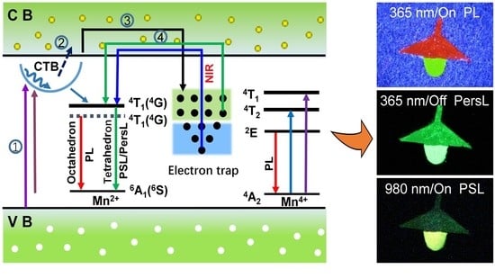
As a result, the hydrothermal approach is an effective method for the preparation of afterglow phosphors. The stored energy of Zn3Li0.4Ga1.6GeO8:Mn and Zn2.4Li0.6Ga2GeO8:Mn PersL phosphors can also be triggered by heating, which helps to release the absorbed energy and provide insights into PersL mechanism. TL curves monitored at 540 nm are shown in Figure 6b. The TL curves for Zn3Li0.4Ga1.6GeO8:Mn (Figure 6b, green curve) and Zn2.4Li0.6Ga2GeO8:Mn (Figure 6b, red curve) are similar and can be divided into two broad bands at 358 K and 376 K, and at 363 K and 423 K, respectively. These results indicate the presence of a shallow trap and deep trap in the two matrix materials, and the existence of the deep traps provides the conditions for generating PersL. Compared to Zn3Li0.4Ga1.6GeO8:Mn phosphor, the traps of Zn2.4Li0.6Ga2GeO8:Mn phosphor are deeper, which may explain why Zn2.4Li0.6Ga2GeO8:Mn phosphor has a long green afterglow duration. The possible luminescence mechanisms, including green PL, PersL and PSL from Mn2+, and the two red PL from Mn2+ and Mn4+ are proposed and shown in Figure 7 [8,22,32,33].
As shown in Figure 7, under UV excitation, electrons are first excited to the Mn2+–O2− CTB or the excited state of Mn ions, some electrons reach the conduction band through photoelectric separation and are trapped by traps, and some electrons are relaxed to 4T1 and 2E levels through lattice vibration or non-radiation, resulting in 4T1 (4G)→6A1 (6S) (540 nm) transitions of the Mn2+-occupied tetrahedral site, 4T1 (4G)→6A1 (6S) (701 nm) transitions of Mn2+ at the octahedral site, and 2E→4A2 (701 nm) transitions of Mn4+ at the octahedral site. When the excitation is stopped, the trapped electrons in the trap reach the conduction band through lattice thermal vibration and are released to the 4T1 energy level of Mn2+ ions, resulting in green continuous fluorescence from 4T1 (4G)→6A1 (6S) (540 nm).
The luminescent emission color response to excitation wavelength and excitation power is convenient for various applications [8,34,35]. To verify the feasibility of the phosphors in the anti-counterfeiting fields, we used these phosphors as inks printed the table lamp patterns in Figure 8. Figure 8a depicts the schematic diagram of these patterns. The lampshade and bulb are printed with Zn3Li0.4Ga1.6GeO8:Mn and Zn2.4Li0.6Ga2GeO8:Mn phosphor synthesized by a hydrothermal approach, respectively.
Upon the irradiation of 254 nm UV light, the lampshade became blue–white due to the color mixture of the Mn PL and the defect fluorescence of matrix [36] (Figure 8b), while the bulb became blue–green (Figure 8b). After the stoppage of excitation light, both the lampshade (dark green) and the bulb (bright green) emitted green PersL (Figure 8c), while upon the irradiation of 365 nm UV light, the lampshade and the bulb emitted bright red PL and green PL, respectively (Figure 8d). After the stoppage of UV excitation, these two patterns both showed green PersL colors with different saturations (Figure 8e). After these two patterns disappeared, the entire ‘desk lamp’ pattern was illuminated by a 980 nm laser diode; the lampshade and the bulb were lit again (Figure 8f), with the lampshade emitting a dark green color and the bulb emitting a yellow–green color (for a 980 nm illumination at 0.5 W), which is consistent with spectral characterization. Therefore, Mn-activated Zn3Ga2GeO8 phosphors hold a promise for multi-chrome (green, yellow and red) and multi-mode (PL, PersL, and PSL) anti-counterfeiting applications [37,38,39]. In addition, the PSL and PL of nanomaterials can be used for photodynamic activation [40], dosimetry [41], thermometry [42] and solid-state lighting [43].
Additionally, we need to point out that, for practical applications, the possibility of the power of the laser diodes (2 and 5 W for the 808 nm and 980 nm lasers, respectively) to generate even secondary heating effects on the samples should be considered. It is true that NIR lasers, such as 808 nm or 980 nm lasers, have been widely used for photothermal therapy (PTT) [44,45,46]. However, for PTT, the materials have strong absorptions for NIR, but they do not have luminescence. So, the energy absorbed is released as heat, while in the luminescence materials, the absorbed energy is released as luminescence [47,48]; therefore, heating is not a critical issue, even though it is unavoidable. Heating is always an issue for many applications with NIR lasers.

4. Conclusions

In summary, manganese-activated gallium germanate phosphors were successfully synthesized in two approaches, including a high-temperature solid-state reaction approach and a hydrothermal method. We found, for the first time, that these Mn-activated Zn3Ga2GeO8 phosphors prepared by a hydrothermal method and solid-state reaction method have different crystal phase structures and spectral properties. The phosphors prepared by the hydrothermal method exhibited a double-peak emission, including green PL with a PersL feature and red PL without a PersL effect, due to simultaneously having two kinds of occupancy sites. While the Mn-activated Zn3Ga2GeO8 phosphors prepared by a solid-state reaction method only exhibited red PL. In addition, Li+ ions were selectively substituted for the tetrahedral Zn site or the octahedral Ga site, leading to green light emission at 540 nm (4T1(G)→6A1(S) transition of Mn2+) with an afterglow and red light centered at 701 nm (2E→4A2 transition of Mn4+) in the absence of afterglow. Particularly, Mn-activated Zn3Ga2GeO8 phosphors synthesized by a hydrothermal approach via Li+ replacing the Zn2+ or Ga3+ sites exhibit dynamic and multicolor emissions as luminescence labels for multiple anti-counterfeiting, thus revealing the great potential of these phosphors in multicolor display, anti-forgery, and other potential applications.

Author Contributions

Conceptualization, D.G. and W.C.; methodology, D.G. and P.W.; validation, P.W., F.G. and W.N.; formal analysis, D.G. and W.C.; investigation, P.W., F.G. and W.N.; resources, D.G. and W.C.; data curation, P.W.; writing—original draft preparation, D.G.; writing—review and editing, W.N., D.G. and W.C.; visualization, D.G. and P.W.; supervision, D.G.; project administration, D.G. and F.G.; funding acquisition, G.D and W.C. All authors have read and agreed to the published version of the manuscript.

Funding

This research was funded by National Natural Science Foundation of China, grant number 11604253 and Natural Science Foundation of Shaanxi Province of China, grant number 2018JM1036.

Data Availability Statement

Not applicable.

Acknowledgments

We would like to acknowledge the support from Guangxi Jialouyuan Medical Inc., Solgro, and the distinguished award from UT Arlington.

Conflicts of Interest

The authors declare no conflict of interest.

References

  1. Thejas, K.K.; Abraham, M.; Kunti, A.K.; Tchernycheva, M.; Ahmad, S.; Das, S. Review on deep red-emitting rare-earth free germanates and their efficiency as well as adaptability for various applications. Appl. Mater. Today 2021, 24, 101094. [Google Scholar] [CrossRef]
  2. Wang, T.; Wang, S.F.; Liu, Z.Y.; He, Z.Y.; Yu, P.; Zhao, M.Y.; Zhang, H.X.; Lu, L.F.; Wang, Z.; Wang, Z.; et al. A hybrid erbium(III)–bacteriochlorin near-infrared probe for multiplexed biomedical imaging. Nat. Mater. 2021, 20, 1571–1578. [Google Scholar] [CrossRef]
  3. Zhang, Y.; Zhu, X.H.; Zhang, Y. Exploring heterostructured upconversion nanoparticles: From rational engineering to diverse applications. ACS Nano 2021, 15, 3709–3735. [Google Scholar] [CrossRef] [PubMed]
  4. Cui, X.Y.; Shi, W.Y.; Lu, C. Control of multicolor and white emission by triplet energy transfer. J. Phys. Chem. A 2021, 125, 4209–4215. [Google Scholar] [CrossRef] [PubMed]
  5. Shi, S.C.; Bai, W.H.; Xuan, T.T.; Zhou, T.L.; Dong, G.Y.; Xie, R.J. In situ inkjet printing patterned lead halide perovskite quantum dot color conversion films by using cheap and eco-friendly aqueous inks. Small Methods 2021, 5, 2000889. [Google Scholar] [CrossRef] [PubMed]
  6. Pan, Z.W.; Lu, Y.Y.; Liu, F. Sunlight-activated long-persistent luminescence in the near-infrared from Cr3+-doped zinc gallogermanates. Nat. Mater. 2012, 11, 58–63. [Google Scholar] [CrossRef] [PubMed]
  7. Chuang, Y.J.; Zhen, Z.P.; Zhang, F.; Liu, F.; Mishra, J.P.; Tang, W.; Chen, H.M.; Huang, X.L.; Wang, L.C.; Chen, X.Y.; et al. Photostimulable near-infrared persistent luminescent nanoprobes for ultrasensitive and longitudinal deep-tissue bio-imaging. Theranostics 2014, 4, 1112–1122. [Google Scholar] [CrossRef] [PubMed] [Green Version]
  8. Gao, D.L.; Gao, J.; Gao, F.; Kuang, Q.Q.; Pan, Y.; Chen, Y.F.; Pan, Z.W. Quintuple-mode dynamic anti-counterfeiting using multi-mode persistent phosphors. J. Mater. Chem. C 2021, 9, 16634–16644. [Google Scholar] [CrossRef]
  9. Zhou, Q.; Dolgov, L.; Srivastava, A.M.; Zhou, L.; Wang, Z.L.; Shi, J.X.; Dramićanin, M.D.; Brik, M.G.; Wu, M.M. Mn2+ and Mn4+ red phosphors: Synthesis, luminescence and applications in WLEDs. A review. J. Mater. Chem. C 2018, 6, 2652–2671. [Google Scholar] [CrossRef]
  10. Kanzaki, M. Crystal structures of Zn2GeO4 cubic/tetragonal spinel and Zn2SiO4 modified spinel phases. J. Miner. Petrol. Sci. 2018, 113, 41–46. [Google Scholar] [CrossRef] [Green Version]
  11. Gao, D.L.; Ma, K.W.; Wang, P.; Zhang, X.Y.; Pang, Q.; Xin, H.; Zhang, Z.H.; Jiao, H. Tuning multicolour emission of Zn2GeO4: Mn phosphors by Li+ doping for information encryption and anti-counterfeiting applications. Dalton Trans. 2022, 51, 553–561. [Google Scholar] [CrossRef] [PubMed]
  12. Dong, L.P.; Zhang, L.; Jia, Y.C.; Xu, Y.H.; Yin, S.W.; You, H.P. ZnGa2−yAlyO4: Mn2+, Mn4+ thermochromic phosphors: Valence state control and optical temperature sensing. Inorg. Chem. 2020, 59, 15969–15976. [Google Scholar] [CrossRef]
  13. Chen, X.; Yao, W.; Wang, Q.; Wu, W. Designing multicolor dual-mode lanthanide-doped NaLuF4/Y2O3 composites for advanced anticounterfeiting. Adv. Opt. Mater. 2020, 8, 1901209. [Google Scholar] [CrossRef]
  14. Wang, Y.J.; Włodarczyk, D.; Brik, M.G.; Barzowska, J.; Shekhovtsov, A.N.; Belikov, K.N.; Paszkowicz, W.; Li, L.; Zhou, X.J.; Suchocki, A. Effect of temperature and high pressure on luminescence properties of Mn3+ ions in Ca3Ga2Ge3O12 single crystals. J. Phys. Chem. C 2021, 125, 5146–5157. [Google Scholar] [CrossRef]
  15. López, S.; Romero, A.H.; Rodríguez-Hernández, P.; Munoz, A. First-principles study of the high-pressure phase transition in ZnAl2O4 and ZnGa2O4: From cubic spinel to orthorhombic post-spinel structures. Phys. Rev. B 2009, 79, 214103. [Google Scholar] [CrossRef]
  16. Liu, F.; Liang, Y.; Pan, Z. Detection of up-converted persistent luminescence in the near infrared emitted by the Zn3Ga2GeO8: Cr3+, Yb3+, Er3+ phosphor. Phys. Rev. Lett. 2014, 113, 177401. [Google Scholar] [CrossRef]
  17. Allix, M.; Chenu, S.; Véron, E.; Poumeyrol, T.; Kouadri-Boudjelthia, E.A.; Alahrache, S.; Porcher, F.; Massiot, D.; Fayon, F. Considerable improvement of long-persistent luminescence in germanium and tin substituted ZnGa2O4. Chem. Mater. 2013, 25, 1600–1606. [Google Scholar] [CrossRef]
  18. Huang, K.; Dou, X.; Zhang, Y.F.; Gou, X.P.; Liu, J.; Qu, J.L.; Li, Y.; Huang, P.; Han, G. Enhancing light and X-ray charging in persistent luminescence nanocrystals for orthogonal afterglow anti-counterfeiting. Adv. Funct. Mater. 2021, 31, 2009920. [Google Scholar] [CrossRef]
  19. Ma, Q.; Wang, J.; Zheng, W.; Wang, Q.; Li, Z.H.; Cong, H.J.; Liu, H.J.; Chen, X.Y.; Yuan, Q. Controlling disorder in host lattice by hetero-valence ion doping to manipulate luminescence in spinel solid solution phosphors. Sci. China Chem. 2018, 61, 1624–1629. [Google Scholar] [CrossRef]
  20. Srivastava, B.B.; Gupta, S.K.; Li, Y.; Mao, Y. Bright persistent green emitting water-dispersible Zn2GeO4: Mn nanorods. Dalton Trans. 2020, 49, 7328–7340. [Google Scholar] [CrossRef]
  21. Ma, Z.D.; Zhou, J.Y.; Zhang, J.C.; Zeng, S.H.; Zhou, H.; Smith, A.T.; Wang, W.X.; Sun, L.Y.; Wang, Z.F. Mechanics-induced triple-mode anticounterfeiting and moving tactile sensing by simultaneously utilizing instantaneous and persistent mechanoluminescence. Mater. Horizons 2019, 6, 2003–2008. [Google Scholar] [CrossRef]
  22. Lin, S.S.; Lin, H.; Ma, C.G.; Cheng, Y.; Ye, S.Z.; Lin, F.L.; Li, R.F.; Xu, J.; Wang, Y.S. High-security-level multi-dimensional optical storage medium: Nanostructured glass embedded with LiGa5O8: Mn2+ with photostimulated luminescence. Light Sci. Appl. 2020, 9, 22. [Google Scholar] [CrossRef] [PubMed] [Green Version]
  23. Dong, L.; Zhang, L.; Jia, Y.; Shao, B.; Lü, W.; Zhao, S.; You, H. Enhancing luminescence and controlling the Mn valence state of Gd3Ga5−x−δAlx−y+δO12:yMn phosphors by the design of the garnet structure. ACS Appl. Mater. Interfaces 2020, 12, 7334–7344. [Google Scholar] [CrossRef] [PubMed]
  24. Du, J.R.; Li, K.; Deun, R.V.; Poelman, D.; Lin, H.W. Near-infrared persistent luminescence and trap reshuffling in Mn4+ doped alkali-earth metal tungstates. Adv. Opt. Mater. 2022, 10, 2101714. [Google Scholar] [CrossRef]
  25. Liu, F.; Yan, W.Z.; Chuang, Y.-J.; Zhen, Z.P.; Xie, J.; Pan, Z.W. Photostimulated near-infrared persistent luminescence as a new optical read-out from Cr3+ doped LiGa5O8. Sci. Rep. 2013, 3, 1554. [Google Scholar] [CrossRef] [Green Version]
  26. Zheng, S.H.; Shi, J.P.; Fu, X.Y.; Wang, C.C.; Sun, X.; Chen, C.J.; Zhuang, Y.X.; Zou, X.Y.; Li, Y.C.; Zhang, H.W. X-ray recharged long afterglow luminescent nanoparticles MgGeO3: Mn2+, Yb3+, Li+ in the first and second biological windows for long-term bioimaging. Nanoscale 2020, 12, 14037–14046. [Google Scholar] [CrossRef]
  27. Chen, X.; Li, Y.; Huang, K.; Huang, L.; Tian, X.; Dong, H.; Kang, R.; Hu, Y.; Nie, J.; Qiu, J.; et al. Trap energy upconversion-like near-infrared to near-infrared light rejuvenateable persistent luminescence. Adv. Mater. 2021, 33, 2008722. [Google Scholar] [CrossRef]
  28. Adachi, S. Review—Mn4+-activated red and deep red-emitting phosphors. ECS J. Solid State Sci. Technol. 2020, 9, 016001. [Google Scholar] [CrossRef]
  29. Brik, M.G.; Camardello, S.J.; Srivastava, A.M. Influence of covalency on the Mn4+ 2Eg4A2g emission energy in crystals. ECS J. Solid State Sci. Technol. 2015, 4, R39. [Google Scholar] [CrossRef]
  30. Brik, M.G.; Srivastava, A.M. Critical review—A review of the electronic structure and optical properties of ions with d3 electron configuration (V2+, Cr3+, Mn4+, Fe5+) and main related misconceptions. ECS J. Solid State Sci. Technol. 2017, 7, R3079. [Google Scholar] [CrossRef]
  31. Si, T.; Zhu, Q.; Xiahou, J.Q.; Sun, X.D.; Li, J.-G. Regulating Mn2+/Mn4+ activators in ZnGa2O4 via Mg2+/Ge4+ doping to generate multimode luminescence for advanced anti-counterfeiting. ACS Appl. Electron. Mater. 2021, 3, 2005–2016. [Google Scholar] [CrossRef]
  32. Sun, X.Y.; Zhang, J.H.; Zhang, X.; Luo, Y.S.; Hao, Z.D.; Wang, X.J. Effect of retrapping on photostimulated luminescence in Sr3SiO5: Eu2+, Dy3+ phosphor. J. Appl. Phys. 2009, 105, 013501. [Google Scholar] [CrossRef] [Green Version]
  33. Ueda, J.; Maki, R.; Tanabe, S. Vacuum referred binding energy (VRBE)-guided design of orange persistent Ca3Si2O7: Eu2+ phosphors. Inorg. Chem. 2017, 56, 10353–10360. [Google Scholar] [CrossRef] [PubMed]
  34. Gao, D.L.; Gao, J.; Zhao, D.; Pang, Q.; Xiao, G.Q.; Wang, L.L.; Ma, K.W. Enhancing the red upconversion luminescence of hybrid porous microtubes via an in situ O-substituted reaction through heat treatment. J. Mater. Chem. C 2020, 8, 17318–17324. [Google Scholar] [CrossRef]
  35. Gao, D.L.; Zhao, D.; Pan, Y.; Chai, R.P.; Pang, Q.; Zhang, X.Y.; Chen, W. Extending the color response range of Yb3+ concentration-dependent multimodal luminescence in Yb/Er doped fluoride microrods by annealing treatment. Ceram. Int. 2021, 47, 32000–32007. [Google Scholar] [CrossRef]
  36. Wang, J.; Ma, Q.Q.; Zheng, W.; Liu, H.Y.; Yin, C.Q.; Wang, F.B.; Chen, X.Y.; Yuan, Q.; Tan, W.H. One-dimensional luminous nanorods featuring tunable persistent luminescence for autofluorescence-free biosensing. ACS Nano 2017, 11, 8185–8191. [Google Scholar] [CrossRef]
  37. You, W.W.; Tu, D.T.; Li, R.F.; Zheng, W.; Chen, X.Y. “Chameleon-like” optical behavior of lanthanide-doped fluoride nanoplates for multilevel anti-counterfeiting applications. Nano Res. 2019, 12, 1417–1422. [Google Scholar] [CrossRef]
  38. Ding, M.Y.; Dong, B.; Lu, Y.; Yang, X.F.; Yuan, Y.J.; Bai, W.F.; Wu, S.T.; Ji, Z.Z.; Lu, C.H.; Zhang, K.; et al. Energy manipulation in lanthanide-doped core-shell nanoparticles for tunable dual-mode luminescence toward advanced anti-counterfeiting. Adv. Mater. 2020, 32, 2002121. [Google Scholar] [CrossRef]
  39. Zhang, Y.; Huang, R.; Li, H.L.; Lin, Z.X.; Hou, D.J.; Guo, Y.Q.; Song, J.; Song, C.; Lin, Z.W.; Zhang, W.X.; et al. Triple-mode emissions with invisible near-infrared after-glow from Cr3+-doped zinc aluminum germanium nanoparticles for advanced anti-counterfeiting applications. Small 2020, 16, 2003121. [Google Scholar] [CrossRef]
  40. Ma, L.; Zou, X.; Bui, B.; Chen, W.; Song, K.H.; Solberg, T. X-ray excited ZnS:Cu,Co afterglow nanoparticles for photodynamic activation. Appl. Phys. Lett. 2014, 105, 13702. [Google Scholar] [CrossRef]
  41. Chen, W.; Wang, S.P.; Westcott, S.; Liu, Y. Dose Dependent X-Ray Luminescence in MgF2: Eu2+, Mn2+ Phosphors. J. Appl. Phys. 2008, 103, 113103. [Google Scholar] [CrossRef]
  42. Wang, S.P.; Westcott, L.S.; Chen, W. Nanoparticle Luminescence Thermometry. J. Phys. Chem. B 2002, 106, 11203–11209. [Google Scholar] [CrossRef]
  43. Chen, W.; Joly, A.; Malm, J.O.; Bovin, J.O.; Wang, S.P. Full-Color Emission and Temperature Dependence of The Luminescence In Poly-P-Phenylene Ethynylene-ZnS:Mn2+ Composite Particles. J. Phys. Chem. B 2003, 107, 6544–6551. [Google Scholar] [CrossRef]
  44. Li, Y.; Lu, W.; Huang, Q.; Huang, M.; Li, C.; Chen, W. Copper sulfide nanoparticles for photothermal ablation of tumor cells. Nanomedicine 2010, 5, 1161–1171. [Google Scholar] [CrossRef] [PubMed] [Green Version]
  45. Lakshmanan, S.B.; Zou, X.; Hossu, M.; Ma, L.; Yang, C.; Chen, W. Local field enhanced Au/CuS nanocomposites as efficient photothermal transducer agents for cancer treatment. J. Biomed. Nanotechnol. 2012, 8, 883–890. [Google Scholar] [CrossRef] [PubMed]
  46. Chu, X.; Li, K.; Guo, H.; Zheng, H.; Shuda, S.; Wang, X.; Zhang, J.; Chen, W.; Zhang, Y. Exploration of Graphitic-C3N4 Quantum Dots for Microwave-Induced Photodynamic Therapy. ACS Biomater. Sci. Eng. 2017, 3, 1836–1844. [Google Scholar] [CrossRef]
  47. Ma, L.; Chen, W. ZnS:Cu,Co water-soluble afterglow nanoparticles: Synthesis, luminescence and potential applications. Nanotechnology 2010, 21, 385604. [Google Scholar] [CrossRef]
  48. Tang, X.; Yu, H.; Bui, B.; Wang, L.; Xing, C.; Wang, S.; Chen, M.; Hu, Z.; Chen, W. Nitrogen-doped fluorescence carbon dots as multi-mechanism detection for iodide and curcumin in biological and food samples. Bioact. Mater. 2020, 6, 1541–1554. [Google Scholar] [CrossRef]
Figure 1. XRD patterns of Zn3Li0.4Ga1.6GeO8:Mn, Zn2.4Li0.6Ga2GeO8:Mn and Zn3Ga2GeO8:Mn phosphors. (a) Standard data for rhombohedral Zn2GeO4 (JCPDS No. 04-007-5691); (b,c) Zn3Li0.4Ga1.6GeO8:Mn and Zn2.4Li0.6Ga2GeO8:Mn phosphors synthesized by hydrothermal approach; (d,e) Zn2.4Li0.6Ga2GeO8:Mn and Zn3Ga2GeO8:Mn phosphors synthesized by solid-state reaction; (f,g) Standard data for cubic ZnGa2O4 (JCPDS No. 01-071-0843) and cubic Zn2GeO4 (JCPDS No. 04-007-9080).
Nanomaterials 12 02029 g001
Figure 2. SEM images (ad) and EDX mapping (e,f) of Zn3Ga2GeO8 phosphors annealed at 1100 °C. (a,b) Zn3Li0.4Ga1.6GeO8:Mn phosphor in (a) and Zn2.4Li0.6Ga2GeO8:Mn in (b) synthesized by hydrothermal approach; (c,d) Zn2.4Li0.6Ga2GeO8:Mn in (c) and Zn3Ga2GeO8:Mn in (d) synthesized by solid-state reaction; (e) EDX mapping of Zn3Li0.4Ga1.6GeO8:Mn synthesized by hydrothermal approach; and (f) EDX mapping of Zn3Ga2GeO8:Mn synthesized by solid-state reaction.
Nanomaterials 12 02029 g002
Figure 3. Excited wavelength-dependent fluorescence characteristics of Zn3Li0.4Ga1.6GeO8:Mn phosphors synthesized by hydrothermal approach. (a) PL spectra under various selective excitation and PSL upon 980 nm laser diode (0.5 W) irradiation; (b) PLE spectra (monitored at 540 or 701 nm); (c) PersL decay curves (monitoring at 540 nm) after irradiation by using 254 nm or 357 nm light for 5 min, the inset is the PersL emission spectrum achieved at 7 s of the decay.
Nanomaterials 12 02029 g003
Figure 4. Excited wavelength-dependent fluorescence characteristics of Zn2.4Li0.6Ga2GeO8:Mn phosphors prepared by hydrothermal approach. (a) PL spectra under various selective excitation and PSL upon 980 nm laser diode (0.5 W) irradiation; (b) Excitation spectra monitoring at 540 and 701 nm; (c) PersL decay curves (monitoring at 540 nm) after irradiation by using 254 nm or 337 nm light for 5 min, the inset is the PersL emission spectrum achieved at 7 s of the decay.
Nanomaterials 12 02029 g004
Figure 5. PLE and PL spectra and the corresponding digital PL images of Zn2.4Li0.6Ga2GeO8:Mn and Zn3Ga2GeO8:Mn phosphors synthesized by solid-state reaction. (a) PLE spectra monitored at 701 nm and PL spectra under 365 nm UV lamp excitation; (b) Digital PL photographs under 365 nm irradiation. The camera parameters are manual/ISO 3200/1 s. The slight difference in PL spectra can be seen in the cyan dash dot ellipse.
Nanomaterials 12 02029 g005
Figure 6. (a) PL (excitation at 254 nm) and PLE (monitoring at 540 nm) spectra of undoped Zn3Li0.4Ga1.6GeO8 phosphors synthesized by hydrothermal approach; (b) TL spectra obtained after 5 min illumination for Zn3Li0.4Ga1.6GeO8:Mn (green curve) and Zn2.4Li0.6Ga2GeO8:Mn phosphors (red curve) prepared by hydrothermal method.
Nanomaterials 12 02029 g006
Figure 7. The proposed PL, PersL and PSL schematic diagram for green and red luminescence of Mn2+/Mn4+ in the Zn3Ga2GeO8 phosphors. Therein, ① UV light excitation, ② energy (or electron) transfer processes, ③ trapping, ④ release. The straight-line arrows and curved-line arrows stand for optical transitions and energy (or electron) transfer processes, respectively. Note that the solid and dashed lines represent the 4T1 energy levels of Mn2+ in the weak tetrahedral crystal field and strong octahedral crystal field, respectively.
Nanomaterials 12 02029 g007
Figure 8. Digital photographs of a triple-mode (PL, PersL, and PSL modes) ‘chandelier’ pattern printed used Zn3Li0.4Ga1.6GeO8:Mn phosphor (lampshade) and Zn2.4Li0.6Ga2GeO8:Mn phosphor (light bulb) synthesized by hydrothermal approach. (a) The design of the ‘chandelier’ pattern; (b,c) PL and PersL images acquired upon and after 254 nm UV light excitation (for 5 min); (d,e) PL and PersL images acquired upon and after 365 nm UV light excitation (for 5 min); (f) PSL images upon a 980 nm laser diode irradiation (at 0.5 W). The PSL imaging was achieved until the disappearance of chandelier to the naked eye. The camera parameters are manual/ISO 3200/4 s.
Nanomaterials 12 02029 g008
Publisher’s Note: MDPI stays neutral with regard to jurisdictional claims in published maps and institutional affiliations.