
Recent Advances in Gadolinium Based Contrast Agents for Bioimaging Applications


 ,
,  ,
,
Abstract
:1. Introduction
2. Bioimaging Techniques
2.1. Magnetic Resonance Imaging (MRI)
2.2. Optical Imaging (OI)
2.3. Computed Tomography (CT) Imaging
2.4. Ultrasound (US) Imaging
2.5. Positron Emission Tomography (PET), Single Photon Emission Computed Tomography (SPECT) Imaging
3. Nanoparticles in Molecular Imaging
4. Functionalization of Gd-NPs
4.1. Naturally Derived Polymers
4.2. Saccharides and Their Derivatives
4.3. Lipids and Their Derivatives
4.4. Synthetic Polymers
4.5. Organic and Inorganic Molecules
5. Conclusions and Future Perspectives
Author Contributions
Funding
Data Availability Statement
Conflicts of Interest
References
- Ferrari, M. Cancer nanotechnology: Opportunities and challenges. Nat. Rev. Cancer 2005, 5, 161–171. [Google Scholar] [CrossRef]
- Brown, M.A.; Semelka, R.C. MRI: Basic Principles and Applications; John Wiley & Sons: Hoboken, NJ, USA, 2011; ISBN 0470920866. [Google Scholar]
- Weissleder, R. Molecular imaging in cancer. Science 2006, 312, 1168–1171. [Google Scholar] [CrossRef] [Green Version]
- Greish, K. Enhanced permeability and retention of macromolecular drugs in solid tumors: A royal gate for targeted anticancer nanomedicines. J. Drug Target. 2007, 15, 457–464. [Google Scholar] [CrossRef]
- Hawley, A.E.; Illum, L.; Davis, S.S. Preparation of biodegradable, surface engineered PLGA nanospheres with enhanced lymphatic drainage and lymph node uptake. Pharm. Res. 1997, 14, 657–661. [Google Scholar] [CrossRef]
- Davis, M.E.; Chen, Z.; Shin, D.M. Nanoparticle therapeutics: An emerging treatment modality for cancer. Nanosci. Technol. A Collect. Rev. Nat. J. 2010, 239–250. [Google Scholar] [CrossRef]
- Sharma, P.; Brown, S.; Walter, G.; Santra, S.; Moudgil, B. Nanoparticles for bioimaging. Adv. Colloid Interface Sci. 2006, 123, 471–485. [Google Scholar] [CrossRef] [PubMed]
- Zhang, L.; Liu, R.; Peng, H.; Li, P.; Xu, Z.; Whittaker, A.K. The evolution of gadolinium based contrast agents: From single-modality to multi-modality. Nanoscale 2016, 8, 10491–10510. [Google Scholar] [PubMed]
- Donato, H.; França, M.; Candelária, I.; Caseiro-Alves, F. Liver MRI: From basic protocol to advanced techniques. Eur. J. Radiol. 2017, 93, 30–39. [Google Scholar] [CrossRef] [PubMed] [Green Version]
- Foucault-Collet, A.; Gogick, K.A.; White, K.A.; Villette, S.; Pallier, A.; Collet, G.; Kieda, C.; Li, T.; Geib, S.J.; Rosi, N.L. Lanthanide near infrared imaging in living cells with Yb3+ nano metal organic frameworks. Proc. Natl. Acad. Sci. USA 2013, 110, 17199–17204. [Google Scholar] [CrossRef] [PubMed] [Green Version]
- Cai, Z.; Ye, Z.; Yang, X.; Chang, Y.; Wang, H.; Liu, Y.; Cao, A. Encapsulated enhanced green fluorescence protein in silica nanoparticle for cellular imaging. Nanoscale 2011, 3, 1974–1976. [Google Scholar] [CrossRef]
- Genovese, D.; Bonacchi, S.; Juris, R.; Montalti, M.; Prodi, L.; Rampazzo, E.; Zaccheroni, N. Prevention of self-quenching in fluorescent silica nanoparticles by efficient energy transfer. Angew. Chem. Int. Ed. 2013, 52, 5965–5968. [Google Scholar] [CrossRef] [PubMed]
- Grebenik, E.A.; Nadort, A.; Generalova, A.N.; Nechaev, A.V.; Sreenivasan, V.K.A.; Khaydukov, E.V.; Semchishen, V.A.; Popov, A.P.; Sokolov, V.I.; Akhmanov, A.S. Feasibility study of the optical imaging of a breast cancer lesion labeled with upconversion nanoparticle biocomplexes. J. Biomed. Opt. 2013, 18, 76004. [Google Scholar] [CrossRef] [PubMed] [Green Version]
- Lee, S.; Cha, E.; Park, K.; Lee, S.; Hong, J.; Sun, I.; Kim, S.Y.; Choi, K.; Kwon, I.C.; Kim, K. A near-infrared-fluorescence-quenched gold-nanoparticle imaging probe for in vivo drug screening and protease activity determination. Angew. Chem. 2008, 120, 2846–2849. [Google Scholar] [CrossRef]
- Muthukumar, T.; Chamundeeswari, M.; Prabhavathi, S.; Gurunathan, B.; Chandhuru, J.; Sastry, T.P. Carbon nanoparticle from a natural source fabricated for folate receptor targeting, imaging and drug delivery application in A549 lung cancer cells. Eur. J. Pharm. Biopharm. 2014, 88, 730–736. [Google Scholar] [CrossRef] [PubMed]
- Wang, Y.; Zhou, K.; Huang, G.; Hensley, C.; Huang, X.; Ma, X.; Zhao, T.; Sumer, B.D.; DeBerardinis, R.J.; Gao, J. A nanoparticle-based strategy for the imaging of a broad range of tumours by nonlinear amplification of microenvironment signals. Nat. Mater. 2014, 13, 204–212. [Google Scholar] [CrossRef] [Green Version]
- Markovic, S.; Belz, J.; Kumar, R.; Cormack, R.A.; Sridhar, S.; Niedre, M. Near-infrared fluorescence imaging platform for quantifying in vivo nanoparticle diffusion from drug loaded implants. Int. J. Nanomed. 2016, 11, 1213. [Google Scholar] [CrossRef] [Green Version]
- Dubreil, L.; Leroux, I.; Ledevin, M.; Schleder, C.; Lagalice, L.; Lovo, C.; Fleurisson, R.; Passemard, S.; Kilin, V.; Gerber-Lemaire, S. Multi-harmonic imaging in the second near-infrared window of nanoparticle-labeled stem cells as a monitoring tool in tissue depth. ACS Nano 2017, 11, 6672–6681. [Google Scholar] [CrossRef]
- Ghaghada, K.B.; Badea, C.T.; Karumbaiah, L.; Fettig, N.; Bellamkonda, R.V.; Johnson, G.A.; Annapragada, A. Evaluation of tumor microenvironment in an animal model using a nanoparticle contrast agent in computed tomography imaging. Acad. Radiol. 2011, 18, 20–30. [Google Scholar] [CrossRef] [Green Version]
- Bonitatibus, P.J., Jr.; Torres, A.S.; Goddard, G.D.; FitzGerald, P.F.; Kulkarni, A.M. Synthesis, characterization, and computed tomography imaging of a tantalum oxide nanoparticle imaging agent. Chem. Commun. 2010, 46, 8956–8958. [Google Scholar] [CrossRef]
- Hu, Y.; Wang, Y.; Jiang, J.; Han, B.; Zhang, S.; Li, K.; Ge, S.; Liu, Y. Preparation and characterization of novel perfluorooctyl bromide nanoparticle as ultrasound contrast agent via layer-by-layer self-assembly for folate-receptor-mediated tumor imaging. Biomed. Res. Int. 2016, 2016, 6381464. [Google Scholar] [CrossRef] [Green Version]
- Wang, X.; Chen, H.; Zheng, Y.; Ma, M.; Chen, Y.; Zhang, K.; Zeng, D.; Shi, J. Au-nanoparticle coated mesoporous silica nanocapsule-based multifunctional platform for ultrasound mediated imaging, cytoclasis and tumor ablation. Biomaterials 2013, 34, 2057–2068. [Google Scholar] [CrossRef]
- Seo, M.; Gorelikov, I.; Williams, R.; Matsuura, N. Microfluidic assembly of monodisperse, nanoparticle-incorporated perfluorocarbon microbubbles for medical imaging and therapy. Langmuir 2010, 26, 13855–13860. [Google Scholar] [CrossRef] [PubMed]
- Pressly, E.D.; Pierce, R.A.; Connal, L.A.; Hawker, C.J.; Liu, Y. Nanoparticle PET/CT imaging of natriuretic peptide clearance receptor in prostate cancer. Bioconjug. Chem. 2013, 24, 196–204. [Google Scholar] [CrossRef] [PubMed]
- Ahmad, M.Y.; Ahmad, M.W.; Cha, H.; Oh, I.; Tegafaw, T.; Miao, X.; Ho, S.L.; Marasini, S.; Ghazanfari, A.; Yue, H. Cyclic RGD-coated ultrasmall Gd2O3 nanoparticles as tumor-targeting positive magnetic resonance imaging contrast agents. Eur. J. Inorg. Chem. 2018, 2018, 3070–3079. [Google Scholar] [CrossRef]
- Petrik, M.; Weigel, C.; Kirsch, M.; Hosten, N. No detectable nephrotoxic side effect using a dimer, non-ionic contrast media in cerebral perfusion computed tomography in case of suspected brain ischemia. RoFo Fortschr. Geb. Rontgenstrahlen Nukl. 2005, 177, 1242–1249. [Google Scholar] [CrossRef]
- Oh, I.; Min, H.S.; Li, L.; Tran, T.H.; Lee, Y.; Kwon, I.C.; Choi, K.; Kim, K.; Huh, K.M. Cancer cell-specific photoactivity of pheophorbide a–glycol chitosan nanoparticles for photodynamic therapy in tumor-bearing mice. Biomaterials 2013, 34, 6454–6463. [Google Scholar] [CrossRef]
- Hoshyar, N.; Gray, S.; Han, H.; Bao, G. The effect of nanoparticle size on in vivo pharmacokinetics and cellular interaction. Nanomedicine 2016, 11, 673–692. [Google Scholar] [CrossRef] [Green Version]
- Scott, R.P.; Quaggin, S.E. The cell biology of renal filtration. J. Cell Biol. 2015, 209, 199–210. [Google Scholar] [CrossRef] [PubMed]
- Longmire, M.; Choyke, P.L.; Kobayashi, H. Clearance properties of nano-sized particles and molecules as imaging agents: Considerations and caveats. Nanomedicine 2008, 3, 703–717. [Google Scholar] [CrossRef] [PubMed] [Green Version]
- Zhou, Y.; Dai, Z. New strategies in the design of nanomedicines to oppose uptake by the mononuclear phagocyte system and enhance cancer therapeutic efficacy. Chem. Asian J. 2018, 13, 3333–3340. [Google Scholar] [CrossRef]
- Huang, Y.; He, S.; Cao, W.; Cai, K.; Liang, X.-J. Biomedical nanomaterials for imaging-guided cancer therapy. Nanoscale 2012, 4, 6135–6149. [Google Scholar] [CrossRef]
- Al-Jamal, W.; Al-Jamal, K.T.; Bomans, P.H.; Frederik, P.M.; Kostarelos, K. Functionalized-quantum-dot-liposome hybrids as multimodal nanoparticles for cancer. Small 2008, 4, 1406–1415. [Google Scholar] [CrossRef]
- Soo Choi, H.; Liu, W.; Misra, P.; Tanaka, E.; Zimmer, J.P.; Itty Ipe, B.; Bawendi, M.G.; Frangioni, J.V. Renal clearance of quantum dots. Nat. Biotechnol. 2007, 25, 1165–1170. [Google Scholar] [CrossRef] [Green Version]
- Selim, K.M.K.; Ha, Y.-S.; Kim, S.-J.; Chang, Y.; Kim, T.-J.; Lee, G.H.; Kang, I.-K. Surface modification of magnetite nanoparticles using lactobionic acid and their interaction with hepatocytes. Biomaterials 2007, 28, 710–716. [Google Scholar] [CrossRef] [PubMed]
- Park, J.Y.; Choi, E.S.; Baek, M.J.; Lee, G.H.; Woo, S.; Chang, Y. Water-soluble Ultra Small paramagnetic or superparamagnetic metal oxide nanoparticles for molecular MR imaging. Eur. J. Inorg. Chem. 2009, 2477–2481. [Google Scholar] [CrossRef]
- McCarthy, J.R.; Weissleder, R. Multifunctional magnetic nanoparticles for targeted imaging and therapy. Adv. Drug Deliv. Rev. 2008, 60, 1241–1251. [Google Scholar] [CrossRef] [PubMed] [Green Version]
- Lee, E.J.; Heo, W.C.; Park, J.W.; Chang, Y.; Bae, J.-E.; Chae, K.S.; Kim, T.J.; Park, J.A.; Lee, G.H. D-Glucuronic Acid Coated Gd(IO3)3·2H2O Nanomaterial as a Potential T1 MRI-CT Dual Contrast Agent. Eur. J. Inorg. Chem. 2013, 2013, 2858–2866. [Google Scholar] [CrossRef]
- Byrne, J.D.; Betancourt, T.; Brannon-Peppas, L. Active targeting schemes for nanoparticle systems in cancer therapeutics. Adv. Drug Deliv. Rev. 2008, 60, 1615–1626. [Google Scholar] [CrossRef] [PubMed]
- Patel, D.; Chang, Y.; Lee, G.H. Amino acid functionalized magnetite nanoparticles in saline solution. Curr. Appl. Phys. 2009, 9, S32–S34. [Google Scholar] [CrossRef]
- Kim, S.J.; Xu, W.; Ahmad, M.W.; Baeck, J.S.; Chang, Y.; Bae, J.E.; Chae, K.S.; Kim, T.J.; Park, J.A.; Lee, G.H. Synthesis of nanoparticle CT contrast agents: In vitro and in vivo studies. Sci. Technol. Adv. Mater. 2015, 16. [Google Scholar] [CrossRef] [Green Version]
- Kattel, K.; Park, J.Y.; Xu, W.; Bony, B.A.; Heo, W.C.; Tegafaw, T.; Kim, C.R.; Ahmad, M.W.; Jin, S.; Baeck, J.S.; et al. Surface coated Eu(OH)3 nanorods: A facile synthesis, characterization, MR relaxivities and in vitro cytotoxicity. J. Nanosci. Nanotechnol. 2013, 13, 7214–7219. [Google Scholar] [CrossRef]
- Popovtzer, R.; Agrawal, A.; Kotov, N.A.; Popovtzer, A.; Balter, J.; Carey, T.E.; Kopelman, R. Targeted gold nanoparticles enable molecular CT imaging of cancer. Nano Lett. 2008, 8, 4593–4596. [Google Scholar] [CrossRef] [PubMed] [Green Version]
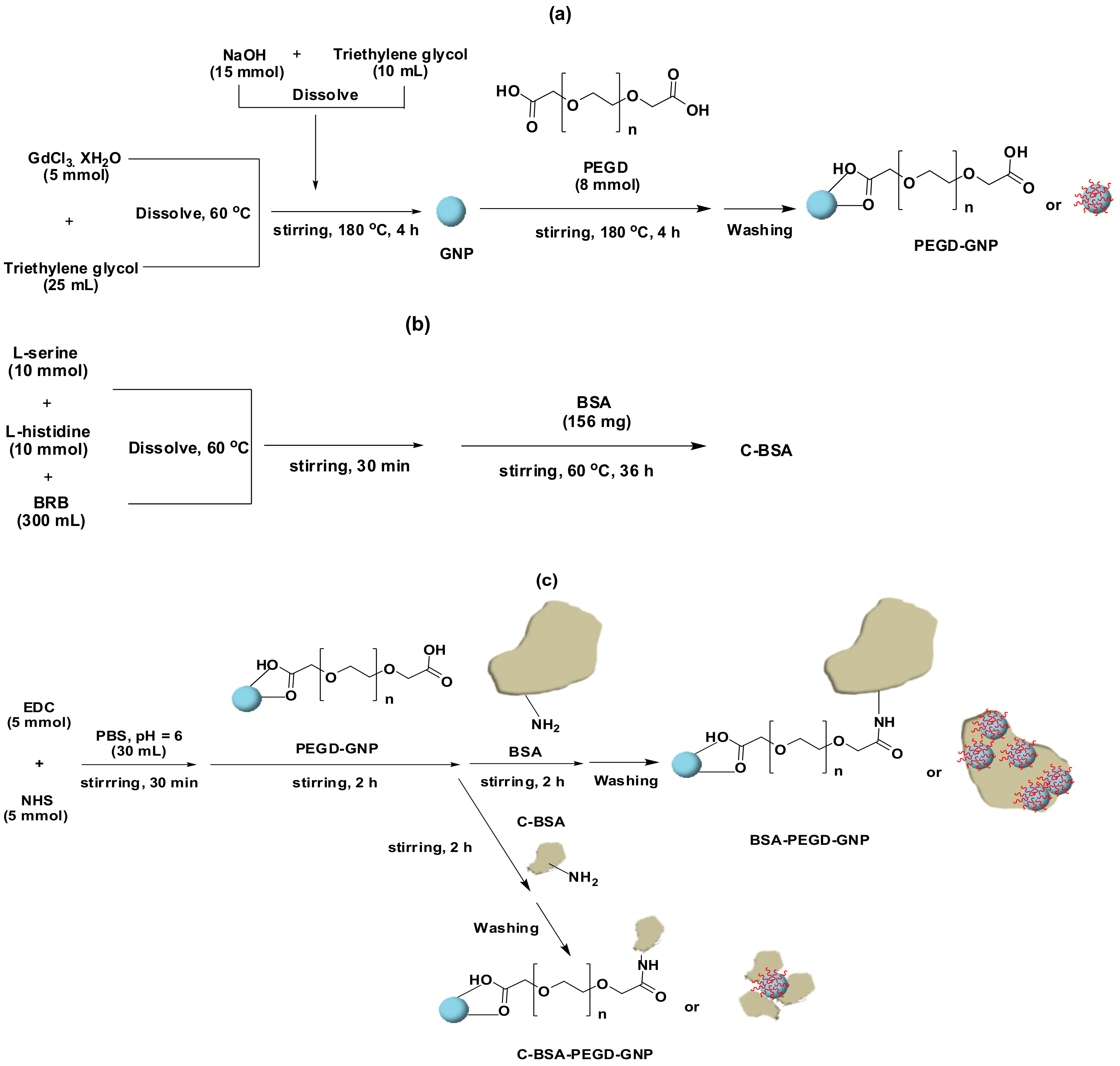
- Ahmad, M.W.; Kim, C.R.; Baeck, J.S.; Chang, Y.; Kim, T.J.; Bae, J.E.; Chae, K.S.; Lee, G.H. Bovine serum albumin (BSA) and cleaved-BSA conjugated ultrasmall Gd2O3 nanoparticles: Synthesis, characterization, and application to MRI contrast agents. Colloids Surf. A Physicochem. Eng. Asp. 2014, 450, 67–75. [Google Scholar] [CrossRef]
- Tegafaw, T.; Xu, W.; Ahmad, M.W.; Xu, M.; Chang, Y.; Chae, K.S.; Kim, T.J.; Lee, G.H. Fluorescent Brightener 28-Coated Fe3O4 Nanoparticles: Synthesis, Characterization, and Fluorescent Properties. J. Nanosci. Nanotechnol. 2016, 16, 10986–10990. [Google Scholar] [CrossRef]
- Lartigue, L.; Coupeau, M.; Lesault, M. Luminophore and magnetic multicore nanoassemblies for dual-mode MRI and fluorescence imaging. Nanomaterials 2020, 10, 28. [Google Scholar] [CrossRef] [Green Version]
- Krasia-Christoforou, T.; Socoliuc, V.; Knudsen, K.D.; Tombácz, E.; Turcu, R.; Vékás, L. From single-core nanoparticles in ferrofluids to multi-core magnetic nanocomposites: Assembly strategies, structure, and magnetic behavior. Nanomaterials 2020, 10, 2178. [Google Scholar] [CrossRef]
- Li, J.; Khalid, A.; Verma, R.; Abraham, A.; Qazi, F.; Dong, X.; Liang, G.; Tomljenovic-Hanic, S. Silk fibroin coated magnesium oxide nanospheres: A biocompatible and biodegradable tool for noninvasive bioimaging applications. Nanomaterials 2021, 11, 695. [Google Scholar] [CrossRef]
- Mnasri, W.; Parvizian, M.; Ammar-Merah, S. Design and Synthesis of Luminescent Lanthanide-Based Bimodal Nanoprobes for Dual Magnetic Resonance (MR) and Optical Imaging. Nanomaterials 2021, 11, 354. [Google Scholar] [CrossRef]
- Kharisov, B.I.; Dias, H.V.R.; Kharissova, O.V.; Vázquez, A.; Pena, Y.; Gomez, I. Solubilization, dispersion and stabilization of magnetic nanoparticles in water and non-aqueous solvents: Recent trends. RSC Adv. 2014, 4, 45354–45381. [Google Scholar] [CrossRef]
- Patel, D.; Moon, J.Y.; Chang, Y.; Kim, T.J.; Lee, G.H. Poly(d,l-lactide-co-glycolide) coated superparamagnetic iron oxide nanoparticles: Synthesis, characterization and in vivo study as MRI contrast agent. Colloids Surf. A Physicochem. Eng. Asp. 2008, 313–314, 91–94. [Google Scholar] [CrossRef]
- De Castro, K.C.; Costa, J.M.; Campos, M.G.N. Drug-loaded polymeric nanoparticles: A review. Int. J. Polym. Mater. Polym. Biomater. 2020, 1–13. [Google Scholar] [CrossRef]
- Huang, J.; Wang, L.; Lin, R.; Wang, A.Y.; Yang, L.; Kuang, M.; Qian, W.; Mao, H. Casein-coated iron oxide nanoparticles for high MRI contrast enhancement and efficient cell targeting. ACS Appl. Mater. Interfaces 2013, 5, 4632–4639. [Google Scholar] [CrossRef] [Green Version]
- Jurado, R.; Gálvez, N. Apoferritin amyloid-fibril directed the in situ assembly and/or synthesis of optical and magnetic nanoparticles. Nanomaterials 2021, 11, 146. [Google Scholar] [CrossRef]
- Villaraza, A.J.L.; Bumb, A.; Brechbiel, M.W. Macromolecules, dendrimers, and nanomaterials in magnetic resonance imaging: The interplay between size, function, and pharmacokinetics. Chem. Rev. 2010, 110, 2921–2959. [Google Scholar] [CrossRef] [Green Version]
- Bellin, M.-F. MR contrast agents, the old and the new. Eur. J. Radiol. 2006, 60, 314–323. [Google Scholar] [CrossRef] [PubMed]
- Weinmann, H.J.; Brasch, R.C.; Press, W.R.; Wesbey, G.E. Characteristics of gadolinium-DTPA complex: A potential NMR contrast agent. Am. J. Roentgenol. 1984, 142, 619–624. [Google Scholar] [CrossRef] [PubMed] [Green Version]
- Rose, T.A., Jr.; Choi, J.W. Intravenous imaging contrast media complications: The basics that every clinician needs to know. Am. J. Med. 2015, 128, 943–949. [Google Scholar] [CrossRef] [PubMed]
- Chopra, T.; Kandukurti, K.; Shah, S.; Ahmed, R.; Panesar, M. Understanding nephrogenic systemic fibrosis. Int. J. Nephrol. 2012, 2012, 912189. [Google Scholar] [CrossRef] [Green Version]
- Marckmann, P.; Skov, L.; Rossen, K.; Dupont, A.; Damholt, M.B.; Heaf, J.G.; Thomsen, H.S. Nephrogenic systemic fibrosis: Suspected causative role of gadodiamide used for contrast-enhanced magnetic resonance imaging. J. Am. Soc. Nephrol. 2006, 17, 2359–2362. [Google Scholar] [CrossRef] [PubMed] [Green Version]
- Caravan, P.; Ellison, J.J.; McMurry, T.J.; Lauffer, R.B. Gadolinium(III) chelates as MRI contrast agents: Structure, dynamics, and applications. Chem. Rev. 1999, 99, 2293–2352. [Google Scholar] [CrossRef]
- Rowe, M.D.; Thamm, D.H.; Kraft, S.L.; Boyes, S.G. Polymer-modified gadolinium metal-organic framework nanoparticles used as multifunctional nanomedicines for the targeted imaging and treatment of cancer. Biomacromolecules 2009, 10, 983–993. [Google Scholar] [CrossRef]
- Aime, S.; Botta, M.; Fasano, M.; Terreno, E. Lanthanide (III) chelates for NMR biomedical applications. Chem. Soc. Rev. 1998, 27, 19–29. [Google Scholar] [CrossRef]
- Aime, S.; Barge, A.; Cabella, C.; Crich, S.G.; Gianolio, E. Targeting cells with MR imaging probes based on paramagnetic Gd (III) chelates. Curr. Pharm. Biotechnol. 2004, 5, 509–518. [Google Scholar] [CrossRef]
- Aime, S.; Crich, S.G.; Gianolio, E.; Giovenzana, G.B.; Tei, L.; Terreno, E. High sensitivity lanthanide (III) based probes for MR-medical imaging. Coord. Chem. Rev. 2006, 250, 1562–1579. [Google Scholar] [CrossRef]
- Zhou, Z.; Huang, D.; Bao, J.; Chen, Q.; Liu, G.; Chen, Z.; Chen, X.; Gao, J. A synergistically enhanced T1–T2 dual-modal contrast agent. Adv. Mater. 2012, 24, 6223–6228. [Google Scholar] [CrossRef] [PubMed] [Green Version]
- Ahmad, M.W.; Xu, W.; Kim, S.J.; Baeck, J.S.; Chang, Y.; Bae, J.E.; Chae, K.S.; Park, J.A.; Kim, T.J.; Lee, G.H. Potential dual imaging nanoparticle: Gd2O3 nanoparticle. Sci. Rep. 2015, 5, 8549. [Google Scholar] [CrossRef] [PubMed] [Green Version]
- Ananta, J.S.; Godin, B.; Sethi, R.; Moriggi, L.; Liu, X.; Serda, R.E.; Krishnamurthy, R.; Muthupillai, R.; Bolskar, R.D.; Helm, L. Geometrical confinement of gadolinium-based contrast agents in nanoporous particles enhances T 1 contrast. Nat. Nanotechnol. 2010, 5, 815–821. [Google Scholar] [CrossRef] [Green Version]
- Bridot, J.-L.; Faure, A.-C.; Laurent, S.; Rivière, C.; Billotey, C.; Hiba, B.; Janier, M.; Josserand, V.; Coll, J.-L.; Elst, L.V.; et al. Hybrid gadolinium oxide nanoparticles: Multimodal contrast agents for in vivo imaging. J. Am. Chem. Soc. 2007, 129, 5076–5084. [Google Scholar] [CrossRef] [PubMed]
- Park, J.Y.; Baek, M.J.; Choi, E.S.; Woo, S.; Kim, J.H.; Kim, T.J.; Jung, J.C.; Chae, K.S.; Chang, Y.; Lee, G.H. Paramagnetic ultrasmall gadolinium oxide nanoparticles as advanced T1 MRI contrast agent: Account for large longitudinal relaxivity, optimal particle diameter, and in vivo T1 MR images. ACS Nano 2009, 3, 3663–3669. [Google Scholar] [CrossRef]
- Kim, C.R.; Baeck, J.S.; Chang, Y.; Bae, J.E.; Chae, K.S.; Lee, G.H. Ligand-size dependent water proton relaxivities in ultrasmall gadolinium oxide nanoparticles and in vivo T1 MR images in a 1.5 T MR field. Phys. Chem. Chem. Phys. 2014, 16, 19866–19873. [Google Scholar] [CrossRef]
- Tegafaw, T.; Xu, W.; Lee, S.H.; Chae, K.S.; Cha, H.; Chang, Y.; Lee, G.H. Ligand-size and ligand-chain hydrophilicity effects on the relaxometric properties of ultrasmall Gd2O3 nanoparticles. AIP Adv. 2016, 6, 065114. [Google Scholar] [CrossRef] [Green Version]
- Bony, B.A.; Baeck, J.S.; Chang, Y.; Bae, J.E.; Chae, K.S.; Lee, G.H. A Highly efficient new T1 MRI contrast agent with r2/r1 ≈ 1.0: Mixed Cu(II)/Gd(III) oxide nanoparticle. Bull. Korean Chem. Soc. 2015, 36, 1203–1208. [Google Scholar]
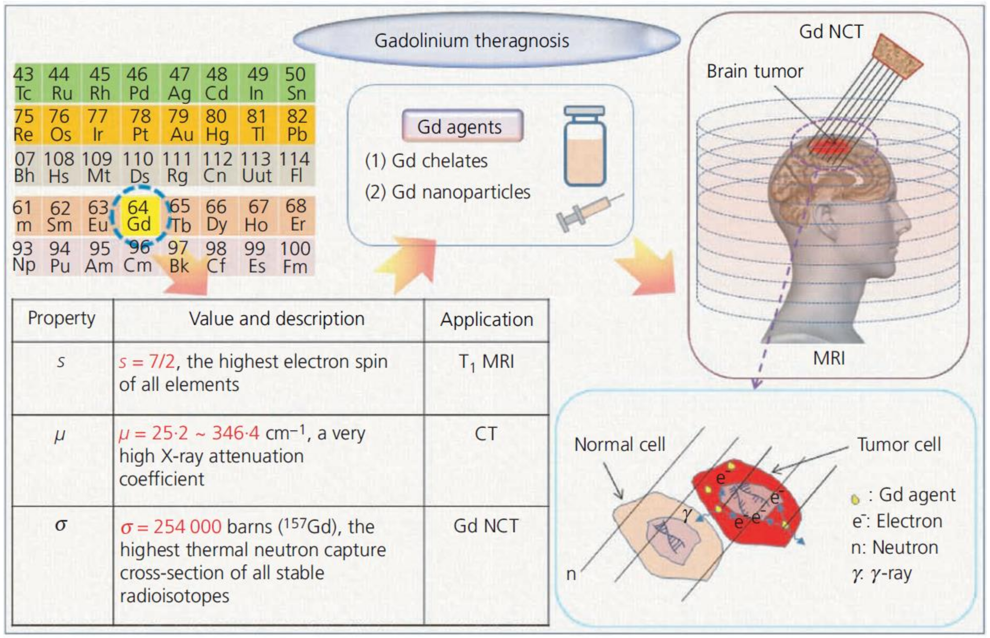
- Chang, Y.; Chae, K.S.; Lee, G.H. Gadolinium agents for theragnosis of malignant tumors. Bioinspired Biomim. Nanobiomater. 2016, 5, 167–170. [Google Scholar] [CrossRef]
- Ghaghada, K.B.; Starosolski, Z.A.; Bhayana, S.; Stupin, I.; Patel, C.V.; Bhavane, R.C.; Gao, H.; Bednov, A.; Yallampalli, C.; Belfort, M. Pre-clinical evaluation of a nanoparticle-based blood-pool contrast agent for MR imaging of the placenta. Placenta 2017, 57, 60–70. [Google Scholar] [CrossRef] [PubMed]
- Rotz, M.W.; Culver, K.S.B.; Parigi, G.; MacRenaris, K.W.; Luchinat, C.; Odom, T.W.; Meade, T.J. High relaxivity Gd (III)–DNA gold nanostars: Investigation of shape effects on proton relaxation. ACS Nano 2015, 9, 3385–3396. [Google Scholar] [CrossRef] [PubMed] [Green Version]
- Beija, M.; Li, Y.; Duong, H.T.; Laurent, S.; Vander Elst, L.; Muller, R.N.; Lowe, A.B.; Davis, T.P.; Boyer, C. Polymer–gold nanohybrids with potential use in bimodal MRI/CT: Enhancing the relaxometric properties of Gd (III) complexes. J. Mater. Chem. 2012, 22, 21382–21386. [Google Scholar] [CrossRef]
- Huang, C.-L.; Huang, C.-C.; Mai, F.-D.; Yen, C.-L.; Tzing, S.-H.; Hsieh, H.-T.; Ling, Y.-C.; Chang, J.-Y. Application of paramagnetic graphene quantum dots as a platform for simultaneous dual-modality bioimaging and tumor-targeted drug delivery. J. Mater. Chem. B 2015, 3, 651–664. [Google Scholar] [CrossRef]
- Abou, D.S.; Thorek, D.L.J.; Ramos, N.N.; Pinkse, M.W.H.; Wolterbeek, H.T.; Carlin, S.D.; Beattie, B.J.; Lewis, J.S. 89 Zr-labeled paramagnetic octreotide-liposomes for PET-MR imaging of cancer. Pharm. Res. 2013, 30, 878–888. [Google Scholar] [CrossRef]
- Park, J.-A.; Kim, J.Y.; Lee, Y.J.; Lee, W.; Lim, S.M.; Kim, T.-J.; Yoo, J.; Chang, Y.; Kim, K.M. Gadolinium complex of 125I/127I-RGD-DOTA conjugate as a tumor-targeting SPECT/MR bimodal imaging probe. ACS Med. Chem. Lett. 2013, 4, 216–219. [Google Scholar] [CrossRef] [Green Version]
- Aryal, M.; Papademetriou, I.; Zhang, Y.-Z.; Power, C.; McDannold, N.; Porter, T. MRI monitoring and quantification of ultrasound-mediated delivery of liposomes dually Labeled with gadolinium and fluorophore through the blood-brain barrier. Ultrasound Med. Biol. 2019, 45, 1733–1742. [Google Scholar] [CrossRef]
- Hu, D.-H.; Sheng, Z.-H.; Zhang, P.-F.; Yang, D.-Z.; Liu, S.-H.; Gong, P.; Gao, D.-Y.; Fang, S.-T.; Ma, Y.-F.; Cai, L.-T. Hybrid gold–gadolinium nanoclusters for tumor-targeted NIRF/CT/MRI triple-modal imaging in vivo. Nanoscale 2013, 5, 1624–1628. [Google Scholar] [CrossRef] [PubMed]
- Arifin, D.R.; Long, C.M.; Gilad, A.A.; Alric, C.; Roux, S.; Tillement, O.; Link, T.W.; Arepally, A.; Bulte, J.W.M. Trimodal gadolinium-gold microcapsules containing pancreatic islet cells restore normoglycemia in diabetic mice and can be tracked by using US, CT, and positive-contrast MR imaging. Radiology 2011, 260, 790–798. [Google Scholar] [CrossRef] [PubMed] [Green Version]
- Wu, B.; Lu, S.-T.; Yu, H.; Liao, R.-F.; Li, H.; Zafitatsimo, B.V.L.; Li, Y.-S.; Zhang, Y.; Zhu, X.-L.; Liu, H.-G. Gadolinium-chelate functionalized bismuth nanotheranostic agent for in vivo MRI/CT/PAI imaging-guided photothermal cancer therapy. Biomaterials 2018, 159, 37–47. [Google Scholar] [CrossRef]
- Hüber, M.M.; Staubli, A.B.; Kustedjo, K.; Gray, M.H.B.; Shih, J.; Fraser, S.E.; Jacobs, R.E.; Meade, T.J. Fluorescently detectable magnetic resonance imaging agents. Bioconjug. Chem. 1998, 9, 242–249. [Google Scholar] [CrossRef] [PubMed]
- Yan, G.-P.; Liu, M.-L.; Li, L.-Y. Polyaspartamide gadolinium complexes containing sulfadiazine groups as potential macromolecular MRI contrast agents. Bioconjug. Chem. 2005, 16, 967–971. [Google Scholar] [CrossRef]
- Bull, S.R.; Guler, M.O.; Bras, R.E.; Venkatasubramanian, P.N.; Stupp, S.I.; Meade, T.J. Magnetic resonance imaging of self-assembled biomaterial scaffolds. Bioconjug. Chem. 2005, 16, 1343–1348. [Google Scholar] [CrossRef]
- Bull, S.R.; Guler, M.O.; Bras, R.E.; Meade, T.J.; Stupp, S.I. Self-assembled peptide amphiphile nanofibers conjugated to MRI contrast agents. Nano Lett. 2005, 5, 1–4. [Google Scholar] [CrossRef]
- Anderson, E.A.; Isaacman, S.; Peabody, D.S.; Wang, E.Y.; Canary, J.W.; Kirshenbaum, K. Viral nanoparticles donning a paramagnetic coat: Conjugation of MRI contrast agents to the MS2 capsid. Nano Lett. 2006, 6, 1160–1164. [Google Scholar] [CrossRef]
- Langereis, S.; de Lussanet, Q.G.; van Genderen, M.H.P.; Backes, W.H.; Meijer, E.W. Multivalent contrast agents based on gadolinium−diethylenetriaminepentaacetic acid-terminated poly (propylene imine) dendrimers for magnetic resonance imaging. Macromolecules 2004, 37, 3084–3091. [Google Scholar] [CrossRef]
- Talanov, V.S.; Regino, C.A.S.; Kobayashi, H.; Bernardo, M.; Choyke, P.L.; Brechbiel, M.W. Dendrimer-based nanoprobe for dual modality magnetic resonance and fluorescence imaging. Nano Lett. 2006, 6, 1459–1463. [Google Scholar] [CrossRef]
- Mulder, W.J.M.; Strijkers, G.J.; Griffioen, A.W.; van Bloois, L.; Molema, G.; Storm, G.; Koning, G.A.; Nicolay, K. A liposomal system for contrast-enhanced magnetic resonance imaging of molecular targets. Bioconjug. Chem. 2004, 15, 799–806. [Google Scholar] [CrossRef]
- Frias, J.C.; Williams, K.J.; Fisher, E.A.; Fayad, Z.A. Recombinant HDL-like nanoparticles: A specific contrast agent for MRI of atherosclerotic plaques. J. Am. Chem. Soc. 2004, 126, 16316–16317. [Google Scholar] [CrossRef]
- Accardo, A.; Tesauro, D.; Roscigno, P.; Gianolio, E.; Paduano, L.; D’Errico, G.; Pedone, C.; Morelli, G. Physicochemical properties of mixed micellar aggregates containing CCK peptides and Gd complexes designed as tumor specific contrast agents in MRI. J. Am. Chem. Soc. 2004, 126, 3097–3107. [Google Scholar] [CrossRef]
- Turner, J.L.; Pan, D.; Plummer, R.; Chen, Z.; Whittaker, A.K.; Wooley, K.L. Synthesis of gadolinium-labeled shell-crosslinked nanoparticles for magnetic resonance imaging applications. Adv. Funct. Mater. 2005, 15, 1248–1254. [Google Scholar] [CrossRef]
- Platas-Iglesias, C.; Vander Elst, L.; Zhou, W.; Muller, R.N.; Geraldes, C.F.G.C.; Maschmeyer, T.; Peters, J.A. Zeolite GdNaY nanoparticles with very high relaxivity for application as contrast agents in magnetic resonance imaging. Chem. Eur. J. 2002, 8, 5121–5131. [Google Scholar] [CrossRef]
- Bolskar, R.D.; Benedetto, A.F.; Husebo, L.O.; Price, R.E.; Jackson, E.F.; Wallace, S.; Wilson, L.J.; Alford, J.M. First soluble M@ C60 derivatives provide enhanced access to metallofullerenes and permit in vivo evaluation of Gd@ C60 [C (COOH) 2] 10 as a MRI contrast agent. J. Am. Chem. Soc. 2003, 125, 5471–5478. [Google Scholar] [CrossRef] [PubMed]
- Sitharaman, B.; Kissell, K.R.; Hartman, K.B.; Tran, L.A.; Baikalov, A.; Rusakova, I.; Sun, Y.; Khant, H.A.; Ludtke, S.J.; Chiu, W. Superparamagnetic gadonanotubes are high-performance MRI contrast agents. Chem. Commun. 2005, 3915–3917. [Google Scholar] [CrossRef] [PubMed]
- Balkus, K.J.; Shi, J. A study of suspending agents for gadolinium (III)-exchanged hectorite. An oral magnetic resonance imaging contrast agent. Langmuir 1996, 12, 6277–6281. [Google Scholar] [CrossRef]
- Lin, Y.-S.; Hung, Y.; Su, J.-K.; Lee, R.; Chang, C.; Lin, M.-L.; Mou, C.-Y. Gadolinium (III)-incorporated nanosized mesoporous silica as potential magnetic resonance imaging contrast agents. J. Phys. Chem. B 2004, 108, 15608–15611. [Google Scholar] [CrossRef]
- Mulder, W.J.M.; Koole, R.; Brandwijk, R.J.; Storm, G.; Chin, P.T.K.; Strijkers, G.J.; de Mello Donegá, C.; Nicolay, K.; Griffioen, A.W. Quantum dots with a paramagnetic coating as a bimodal molecular imaging probe. Nano Lett. 2006, 6, 1–6. [Google Scholar] [CrossRef] [PubMed] [Green Version]
- Vuu, K.; Xie, J.; McDonald, M.A.; Bernardo, M.; Hunter, F.; Zhang, Y.; Li, K.; Bednarski, M.; Guccione, S. Gadolinium-rhodamine nanoparticles for cell labeling and tracking via magnetic resonance and optical imaging. Bioconjug. Chem. 2005, 16, 995–999. [Google Scholar] [CrossRef]
- Alric, C.; Serduc, R.; Mandon, C.; Taleb, J.; Le Duc, G.; Le Meur-Herland, A.; Billotey, C.; Perriat, P.; Roux, S.; Tillement, O. Gold nanoparticles designed for combining dual modality imaging and radiotherapy. Gold Bull. 2008, 41, 90–97. [Google Scholar] [CrossRef] [Green Version]
- Engström, M.; Klasson, A.; Pedersen, H.; Vahlberg, C.; Käll, P.-O.; Uvdal, K. High proton relaxivity for gadolinium oxide nanoparticles. Magn. Reson. Mater. Phys. Biol. Med. 2006, 19, 180–186. [Google Scholar] [CrossRef]
- McDonald, M.A.; Watkin, K.L. Small particulate gadolinium oxide and gadolinium oxide albumin microspheres as multimodal contrast and therapeutic agents. Investig. Radiol. 2003, 38, 305–310. [Google Scholar] [CrossRef]
- Roberts, D.; Zhu, W.L.; Frommen, C.M.; Rosenzweig, Z. Synthesis of gadolinium oxide magnetoliposomes for magnetic resonance imaging. J. Appl. Phys. 2000, 87, 6208–6210. [Google Scholar] [CrossRef]
- Evanics, F.; Diamente, P.R.; Van Veggel, F.; Stanisz, G.J.; Prosser, R.S. Water-soluble GdF3 and GdF3/LaF3 nanoparticles physical characterization and NMR relaxation properties. Chem. Mater. 2006, 18, 2499–2505. [Google Scholar] [CrossRef]
- Hu, K.-W.; Jhang, F.-Y.; Su, C.-H.; Yeh, C.-S. Fabrication of Gd2O(CO3)2·H2O/silica/gold hybrid particles as a bifunctional agent for MR imaging and photothermal destruction of cancer cells. J. Mater. Chem. 2009, 19, 2147–2153. [Google Scholar] [CrossRef]
- Li, I.; Su, C.; Sheu, H.; Chiu, H.; Lo, Y.; Lin, W.; Chen, J.; Yeh, C. Gd2O (CO3)2H2O particles and the corresponding Gd2O3: Synthesis and applications of magnetic resonance contrast agents and template particles for hollow spheres and hybrid composites. Adv. Funct. Mater. 2008, 18, 766–776. [Google Scholar] [CrossRef]
- Nwe, K.; Milenic, D.; Bryant, L.H.; Regino, C.A.S.; Brechbiel, M.W. Preparation, characterization and in vivo assessment of Gd-albumin and Gd-dendrimer conjugates as intravascular contrast-enhancing agents for MRI. J. Inorg. Biochem. 2011, 105, 722–727. [Google Scholar] [CrossRef] [Green Version]
- Ma, X.-H.; Gong, A.; Xiang, L.-C.; Chen, T.-X.; Gao, Y.-X.; Liang, X.-J.; Shen, Z.-Y.; Wu, A.-G. Biocompatible composite nanoparticles with large longitudinal relaxivity for targeted imaging and early diagnosis of cancer. J. Mater. Chem. B 2013, 1, 3419–3428. [Google Scholar] [CrossRef]
- Ahmad, M.Y.; Cha, H.; Oh, I.-T.; Tegafaw, T.; Miao, X.; Ho, S.L.; Marasini, S.; Ghazanfari, A.; Yue, H.; Chae, K.S.; et al. Synthesis, characterization, and enhanced cancer-imaging application of trans-activator of transcription peptide-conjugated ultrasmall gadolinium oxide nanoparticles. Bull. Korean Chem. Soc. 2018, 39, 435–441. [Google Scholar] [CrossRef]
- Park, J.; Lee, J.; Jung, J.; Yu, D.; Oh, C.; Ha, S.; Kim, T.; Chang, Y. Gd-DOTA conjugate of RGD as a potential tumor-targeting MRI contrast agent. ChemBioChem 2008, 9, 2811–2813. [Google Scholar] [CrossRef] [PubMed]
- Yang, Z.; He, W.; Zheng, H.; Wei, J.; Liu, P.; Zhu, W.; Lin, L.; Zhang, L.; Yi, C.; Xu, Z. One-pot synthesis of albumin-gadolinium stabilized polypyrrole nanotheranostic agent for magnetic resonance imaging guided photothermal therapy. Biomaterials 2018, 161, 1–10. [Google Scholar] [CrossRef] [PubMed]
- Bertozzi, C.R.; Kiessling, L.L. Chemical glycobiology. Science 2001, 291, 2357–2364. [Google Scholar] [CrossRef] [Green Version]
- Martinelli, J.; Fekete, M.; Tei, L.; Botta, M. Cleavable β-cyclodextrin nanocapsules incorporating Gd III-chelates as bioresponsive MRI probes. Chem. Commun. 2011, 47, 3144–3146. [Google Scholar] [CrossRef] [PubMed]
- Gambino, G.; Engelmann, J.; Tei, L.; Botta, M.; Logothetis, N.K.; Mamedov, I. Multimodal contrast agents for in vivo neuroanatomical analysis of monosynaptic connections. Biomaterials 2013, 34, 7135–7142. [Google Scholar] [CrossRef] [PubMed]
- Zhang, W.; Chen, Y.; Huang, Z.W.; Cai, L.; He, L. The synthesis of a D-glucosamine contrast agent, Gd-DTPA-DG, and its application in cancer molecular imaging with MRI. Eur. J. Radiol. 2011, 79, 369–374. [Google Scholar] [CrossRef]
- Termsarasab, U.; Cho, H.-J.; Moon, H.T.; Park, J.-H.; Yoon, I.-S.; Kim, D.-D. Self-assembled magnetic resonance imaging nanoprobes based on arachidyl chitosan for cancer diagnosis. Colloids Surf. B Biointerfaces 2013, 109, 280–286. [Google Scholar] [CrossRef]
- Mortezazadeh, T.; Gholibegloo, E.; Alam, N.R.; Dehghani, S.; Haghgoo, S.; Ghanaati, H.; Khoobi, M. Gadolinium (III) oxide nanoparticles coated with folic acid-functionalized poly (β-cyclodextrin-co-pentetic acid) as a biocompatible targeted nano-contrast agent for cancer diagnostic: In vitro and in vivo studies. Magn. Reson. Mater. Phys. Biol. Med. 2019, 32, 487–500. [Google Scholar] [CrossRef] [PubMed]
- Bony, B.A.; Baeck, J.S.; Chang, Y.; Bae, J.E.; Chae, K.S.; Lee, G.H. Water-soluble D-glucuronic acid coated ultrasmall mixed Ln/Mn (Ln = Gd and Dy) oxide nanoparticles and their application to magnetic resonance imaging. Biomater. Sci. 2014, 2, 1287–1295. [Google Scholar] [CrossRef] [PubMed]
- Xu, W.; Miao, X.; Oh, I.; Chae, K.S.; Cha, H.; Chang, Y.; Lee, G.H. Dextran-coated ultrasmall Gd2O3 nanoparticles as potential T1 MRI contrast agent. ChemistrySelect 2016, 1, 6086–6091. [Google Scholar] [CrossRef]
- Hifumi, H.; Yamaoka, S.; Tanimoto, A.; Akatsu, T.; Shindo, Y.; Honda, A.; Citterio, D.; Oka, K.; Kuribayashi, S.; Suzuki, K. Dextran coated gadolinium phosphate nanoparticles for magnetic resonance tumor imaging. J. Mater. Chem. 2009, 19, 6393–6399. [Google Scholar] [CrossRef]
- Ahmad, M.Y.; Ahmad, M.; Yue, H.; Ho, S.L.; Cha, H.; Marasini, S.; Tegafaw, T.; Liu, S.; Ghazanfari, A.; Chae, K.-S. Chitosan oligosaccharide lactate-coated ultrasmall gadolinium oxide nanoparticles: Synthesis, in vitro cytotoxicity, and relaxometric properties. J. Nanosci. Nanotechnol. 2021, 21, 4145–4150. [Google Scholar] [CrossRef]
- Cheng, Z.; Al Zaki, A.; Jones, I.W.; Hall, H.K.; Aspinwall, C.A.; Tsourkas, A. Stabilized porous liposomes with encapsulated Gd-labeled dextran as a highly efficient MRI contrast agent. Chem. Commun. 2014, 50, 2502–2504. [Google Scholar] [CrossRef] [Green Version]
- Hossann, M.; Wang, T.; Syunyaeva, Z.; Wiggenhorn, M.; Zengerle, A.; Issels, R.D.; Reiser, M.; Lindner, L.H.; Peller, M. Non-ionic Gd-based MRI contrast agents are optimal for encapsulation into phosphatidyldiglycerol-based thermosensitive liposomes. J. Control. Release 2013, 166, 22–29. [Google Scholar] [CrossRef]
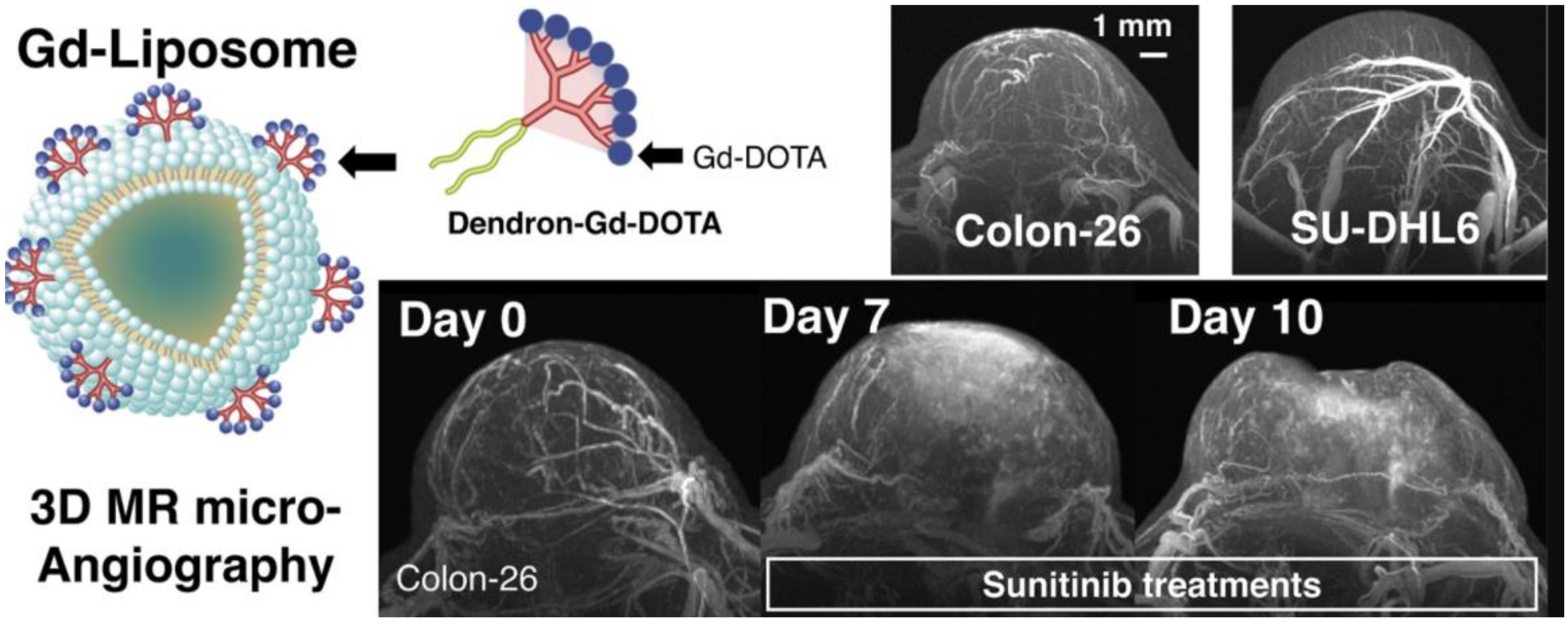
- Nitta, N.; Takakusagi, Y.; Kokuryo, D.; Shibata, S.; Tomita, A.; Higashi, T.; Aoki, I.; Harada, M. Intratumoral evaluation of 3D microvasculature and nanoparticle distribution using a gadolinium-dendron modified nano-liposomal contrast agent with magnetic resonance micro-imaging. Nanomed. Nanotechnol. Biol. Med. 2018, 14, 1315–1324. [Google Scholar] [CrossRef]
- Machová, E.; O’Regan, S.; Newcombe, J.; Meunier, F.; Prentice, J.; Dove, R.; Lisá, V.; Doležal, V. Detection of choline transporter-like 1 protein CTL1 in neuroblastoma× glioma cells and in the CNS, and its role in choline uptake. J. Neurochem. 2009, 110, 1297–1309. [Google Scholar] [CrossRef]
- Lattuada, L.; Lux, G. Synthesis of Gd-DTPA-cholesterol: A new lipophilic gadolinium complex as a potential MRI contrast agent. Tetrahedron Lett. 2003, 44, 3893–3895. [Google Scholar] [CrossRef]
- Rui, M.; Guo, W.; Ding, Q.; Wei, X.; Xu, J.; Xu, Y. Recombinant high-density lipoprotein nanoparticles containing gadolinium-labeled cholesterol for morphologic and functional magnetic resonance imaging of the liver. Int. J. Nanomed. 2012, 7, 3751. [Google Scholar] [CrossRef] [Green Version]
- Ye, F.; Ke, T.; Jeong, E.-K.; Wang, X.; Sun, Y.; Johnson, M.; Lu, Z.-R. Noninvasive visualization of in vivo drug delivery of poly (L-glutamic acid) using contrast-enhanced MRI. Mol. Pharm. 2006, 3, 507–515. [Google Scholar] [CrossRef] [PubMed] [Green Version]
- Shiraishi, K.; Kawano, K.; Maitani, Y.; Yokoyama, M. Polyion complex micelle MRI contrast agents from poly (ethylene glycol)-b-poly (L-lysine) block copolymers having Gd-DOTA; preparations and their control of T1-relaxivities and blood circulation characteristics. J. Control. Release 2010, 148, 160–167. [Google Scholar] [CrossRef] [PubMed]
- Liu, Q.; Zhu, H.; Qin, J.; Dong, H.; Du, J. Theranostic vesicles based on bovine serum albumin and poly (ethylene glycol)-block-poly (L-lactic-co-glycolic acid) for magnetic resonance imaging and anticancer drug delivery. Biomacromolecules 2014, 15, 1586–1592. [Google Scholar] [CrossRef] [PubMed]
- Liu, Y.; Chen, Z.; Liu, C.; Yu, D.; Lu, Z.; Zhang, N. Gadolinium-loaded polymeric nanoparticles modified with Anti-VEGF as multifunctional MRI contrast agents for the diagnosis of liver cancer. Biomaterials 2011, 32, 5167–5176. [Google Scholar] [CrossRef]
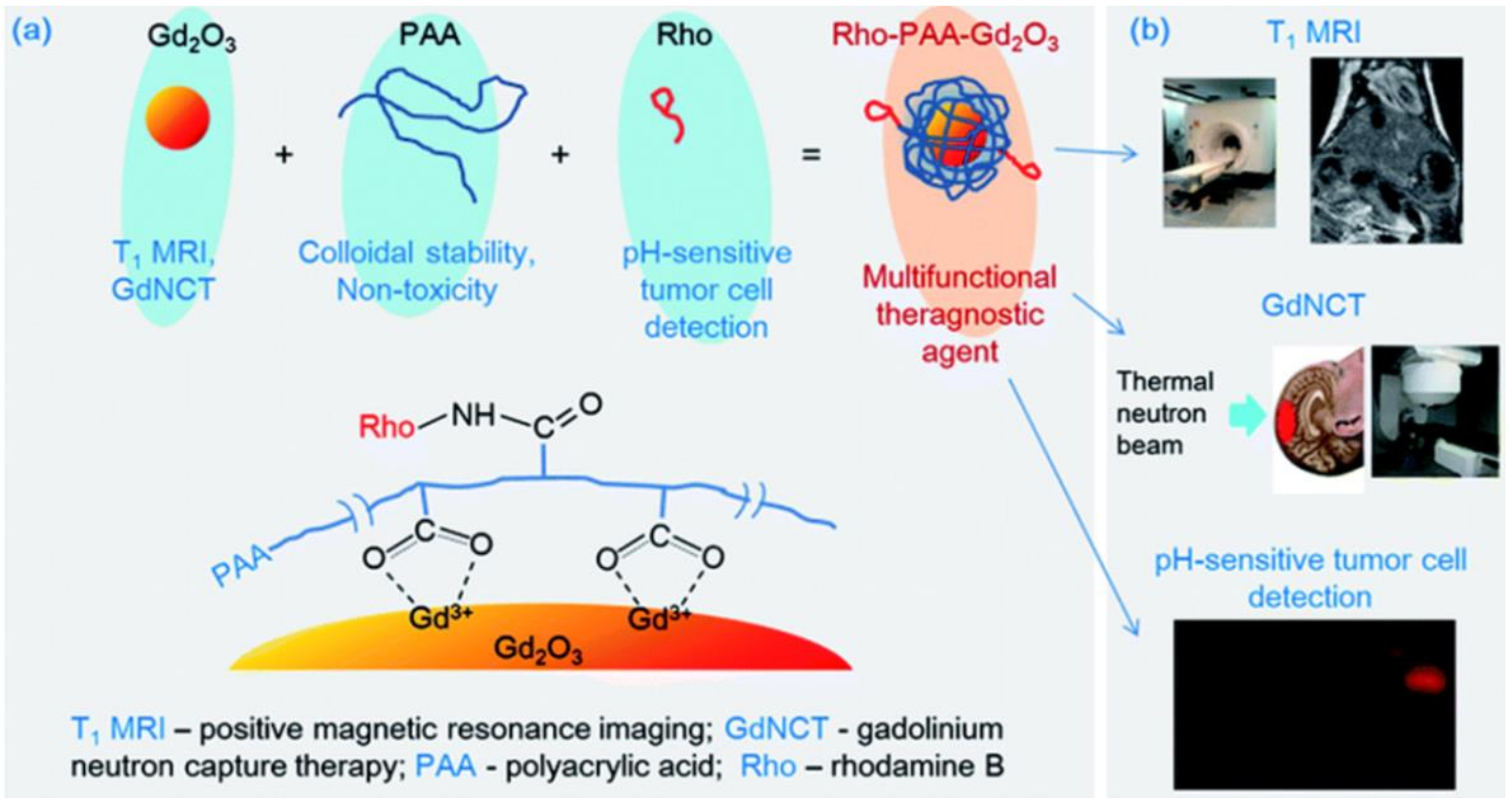
- Ho, S.L.; Cha, H.; Oh, I.T.; Jung, K.-H.; Kim, M.H.; Lee, Y.J.; Miao, X.; Tegafaw, T.; Ahmad, M.Y.; Chae, K.S.; et al. Magnetic resonance imaging, gadolinium neutron capture therapy, and tumor cell detection using ultrasmall Gd2O3 nanoparticles coated with polyacrylic acid-rhodamine B as a multifunctional tumor theragnostic agent. RSC Adv. 2018, 8, 12653–12665. [Google Scholar] [CrossRef] [Green Version]
- Jang, Y.-J.; Liu, S.; Yue, H.; Park, J.; Cha, H.; Ho, S.L.; Marasini, S.; Ghazanfari, A.; Ahmad, M.Y.; Miao, X. Hydrophilic biocompatible poly (acrylic acid-co-maleic acid) polymer as a surface-coating ligand of ultrasmall Gd2O3 nanoparticles to obtain a high r1 value and T1 MR images. Diagnostics 2021, 11, 2. [Google Scholar] [CrossRef]
- Ahmad, M.Y.; Ahmad, M.W.; Yue, H.; Ho, S.L.; Park, J.A.; Jung, K.-H.; Cha, H.; Marasini, S.; Ghazanfari, A.; Liu, S.; et al. In vivo positive magnetic resonance imaging applications of poly(methyl vinyl ether-alt-maleic acid)-coated ultra-small paramagnetic gadolinium oxide nanoparticles. Molecules 2020, 25, 1159. [Google Scholar] [CrossRef] [Green Version]
- Yon, M.; Gineste, S.; Parigi, G.; Lonetti, B.; Gibot, L.; Talham, D.R.; Marty, J.-D.; Mingotaud, C. Hybrid Polymeric Nanostructures Stabilized by Zirconium and Gadolinium Ions for Use as Magnetic Resonance Imaging Contrast Agents. ACS Appl. Nano Mater. 2021, 4, 4974–4982. [Google Scholar] [CrossRef]
- Hu, X.; Tang, Y.; Hu, Y.; Lu, F.; Lu, X.; Wang, Y.; Li, J.; Li, Y.; Ji, Y.; Wang, W. Gadolinium-chelated conjugated polymer-based nanotheranostics for photoacoustic/magnetic resonance/NIR-II fluorescence imaging-guided cancer photothermal therapy. Theranostics 2019, 9, 4168. [Google Scholar] [CrossRef] [PubMed]
- Grogna, M.; Cloots, R.; Luxen, A.; Jérôme, C.; Desreux, J.-F.; Detrembleur, C. Design and synthesis of novel DOTA (Gd 3+)–polymer conjugates as potential MRI contrast agents. J. Mater. Chem. 2011, 21, 12917–12926. [Google Scholar] [CrossRef]
- Kang, M.-K.; Lee, G.H.; Jung, K.-H.; Jung, J.-C.; Kim, H.-K.; Kim, Y.-H.; Lee, J.; Ryeom, H.-K.; Kim, T.-J.; Chang, Y. Gadolinium Nanoparticles Conjugated with Therapeutic Bifunctional Chelate as a Potential T1 Theranostic Magnetic Resonance Imaging Agent. J. Biomed. Nanotechnol. 2016, 12, 894–908. [Google Scholar] [CrossRef]
- Kim, H.-K.; Kang, M.-K.; Jung, K.-H.; Kang, S.-H.; Kim, Y.-H.; Jung, J.-C.; Lee, G.H.; Chang, Y.; Kim, T.-J. Gadolinium complex of DO3A-benzothiazole aniline (BTA) conjugate as a theranostic agent. J. Med. Chem. 2013, 56, 8104–8111. [Google Scholar] [CrossRef]
- Yue, H.; Marasini, S.; Ahmad, M.Y.; Ho, S.L.; Cha, H.; Liu, S.; Jang, Y.J.; Tegafaw, T.; Ghazanfari, A.; Miao, X.; et al. Carbon-coated ultrasmall gadolinium oxide (Gd2O3@C) nanoparticles: Application to magnetic resonance imaging and fluorescence properties resonance imaging and fluorescence properties. Colloids Surfaces A Physicochem. Eng. Asp. 2020, 586, 124261. [Google Scholar] [CrossRef]
- Wang, F.; Peng, E.; Zheng, B.; Li, S.F.Y.; Xue, J.M. Synthesis of water-dispersible Gd2O3/GO nanocomposites with enhanced MRI T 1 relaxivity. J. Phys. Chem. C 2015, 119, 23735–23742. [Google Scholar] [CrossRef]
- Lee, B.H.; Hasan, M.T.; Lichthardt, D.; Gonzalez-Rodriguez, R.; Naumov, A. V Manganese–nitrogen and gadolinium–nitrogen Co-doped graphene quantum dots as bimodal magnetic resonance and fluorescence imaging nanoprobes. Nanotechnology 2020, 32, 95103. [Google Scholar] [CrossRef] [PubMed]
- Li, Y.; Dong, H.; Tao, Q.; Ye, C.; Yu, M.; Li, J.; Zhou, H.; Yang, S.; Ding, G.; Xie, X. Enhancing the magnetic relaxivity of MRI contrast agents via the localized superacid microenvironment of graphene quantum dots. Biomaterials 2020, 250, 120056. [Google Scholar] [CrossRef]
- Carniato, F.; Muñoz-Úbeda, M.; Tei, L.; Botta, M. Selective functionalization of mesoporous silica nanoparticles with ibuprofen and Gd (III) chelates: A new probe for potential theranostic applications. Dalton Trans. 2015, 44, 17927–17931. [Google Scholar] [CrossRef]
- He, K.; Li, J.; Shen, Y.; Yu, Y. pH-Responsive polyelectrolyte coated gadolinium oxide-doped mesoporous silica nanoparticles (Gd2O3@MSNs) for synergistic drug delivery and magnetic resonance imaging enhancement. J. Mater. Chem. B 2019, 7, 6840–6854. [Google Scholar] [CrossRef]
- Liu, J.; Xiong, Z.; Zhang, J.; Peng, C.; Klajnert-Maculewicz, B.; Shen, M.; Shi, X. Zwitterionic gadolinium (III)-complexed dendrimer-entrapped gold nanoparticles for enhanced computed tomography/magnetic resonance imaging of lung cancer metastasis. ACS Appl. Mater. Interfaces 2019, 11, 15212–15221. [Google Scholar] [CrossRef] [PubMed]
- Tegafaw, T.; Xu, W.; Ahmad, M.W.; Baeck, J.S.; Chang, Y.; Bae, J.E.; Chae, K.S.; Kim, T.J.; Lee, G.H. Dual-mode T1 and T2 magnetic resonance imaging contrast agent based on ultrasmall mixed gadolinium-dysprosium oxide nanoparticles: Synthesis, characterization, and in vivo application. Nanotechnology 2015, 26, 365102. [Google Scholar] [CrossRef]
- Choi, E.S.; Park, J.Y.; Baek, M.J.; Xu, W.; Kattel, K.; Kim, J.H.; Lee, J.J.; Chang, Y.; Kim, T.J.; Bae, J.E.; et al. Water-soluble ultra-small manganese oxide surface doped gadolinium oxide (Gd2O3@MnO) nanoparticles for MRI contrast agent. Eur. J. Inorg. Chem. 2010, 4555–4560. [Google Scholar] [CrossRef]
- Tegafaw, T.; Bony, B.A.; Xu, W.; Cha, H.; Chang, Y.; Lee, S.-H.; Chae, K.S.; Lee, G.H. Longitudinal water proton relaxivity and in vivo T 1 MR images of mixed Zn(II)/Gd(III) oxide nanoparticles. J. Nanosci. Nanotechnol. 2017, 17, 2423–2430. [Google Scholar] [CrossRef]
- Xu, W.; Park, J.Y.; Kattel, K.; Bony, B.A.; Heo, W.C.; Jin, S.; Park, J.W.; Chang, Y.; Do, J.Y.; Chae, K.S.; et al. A T1, T2 magnetic resonance imaging (MRI)-fluorescent imaging (FI) by using ultrasmall mixed gadolinium–europium oxide nanoparticles. New J. Chem. 2012, 36, 2361–2367. [Google Scholar] [CrossRef]
- Kim, H.-K.; Lee, J.-J.; Choi, G.; Sung, B.; Kim, Y.-H.; Baek, A.R.; Kim, S.; Song, H.; Kim, M.; Cho, A.E.; et al. Gadolinium-based neuroprognostic magnetic resonance imaging agents suppress COX-2 for prevention of reperfusion injury after stroke. J. Med. Chem. 2020, 63, 6909–6923. [Google Scholar] [CrossRef]
- Choi, G.; Kim, H.-K.; Baek, A.R.; Kim, S.; Kim, M.J.; Kim, M.; Cho, A.E.; Lee, G.-H.; Jung, H.; Yang, J.-U.; et al. Multifunctional imaging of amyloid-beta peptides with a new gadolinium-based contrast agent in Alzheimer’s disease. J. Ind. Eng. Chem. 2020, 83, 214–223. [Google Scholar] [CrossRef]
- Park, J.Y.; Kim, S.J.; Lee, G.H.; Jin, S.; Chang, Y.; Bae, J.E.; Chae, K.S. Various ligand-coated ultrasmall gadolinium-oxide nanoparticles: Water proton relaxivity and in-vivo T1 MR image. J. Korean Phys. Soc. 2015, 66, 1295–1302. [Google Scholar] [CrossRef]
- Baek, A.R.; Kim, H.-K.; Park, S.; Lee, G.H.; Kang, H.J.; Jung, J.-C.; Park, J.-S.; Ryeom, H.-K.; Kim, T.-J.; Chang, Y. Gadolinium complex of 1,4,7,10-tetraazacyclododecane-1,4,7-trisacetic acid (DO3A)-ethoxybenzyl (EOB) conjugate as a new macrocyclic hepatobiliary MRI contrast agent. J. Med. Chem. 2017, 60, 4861–4868. [Google Scholar] [CrossRef]
- Miao, X.; Xu, W.; Cha, H.; Chang, Y.; Oh, I.T.; Chae, K.S.; Lee, G.H. Application of dye-coated ultrasmall gadolinium oxide nanoparticles for biomedical dual imaging. Bull. Korean Chem. Soc. 2017, 38, 1058–1068. [Google Scholar] [CrossRef]
- Xu, W.; Park, J.Y.; Kattel, K.; Ahmad, M.W.; Bony, B.A.; Heo, W.C.; Jin, S.; Park, J.W.; Chang, Y.; Kim, T.J.; et al. Fluorescein-polyethyleneimine coated gadolinium oxide nanoparticles as T1 magnetic resonance imaging (MRI)–cell labeling (CL) dual agents. RSC Adv. 2012, 2, 10907–10915. [Google Scholar] [CrossRef]
- Jung, K.-H.; Kim, H.-K.; Lee, G.H.; Kang, D.-S.; Park, J.-A.; Kim, K.M.; Chang, Y.; Kim, T.-J. Gd complexes of macrocyclic diethylenetriaminepentaacetic acid (DTPA) biphenyl-2,2′-bisamides as strong blood-pool magnetic resonance imaging contrast agents. J. Med. Chem. 2011, 54, 5385–5394. [Google Scholar] [CrossRef] [PubMed]
- Gu, S.; Kim, H.-K.; Lee, G.H.; Kang, B.-S.; Chang, Y.; Kim, T.-J. Gd-complexes of 1,4,7,10-tetraazacyclododecane- N, N′, N″, N‴-1,4,7,10-tetraacetic acid (DOTA) conjugates of tranexamates as a new class of blood-pool magnetic resonance imaging contrast agents. J. Med. Chem. 2011, 54, 143–152. [Google Scholar] [CrossRef] [PubMed]


| Imaging Technique | Detection | Imaging Probes | Common CAs | Some Clinical Applications | Advantages | Disadvantages |
|---|---|---|---|---|---|---|
| Optical imaging | Visible, ultraviolet, and infrared light | Organic dyes, QDs, Lanthanide ion | Fluorescein, cresyl violet acetate, indocyanine green, toluidine blue | Optical microscopy, endoscopy, scanning laser ophthalmoscopy | High sensitivity | Low resolution and poor tissue penetration |
| Computed tomography | X-rays | Iodine, Lanthanide, Gold compounds | Iopamidol, ioxaglate | Cerebral infarction, angiography | High spatial resolution | Costly with poor soft tissue imaging |
| Magnetic resonance imaging | Magnetic field | Gd, Fe, Mn compounds | Gadoteridol, gadopentetate dimeglumine | Cerebral and coronary angiography | High-resolution Excellent signal in soft tissues | Costly with low sensitivity |
| Positron emission tomography | Gamma-rays | Radioactive elements: 18F, 15O, 64Cu 68Ga etc. | 18FDG, 15H2O, 68Ga-EDTA, 11C-methionine | Degenerative diseases and cerebral blood flow | Quantitative | Exposure to radiation with poor resolution |
| Single photon emission computed tomography | Gamma-rays | Radioactive elements: 18F, 11C, 15O, 68Ga, 64Cu etc. | 99mTc-HMPAO, 99mTc-ECD, 111In-octreotide | Dementia, cardiac imaging and cerebral infarction | Quantitative | Exposure to radiation with poor resolution |
| Ultra- sonography | Ultrasonic waves | Microbubbles | Microbubbles | Congenital conditions and echocardiography | Cost-effective, simple, and fast. | Poor resolution |
| Imaging Modality | Nanoparticles * | Applications | Ref. |
|---|---|---|---|
| MRI | Liposomal Gd | Imaging placenta | [75] |
| MRI | Gd(III)-thiolated DNA–Au nanostars (DNA-Gd@stars) | Imaging pancreatic cancer cells | [76] |
| MRI/CT | Gd(III)-decorated Au NPs | Enhancing the relaxometric properties of Gd(iii) complexes | [77] |
| MRI/OI | GQDs-folate-DOX-Gd | Bioimaging and tumor targeted drug delivery | [78] |
| MRI/PET | Pegylated liposome (LP)-(Gd)-positron-emitting 89Zr | Imaging of Cancer | [79] |
| MRI/SPECT | Gd Complex of 125I/127I-RGD-DOTA Conjugate | Tumor targeting | [80] |
| MRI/US | Liposomes-Gd-rhodamine | MRI Monitoring and Quantification of US-Mediated Delivery | [81] |
| NIRF/CT/MRI | Gold–Gd nanoclusters (NCs) | Tumor targeting and low body residues | [82] |
| MRI/US/CT | Gd- Gold Microcapsules | Multimodal cellular imaging of transplanted islet cells | [83] |
| MRI/CT/PAI | Bismuth-Gd-PEG NPs | Imaging-guided photothermal cancer therapy | [84] |
| S. No. | CAs | Magnetic Relaxivity (r1) (s−1·mM−1) | Particle Size (davg) (nm) | Specific Characteristics | Modality | Ref. |
|---|---|---|---|---|---|---|
| 1 | Gd–albumin conjugates | 9~10.5 | ~5–6 | Blood clearance half-lives = 40–47 min | T1 MRI | [110] |
| 2 | Gd–albumin-Folic acid conjugates | 10.8 | ~201–215 | Almost non-cytotoxic, good biocompatibility | T1 MRI | [111] |
| 3 | TAT-Gd-NPs | 18.2 | 1.5 | Good in vitro cell viability, non-toxic upto 20 μM Gd | T1 MRI | [112] |
| 4 | Cyclic RGD-conjugated Gd-NPs | 10.0–18.7 | 1.0–2.5 | Nontoxic up to 10 μM Gd | T1 MRI | [25] |
| 5 | PPy@BSA-Gd | 10.203 | 50 | Good cytocompatibility, Phototherma therapy | T1 MRI | [114] |
| 6 | D-glucuronic acid coated Gd-NPs | 12.2 | 2.0 | Highly water-dispersible and non-toxic | T1 MRI | [122] |
| 7 | Chitosan oligosaccharide lactate (COL)-Gd-NPs | 13.0 | 1.9 | Non-toxic up to 500 μM Gd | T1 MRI | [124] |
| 8 | Gd-NPs-polyacrylic acid (PAA)-rhodamine B (Rho) | 22.6 | 1.5 | High cell viabilities up to 500 μM Gd and good biocompatibility | T1 MRI-NCT-FI | [135] |
| 9 | PAAMA coated Gd-NPs | 40.6 | 1.8 | Exceptionally low cellular toxicity | T1 MRI | [136] |
| 10 | PMVEMA-coated Gd-NPs | 36.2 | 1.9 | Excellent colloidal stability in aqueous solution and appreciable biocompatibility | T1 MRI | [137] |
| 11 | PFTQ-PEG-Gd-NPs | 10.95 | 95 ± 4.6 | Low biotoxicity and outstanding chemical and optical stability | T1 MRI | [139] |
| 12 | Gd-NPs@SiO2-DO3A and Gd-NPs@SiO2-DO2A-benzothiazoles (BTA) | Gd@SiO2-DO3A = 5.47 Gd@SiO2-DO2A-BTA = 7.99 | 50–60 | High water solubility and colloidal stability, anticancer characteristics | T1 MRI | [141] |
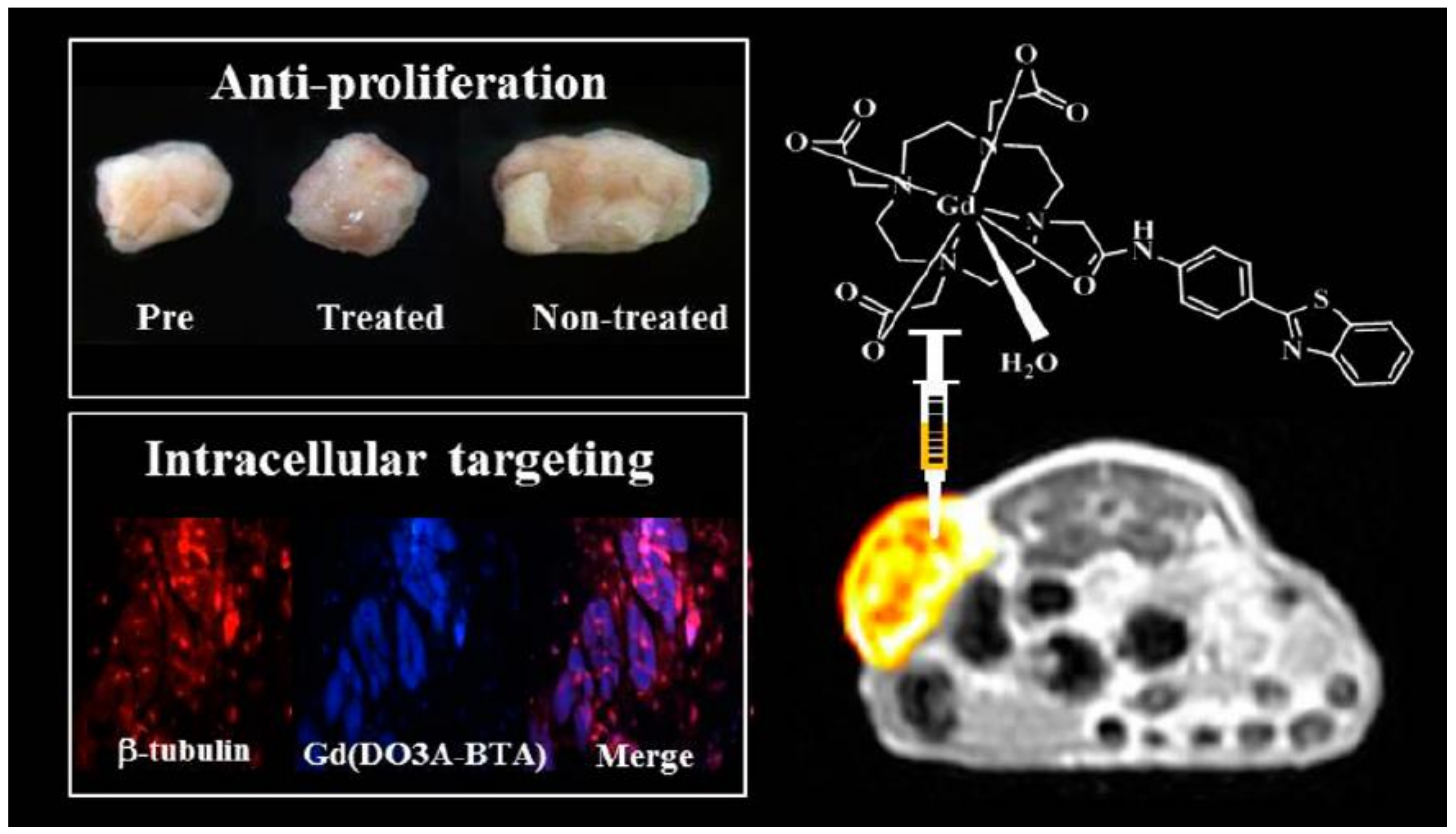
| 13 | Gd(DO3A-BTAA)(H2O) chelates | 3.84 | - | Tumour-specific, antiproliferative activities | T1 MRI | [142] |
| 14 | Gd-NPs@C | 16.26 | 3.1 | Cell viabilities up to 500 μM Gd, good biocompatibility | T1 MRI-FI | [143] |
| 15 | Gd-NPs/GO-NCs | 34.48 | 2.89 | Water dispersible with good biocompatibility | T1 MRI | [144] |
| 16 | Gd-PEG-GO-QDs | 210.9 at 114 μT | 4.0 | Low biotoxicities | T1 MRI-FI | [146] |
| 17 | Gd(III)-loaded AuNPs-DEN modified with arginine-glycine-aspartic acid peptide Complex | 13.17 | - | Satisfactory cytocompatibility | T1 MRI-CT | [149] |
| 18 | Gd-dysprosium oxide nanoparticles (Gd-DONPs) D-glucuronic acid coated Gd-DONPs | 6.0 | 1.0 | Non-toxic up to 200 μM | T1, T2 MRI | [150] |
| 19 | D-glucuronic acid coated Zn(II)/Gd(III) oxide NPs | 12.3 | 2.1 | Slightly cytotoxic in DU145 cell lines, negligible cytotoxicity in NCTC1469 cell lines up to 200 M (Gd + Zn) | T1 MRI | [152] |
| 20 | LA coated Gd–europium oxide NPs | 11.9 | 1.75 | Non-toxic up to 279 mM Gd and Eu | T1,T2 MRI-FI | [153] |
| 21 | Gd compounds –NSAIDs chelates | 5.0–7.0 | - | Neuroprognostic | T1 MRI | [154] |
| 22 | Gd-chelate conjugated with Chal (Gd-DO3A-Chal | 4.95 | - | Aβ-targeting | MRI-FI | [155] |
| 23 | Fluorescein/FITC-Gd-NPs | Fluorescein-coated = 9.8 FITC-coated = 12.3 | Fluorescein-coated = 1.6 FITC-coated = 1.4 | Good cell viability up to 100 μM Gd | T1 MRI-FI | [158] |
Publisher’s Note: MDPI stays neutral with regard to jurisdictional claims in published maps and institutional affiliations. |
© 2021 by the authors. Licensee MDPI, Basel, Switzerland. This article is an open access article distributed under the terms and conditions of the Creative Commons Attribution (CC BY) license (https://creativecommons.org/licenses/by/4.0/).
Share and Cite
Fatima, A.; Ahmad, M.W.; Al Saidi, A.K.A.; Choudhury, A.; Chang, Y.; Lee, G.H. Recent Advances in Gadolinium Based Contrast Agents for Bioimaging Applications. Nanomaterials 2021, 11, 2449. https://doi.org/10.3390/nano11092449
Fatima A, Ahmad MW, Al Saidi AKA, Choudhury A, Chang Y, Lee GH. Recent Advances in Gadolinium Based Contrast Agents for Bioimaging Applications. Nanomaterials. 2021; 11(9):2449. https://doi.org/10.3390/nano11092449
Chicago/Turabian StyleFatima, Atiya, Md. Wasi Ahmad, Abdullah Khamis Ali Al Saidi, Arup Choudhury, Yongmin Chang, and Gang Ho Lee. 2021. "Recent Advances in Gadolinium Based Contrast Agents for Bioimaging Applications" Nanomaterials 11, no. 9: 2449. https://doi.org/10.3390/nano11092449