
Green Synthesis of Silver Nanoparticles with Antibacterial Activity Using Various Medicinal Plant Extracts: Morphology and Antibacterial Efficacy

 , , ,
, , ,
Abstract
:1. Introduction
2. Materials and Methods
2.1. Materials and Chemicals
2.2. Antioxidant Activity—DPPH Method
2.3. Total Phenolic Content (TPC)
2.4. Preparation of Plant Extracts
2.5. Biosynthesis of AgNPs
2.6. Characterisation Techniques
2.7. Antibacterial Activity Using Agar Well-Diffusion Assay
2.8. Statistical Analysis
3. Results and Discussion
3.1. Characterisation of Plant Extracts
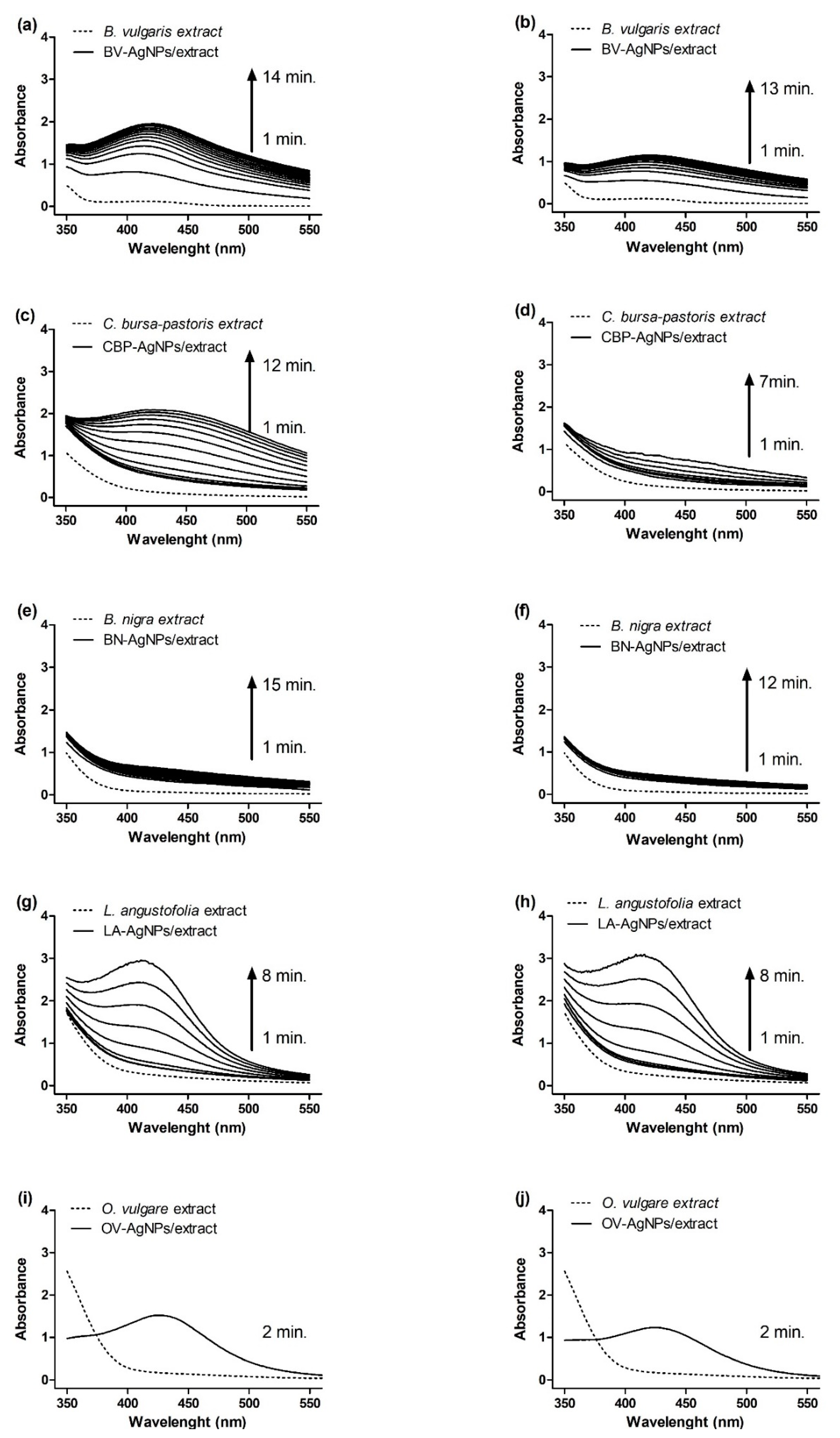
3.2. Synthesis and Optical Studies of AgNPs
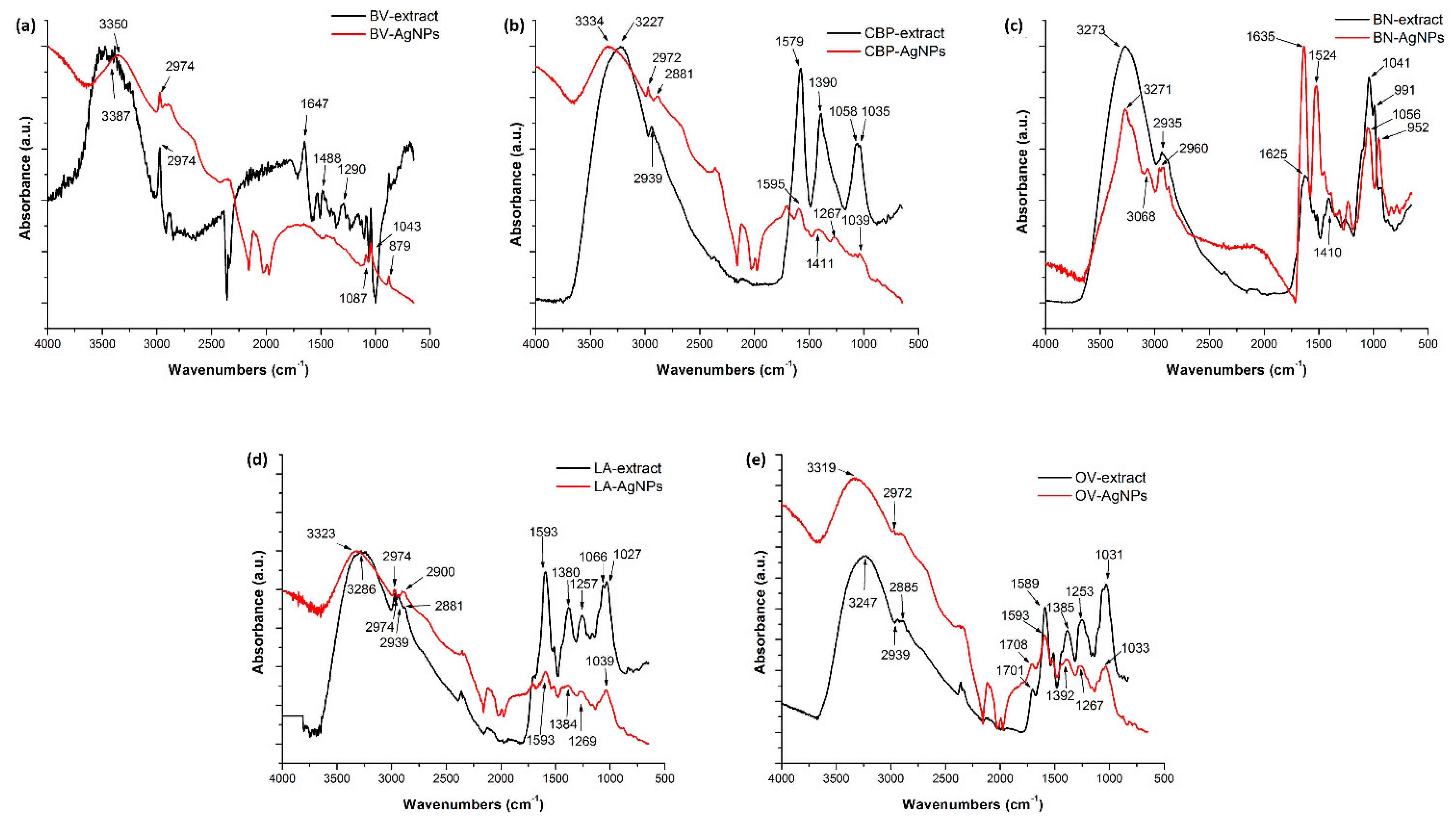
3.3. Structural Characterisation of AgNPs Obtained from 5 mM AgNO3
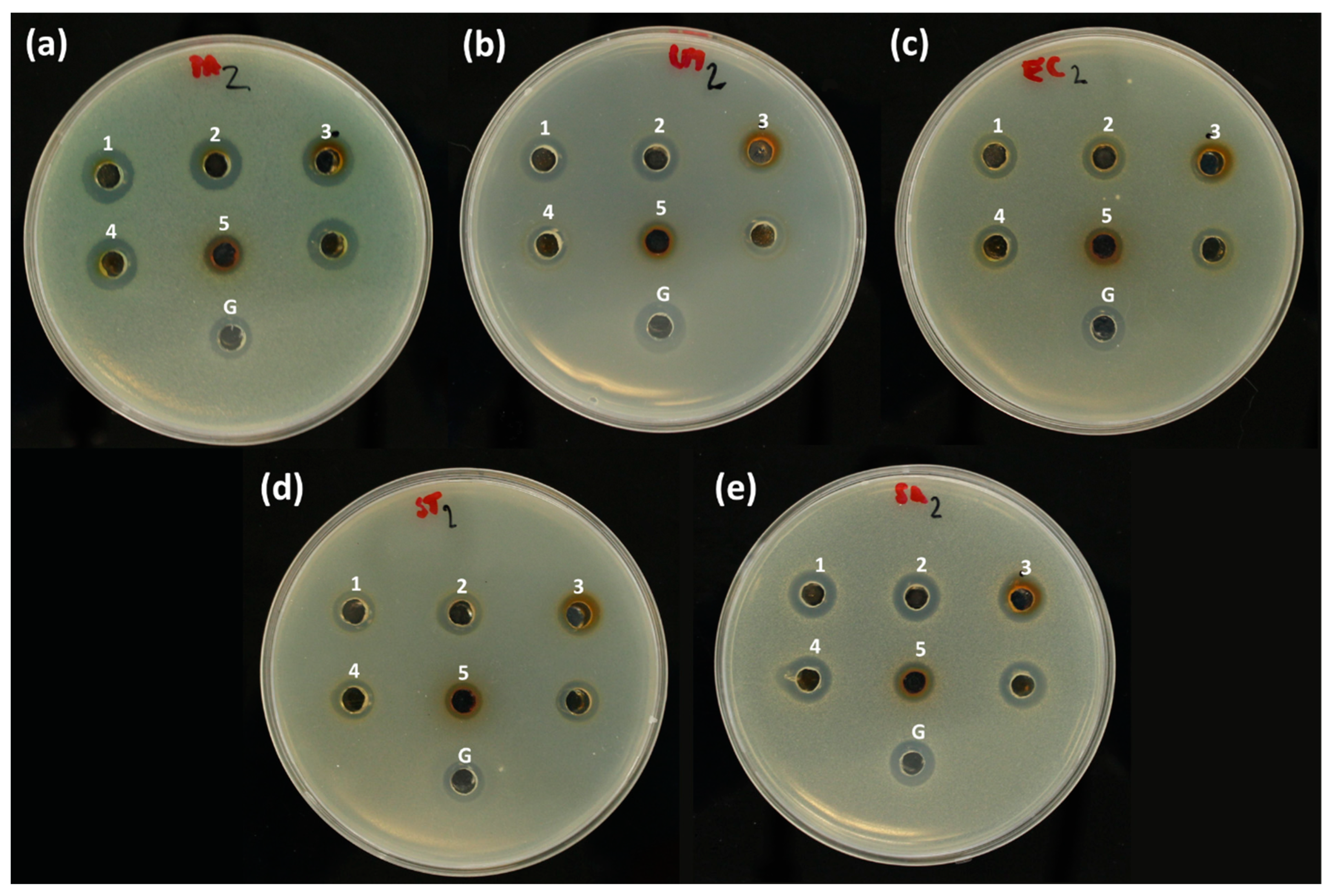
3.4. Antibacterial Activity of AgNPs
4. Conclusions
Supplementary Materials
Author Contributions
Funding
Data Availability Statement
Acknowledgments
Conflicts of Interest
References
- Hemeg, H.A. Nanomaterials for alternative antibacterial therapy. Int. J. Nanomed. 2017, 12, 8211–8225. [Google Scholar] [CrossRef] [PubMed] [Green Version]
- Franci, G.; Falanga, A.; Galdiero, S.; Palomba, L.; Rai, M.; Morelli, G.; Galdiero, M. Silver Nanoparticles as Potential Antibacterial Agents. Molecules 2015, 20, 8856–8874. [Google Scholar] [CrossRef] [Green Version]
- Dakal, T.C.; Kumar, A.; Majumdar, R.S.; Yadav, V. Mechanistic Basis of Antimicrobial Actions of Silver Nanoparticles. Front. Microbiol. 2016, 7, 1–17. [Google Scholar] [CrossRef] [PubMed] [Green Version]
- Ismail, M.; Gul, S.; Khan, M.A.; Khan, M.I. Plant Mediated Green Synthesis of Anti- Microbial Silver Nanoparticles—A Review on Recent Trends. Rev. Nanosci. Nanotechnol. 2016, 5, 119–135. [Google Scholar] [CrossRef]
- Shanmuganathan, R.; Karuppusamy, I.; Saravanan, M.; Muthukumar, H.; Ponnuchamy, K.; Ramkumar, V.S.; Pugazhendhi, A. Synthesis of Silver Nanoparticles and their Biomedical Applications—A Comprehensive Review. Curr. Pharm. Des. 2019, 25, 2650–2660. [Google Scholar] [CrossRef] [PubMed]
- Salem, S.S.; Fouda, A. Green Synthesis of Metallic Nanoparticles and Their Prospective Biotechnological Applications: An Overview. Biol. Trace Elem. Res. 2021, 199, 344–370. [Google Scholar] [CrossRef] [PubMed]
- Das, C.G.A.; Kumar, V.G.; Dhas, T.S.; Karthick, V.; Govindaraju, K.; Joselin, J.M.; Baalamurugan, J. Antibacterial activity of silver nanoparticles (biosynthesis): A short review on recent advances. Biocatal. Agric. Biotechnol. 2020, 27, 101593. [Google Scholar] [CrossRef]
- Alsharif, S.M.; Salem, S.S.; Abdel-Rahman, M.A.; Fouda, A.; Eid, A.M.; El-Din Hassan, S.; Awad, M.A.; Mohamed, A.A. Multifunctional properties of spherical silver nanoparticles fabricated by different microbial taxa. Heliyon 2020, 6, e03943. [Google Scholar] [CrossRef]
- Salleh, A.; Naomi, R.; Utami, N.D.; Mohammad, A.W.; Mahmoudi, E.; Mustafa, N.; Fauzi, M.B. The Potential of Silver Nanoparticles for Antiviral and Antibacterial Applications: A Mechanism of Action. Nanomaterials 2020, 10, 1566. [Google Scholar] [CrossRef]
- Jeremiah, S.S.; Miyakawa, K.; Morita, T.; Yamaoka, Y.; Ryo, A. Potent antiviral effect of silver nanoparticles on SARS-CoV-2. Biochem. Biophys. Res. Commun. 2020, 533, 195–200. [Google Scholar] [CrossRef]
- Song, K.C.; Lee, S.M.; Park, T.S.; Lee, B.S. Preparation of colloidal silver nanoparticles by chemical reduction method. Korean J. Chem. Eng. 2009, 26, 153–155. [Google Scholar] [CrossRef]
- Lee, D.K.; Kang, Y.S. Synthesis of silver nanocrystallites by a new thermal decomposition method and their characterization. ETRI J. 2004, 26, 252–256. [Google Scholar] [CrossRef]
- Pluym, T.C.; Powell, Q.H.; Gurav, A.S.; Ward, T.L.; Kodas, T.T.; Wang, L.M.; Glicksman, H.D. Solid silver particle production by spray pyrolysis. J. Aerosol Sci. 1993, 24, 383–392. [Google Scholar] [CrossRef]
- Alahmad, A.; Feldhoff, A.; Bigall, N.C.; Rusch, P.; Scheper, T.; Walter, J.-G. Hypericum perforatum L.-Mediated Green Synthesis of Silver Nanoparticles Exhibiting Antioxidant and Anticancer Activities. Nanomaterials 2021, 11, 487. [Google Scholar] [CrossRef]
- Bedlovičová, Z.; Salayová, A. Green-Synthesized Silver Nanoparticles and Their Potential for Antibacterial Applications. In Bacterial Pathogenesis and Antibacterial Control; Kırmusaoğlu, S., Ed.; IntechOpen: London, UK, 2018; pp. 73–94. [Google Scholar]
- Rafique, M.; Sadaf, I.; Rafique, M.S.; Tahir, M.B. A review on green synthesis of silver nanoparticles and their applications. Artif. Cells Nanomed. Biotechnol. 2017, 45, 1272–1291. [Google Scholar] [CrossRef]
- Rajeshkumar, S.; Bharath, L.V. Mechanism of plant-mediated synthesis of silver nanoparticles—A review on biomolecules involved, characterisation and antibacterial activity. Chem. Biol. Interact. 2017, 273, 219–227. [Google Scholar] [CrossRef]
- Ovais, M.; Khalil, A.T.; Raza, A.; Khan, M.A.; Ahmad, I.; Islam, N.U.; Saravanan, M.; Ubaid, M.F.; Ali, M.; Shinwari, Z.K. Green synthesis of silver nanoparticles via plant extracts: Beginning a new era in cancer theranostics. Nanomedicine 2016, 11, 3157–3177. [Google Scholar] [CrossRef]
- Makarov, V.V.; Love, A.J.; Sinitsyna, O.V.; Makarova, S.S.; Yaminsky, I.V.; Taliansky, M.E.; Kalinina, N.O. “Green” nanotechnologies: Synthesis of metal nanoparticles using plants. Acta Nat. 2014, 6, 35–44. [Google Scholar] [CrossRef] [Green Version]
- Rajan, R.; Chandran, K.; Harper, S.L.; Yun, S.-I.; Kalaichelvan, P.T. Plant extract synthesized silver nanoparticles: An ongoing source of novel biocompatible materials. Ind. Crops Prod. 2015, 70, 356–373. [Google Scholar] [CrossRef]
- Saravanan, M.; Barik, S.K.; MubarakAli, D.; Prakash, P.; Pugazhendhi, A. Synthesis of silver nanoparticles from Bacillus brevis (NCIM 2533) and their antibacterial activity against pathogenic bacteria. Microb. Pathog. 2018, 116, 221–226. [Google Scholar] [CrossRef]
- Samuel, M.S.; Jose, S.; Selvarajan, E.; Mathimani, T.; Pugazhendhi, A. Biosynthesized silver nanoparticles using Bacillus amyloliquefaciens; Application for cytotoxicity effect on A549 cell line and photocatalytic degradation of p-nitrophenol. J. Photochem. Photobiol. B Biol. 2020, 202, 111642. [Google Scholar] [CrossRef]
- Saravanan, M.; Arokiyaraj, S.; Lakshmi, T.; Pugazhendhi, A. Synthesis of silver nanoparticles from Phenerochaete chrysosporium (MTCC-787) and their antibacterial activity against human pathogenic bacteria. Microb. Pathog. 2018, 117, 68–72. [Google Scholar] [CrossRef]
- Pugazhendhi, A.; Prabakar, D.; Jacob, J.M.; Karuppusamy, I.; Saratale, R.G. Synthesis and characterization of silver nanoparticles using Gelidium amansii and its antimicrobial property against various pathogenic bacteria. Microb. Pathog. 2018, 114, 41–45. [Google Scholar] [CrossRef]
- Kováčová, M.; Daneu, N.; Tkáčiková, Ľ.; Búreš, R.; Dutková, E.; Stahorský, M.; Bujňáková, Z.L.; Baláž, M. Sustainable One-Step Solid-State Synthesis of Antibacterially Active Silver Nanoparticles Using Mechanochemistry. Nanomaterials 2020, 10, 2119. [Google Scholar] [CrossRef] [PubMed]
- Bordoloi, M.; Sahoo, R.K.; Tamuli, K.J.; Saikia, S.; Dutta, P.P. Plant Extracts Promoted Preparation of Silver and Gold Nanoparticles: A Systematic Review. Nano 2020, 15, 2030001. [Google Scholar] [CrossRef] [Green Version]
- Rafique, M.; Sadaf, I.; Tahir, M.B.; Rafique, M.S.; Nabi, G.; Iqbal, T.; Sughra, K. Novel and facile synthesis of silver nanoparticles using Albizia procera leaf extract for dye degradation and antibacterial applications. Mater. Sci. Eng. C 2019, 99, 1313–1324. [Google Scholar] [CrossRef] [PubMed]
- Srikar, S.K.; Giri, D.D.; Pal, D.B.; Mishra, P.K.; Upadhyay, S.N. Green Synthesis of Silver Nanoparticles: A Review. Green Sustain. Chem. 2016, 6, 34–56. [Google Scholar] [CrossRef] [Green Version]
- Pandit, R. Green synthesis of silver nanoparticles from seed extract of Brassica nigra and its antibacterial activity. Nusant. Biosci. 2015, 7, 15–19. [Google Scholar] [CrossRef]
- Sankar, R.; Karthik, A.; Prabu, A.; Karthik, S.; Shivashangari, K.S.; Ravikumar, V. Origanum vulgare mediated biosynthesis of silver nanoparticles for its antibacterial and anticancer activity. Colloids Surfaces B Biointerf. 2013, 108, 80–84. [Google Scholar] [CrossRef]
- Baláž, M.; Balážová, Ľ.; Daneu, N.; Dutková, E.; Balážová, M.; Bujňáková, Z.; Shpotyuk, Y. Plant-Mediated Synthesis of Silver Nanoparticles and Their Stabilization by Wet Stirred Media Milling. Nanoscale Res. Lett. 2017, 12, 83. [Google Scholar] [CrossRef] [Green Version]
- Shaik, M.R.; Khan, M.; Kuniyil, M.; Al-Warthan, A.; Alkhathlan, H.Z.; Siddiqui, M.R.H.; Shaik, J.P.; Ahamed, A.; Mahmood, A.; Khan, M.; et al. Plant-Extract-Assisted Green Synthesis of Silver Nanoparticles Using Origanum vulgare L. Extract and Their Microbicidal Activities. Sustainability 2018, 10, 913. [Google Scholar] [CrossRef] [Green Version]
- Baláž, M.; Balážová, Ľ.; Kováčová, M.; Daneu, N.; Salayová, A.; Bedlovičová, Z.; Tkáčiková, Ľ. The relationship between precursor concentration and antibacterial activity of biosynthesized Ag nanoparticles. Adv. Nano Res. 2019, 7, 125–134. [Google Scholar] [CrossRef]
- Behravan, M.; Panahi, A.H.; Naghizadeh, A.; Ziaee, M.; Mahdavi, R.; Mirzapour, A. Facile green synthesis of silver nanoparticles using Berberis vulgaris leaf and root aqueous extract and its antibacterial activity. Int. J. Biol. Macromol. 2019, 124, 148–154. [Google Scholar] [CrossRef] [PubMed]
- Villalpando, M.; Rosas, G. Effect of Magnetic Stirring on the Green Synthesis of Silver Nanoparticles Using Lavandula angustifolia Plant Extract. Microsc. Microanal. 2019, 25, 1976–1977. [Google Scholar] [CrossRef] [Green Version]
- Belova, M.M.; Shipunova, V.O.; Kotelnikova, P.A.; Babenyshev, A.V.; Rogozhin, E.A.; Cherednichenko, M.Y.; Deyev, S.M. “Green” synthesis of cytotoxic silver nanoparticles based on secondary metabolites of Lavandula angustifolia mill. Acta Nat. 2019, 11, 47–53. [Google Scholar] [CrossRef]
- Bunghez, I.R.; Fierascu, R.C.; Dumitrescu, O.; Fierascu, I.; Ion, R.M. Characterization of silver nanoparticles obtained by Lavandula angustifolia extract. Rev. Roum. Chim. 2015, 60, 515–519. [Google Scholar]
- Adam, K.; Sivropoulou, A.; Kokkini, S.; Lanaras, T.; Arsenakis, M. Antifungal activities of Origanum vulgare subsp.Hirtum, Mentha spicata, Lavandula angustifolia and Salvia fruiticosa essential oil against human pathogenic fungi. J. Agric. Food Chem. 1998, 46, 1739–1745. [Google Scholar] [CrossRef]
- Daferera, D.J.; Ziogas, B.N.; Polissiou, M.G. GC-MS Analysis of Essential Oils from Some Greek Aromatic Plants and Their Fungitoxicity on Penicillium digitatum. J. Agric. Food Chem. 2000, 48, 2576–2581. [Google Scholar] [CrossRef]
- Dănilă, E.; Moldovan, Z.; Popa, M.; Chifiriuc, M.C.; Kaya, A.D.; Kaya, M.A. Chemical composition, antimicrobial and antibiofilm efficacy of C. limon and L. angustifolia EOs and of their mixtures against Staphylococcus epidermidis clinical strains. Ind. Crops Prod. 2018, 122, 483–492. [Google Scholar] [CrossRef]
- Hammer, K.A.; Carson, C.F.; Riley, T.V. Antimicrobial activity of essential oils and other plant extracts. J. Appl. Microbiol. 1999, 86, 985–990. [Google Scholar] [CrossRef] [Green Version]
- Rodriguez-Garcia, I.; Silva-Espinoza, B.A.; Ortega-Ramirez, L.A.; Leyva, J.M.; Siddiqui, M.W.; Cruz-Valenzuela, M.R.; Gonzalez-Aguilar, G.A.; Ayala-Zavala, J.F. Oregano Essential Oil as an Antimicrobial and Antioxidant Additive in Food Products. Crit. Rev. Food Sci. Nutr. 2016, 56, 1717–1727. [Google Scholar] [CrossRef]
- Liang, C.-H.; Chan, L.-P.; Ding, H.-Y.; So, E.C.; Lin, R.-J.; Wang, H.-M.; Chen, Y.-G.; Chou, T.-H. Free radical scavenging activity of 4-(3,4-Dihydroxybenzoyloxymethyl)phenyl-O-β-D-glucopyranoside from Origanum vulgare and its protection against oxidative damage. J. Agric. Food Chem. 2012, 60, 7690–7696. [Google Scholar] [CrossRef] [PubMed]
- Al-Snafi, A.E. The chemical constituents and pharmacological effects of Capsella bursa-pastoris- A review. Int. J. Pharmacol. Toxicol. 2015, 5, 76–81. [Google Scholar]
- Al-Snafi, A.E. The pharmacological importance of Brassica nigra and Brassica rapa grown in Iraq. J. Pharm. Biol. 2015, 5, 240–253. [Google Scholar]
- Shamsa, F.; Ahmadiani, A.; Khosrokhavar, R. Antihistaminic and anticholinergic activity of barberry fruit (Berberis vulgaris) in the guinea-pig ileum. J. Ethnopharmacol. 1999, 64, 161–166. [Google Scholar] [CrossRef]
- Abushouk, A.I.; Salem, A.M.A.; Abdel-Daim, M.M. Berberis vulgaris for cardiovascular disorders: A scoping literature review. Iran. J. Basic Med. Sci. 2017, 20, 503–510. [Google Scholar] [CrossRef]
- Tahmasebi, L.; Zakerkish, M.; Golfakhrabadi, F.; Namjoyan, F. Randomised clinical trial of Berberis vulgaris root extract on glycemic and lipid parameters in type 2 diabetes mellitus patients. Eur. J. Integr. Med. 2019, 32, 100998. [Google Scholar] [CrossRef]
- Shidfar, F.; Ebrahimi, S.S.; Hosseini, S.; Heydari, I.; Shidfar, S.; Hajhassani, G. The effects of Berberis vulgaris fruit extract on serum lipoproteins, apoB, apoA-I, homocysteine, glycemic control and total antioxidant capacity in type 2 diabetic patients. Iran. J. Pharm. Res. 2012, 11, 643–652. [Google Scholar] [CrossRef]
- Cazzola, R.; Cestaro, B. Antioxidant Spices and Herbs Used in Diabetes. In Diabetes: Oxidative Stress and Dietary Antioxidants; Elsevier: Amsterdam, The Netherlands, 2013; pp. 89–97. ISBN 9780124058859. [Google Scholar]
- Srinivasan, K. Plant foods in the management of diabetes mellitus: Spices as beneficial antidiabetic food adjuncts. Int. J. Food Sci. Nutr. 2005, 56, 399–414. [Google Scholar] [CrossRef]
- Brand-Williams, W.; Cuvelier, M.E.; Berset, C. Use of a free radical method to evaluate antioxidant activity. LWT Food Sci. Technol. 1995, 28, 25–30. [Google Scholar] [CrossRef]
- Folin, O.; Ciocalteu, V. On Tyrosine and Tryptophane Determinations in Proteins. J. Biol. Chem. 1927, LXXIII, 627–650. [Google Scholar] [CrossRef]
- Rojas, J.J.; Ochoa, V.J.; Ocampo, S.A.; Muñoz, J.F. Screening for antimicrobial activity of ten medicinal plants used in Colombian folkloric medicine: A possible alternative in the treatment of non-nosocomial infections. BMC Complement. Altern. Med. 2006, 6, 1–6. [Google Scholar] [CrossRef] [PubMed] [Green Version]
- Bhutto, A.A.; Kalay, Ş.; Sherazi, S.T.H.; Culha, M. Quantitative structure–activity relationship between antioxidant capacity of phenolic compounds and the plasmonic properties of silver nanoparticles. Talanta 2018, 189, 174–181. [Google Scholar] [CrossRef] [PubMed]
- Liu, Y.-S.; Chang, Y.-C.; Chen, H.-H. Silver nanoparticle biosynthesis by using phenolic acids in rice husk extract as reducing agents and dispersants. J. Food Drug Anal. 2018, 26, 649–656. [Google Scholar] [CrossRef] [PubMed] [Green Version]
- Olteanu, R.L.; Nicolescu, C.M.; Bumbac, M. Influence of Phytochemical Reductive Capacity on Ultraviolet-Visible Spectroscopic Behavior of Silver Nanoparticles. Anal. Lett. 2017, 50, 2786–2801. [Google Scholar] [CrossRef]
- Lee, Y.H.; Choo, C.; Waisundara, V.Y. Determination of the Total Antioxidant Capacity and Quantification of Phenolic Compounds of Different Solvent Extracts of Black Mustard Seeds (Brassica nigra). Int. J. Food Prop. 2015, 18, 2500–2507. [Google Scholar] [CrossRef]
- Alam, M.B.; Hossain, M.S.; Haque, E.M. Antioxidant and anti-inflammatory activities of the leaf extract of Brassica nigra. Int. J. Pharm. Sci. Res. 2011, 2, 303–310. [Google Scholar]
- Bedlovičová, Z.; Strapáč, I.; Baláž, M.; Salayová, A. A Brief Overview on Antioxidant Activity Determination of Silver Nanoparticles. Molecules 2020, 25, 3191. [Google Scholar] [CrossRef] [PubMed]
- Upadhyay, L.S.B.; Verma, N. Recent Developments and Applications in Plant-extract Mediated Synthesis of Silver Nanoparticles. Anal. Lett. 2015, 48, 2676–2692. [Google Scholar] [CrossRef]
- De Leersnyder, I.; De Gelder, L.; Van Driessche, I.; Vermeir, P. Revealing the importance of aging, environment, size and stabilization mechanisms on the stability of metal nanoparticles: A case study for silver nanoparticles in a minimally defined and complex undefined bacterial growth medium. Nanomaterials 2019, 9, 1684. [Google Scholar] [CrossRef] [Green Version]
- Rajawat, S.; Qureshi, M.S. Electrolytic Deposition of Silver Nanoparticles Under “Principles of Green Chemistry”. Arab. J. Sci. Eng. 2014, 39, 563–568. [Google Scholar] [CrossRef]
- Mourdikoudis, S.; Pallares, R.M.; Thanh, N.T.K. Characterization techniques for nanoparticles: Comparison and complementarity upon studying nanoparticle properties. Nanoscale 2018, 10, 12871–12934. [Google Scholar] [CrossRef] [Green Version]
- Baláž, M.; Daneu, N.; Balážová, Ľ.; Dutková, E.; Tkáčiková, Ľ.; Briančin, J.; Vargová, M.; Balážová, M.; Zorkovská, A.; Baláž, P. Bio-mechanochemical synthesis of silver nanoparticles with antibacterial activity. Adv. Powder Technol. 2017, 28, 3307–3312. [Google Scholar] [CrossRef]
- Baláž, M.; Bedlovičová, Z.; Kováčová, M.; Salayová, A.; Balážová, L’. Green and Bio-Mechanochemical Approach to Silver Nanoparticles Synthesis, Characterization and Antibacterial Potential. In Nanostructures for Antimicrobial and Antibiofilm Applications; Springer Natura: Berlin/Heidelberg, Germany, 2020; pp. 145–183. ISBN 978-3-030-40336-2. [Google Scholar]
- Lee, S.H.; Jun, B.H. Silver nanoparticles: Synthesis and application for nanomedicine. Int. J. Mol. Sci. 2019, 20, 865. [Google Scholar] [CrossRef] [PubMed] [Green Version]
- Kokila, T.; Ramesh, P.S.; Geetha, D. Biosynthesis of AgNPs using Carica Papaya peel extract and evaluation of its antioxidant and antimicrobial activities. Ecotoxicol. Environ. Saf. 2016, 134, 467–473. [Google Scholar] [CrossRef]
- Elemike, E.E.; Onwudiwe, D.C.; Ekennia, A.C.; Katata-Seru, L. Biosynthesis, characterization, and antimicrobial effect of silver nanoparticles obtained using Lavandula × intermedia. Res. Chem. Intermed. 2017, 43, 1383–1394. [Google Scholar] [CrossRef]


| Plant Species | Concentration of AgNO3 | Characterisation | Activity | Size of AgNPs (nm) | Shape of AgNPs | Ref. |
|---|---|---|---|---|---|---|
| B. nigra | 1 mM | UV-Vis | antibacterial | 41 | - | [29] |
| NTA | ||||||
| FTIR | ||||||
| DLS | ||||||
| O. vulgare | 1 mM | UV-Vis | antibacterial | 63–85 | spherical | [30] |
| FTIR | ||||||
| FE-SEM | ||||||
| XRD | ||||||
| O. vulgare | 0.5 mM | UV-Vis | antibacterial | 2–25 | spherical | [32] |
| XRD | ||||||
| TEM | ||||||
| EDX | ||||||
| FTIR | ||||||
| O. vulgare | 1, 2.5, 5, 10 and 100 mM | UV-Vis | antibacterial | 151–4819 | polygonal | [33] |
| PCCS | ||||||
| TEM | ||||||
| B. vulgaris | 0.5, 1, 3 and 10 mM | UV-Vis | antibacterial | 30–70 | spherical | [34] |
| TEM | ||||||
| XRD | ||||||
| DLS | ||||||
| L. angustifolia | 10 mM | UV-Vis | - | 20 | spherical, triangular | [35] |
| SEM | ||||||
| XRD | ||||||
| TEM | ||||||
| EDS | ||||||
| L. angustifolia fractions | 1 g/L | UV-Vis | cytotoxic | 35.4 ± 1.6 and 56.4 ± 2.4 | round- shape, tetrahedron, polyhedron | [36] |
| DLS | ||||||
| SEM | ||||||
| L. angustifolia | 1 mM | UV-Vis | antioxidant | 10–30 | spherical | [37] |
| EDXRF | ||||||
| SEM | ||||||
| B. vulgaris | 2 and 5 mM | UV-Vis | antibacterial antioxidant | 75.7 ± 17.1 | spherical | This study |
| B. nigra | TEM | 14.7 ± 7.9 | spherical | |||
| C. bursa-pastoris | ZP | 16.2 ± 8.4 | spherical | |||
| L. angustifolia | PCCS | 37.8 ± 10.7 | spherical | |||
| O. vulgare | EDS | 46.1 ± 19.7 | smaller-spherical, larger-truncated octahedral |
| Plant Extract | TPC (mg GAE/mL) ± SD | DPPH (%) ± SD |
|---|---|---|
| Berberis vulgaris | 0.234 ± 0.009 | 61.78 ± 1.80 |
| Brassica nigra | 0.297 ± 0.028 | 73.77 ± 3.47 |
| Capsella bursa-pastoris | 0.464 ± 0.031 | 89.68 ± 3.18 |
| Lavandula angustifolia | 0.884 ± 0.022 | 92.97 ± 0.04 |
| Origanum vulgare | 4.569 ± 0.766 | 94.98 ± 0.08 |
| Plant-AgNPs | SPR Peak Position (nm) | Time (min) 2/5 mM AgNO3 |
|---|---|---|
| BV-AgNPs | 421 nm | 14/12 |
| CBP-AgNPs | 422 nm | 12/7 |
| BN-AgNPs | Not found | 15/12 |
| LA-AgNPs | 412 nm | 8/8 |
| OV-AgNPs | 426 nm | 2/2 |
| Phase | Hkl | D (nm) | I/I0-Ag (%) | I/I0-AgCl (%) |
|---|---|---|---|---|
| AgCl | 111 | 3.2037 | 51.3 | |
| AgCl | 200 | 2.7745 | 100.0 | |
| Ag | 111 | 2.3592 | 100.0 | |
| Ag | 200 | 2.0431 | 48.2 | |
| AgCl | 220 | 1.9618 | 67.0 | |
| AgCl | 113 | 1.6730 | 23.0 | |
| AgCl | 222 | 1.6018 | 22.3 | |
| Ag | 220 | 1.4447 | 29.8 | |
| AgCl | 400 | 1.3872 | 10.5 | |
| AgCl | 331 | 1.2730 | 10.0 | |
| AgCl | 420 | 1.2407 | 31.8 | |
| Ag | 113 | 1.2326 | 29.8 |
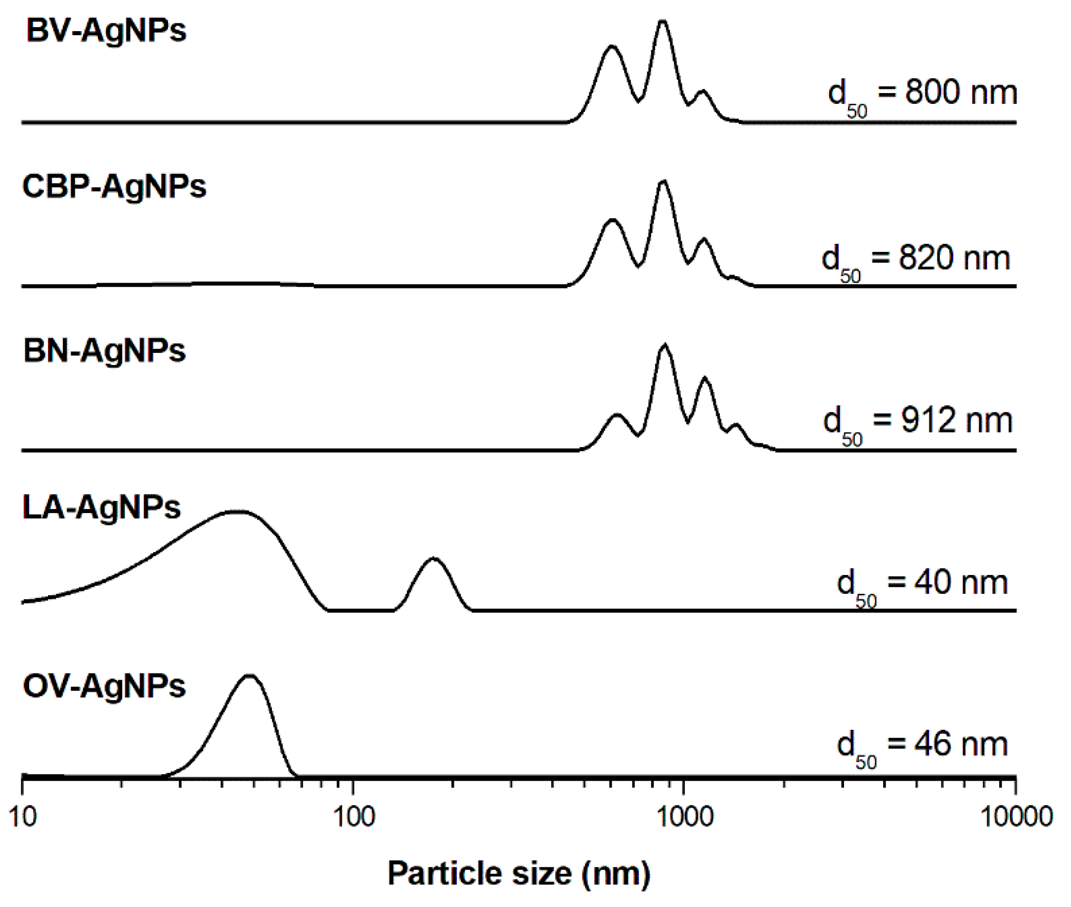
| Sample | Coarse Fraction |
|---|---|
| OV-AgNPs | 46.1 ± 19.7 nm |
| LA-AgNPs | 37.8 ± 10.7 nm |
| BN-AgNPs | 14.7 ± 7.9 nm |
| CBP-AgNPs | 16.2 ± 8.4 nm |
| BV-AgNPs | 75.7 ± 17.1 nm |
| Sample | Zeta Potential (mV) | pH |
|---|---|---|
| OV-AgNPs | −11.9 | 3.20 |
| LA-AgNPs | −15.8 | 3.31 |
| BN-AgNPs | −6.5 | 4.28 |
| CBP-AgNPs | −18.1 | 4.81 |
| BV-AgNPs | 0.38 | 3.77 |
| Plant Extract-AgNPs | Bacteria Type | P. aeruginosa | L. monocytogenes | E. coli | S. enterica ser. Typhimurium | S. aureus |
|---|---|---|---|---|---|---|
| Conc. of AgNO3 | RIZD (% ± SD) | |||||
| CBP-AgNPs | 2 mM | 92.61 ± 10.07 | 55.17 ± 12.40 | 80.76 ± 2.94 | 80.10 ± 4.72 | 96.03 ± 5.90 |
| 5 mM | 148.33 ± 1.50 | 59.88 ± 4.15 | 87.78 ± 5.11 | 98.16 ± 8.63 | 102.27 ± 5.88 | |
| BN-AgNPs | 2 mM | 73.87 ± 12.45 | 60.65 ± 1.80 | 83.31 ± 3.45 | 77.33 ± 7.25 | 93.12 ± 14.77 |
| 5 mM | 152.53 ± 8.60 | 84.44 ± 9.21 | 99.09 ± 15.35 | 99.05 ± 5.72 | 104.80 ± 4.35 | |
| LA-AgNPs | 2 mM | 97.78 ± 12.20 | 21.79 ± 15.48 | 82.77 ± 1.03 | 82.31 ± 2.82 | 89.91 ± 4.78 |
| 5 mM | 146.25 ± 10.62 | 80.06 ± 3.33 | 93.81 ± 12.41 | 97.25 ± 12.05 | 101.29 ± 6.33 | |
| BV-AgNPs | 2 mM | 77.57 ± 0.97 | 72.18 ± 8.99 | 89.21 ± 6.48 | 88.78 ± 9.04 | 93.64 ± 7.07 |
| 5 mM | 151.12 ± 18.28 | 83.67 ± 6.47 | 94.99 ± 5.14 | 99.69 ± 18.35 | 101.93 ± 8.33 | |
| OV-AgNPs | 2 mM | 0.00 ± 0.00 * | 23.73 ± 2.93 * | 0.00 ± 0.00 * | 0.00 ± 0.00 * | 0.00 ± 0.00 * |
| 5 mM | 97.75 ± 2.05 | 58.84 ± 7.38 | 71.53 ± 8.95 | 66.61 ± 7.05 | 74.14 ± 5.59 | |
| AgNO3 | 2 mM | 96.92 ± 19.44 | 67.80 ± 10.74 | 89.25 ± 8.02 | 86.25 ± 9.24 | 90.66 ± 6.34 |
| 5 mM | 111.37 ± 10.52 | 90.47 ± 12.41 | 99.78 ± 4.84 | 104.00 ± 7.27 | 117.81 ± 2.11 | |
Publisher’s Note: MDPI stays neutral with regard to jurisdictional claims in published maps and institutional affiliations. |
© 2021 by the authors. Licensee MDPI, Basel, Switzerland. This article is an open access article distributed under the terms and conditions of the Creative Commons Attribution (CC BY) license (https://creativecommons.org/licenses/by/4.0/).
Share and Cite
Salayová, A.; Bedlovičová, Z.; Daneu, N.; Baláž, M.; Lukáčová Bujňáková, Z.; Balážová, Ľ.; Tkáčiková, Ľ. Green Synthesis of Silver Nanoparticles with Antibacterial Activity Using Various Medicinal Plant Extracts: Morphology and Antibacterial Efficacy. Nanomaterials 2021, 11, 1005. https://doi.org/10.3390/nano11041005
Salayová A, Bedlovičová Z, Daneu N, Baláž M, Lukáčová Bujňáková Z, Balážová Ľ, Tkáčiková Ľ. Green Synthesis of Silver Nanoparticles with Antibacterial Activity Using Various Medicinal Plant Extracts: Morphology and Antibacterial Efficacy. Nanomaterials. 2021; 11(4):1005. https://doi.org/10.3390/nano11041005
Chicago/Turabian StyleSalayová, Aneta, Zdenka Bedlovičová, Nina Daneu, Matej Baláž, Zdenka Lukáčová Bujňáková, Ľudmila Balážová, and Ľudmila Tkáčiková. 2021. "Green Synthesis of Silver Nanoparticles with Antibacterial Activity Using Various Medicinal Plant Extracts: Morphology and Antibacterial Efficacy" Nanomaterials 11, no. 4: 1005. https://doi.org/10.3390/nano11041005