Biological Safety and Biodistribution of Chitosan Nanoparticles

 , ,
, ,  , ,
, ,
Abstract
:1. Introduction
2. Materials and Methods
2.1. Characterization of Chitosan Sample
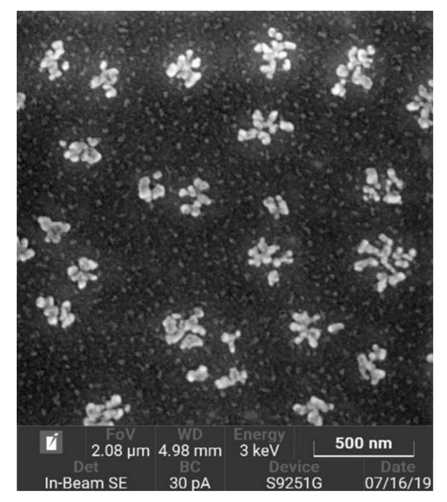
2.2. Preparation of Chitosan Nanoparticles
2.3. In Vitro Experiments
2.3.1. Assessment of Hemolytic Activity
2.3.2. Assessment of Anticoagulant Activity
2.3.3. Assessment of Antiplatelet Activity
2.3.4. Study of Cytotoxic Properties
2.4. In Vivo Experiments
2.4.1. Biosafety Study of Single Intravenous CNP Administration
Assessment of CNP Effect on Hemodynamic Parameters
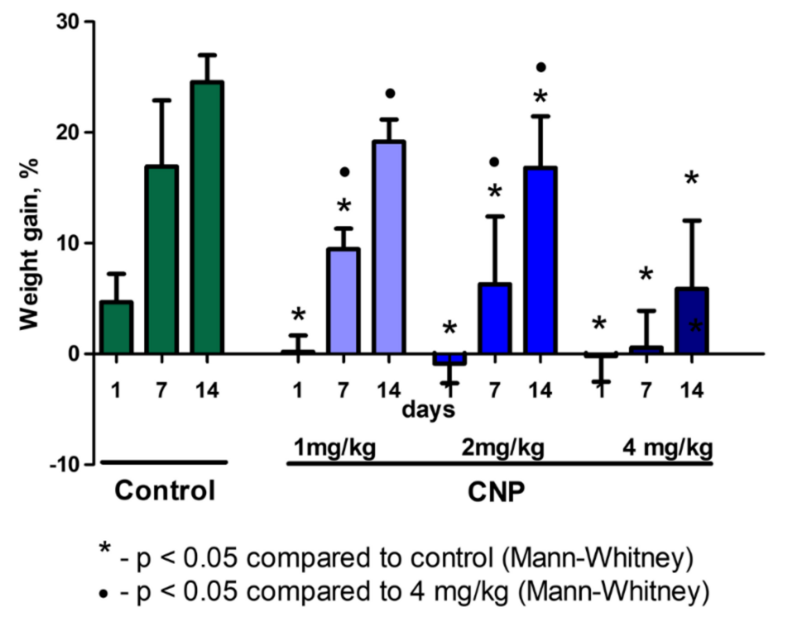
Assessment of General Toxicity
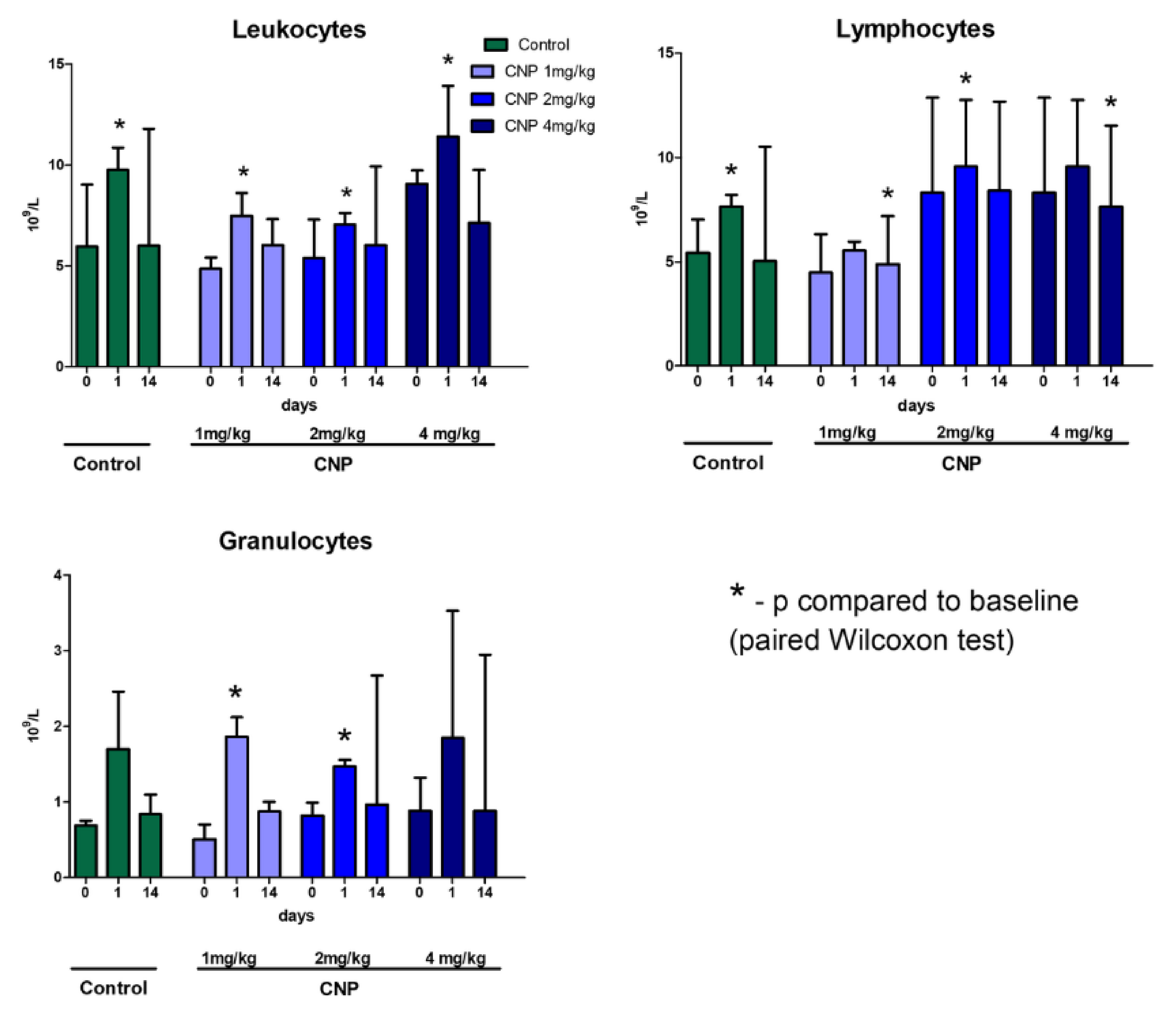
Hematological Analysis
Blood Chemistry Analysis
2.4.2. Study of CNP Biodistribution in Rats
Preparation of ICG-CNPs
Assessment of ICG-CNP Biodistribution
Histological Examinations
Morphological Visualization of CNP Internalization
Immunohistochemical Analysis
2.5. Statistical Analysis
3. Results
3.1. In Vitro Assessment of CNP Hemocompatibility
3.1.1. Hemolytic Activity
3.1.2. Effect of CNP Suspension on Coagulation Hemostasis
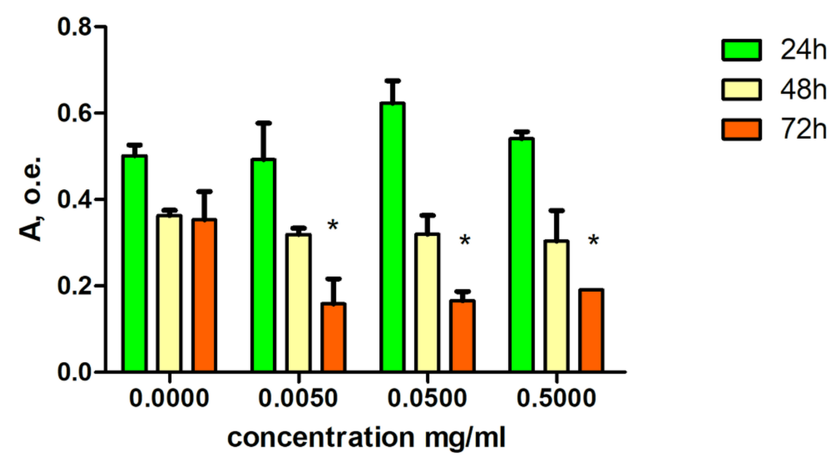
3.1.3. Cytotoxic CNP Properties
3.2. In Vivo Results of Acute Experiments
3.2.1. Effect of CNP Suspension on Systemic Hemodynamics
3.2.2. In Vivo Biodistribution of CNPs
3.3. General Toxicity of CNP Suspension
3.3.1. Results of Hematological Analysis
3.3.2. Results of Biochemical Blood Tests
3.3.3. Results of Histological Analysis
Liver
Lungs
3.3.4. Results of Immunohistochemical Analysis
3.3.5. Histological Verification of CNP Internalization
4. Discussion
5. Conclusions
Author Contributions
Funding
Conflicts of Interest
References
- Kato, Y.; Onishi, H.; Machida, Y. Contribution of chitosan and its derivatives to cancer chemotherapy. In Vivo 2005, 19, 301–310. [Google Scholar] [PubMed]
- Cheung, R.C.; Ng, T.B.; Wong, J.H.; Chan, W.Y. Chitosan: An update on potential biomedical and pharmaceutical applications. Mar. Drugs 2015, 13, 5156–5186. [Google Scholar] [CrossRef] [PubMed]
- Muxika, A.; Etxabide, A.; Uranga, J.; Guerrero, P.; de la Caba, K. Chitosan as a bioactive polymer: Processing, properties and applications. Int. J. Biol. Macromol. 2017, 105, 1358–1368. [Google Scholar] [CrossRef] [PubMed]
- Shariatinia, Z. Pharmaceutical applications of chitosan. Adv. Colloid Interface Sci. 2019, 263, 131–194. [Google Scholar] [CrossRef]
- Dubashynskaya, N.V.; Poshina, D.N.; Raik, S.V.; Urtti, A.; Skorik, Y.A. Polysaccharides in ocular drug delivery. Pharmaceutics 2020, 12, 22. [Google Scholar] [CrossRef] [Green Version]
- Carreno-Gómez, B.; Duncan, R. Valuation of the biological properties of soluble chitosan and chitosan microspheres. Int. J. Pharm. 1997, 148, 231–240. [Google Scholar] [CrossRef]
- Kazemzadeh-Narbat, M.; Reid, M.; Brooks, M.S.; Ghanem, A. Chitosan nanoparticles as adenosine carriers. J. Microencapsul. 2015, 32, 460–466. [Google Scholar] [CrossRef]
- Panevin, A.A.; Golyshev, A.A.; Skorik, Y.A.; Zhuravskii, S.G.; Sonin, D.L. Conjugation of succinate to chitosan increases the cochlear cytoprotective effect. Pharm. Chem. J. 2017, 50, 711–714. [Google Scholar] [CrossRef]
- Kritchenkov, A.S.; Andranovits, S.; Skorik, Y.A. Chitosan and its derivatives: Vectors in gene therapy. Russ. Chem. Rev. 2017, 86, 231–239. [Google Scholar] [CrossRef]
- Berezin, A.S.; Lomkova, E.A.; Skorik, Y.A. Chitosan conjugates with biologically active compounds: Design strategies, properties, and targeted drug delivery. Russ. Chem. Bull. 2012, 61, 781–795. [Google Scholar]
- Panzarasa, G.; Osypova, A.; Sicher, A.; Bruinink, A.; Dufresne, E.R. Controlled formation of chitosan particles by a clock reaction. Soft Matter. 2018, 14, 6415–6418. [Google Scholar] [CrossRef] [PubMed]
- Alexis, F.; Pridgen, E.; Molnar, L.K.; Farokhzad, O.C. Factors affecting the clearance and biodistribution of polymeric nanoparticles. Mol. Pharm. 2008, 5, 505–515. [Google Scholar] [CrossRef] [PubMed] [Green Version]
- He, C.; Hu, Y.; Yin, L.; Tang, C.; Yin, C. Effects of particle size and surface charge on cellular uptake and biodistribution of polymeric nanoparticles. Biomaterials 2010, 31, 3657–3666. [Google Scholar] [CrossRef] [PubMed]
- Dash, B.C.; Réthoré, G.; Monaghan, M.; Fitzgerald, K.; Gallagher, W.; Pandit, A. The influence of size and charge of chitosan/polyglutamic acid hollow spheres on cellular internalization, viability and blood compatibility. Biomaterials 2010, 31, 8188–8197. [Google Scholar] [CrossRef] [Green Version]
- Ernsting, M.J.; Murakami, M.; Roy, A.; Li, S.D. Factors controlling the pharmacokinetics, biodistribution and intratumoral penetration of nanoparticles. J. Control. Release 2013, 172, 782–794. [Google Scholar] [CrossRef] [Green Version]
- Shin, S.W.; Song, I.H.; Um, S.H. Role of physicochemical properties in nanoparticle toxicity. Nanomaterials 2015, 5, 1351–1365. [Google Scholar] [CrossRef] [Green Version]
- Fröhlich, E. The role of surface charge in cellular uptake and cytotoxicity of medical nanoparticles. Int. J. Nanomed. 2012, 7, 5577–5591. [Google Scholar] [CrossRef] [Green Version]
- Tanaka, Y.; Tanioka, S.; Tanaka, M.; Tanigawa, T.; Kitamura, Y.; Minami, S.; Okamoto, Y.; Miyashita, M.; Nanno, M. Effects of chitin and chitosan particles on BALB/c mice by oral and parenteral administration. Biomaterials 1997, 18, 591–595. [Google Scholar] [CrossRef]
- Baldrick, P. The safety of chitosan as a pharmaceutical excipient. Regul. Toxicol. Pharmacol. 2010, 56, 290–299. [Google Scholar] [CrossRef]
- Hoemann, C.D.; Fong, D. Immunological responses to chitosan for biomedical applications. In Chitosan Based Biomaterials; Jennings, J.A., Bumgardner, J.D., Eds.; Woodhead Publishing: Cambridge, UK, 2017; Volume 1, pp. 45–79. [Google Scholar]
- Nadesh, R.; Narayanan, D.; Sreerekha, P.R.; Vadakumpully, S.; Mony, U.; Koyakkutty, M.; Nair, S.V.; Menon, D. Hematotoxicological analysis of surface-modified and -unmodified chitosan nanoparticles. J. Biomed. Mater. Res. A 2013, 101, 2957–2966. [Google Scholar] [CrossRef]
- Zhou, X.; Zhang, X.; Zhou, J.; Li, L. An investigation of chitosan and its derivatives on red blood cell agglutination. RSC Adv. 2017, 7, 12247–12254. [Google Scholar] [CrossRef] [Green Version]
- Hu, Z.; Zhang, D.Y.; Lu, S.T.; Li, P.W.; Li, S.D. Chitosan-based composite materials for prospective hemostatic applications. Mar. Drugs 2018, 16, 273. [Google Scholar] [CrossRef] [PubMed] [Green Version]
- Stefan, J.; Lorkowska-Zawicka, B.; Kaminski, K.; Szczubialka, K.; Nowakowska, M.; Korbut, R. The current view on biological potency of cationically modified chitosan. J. Physiol. Pharmacol. 2014, 65, 341–347. [Google Scholar]
- Yan, C.; Chen, D.; Gu, J.; Hu, H.; Zhao, X.; Qiao, M. Preparation of N-succinyl-chitosan and its physical-chemical properties as a novel excipient. Yakugaku Zasshi 2006, 126, 789–793. [Google Scholar] [CrossRef] [PubMed] [Green Version]
- Yan, C.; Gu, J.; Guo, Y.; Chen, D. In vivo biodistribution for tumor targeting of 5-fluorouracil (5-FU) loaded N-succinyl-chitosan (Suc-Chi) nanoparticles. Yakugaku Zasshi 2010, 130, 801–804. [Google Scholar] [CrossRef] [PubMed] [Green Version]
- Kato, Y.; Onishi, H.; Machida, Y. Biological fate of highly-succinylated N-succinyl-chitosan and antitumor characteristics of its water-soluble conjugate with mitomycin C at i.v. and i.p. administration into tumor-bearing mice. Biol. Pharm. Bull. 2000, 23, 1497–1503. [Google Scholar] [CrossRef] [PubMed] [Green Version]
- Wilson, B.; Samanta, M.K.; Muthu, M.S.; Vinothapooshan, G. Design and evaluation of chitosan nanoparticles as novel drug carrier for the delivery of rivastigmine to treat Alzheimer’s disease. Ther. Deliv. 2011, 2, 599–609. [Google Scholar] [CrossRef]
- Zhu, H.; Liu, F.; Guo, J.; Xue, J.; Qian, Z.; Gu, Y. Folate-modified chitosan micelles with enhanced tumor targeting evaluated by near infrared imaging system. Carbohydr. Polym. 2011, 86, 1118–1129. [Google Scholar] [CrossRef]
- Montero, N.; Pérez, E.; Benito, M.; Teijón, C.; Teijón, J.M.; Olmo, R.; Blanco, M.D. Biocompatibility studies of intravenously administered ionic-crosslinked chitosan-BSA nanoparticles as vehicles for antitumour drugs. Int. J. Pharm. 2019, 554, 337–351. [Google Scholar] [CrossRef]
- Hwang, H.; Kwon, J.; Oh, P.S.; Lee, T.K.; Na, K.S.; Lee, C.M.; Jeong, H.S.; Lim, S.T.; Sohn, M.H.; Jeong, H.J. Peptide-loaded nanoparticles and radionuclide imaging for individualized treatment of myocardial ischemia. Radiology 2014, 273, 160–167. [Google Scholar] [CrossRef] [Green Version]
- Pogodina, N.V.; Pavlov, G.M.; Bushin, S.V.; Mel’nikov, A.B.; Lysenko, Y.B.; Nud’ga, L.A.; Marsheva, V.N.; Marchenko, G.N.; Tsvetkov, V.N. Conformational characteristics of chitosan molecules as demonstrated by diffusion-sedimentation analysis and viscometry. Polym. Sci. USSR 1986, 28, 251–259. [Google Scholar] [CrossRef]
- Salvati, A.M.; Samoggia, P.; Taggi, F.; Tentori, L. Hemoglobinometry: A comparison between the hemiglobincyanide method and the Coulter S counter. Clin. Chim. Acta 1977, 77, 13–20. [Google Scholar] [CrossRef]
- Skorik, Y.A.; Kritchenkov, A.S.; Moskalenko, Y.E.; Golyshev, A.A.; Raik, S.V.; Whaley, A.K.; Vasina, L.V.; Sonin, D.L. Synthesis of N-succinyl- and N-glutaryl-chitosan derivatives and their antioxidant, antiplatelet, and anticoagulant activity. Carbohydr. Polym. 2017, 166, 166–172. [Google Scholar] [CrossRef]
- Riess, H.; Braun, G.; Brehm, G.; Hiller, E. Critical evaluation of platelet aggregation in whole human blood. Am. J. Clin. Pathol. 1986, 85, 50–56. [Google Scholar] [CrossRef] [PubMed] [Green Version]
- Niks, M.; Otto, M. Towards an optimized MTT assay. J. Immunol. Methods 1990, 130, 149–151. [Google Scholar] [CrossRef]
- Sonin, D.L.; Korolev, D.V.; Postnov, V.N.; Naumysheva, E.B.; Pochkaeva, E.I.; Vasyutina, M.L.; Galagudza, M.M. Silicon-containing nanocarriers for targeted drug delivery: Synthesis, physicochemical properties and acute toxicity. Drug Deliv. 2016, 23, 1747–1756. [Google Scholar] [CrossRef] [Green Version]
- Jheng, P.R.; Lu, K.Y.; Yu, S.H.; Mi, F.L. Free DOX and chitosan-N-arginine conjugate stabilized indocyanine green nanoparticles for combined chemophotothermal therapy. Colloids Surf. B Biointerfaces 2015, 136, 402–412. [Google Scholar] [CrossRef]
- Chou, T.C.; Fu, E.; Wu, C.J.; Yeh, J.H. Chitosan enhances platelet adhesion and aggregation. Biochem. Biophys. Res. Commun. 2003, 302, 480–483. [Google Scholar] [CrossRef]
- Okamoto, Y.; Yano, R.; Miyatake, K.; Tomohiro, I.; Shigemasa, Y.; Minami, S. Effects of chitin and chitosan on blood coagulation. Carbohydr. Polym. 2003, 53, 337–342. [Google Scholar] [CrossRef]
- Li, X.; Radomski, A.; Corrigan, O.I.; Tajber, L.; De Sousa Menezes, F.; Endter, S.; Medina, C.; Radomski, M.W. Platelet compatibility of PLGA, chitosan and PLGA-chitosan nanoparticles. Nanomedicine 2009, 4, 735–746. [Google Scholar] [CrossRef]
- Zhang, C.; Qu, G.; Sun, Y.; Wu, X.; Yao, Z.; Guo, Q.; Ding, Q.; Yuan, S.; Shen, Z.; Ping, Q.; et al. Pharmacokinetics, biodistribution, efficacy and safety of N-octyl-O-sulfate chitosan micelles loaded with paclitaxel. Biomaterials 2008, 29, 1233–1241. [Google Scholar] [CrossRef] [PubMed]
- Kean, T.; Thanou, M. Biodegradation, biodistribution and toxicity of chitosan. Adv. Drug Deliv. Rev. 2010, 62, 3–11. [Google Scholar] [CrossRef] [PubMed]
- Lee, J.H.; Lee, D.H.; Son, J.H.; Park, J.K.; Kim, S.K. Optimization of chitosan-alginate encapsulation process using pig hepatocytes for development of bioartificial liver. J. Microbiol. Biotechnol. 2005, 15, 7–13. [Google Scholar]
- Chu, X.H.; Shi, X.L.; Feng, Z.Q.; Gu, Z.Z.; Ding, Y.T. Chitosan nanofiber scaffold enhances hepatocyte adhesion and function. Biotechnol. Lett. 2009, 31, 347–352. [Google Scholar] [CrossRef] [PubMed]
- Loh, J.W.; Yeoh, G.; Saunders, M.; Lim, L.Y. Uptake and cytotoxicity of chitosan nanoparticles in human liver cells. Toxicol. Appl. Pharmacol. 2010, 249, 148–157. [Google Scholar] [CrossRef]
- Moreau, E.; Domurado, M.; Chapon, P.; Vert, M.; Domurad, D. Biocompatibility of polycations: In vitro agglutination and lysis of red blood cells and in vivo toxicity. J. Drug Target. 2002, 10, 161–173. [Google Scholar] [CrossRef]
- Cao, J.; Huang, S.; Chen, Y.; Li, S.; Li, X.; Deng, D.; Qian, Z.; Tang, L.; Gu, Y. Near-infrared light-triggered micelles for fast controlled drug release in deep tissue. Biomaterials 2013, 34, 6272–6283. [Google Scholar] [CrossRef]
- Hirano, S.; Seino, H.; Akiyama, Y.; Nonaka, I. Chitosan: A biocompatible material for oral and intravenous administrations. In Progress in Biomedical Polymers; Gebelein, C.G., Dunn, R.L., Eds.; Springer: Boston, MA, USA, 1990; pp. 283–290. [Google Scholar]
- Banerjee, T.; Singh, A.K.; Sharma, R.K.; Maitra, A.N. Labeling efficiency and biodistribution of Technetium-99m labeled nanoparticles: Interference by colloidal tin oxide particles. Int. J. Pharm. 2005, 289, 189–195. [Google Scholar] [CrossRef]
- Pusateri, A.E.; Holcomb, J.B.; Bhattacharyya, S.N.; Harris, R.A.; Gomez, R.R.; MacPhee, M.J.; Enriquez, J.I.; Delgado, A.V.; Charles, N.C.; Hess, J.R. Different hypotensive responses to intravenous bovine and human thrombin preparations in swine. J. Trauma 2001, 50, 83–90. [Google Scholar] [CrossRef]
- Saxena, V.; Sadoqi, M.; Shao, J. Polymeric nanoparticulate delivery system for indocyanine green: Biodistribution in healthy mice. Int. J. Pharm. 2006, 308, 200–204. [Google Scholar] [CrossRef]
- Ma, Y.; Sadoqi, M.; Shao, J. Biodistribution of indocyanine green-loaded nanoparticles with surface modifications of PEG and folic acid. Int. J. Pharm. 2012, 436, 25–31. [Google Scholar] [CrossRef] [PubMed]
- Snima, K.S.; Jayakumar, R.; Lakshmanan, V.K. In vitro and in vivo biological evaluation of O-carboxymethyl chitosan encapsulated metformin nanoparticles for pancreatic cancer therapy. Pharm. Res. 2014, 31, 3361–3370. [Google Scholar] [CrossRef] [PubMed]
- Desmettre, T.; Devoisselle, J.M.; Mordon, S. Fluorescence properties and metabolic features of indocyanine green (ICG) as related to angiography. Surv. Ophthalmol. 2000, 45, 15–27. [Google Scholar] [CrossRef]
- Malbrain, M.L.; Viaene, D.; Kortgen, A.; De Laet, I.; Dits, H.; Van Regenmortel, N.; Schoonheydt, K.; Bauer, M. Relationship between intra-abdominal pressure and indocyanine green plasma disappearance rate: Hepatic perfusion may be impaired in critically ill patients with intra-abdominal hypertension. Ann. Intensive Care 2012, 2, S19. [Google Scholar] [CrossRef] [PubMed] [Green Version]
- Richardson, S.C.; Kolbe, H.V.; Duncan, R. Potential of low molecular mass chitosan as a DNA delivery system: Biocompatibility, body distribution and ability to complex and protect DNA. Int. J. Pharm. 1999, 178, 231–243. [Google Scholar] [CrossRef]
- Lee, S.Y.; Jung, E.; Park, J.H.; Park, J.W.; Shim, C.K.; Kim, D.D.; Yoon, I.S.; Cho, H.J. Transient aggregation of chitosan-modified poly(d,l-lactic-co-glycolic) acid nanoparticles in the blood stream and improved lung targeting efficiency. J. Colloid. Interface Sci. 2016, 480, 102–108. [Google Scholar] [CrossRef]
- Dash, M.; Chiellini, F.; Ottenbrite, R.M.; Chiellini, E. Chitosan—A versatile semi-synthetic polymer in biomedical applications. Prog. Polym. Sci. 2011, 36, 981–1014. [Google Scholar] [CrossRef]
- Shan, X.; Xu, T.; Liu, Z.; Hu, X.; Zhang, Y.D.; Wang, B. Safety and toxicology of the intravenous administration of Ang2-siRNA plasmid chitosan magnetic nanoparticles. Mol. Med. Rep. 2017, 15, 736–742. [Google Scholar] [CrossRef] [Green Version]
- D’Agata, I.D.; Balistreri, W.F. Evaluation of liver disease in the pediatric patient. Pediatr Rev. 1999, 20, 376–390. [Google Scholar] [CrossRef] [Green Version]
- Katona, P.; Katona-Apte, J. The interaction between nutrition and infection. Clin. Infect. Dis. 2008, 46, 1582–1588. [Google Scholar] [CrossRef]
- Nolte, A.; Hossfeld, S.; Post, M.; Niederlaender, J.; Walker, T.; Schlensak, C.; Wendel, H.P. Endotoxins affect diverse biological activity of chitosans in matters of hemocompatibility and cytocompatibility. J. Mater. Sci. Mater. Med. 2014, 25, 2121–2130. [Google Scholar] [CrossRef] [PubMed]


| Parameter | Control n = 5 | CNP 0.25% n = 5 | p |
|---|---|---|---|
| APTT, s N 28–40 | 34.8 ± 1.1 | 31.7 ± 1.2 | 0.09 n/s |
| PI, % N 85–115% | 99.2 ± 1.7 | 60.5 ± 1.8 * | 0.005 |
| TT, s N 14–20 | 16.4 ± 1.7 | 20.3 ± 1.5 | 0.051 n/s |
| Saline | CNP 2 mg/kg | |||
|---|---|---|---|---|
| MAP, mm Hg | HR, bpm | MAP, mm Hg | HR, bpm | |
| Baseline | 136 [134–148] | 396 [372–420] | 121 [105–142] | 396 [366–408] |
| Beginning of administration | 136 [130–151] | 396 [365–420] | 134 [110–146] | 384 [361–402] |
| End of administration | 139 [126–147] | 396 [366–420] | 138 [115–145] | 395 [364–402] |
| 5 min after administration | 126 [117–148] | 384 [344–420] | 143 [120–144] *p = 0.026 | 396 [371–401] |
| 30 min after administration | 134 [110–146] | 384 [361–402] | 136 [118–141] | 396 [383–413] |
| 60 min after administration | – | – | 124 [104–137] | 408 [354–417] |
| Assessment Parameter | Effect of CNP Suspension | |
|---|---|---|
| Platelet adhesion | Decrease | |
| Platelet aggregation | Decrease | |
| Coagulation potential | Decrease | |
| Hemolytic activity | No | |
| Cultural cytotoxicity | Low | |
| Influence on systemic AP | No | |
| Decrease in body weight gain | Yes | |
| Biodistribution | Liver | 93% |
| Lungs | 6% | |
| Intensity of histological reactive changes (granuloma formation) | Liver | Low |
| Lungs | Low | |
© 2020 by the authors. Licensee MDPI, Basel, Switzerland. This article is an open access article distributed under the terms and conditions of the Creative Commons Attribution (CC BY) license (http://creativecommons.org/licenses/by/4.0/).
Share and Cite
Sonin, D.; Pochkaeva, E.; Zhuravskii, S.; Postnov, V.; Korolev, D.; Vasina, L.; Kostina, D.; Mukhametdinova, D.; Zelinskaya, I.; Skorik, Y.; et al. Biological Safety and Biodistribution of Chitosan Nanoparticles. Nanomaterials 2020, 10, 810. https://doi.org/10.3390/nano10040810
Sonin D, Pochkaeva E, Zhuravskii S, Postnov V, Korolev D, Vasina L, Kostina D, Mukhametdinova D, Zelinskaya I, Skorik Y, et al. Biological Safety and Biodistribution of Chitosan Nanoparticles. Nanomaterials. 2020; 10(4):810. https://doi.org/10.3390/nano10040810
Chicago/Turabian StyleSonin, Dmitry, Evgeniia Pochkaeva, Sergei Zhuravskii, Viktor Postnov, Dmitry Korolev, Lyubov Vasina, Daria Kostina, Daria Mukhametdinova, Irina Zelinskaya, Yury Skorik, and et al. 2020. "Biological Safety and Biodistribution of Chitosan Nanoparticles" Nanomaterials 10, no. 4: 810. https://doi.org/10.3390/nano10040810