A Comprehensive Review of the Covalent Immobilization of Biomolecules onto Electrospun Nanofibers

 , ,
, ,
Abstract
:1. Introduction
2. Direct Immobilization
3. Direct Immobilization after Surface Modification
4. EDC/NHS
5. CDI
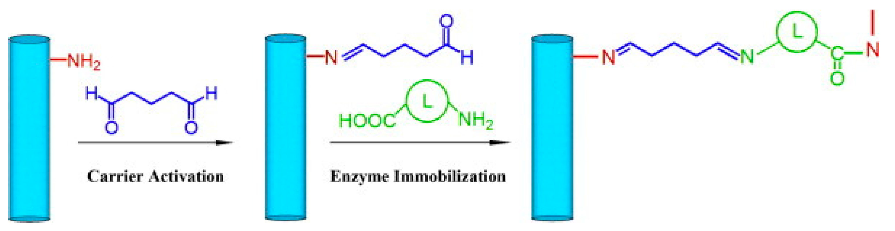
6. Glutaraldehyde
7. Combined Techniques
8. Click Chemistry
9. Future Direction and Conclusions
Author Contributions
Funding
Conflicts of Interest
Abbreviations
| Acronym | Name |
| (P(LA–co–MPC)) | 5-methyl-5-propargyloxycarbonyl-1,3-dioxan-2-one copolymer |
| AChE | acetylcholinesterase |
| AEM | N-(2-aminoethyl)maleimide |
| APTES | 3-aminopropyltriethoxysilane |
| ATRP | atom transfer radical polymerization |
| BMP | bone morphogenetic proteins |
| BPA | bisphenol A |
| BSA | bovine serum albumin |
| CA | covalent conjugation |
| CA-125 | cancer antigen 125 |
| CD44 | cluster of differentiation 44 |
| CDI | 1,1′-carbonyldiimidazole |
| CTC | circulating tumor cell |
| CuAAC | copper-catalyzed azide alkyne cycloaddition |
| Cy5 | streptavidin |
| DBU | 1,8-diazabicyclo[5.4.0]undec-7ene |
| DIBO | 4-dibenzocyclooctynol |
| DNA | deoxyribonucleic acid |
| EDC | 1-ethyl-3-(3-dimethylaminopropyl)carbodiimide |
| EGS | ethylene glycol-bis(sulfosuccinimidyl succinate) |
| ELISA | enzyme-linked immunosorbent assay |
| EPC | enzyme aggregation |
| EpCAM | anti-epithelial cell adhesion molecule |
| ETBFMS | electrospun triple-blend fibrous mats |
| FITC | fluorescein isothiocyanate |
| FMOC | fluorenylmethoxycarbonyl |
| FP | feathered polypeptide |
| GA | glutaraldehyde |
| GRGDS | Gly-Arg-Gly-Asp-Ser |
| hexDA | hexamethylenediamine |
| hIgG | immunoglobulin G |
| HRP | horseradish peroxidase |
| IgG | Immunoglobulin G |
| ITO | indium tin oxide |
| LOD | limit of detection |
| MA | methylacrylate |
| MICs | minimum inhibitory concentrations |
| MNFs | magnetic nanofibers |
| MWCNTS | multiwall carbon nanotubes |
| nHA | nanometric hydroxyapatite |
| NHS | N-Hydroxysuccinimide |
| NHS-Mal | 3-(maleimido) propionic acid N-hydroxysuccinimide ester |
| NT | nitrocellulose |
| OVSEG | poly-(6-O-vinylsebacoyl d-glucose) |
| P3ANA | poly(m-anthranilic acid) |
| PAA | poly(acrylic acid) |
| PAAM | polyacrylamide |
| PAN | polyacrylonitrile |
| PANI | polyaniline |
| PANnFs | polyacrylonitrile nanofibers |
| PBS | phosphate buffered saline |
| pCBMA | poly(carboxybetaine methacrylate) |
| PCL | polycaprolactone |
| PEG | poly(ethylene glycol) |
| PEGDA | PEG diamine |
| PEI | polyethyleneimine |
| PEO | poly(ethylene oxide) |
| PGMA | poly(glycidyl methacrylate) |
| PHB | polyhydroxyalkanoate |
| PIII | plasma-immersion ion implantation |
| PLA | poly lactic acid |
| PLGA | poly(lactic-co-glycolic acid) |
| PLLA | poly(L-lactide) |
| PMMA | poly(methyl methacrylate) |
| PMMA CEA | poly(methyl methacrylate-co-ethyl acrylate) |
| PNIPAM | poly (n-isopropylacrylamide) |
| PS | polystyrene |
| PSBMA | poly(sulfobetaine methacrylate) |
| PSMA | poly(styrene-co-maleic anhydride) |
| PTFE | Polytetrafluoroethylene |
| PVA | polyvinyl alcohol |
| PVP | poly(vinyl pyrrolidone) |
| QDs | quantum dots |
| rGO | reduced graphene oxide |
| rhBMP-2 | recombinant human bone morphogenetic protein-2 |
| rhEGF | recombinant human epidermal growth factor |
| SMCC | succinimidyl 4-(N-maleimidomethyl)cyclohexane-1-carboxylate |
| SPAAC | strain promoted azide-alkyne cycloaddition |
| THF | tetrahydrofuran |
| VD | vitamin-D3 |
| YIGSR | Tyr-Ile-Gly-Ser-Arg |
References
- Portaccio, M.; El-Masry, M.; Rossi Diano, N.; De Maio, A.; Grano, V.; Lepore, M.; Travascio, P.; Bencivenga, U.; Pagliuca, N.; Mita, D.G. An amperometric sensor employing glucose oxidase immobilized on nylon membranes with different pore diameter and grafted with different monomers. J. Mol. Catal. B Enzym. 2002, 18, 49–67. [Google Scholar] [CrossRef]
- Greiner, A.; Wendorff, J.H. Electrospinning: A fascinating method for the preparation of ultrathin fibers. Angew. Chem. Int. Ed. Engl. 2007, 46, 5670–5703. [Google Scholar] [CrossRef] [PubMed]
- Xu, R.; Yuan, J.; Si, Y.; Li, F.; Zhang, B. Estrone removal by horseradish peroxidase immobilized on a nanofibrous support with Fe3O4 nanoparticles. RSC Adv. 2016, 6, 3927–3933. [Google Scholar] [CrossRef]
- Fatarella, E.; Spinelli, D.; Ruzzante, M.; Pogni, R. Nylon 6 film and nanofiber carriers: Preparation and laccase immobilization performance. J. Mol. Catal. B Enzym. 2014, 102, 41–47. [Google Scholar] [CrossRef]
- Talbert, J.N.; Goddard, J.M. Influence of nanoparticle diameter on conjugated enzyme activity. Food Bioprod. Process. 2013, 91, 693–699. [Google Scholar] [CrossRef]
- Illanes, A.; González, J.M.; Gómez, J.M.; Valencia, P.; Wilson, L. Diffusional restrictions in glyoxyl-agarose immobilized penicillin G acylase of different particle size and protein loading. Electron. J. Biotechnol. 2010, 13, 2–3. [Google Scholar] [CrossRef] [Green Version]
- Bailey, J.E.; Cho, Y.K. Immobilization of glucoamylase and glucose oxidase in activated carbon: Effects of particle size and immobilization conditions on enzyme activity and effectiveness. Biotechnol. Bioeng. 1983, 25, 1923–1935. [Google Scholar] [CrossRef]
- Shang, W.; Nuffer, J.H.; Muñiz-Papandrea, V.A.; Colón, W.; Siegel, R.W.; Dordick, J.S. Cytochrome c on Silica Nanoparticles: Influence of Nanoparticle Size on Protein Structure, Stability, and Activity. Small 2009, 5, 470–476. [Google Scholar] [CrossRef]
- Wang, Z.-G.; Wan, L.-S.; Liu, Z.-M.; Huang, X.-J.; Xu, Z.-K. Enzyme immobilization on electrospun polymer nanofibers: An overview. J. Mol. Catal. B Enzym. 2009, 56, 189–195. [Google Scholar] [CrossRef]
- Vasita, R.; Katti, D.S. Nanofibers and their applications in tissue engineering. Int. J. Nanomed. 2006, 1, 15–30. [Google Scholar] [CrossRef]
- Haider, A.; Haider, S.; Kang, I.-K. A comprehensive review summarizing the effect of electrospinning parameters and potential applications of nanofibers in biomedical and biotechnology. Arab. J. Chem. 2018, 11, 1165–1188. [Google Scholar] [CrossRef]
- Fong, H.; Chun, I.; Reneker, D.H. Beaded nanofibers formed during electrospinning. Polymer 1999, 40, 4585–4592. [Google Scholar] [CrossRef]
- Liu, Y.; He, J.-H.; Yu, J.; Zeng, H. Controlling numbers and sizes of beads in electrospun nanofibers. Polym. Int. 2008, 57, 632–636. [Google Scholar] [CrossRef]
- Su, Z.Q.; Li, J.F.; Li, Q.; Ni, T.Y. Chain conformation, crystallization behavior, electrical and mechanical properties of electrospun polymer-carbon nanotube hybrid nanofibers with different orientations. Carbon 2012, 50, 5605–5617. [Google Scholar] [CrossRef]
- Dror, Y.; Salalha, W.; Avrahami, R.; Zussman, E. One-step production of polymeric microtubes by co-electrospinning. Small 2007, 3, 1064. [Google Scholar] [CrossRef] [PubMed]
- Nguyen, T.T.T.; Lee, J.G.; Park, J.S. Fabrication and characterization of coaxial electrospun polyethylene glycol/polyvinylidene fluoride (Core/Sheath) composite non-woven mats. Macromol. Res. 2011, 19, 370–378. [Google Scholar] [CrossRef]
- Reneker, D.H.; Yarin, A.L. Electrospinning jets and polymer nanofibers. Polymer 2008, 49, 2387–2425. [Google Scholar] [CrossRef] [Green Version]
- Wu, Y.; Clark, R.L. Controllable porous polymer particles generated by electrospraying. J. Colloid Interface Sci. 2007, 310, 529–535. [Google Scholar] [CrossRef]
- Canbolat, M.F.; Savas, H.B.; Gultekin, F. Improved catalytic activity by catalase immobilization using γ-cyclodextrin and electrospun PCL nanofibers. J. Appl. Polym. Sci. 2017, 134. [Google Scholar] [CrossRef]
- Wu, L.; Yuan, X.; Sheng, J. Immobilization of cellulase in nanofibrous PVA membranes by electrospinning. J. Membr. Sci. 2005, 250, 167–173. [Google Scholar] [CrossRef]
- Porto, M.D.A.; dos Santos, J.P.; Hackbart, H.; Bruni, G.P.; Fonseca, L.M.; da Rosa Zavareze, E.; Dias, A.R.G. Immobilization of α-amylase in ultrafine polyvinyl alcohol (PVA) fibers via electrospinning and their stability on different substrates. Int. J. Biol. Macromol. 2019, 126, 834–841. [Google Scholar] [CrossRef]
- Wang, Y.; Hsieh, Y.-L. Immobilization of lipase enzyme in polyvinyl alcohol (PVA) nanofibrous membranes. J. Membr. Sci. 2008, 309, 73–81. [Google Scholar] [CrossRef]
- Tanes, M.L.; Xue, J.; Xia, Y. A general strategy for generating gradients of bioactive proteins on electrospun nanofiber mats by masking with bovine serum albumin. J. Mater. Chem. B 2017, 5, 5580–5587. [Google Scholar] [CrossRef]
- Smith, S.; Delaney, M.; Frey, M. Anti-Escherichia coli Functionalized Silver-Doped Carbon Nanofibers for Capture of E. coli in Microfluidic Systems. Polymers 2020, 12, 1117. [Google Scholar] [CrossRef]
- Vonasek, E.; Lu, P.; Hsieh, Y.-L.; Nitin, N. Bacteriophages immobilized on electrospun cellulose microfibers by non-specific adsorption, protein–ligand binding, and electrostatic interactions. Cellulose 2017, 24, 4581–4589. [Google Scholar] [CrossRef]
- Taheran, M.; Naghdi, M.; Brar, S.K.; Knystautas, E.J.; Verma, M.; Surampalli, R.Y. Degradation of chlortetracycline using immobilized laccase on Polyacrylonitrile-biochar composite nanofibrous membrane. Sci. Total Environ. 2017, 605–606, 315–321. [Google Scholar] [CrossRef] [Green Version]
- Zdarta, J.; Jankowska, K.; Wyszowska, M.; Kijeńska-Gawrońska, E.; Zgoła-Grześkowiak, A.; Pinelo, M.; Meyer, A.S.; Moszyński, D.; Jesionowski, T. Robust biodegradation of naproxen and diclofenac by laccase immobilized using electrospun nanofibers with enhanced stability and reusability. Mater. Sci. Eng. C 2019, 103, 109789. [Google Scholar] [CrossRef]
- Zdarta, J.; Staszak, M.; Jankowska, K.; Kaźmierczak, K.; Degórska, O.; Nguyen, L.N.; Kijeńska-Gawrońska, E.; Pinelo, M.; Jesionowski, T. The response surface methodology for optimization of tyrosinase immobilization onto electrospun polycaprolactone–chitosan fibers for use in bisphenol A removal. Int. J. Biol. Macromol. 2020. [Google Scholar] [CrossRef]
- Tański, T.; Jarka, P.; Matysiak, W. Electrospinning Method Used to Create Functional Nanocomposites Films; BoD—Books on Demand: Norderstedt, Germany, 2018; ISBN 978-1-78923-580-7. [Google Scholar]
- Sassolas, A.; Blum, L.J.; Leca-Bouvier, B.D. Immobilization strategies to develop enzymatic biosensors. Biotechnol. Adv. 2012, 30, 489–511. [Google Scholar] [CrossRef]
- Sapountzi, E.; Braiek, M.; Chateaux, J.-F.; Jaffrezic-Renault, N.; Lagarde, F. Recent Advances in Electrospun Nanofiber Interfaces for Biosensing Devices. Sensors 2017, 17, 1887. [Google Scholar] [CrossRef] [PubMed] [Green Version]
- Nguyen, H.H.; Kim, M. An Overview of Techniques in Enzyme Immobilization. Appl. Sci. Converg. Technol. 2017, 26, 157–163. [Google Scholar] [CrossRef]
- Moo-Young, M. Comprehensive Biotechnology: The Principles, Applications, and Regulations of Biotechnology in Industry, Agriculture, and Medicine; Pergamon: Oxford, UK, 1985; ISBN 0-08-026204-X. [Google Scholar]
- Jankowska, K.; Zdarta, J.; Grzywaczyk, A.; Kijeńska-Gawrońska, E.; Biadasz, A.; Jesionowski, T. Electrospun poly(methyl methacrylate)/polyaniline fibres as a support for laccase immobilisation and use in dye decolourisation. Environ. Res. 2020, 184, 109332. [Google Scholar] [CrossRef]
- Taheran, M.; Naghdi, M.; Brar, S.K.; Knystautas, E.J.; Verma, M.; Surampalli, R.Y. Covalent Immobilization of Laccase onto Nanofibrous Membrane for Degradation of Pharmaceutical Residues in Water. ACS Sustain. Chem. Eng. 2017, 5, 10430–10438. [Google Scholar] [CrossRef] [Green Version]
- Das, R.; Mishra, H.; Srivastava, A.; Kayastha, A.M. Covalent immobilization of β-amylase onto functionalized molybdenum sulfide nanosheets, its kinetics and stability studies: A gateway to boost enzyme application. Chem. Eng. J. 2017, 328, 215–227. [Google Scholar] [CrossRef]
- Serafim, B.M.; Leitolis, A.; Crestani, S.; Marcon, B.H.; Foti, L.; Petzhold, C.L.; Radtke, C.; Krieger, M.A.; Saul, C.K. Electrospinning induced surface activation (EISA) of highly porous PMMA microfiber mats for HIV diagnosis. J. Mater. Chem. B 2016, 4, 6004–6011. [Google Scholar] [CrossRef]
- Yang, T.; Hou, P.; Zheng, L.L.; Zhan, L.; Gao, P.F.; Li, Y.F.; Huang, C.Z. Surface-engineered quantum dots/electrospun nanofibers as a networked fluorescence aptasensing platform toward biomarkers. Nanoscale 2017, 9, 17020–17028. [Google Scholar] [CrossRef]
- Lee, S.J.; Tatavarty, R.; Gu, M.B. Electrospun polystyrene–poly(styrene-co-maleic anhydride) nanofiber as a new aptasensor platform. Biosens. Bioelectron. 2012, 38, 302–307. [Google Scholar] [CrossRef]
- Rasouli, R.; Barhoum, A.; Bechelany, M.; Dufresne, A. Nanofibers for Biomedical and Healthcare Applications. Macromol. Biosci. 2019, 19, 1800256. [Google Scholar] [CrossRef]
- Tseng, H.-C.; Lee, A.-W.; Wei, P.-L.; Chang, Y.-J.; Chen, J.-K. Clinical diagnosis of colorectal cancer using electrospun triple-blend fibrous mat-based capture assay of circulating tumor cells. J. Mater. Chem. B 2016, 4, 6565–6580. [Google Scholar] [CrossRef]
- Almyahi, F.; Mahdi, M.; Hassan, J.; Al-Ani, S. Preparation and optical properties of CdS/Epoxy nanocomposites. Int. J. Nanoelectron. Mater. 2012, 5, 57–66. [Google Scholar]
- López-Quintela, A.; Prendes, P.; Pazos-Pellín, M.; Paz, M.; Paz-Abuín, S. Cis/trans reactivity: Epoxy-amine systems. Macromolecules 1998, 31, 4770–4776. [Google Scholar] [CrossRef] [PubMed]
- Riccardi, C.C.; Fraga, F.; Dupuy, J.; Williams, R.J.J. Cure kinetics of diglycidylether of bisphenol A-ethylenediamine revisited using a mechanistic model. J. Appl. Polym. Sci. 2001, 82, 2319–2325. [Google Scholar] [CrossRef]
- Liu, X.; Fang, Y.; Yang, X.; Li, Y.; Wang, C. Electrospun epoxy-based nanofibrous membrane containing biocompatible feather polypeptide for highly stable and active covalent immobilization of lipase. Colloids Surf. B Biointerfaces 2018, 166, 277–285. [Google Scholar] [CrossRef]
- Liu, X.; Fang, Y.; Yang, X.; Li, Y.; Wang, C. Electrospun nanofibrous membranes containing epoxy groups and hydrophilic polyethylene oxide chain for highly active and stable covalent immobilization of lipase. Chem. Eng. J. 2018, 336, 456–464. [Google Scholar] [CrossRef]
- Wong, D.E.; Senecal, K.J.; Goddard, J.M. Immobilization of chymotrypsin on hierarchical nylon 6,6 nanofiber improves enzyme performance. Colloids Surf. B Biointerfaces 2017, 154, 270–278. [Google Scholar] [CrossRef] [PubMed] [Green Version]
- Oktay, B.; Demir, S.; Kayaman-Apohan, N. Immobilization of α-amylase onto poly(glycidyl methacrylate) grafted electrospun fibers by ATRP. Mater. Sci. Eng. C 2015, 50, 386–393. [Google Scholar] [CrossRef]
- Türünç, O.; Kahraman, M.V.; Akdemir, Z.S.; Kayaman-Apohan, N.; Güngör, A. Immobilization of α-amylase onto cyclic carbonate bearing hybrid material. Food Chem. 2009, 112, 992–997. [Google Scholar] [CrossRef]
- Dong, J.; Ghiladi, R.A.; Wang, Q.; Cai, Y.; Wei, Q. Protoporphyrin-IX conjugated cellulose nanofibers that exhibit high antibacterial photodynamic inactivation efficacy. Nanotechnology 2018, 29, 265601. [Google Scholar] [CrossRef]
- Li, Y.; Quan, J.; Branford-White, C.; Williams, G.R.; Wu, J.-X.; Zhu, L.-M. Electrospun polyacrylonitrile-glycopolymer nanofibrous membranes for enzyme immobilization. J. Mol. Catal. B Enzym. 2012, 76, 15–22. [Google Scholar] [CrossRef]
- Huang, X.-J.; Yu, A.-G.; Xu, Z.-K. Covalent immobilization of lipase from Candida rugosa onto poly(acrylonitrile-co-2-hydroxyethyl methacrylate) electrospun fibrous membranes for potential bioreactor application. Bioresour. Technol. 2008, 99, 5459–5465. [Google Scholar] [CrossRef]
- Hermanson, G.T. Bioconjugate Techniques, 3rd ed.; Academic Press: London, UK, 2013. [Google Scholar]
- Stoilova, O.; Ignatova, M.; Manolova, N.; Godjevargova, T.; Mita, D.G.; Rashkov, I. Functionalized electrospun mats from styrene–maleic anhydride copolymers for immobilization of acetylcholinesterase. Eur. Polym. J. 2010, 46, 1966–1974. [Google Scholar] [CrossRef]
- Nair, S.; Kim, J.; Crawford, B.; Kim, S.H. Improving Biocatalytic Activity of Enzyme-Loaded Nanofibers by Dispersing Entangled Nanofiber Structure. Biomacromolecules 2007, 8, 1266–1270. [Google Scholar] [CrossRef] [PubMed]
- Kim, B.C.; Nair, S.; Kim, J.; Kwak, J.H.; Grate, J.W.; Kim, S.H.; Gu, M.B. Preparation of biocatalytic nanofibres with high activity and stability via enzyme aggregate coating on polymer nanofibres. Nanotechnology 2005, 16, S382–S388. [Google Scholar] [CrossRef]
- Jun, S.-H.; Yang, J.; Jeon, H.; Kim, H.S.; Pack, S.P.; Jin, E.; Kim, J. Stabilized and Immobilized Carbonic Anhydrase on Electrospun Nanofibers for Enzymatic CO2 Conversion and Utilization in Expedited Microalgal Growth. Environ. Sci. Technol. 2020, 54, 1223–1231. [Google Scholar] [CrossRef]
- Yoon, J.; Yoon, H.-S.; Shin, Y.; Kim, S.; Ju, Y.; Kim, J.; Chung, S. Ethanol-dispersed and antibody-conjugated polymer nanofibers for the selective capture and 3-dimensional culture of EpCAM-positive cells. Nanomed. Nanotechnol. Biol. Med. 2017, 13, 1617–1625. [Google Scholar] [CrossRef]
- Nataraj, S.K.; Yang, K.S.; Aminabhavi, T.M. Polyacrylonitrile-based nanofibers—A state-of-the-art review. Prog. Polym. Sci. 2012, 37, 487–513. [Google Scholar] [CrossRef]
- Dehghan, S.F.; Golbabaei, F.; Maddah, B.; Latifi, M.; Pezeshk, H.; Hasanzadeh, M.; Akbar-Khanzadeh, F. Optimization of electrospinning parameters for polyacrylonitrile-MgO nanofibers applied in air filtration. J. Air Waste Manag. Assoc. 2016, 66, 912–921. [Google Scholar] [CrossRef] [Green Version]
- Li, S.-F.; Chen, J.-P.; Wu, W.-T. Electrospun polyacrylonitrile nanofibrous membranes for lipase immobilization. J. Mol. Catal. B Enzym. 2007, 47, 117–124. [Google Scholar] [CrossRef]
- Pfaff, D.; Nemecek, G.; Podlech, J. A Lewis acid-promoted Pinner reaction. Beilstein J. Org. Chem. 2013, 9, 1572–1577. [Google Scholar] [CrossRef]
- Handa, T.; Hirose, A.; Yoshida, S.; Tsuchiya, H. The effect of methylacrylate on the activity of glucomylase immobilized on granular polyacrylonitrile. Biotechnol. Bioeng. 1982, 24, 1639–1652. [Google Scholar] [CrossRef]
- Handa, T.; Hirose, A.; Akino, T.; Watanabe, K.; Tsuchiya, H. Preparation of immobilized α-amylase covalently attached to granular polyacrylonitrile. Biotechnol. Bioeng. 1983, 25, 2957–2967. [Google Scholar] [CrossRef]
- Li, S.-F.; Wu, W.-T. Lipase-immobilized electrospun PAN nanofibrous membranes for soybean oil hydrolysis. Biochem. Eng. J. 2009, 45, 48–53. [Google Scholar] [CrossRef]
- Li, S.-F.; Fan, Y.-H.; Hu, J.-F.; Huang, Y.-S.; Wu, W.-T. Immobilization of Pseudomonas cepacia lipase onto the electrospun PAN nanofibrous membranes for transesterification reaction. J. Mol. Catal. B Enzym. 2011, 73, 98–103. [Google Scholar] [CrossRef]
- Xu, R.; Chi, C.; Li, F.; Zhang, B. Laccase–Polyacrylonitrile Nanofibrous Membrane: Highly Immobilized, Stable, Reusable, and Efficacious for 2,4,6-Trichlorophenol Removal. ACS Appl. Mater. Interfaces 2013, 5, 12554–12560. [Google Scholar] [CrossRef]
- Hung, T.-C.; Fu, C.-C.; Su, C.-H.; Chen, J.-Y.; Wu, W.-T.; Lin, Y.-S. Immobilization of cellulase onto electrospun polyacrylonitrile (PAN) nanofibrous membranes and its application to the reducing sugar production from microalgae. Enzym. Microb. Technol. 2011, 49, 30–37. [Google Scholar] [CrossRef]
- Jain, S.; Chattopadhyay, S.; Jackeray, R.; Singh, H. Surface modification of polyacrylonitrile fiber for immobilization of antibodies and detection of analyte. Anal. Chim. Acta 2009, 654, 103–110. [Google Scholar] [CrossRef]
- Kim, U.J.; Kuga, S.; Wada, M.; Okano, T.; Kondo, T. Periodate oxidation of crystalline cellulose. Biomacromolecules 2000, 1, 488–492. [Google Scholar] [CrossRef] [PubMed]
- Ma, Z.; Ramakrishna, S. Electrospun regenerated cellulose nanofiber affinity membrane functionalized with protein A/G for IgG purification. J. Membr. Sci. 2008, 319, 23–28. [Google Scholar] [CrossRef]
- Huang, X.-J.; Chen, P.-C.; Huang, F.; Ou, Y.; Chen, M.-R.; Xu, Z.-K. Immobilization of Candida rugosa lipase on electrospun cellulose nanofiber membrane. J. Mol. Catal. B Enzym. 2011, 70, 95–100. [Google Scholar] [CrossRef]
- Ghasemi-Mobarakeh, L.; Prabhakaran, M.P.; Morshed, M.; Nasr-Esfahani, M.H.; Ramakrishna, S. Bio-functionalized PCL nanofibrous scaffolds for nerve tissue engineering. Mater. Sci. Eng. C 2010, 30, 1129–1136. [Google Scholar] [CrossRef]
- Choi, J.S.; Leong, K.W.; Yoo, H.S. In vivo wound healing of diabetic ulcers using electrospun nanofibers immobilized with human epidermal growth factor (EGF). Biomaterials 2008, 29, 587–596. [Google Scholar] [CrossRef] [PubMed]
- Kurzer, F.; Douraghi-Zadeh, K. Advances in the Chemistry of Carbodiimides. Chem. Rev. 1967, 67, 107–152. [Google Scholar] [CrossRef]
- Sheehan, J.C.; Ledis, S.L. Total synthesis of a monocyclic peptide lactone antibiotic, etamycin. J. Am. Chem. Soc. 1973, 95, 875–879. [Google Scholar] [CrossRef] [PubMed]
- Chen, F.M.F.; Benoiton, N.L. A General Method for Formylating Sensitive Amino Acid Esters. Synthesis 1979, 1979, 709–710. [Google Scholar] [CrossRef]
- Bragg, P.D.; Hou, C. Subunit composition, function, and spatial arrangement in the Ca2+- and Mg2+-activated adenosine triphosphatases of Escherichia coli and Salmonella typhimurium. Arch. Biochem. Biophys. 1975, 167, 311–321. [Google Scholar] [CrossRef]
- Leiro, V.; Parreira, P.; Freitas, S.C.; Martins, M.C.L.; Pêgo, A.P. Chapter 2—Conjugation Chemistry Principles and Surface Functionalization of Nanomaterials. In Biomedical Applications of Functionalized Nanomaterials; Sarmento, B., das Neves, J., Eds.; Micro and Nano Technologies; Elsevier: Amsterdam, The Netherlands, 2018; pp. 35–66. ISBN 978-0-323-50878-0. [Google Scholar]
- Bu, J.; Peng, Z.; Zhao, F.; McLuckey, S.A. Enhanced Reactivity in Nucleophilic Acyl Substitution Ion/Ion Reactions Using Triazole-Ester Reagents. J. Am. Soc. Mass Spectrom. 2017, 28, 1254–1261. [Google Scholar] [CrossRef] [PubMed]
- Campiglio, C.E.; Contessi Negrini, N.; Farè, S.; Draghi, L. Cross-Linking Strategies for Electrospun Gelatin Scaffolds. Materials 2019, 12, 2476. [Google Scholar] [CrossRef] [Green Version]
- Zhao, Y.; Xiong, J.; Shi, X.; Ko, F. Capturing cancer cells using hyaluronic acid-immobilized electrospun random or aligned PLA nanofibers. Colloids Surf. A Physicochem. Eng. Asp. 2019, 583, 123978. [Google Scholar] [CrossRef]
- Zdarta, J.; Jankowska, K.; Bachosz, K.; Kijeńska-Gawrońska, E.; Zgoła-Grześkowiak, A.; Kaczorek, E.; Jesionowski, T. A promising laccase immobilization using electrospun materials for biocatalytic degradation of tetracycline: Effect of process conditions and catalytic pathways. Catal. Today 2019, 348, 127–136. [Google Scholar] [CrossRef]
- Guler, Z.; Silva, J.C.; Sarac, A.S. RGD functionalized poly(ε-caprolactone)/poly(m-anthranilic acid) electrospun nanofibers as high-performing scaffolds for bone tissue engineering RGD functionalized PCL/P3ANA nanofibers. Int. J. Polym. Mater. Polym. Biomater. 2017, 66, 139–148. [Google Scholar] [CrossRef]
- Guler Gokce, Z.; Akalın, P.; Kok, F.N.; Sarac, A.S. Impedimetric DNA biosensor based on polyurethane/poly(m-anthranilic acid) nanofibers. Sens. Actuators B Chem. 2018, 254, 719–726. [Google Scholar] [CrossRef]
- Liu, Y.; Vincent Edwards, J.; Prevost, N.; Huang, Y.; Chen, J.Y. Physico- and bio-activities of nanoscale regenerated cellulose nonwoven immobilized with lysozyme. Mater. Sci. Eng. C 2018, 91, 389–394. [Google Scholar] [CrossRef] [PubMed]
- Matlock-Colangelo, L.; Coon, B.; Pitner, C.L.; Frey, M.W.; Baeumner, A.J. Functionalized electrospun poly(vinyl alcohol) nanofibers for on-chip concentration of E. coli cells. Anal. Bioanal. Chem. 2016, 408, 1327–1334. [Google Scholar] [CrossRef] [PubMed]
- Wan, L.-S.; Ke, B.-B.; Xu, Z.-K. Electrospun nanofibrous membranes filled with carbon nanotubes for redox enzyme immobilization. Enzym. Microb. Technol. 2008, 42, 332–339. [Google Scholar] [CrossRef]
- Wang, Z.-G.; Ke, B.-B.; Xu, Z.-K. Covalent immobilization of redox enzyme on electrospun nonwoven poly(acrylonitrile-co-acrylic acid) nanofiber mesh filled with carbon nanotubes: A comprehensive study. Biotechnol. Bioeng. 2007, 97, 708–720. [Google Scholar] [CrossRef]
- Guex, A.G.; Hegemann, D.; Giraud, M.N.; Tevaearai, H.T.; Popa, A.M.; Rossi, R.M.; Fortunato, G. Covalent immobilisation of VEGF on plasma-coated electrospun scaffolds for tissue engineering applications. Colloids Surf. B Biointerfaces 2014, 123, 724–733. [Google Scholar] [CrossRef]
- Ma, Z.; He, W.; Yong, T.; Ramakrishna, S. Grafting of Gelatin on Electrospun Poly(caprolactone) Nanofibers to Improve Endothelial Cell Spreading and Proliferation and to Control Cell Orientation. Tissue Eng. 2005, 11, 1149–1158. [Google Scholar] [CrossRef]
- Hosseini, S.; Azari, P.; Farahmand, E.; Gan, S.N.; Rothan, H.A.; Yusof, R.; Koole, L.H.; Djordjevic, I.; Ibrahim, F. Polymethacrylate coated electrospun PHB fibers: An exquisite outlook for fabrication of paper-based biosensors. Biosens. Bioelectron. 2015, 69, 257–264. [Google Scholar] [CrossRef]
- Ma, Z.; Kotaki, M.; Yong, T.; He, W.; Ramakrishna, S. Surface engineering of electrospun polyethylene terephthalate (PET) nanofibers towards development of a new material for blood vessel engineering. Biomaterials 2005, 26, 2527–2536. [Google Scholar] [CrossRef]
- Deng, S.; Bai, R.; Chen, J.P. Behaviors and mechanisms of copper adsorption on hydrolyzed polyacrylonitrile fibers. J. Colloid Interface Sci. 2003, 260, 265–272. [Google Scholar] [CrossRef]
- Ye, P.; Xu, Z.-K.; Wu, J.; Innocent, C.; Seta, P. Nanofibrous Membranes Containing Reactive Groups: Electrospinning from Poly(acrylonitrile-co-maleic acid) for Lipase Immobilization. Macromolecules 2006, 39, 1041–1045. [Google Scholar] [CrossRef]
- Chauhan, D.; Gupta, P.K.; Solanki, P.R. Electrochemical immunosensor based on magnetite nanoparticles incorporated electrospun polyacrylonitrile nanofibers for Vitamin-D3 detection. Mater. Sci. Eng. C 2018, 93, 145–156. [Google Scholar] [CrossRef]
- Sangsanoh, P.; Ekapakul, N.; Israsena, N.; Suwantong, O.; Supaphol, P. Enhancement of biocompatibility on aligned electrospun poly(3-hydroxybutyrate) scaffold immobilized with laminin towards murine neuroblastoma Neuro2a cell line and rat brain-derived neural stem cells (mNSCs). Polym. Adv. Technol. 2018, 29, 2050–2063. [Google Scholar] [CrossRef]
- Masaeli, E.; Wieringa, P.A.; Morshed, M.; Nasr-Esfahani, M.H.; Sadri, S.; van Blitterswijk, C.A.; Moroni, L. Peptide functionalized polyhydroxyalkanoate nanofibrous scaffolds enhance Schwann cells activity. Nanomed. Nanotechnol. Biol. Med. 2014, 10, 1559–1569. [Google Scholar] [CrossRef] [PubMed] [Green Version]
- Mahmoudifard, M.; Soudi, S.; Soleimani, M.; Hosseinzadeh, S.; Esmaeili, E.; Vossoughi, M. Efficient protein immobilization on polyethersolfone electrospun nanofibrous membrane via covalent binding for biosensing applications. Mater. Sci. Eng. C 2016, 58, 586–594. [Google Scholar] [CrossRef]
- Khademi, F.; Ai, J.; Soleimani, M.; Verdi, J.; Tavangar, S.M.; Sadroddiny, E.; Massumi, M.; Hashemi, S.M. Improved human endometrial stem cells differentiation into functional hepatocyte-like cells on a glycosaminoglycan/collagen-grafted polyethersulfone nanofibrous scaffold. J. Biomed. Mater. Res. Part B Appl. Biomater. 2017, 105, 2516–2529. [Google Scholar] [CrossRef]
- Heidari-Keshel, S.; Ahmadian, M.; Biazar, E.; Gazmeh, A.; Rabiei, M.; Adibi, M.; Shabani, M. Surface modification of Poly Hydroxybutyrate (PHB) nanofibrous mat by collagen protein and its cellular study. Mater. Technol. 2016, 31, 799–805. [Google Scholar] [CrossRef]
- Rivero, G.; Aldana, A.A.; Frontini Lopez, Y.R.; Liverani, L.; Boccacini, A.R.; Bustos, D.M.; Abraham, G.A. 14-3-3ε protein-immobilized PCL-HA electrospun scaffolds with enhanced osteogenicity. J. Mater. Sci. Mater. Med. 2019, 30, 99. [Google Scholar] [CrossRef]
- Sun, N.; Liu, M.; Wang, J.; Wang, Z.; Li, X.; Jiang, B.; Pei, R. Chitosan Nanofibers for Specific Capture and Nondestructive Release of CTCs Assisted by pCBMA Brushes. Small 2016, 12, 5090–5097. [Google Scholar] [CrossRef]
- Wang, Z.; Sun, N.; Liu, M.; Cao, Y.; Wang, K.; Wang, J.; Pei, R. Multifunctional Nanofibers for Specific Purification and Release of CTCs. ACS Sens. 2017, 2, 547–552. [Google Scholar] [CrossRef]
- Wang, Y.; Hsieh, Y.-L. Enzyme immobilization to ultra-fine cellulose fibers via amphiphilic polyethylene glycol spacers. J. Polym. Sci. Part A Polym. Chem. 2004, 42, 4289–4299. [Google Scholar] [CrossRef]
- Kim, T.G.; Park, T.G. Surface Functionalized Electrospun Biodegradable Nanofibers for Immobilization of Bioactive Molecules. Biotechnol. Prog. 2006, 22, 1108–1113. [Google Scholar] [CrossRef]
- Northrop, B.H.; Frayne, S.H.; Choudhary, U. Thiol–maleimide “click” chemistry: Evaluating the influence of solvent, initiator, and thiol on the reaction mechanism, kinetics, and selectivity. Polym. Chem. 2015, 6, 3415–3430. [Google Scholar] [CrossRef]
- Xiao, Y.; Lin, L.; Shen, M.; Shi, X. Design of DNA Aptamer-Functionalized Magnetic Short Nanofibers for Efficient Capture and Release of Circulating Tumor Cells. Bioconjugate Chem. 2020, 31, 130–138. [Google Scholar] [CrossRef] [PubMed]
- Park, Y.J.; Kim, K.H.; Lee, J.Y.; Ku, Y.; Lee, S.J.; Min, B.M.; Chung, C.P. Immobilization of bone morphogenetic protein-2 on a nanofibrous chitosan membrane for enhanced guided bone regeneration. Biotechnol. Appl. Biochem. 2006, 43, 17. [Google Scholar] [CrossRef]
- Paul, R.; Anderson, G.W. N,N′-Carbonyldiimidazole, a New Peptide Forming Reagent1. J. Am. Chem. Soc. 1960, 82, 4596–4600. [Google Scholar] [CrossRef]
- Vaidyanathan, R.; Kalthod, V.G.; Ngo, D.P.; Manley, J.M.; Lapekas, S.P. Amidations Using N,N′-Carbonyldiimidazole: Remarkable Rate Enhancement by Carbon Dioxide. J. Org. Chem. 2004, 69, 2565–2568. [Google Scholar] [CrossRef]
- Hearn, M.T.W. [7] 1,1′-Carbonyldiimidazole-mediated immobilization of enzymes and affinity ligands. In Methods in Enzymology; Immobilized Enzymes and Cells Part B; Academic Press: Cambridge, MA, USA, 1987; Volume 135, pp. 102–117. [Google Scholar]
- Xu, R.; Si, Y.; Li, F.; Zhang, B. Enzymatic removal of paracetamol from aqueous phase: Horseradish peroxidase immobilized on nanofibrous membranes. Environ. Sci. Pollut. Res. 2015, 22, 3838–3846. [Google Scholar] [CrossRef]
- Baştürk, E.; Demir, S.; Danış, Ö.; Kahraman, M.V. Covalent immobilization of α-amylase onto thermally crosslinked electrospun PVA/PAA nanofibrous hybrid membranes. J. Appl. Polym. Sci. 2013, 127, 349–355. [Google Scholar] [CrossRef]
- Oktay, B.; Kayaman-Apohan, N.; Erdem-Kuruca, S.; Süleymanoğlu, M. Fabrication of collagen immobilized electrospun poly (vinyl alcohol) scaffolds. Polym. Adv. Technol. 2015, 26, 978–987. [Google Scholar] [CrossRef]
- Xu, R.; Tang, R.; Liu, S.; Li, F.; Zhang, B. An environmentally-friendly enzyme-based nanofibrous membrane for 3,3′,5,5′-tetrabromobisphenol removal. RSC Adv. 2015, 5, 64091–64097. [Google Scholar] [CrossRef]
- Çakıroğlu, B.; Çiğil, A.B.; Ogan, A.; Kahraman, M.V.; Demir, S. Covalent immobilization of acetylcholinesterase on a novel polyacrylic acid-based nanofiber membrane. Eng. Life Sci. 2018, 18, 254–262. [Google Scholar] [CrossRef] [PubMed] [Green Version]
- Monier, M.; Ayad, D.M.; Wei, Y.; Sarhan, A.A. Immobilization of horseradish peroxidase on modified chitosan beads. Int. J. Biol. Macromol. 2010, 46, 324–330. [Google Scholar] [CrossRef] [PubMed]
- Pramparo, L.; Stüber, F.; Font, J.; Fortuny, A.; Fabregat, A.; Bengoa, C. Immobilisation of horseradish peroxidase on Eupergit®C for the enzymatic elimination of phenol. J. Hazard. Mater. 2010, 177, 990–1000. [Google Scholar] [CrossRef] [PubMed]
- Hayat, M.A. Principles and Techniques of Scanning Electron Microscopy: Biological Applications; Van Nostrand Reinhold Co.: New York, NY, USA, 1974; ISBN 0422256779. [Google Scholar]
- Sabatini, D.D.; Bensch, K.; Barrnett, R.J. Cytochemistry and Electron Microscopy The Preservation of Cellular Ultrastructure and Enzymatic Activity by Aldehyde Fixation. J. Cell Biol. 1963, 17, 19–58. [Google Scholar] [CrossRef] [PubMed]
- Fein, M.L.; Filachione, E.M. Tanning with Glutaraldehyde. U.S. Patent US2941859A, 21 June 1960. [Google Scholar]
- Richards, F.M.; Knowles, J.R. Glutaraldehyde as a protein cross-linking reagent. J. Mol. Biol. 1968, 37, 231–233. [Google Scholar] [CrossRef]
- Monsan, P. Influence of the conditions of trypsin immobilization onto Spherosil on coupling efficiency. Eur. J. Appl. Microbiol. Biotechnol. 1978, 5, 1–11. [Google Scholar] [CrossRef]
- Migneault, I.; Dartiguenave, C.; Bertrand, M.J.; Waldron, K.C. Glutaraldehyde: Behavior in aqueous solution, reaction with proteins, and application to enzyme crosslinking. BioTechniques 2004, 37, 790–802. [Google Scholar] [CrossRef]
- Xiao, Z.; Xie, Y.; Militz, H.; Mai, C. Effect of glutaraldehyde on water related properties of solid wood. Holzforschung 2010, 64, 483–488. [Google Scholar] [CrossRef]
- Figueiredo, K.C.S.; Alves, T.L.M.; Borges, C.P. Poly(vinyl alcohol) films crosslinked by glutaraldehyde under mild conditions. J. Appl. Polym. Sci. 2009, 111, 3074–3080. [Google Scholar] [CrossRef]
- Rudra, R.; Kumar, V.; Kundu, P.P. Acid catalysed cross-linking of poly vinyl alcohol (PVA) by glutaraldehyde: Effect of crosslink density on the characteristics of PVA membranes used in single chambered microbial fuel cells. RSC Adv. 2015, 5, 83436–83447. [Google Scholar] [CrossRef]
- El-Moghazy, A.Y.; Soliman, E.A.; Ibrahim, H.Z.; Marty, J.-L.; Istamboulie, G.; Noguer, T. Biosensor based on electrospun blended chitosan-poly (vinyl alcohol) nanofibrous enzymatically sensitized membranes for pirimiphos-methyl detection in olive oil. Talanta 2016, 155, 258–264. [Google Scholar] [CrossRef] [PubMed]
- Park, J.-M.; Kim, M.; Park, H.-S.; Jang, A.; Min, J.; Kim, Y.-H. Immobilization of lysozyme-CLEA onto electrospun chitosan nanofiber for effective antibacterial applications. Int. J. Biol. Macromol. 2013, 54, 37–43. [Google Scholar] [CrossRef]
- Lee, K.H.; Ki, C.S.; Baek, D.H.; Kang, G.D.; Ihm, D.-W.; Park, Y.H. Application of electrospun silk fibroin nanofibers as an immobilization support of enzyme. Fibers Polym. 2005, 6, 181–185. [Google Scholar] [CrossRef]
- Jhuang, J.-R.; Lin, S.-B.; Chen, L.-C.; Lou, S.-N.; Chen, S.-H.; Chen, H.-H. Development of immobilized laccase-based time temperature indicator by electrospinning zein fiber. Food Packag. Shelf Life 2020, 23, 100436. [Google Scholar] [CrossRef]
- Palvannan, T.; Saravanakumar, T.; Unnithan, A.R.; Chung, N.-J.; Kim, D.-H.; Park, S.-M. Efficient transformation of phenyl urea herbicide chloroxuron by laccase immobilized on zein polyurethane nanofiber. J. Mol. Catal. B Enzym. 2014, 99, 156–162. [Google Scholar] [CrossRef]
- Huang, X.-J.; Ge, D.; Xu, Z.-K. Preparation and characterization of stable chitosan nanofibrous membrane for lipase immobilization. Eur. Polym. J. 2007, 43, 3710–3718. [Google Scholar] [CrossRef]
- Chen, H.-Y.; Cheng, K.-C.; Hsu, R.-J.; Hsieh, C.-W.; Wang, H.-T.; Ting, Y. Enzymatic degradation of ginkgolic acid by laccase immobilized on novel electrospun nanofiber mat. J. Sci. Food Agric. 2020, 100, 2705–2712. [Google Scholar] [CrossRef]
- Maryšková, M.; Ardao, I.; García-González, C.A.; Martinová, L.; Rotková, J.; Ševců, A. Polyamide 6/chitosan nanofibers as support for the immobilization of Trametes versicolor laccase for the elimination of endocrine disrupting chemicals. Enzym. Microb. Technol. 2016, 89, 31–38. [Google Scholar] [CrossRef]
- Xu, R.; Zhou, Q.; Li, F.; Zhang, B. Laccase immobilization on chitosan/poly(vinyl alcohol) composite nanofibrous membranes for 2,4-dichlorophenol removal. Chem. Eng. J. 2013, 222, 321–329. [Google Scholar] [CrossRef]
- Bösiger, P.; Tegl, G.; Richard, I.M.T.; Le Gat, L.; Huber, L.; Stagl, V.; Mensah, A.; Guebitz, G.M.; Rossi, R.M.; Fortunato, G. Enzyme functionalized electrospun chitosan mats for antimicrobial treatment. Carbohydr. Polym. 2018, 181, 551–559. [Google Scholar] [CrossRef] [PubMed]
- Desai, K.; Kit, K.; Li, J.; Zivanovic, S. Morphological and Surface Properties of Electrospun Chitosan Nanofibers. Biomacromolecules 2008, 9, 1000–1006. [Google Scholar] [CrossRef] [PubMed]
- Li, L.; Hsieh, Y.-L. Chitosan bicomponent nanofibers and nanoporous fibers. Carbohydr. Res. 2006, 341, 374–381. [Google Scholar] [CrossRef]
- Duan, B.; Dong, C.; Yuan, X.; Yao, K. Electrospinning of chitosan solutions in acetic acid with poly(ethylene oxide). J. Biomater. Sci. Polym. Ed. 2004, 15, 797–811. [Google Scholar] [CrossRef]
- Pakravan, M.; Heuzey, M.-C.; Ajji, A. A fundamental study of chitosan/PEO electrospinning. Polymer 2011, 52, 4813–4824. [Google Scholar] [CrossRef]
- Jhuang, J.-R.; Lou, S.-N.; Lin, S.-B.; Chen, S.H.; Chen, L.-C.; Chen, H.-H. Immobilizing laccase on electrospun chitosan fiber to prepare time-temperature indicator for food quality monitoring. Innov. Food Sci. Emerg. Technol. 2020, 102370. [Google Scholar] [CrossRef]
- Xu, R.; Tang, R.; Zhou, Q.; Li, F.; Zhang, B. Enhancement of catalytic activity of immobilized laccase for diclofenac biodegradation by carbon nanotubes. Chem. Eng. J. 2015, 262, 88–95. [Google Scholar] [CrossRef]
- Pavinatto, A.; Mercante, L.A.; Facure, M.H.M.; Pena, R.B.; Sanfelice, R.C.; Mattoso, L.H.C.; Correa, D.S. Ultrasensitive biosensor based on polyvinylpyrrolidone/chitosan/reduced graphene oxide electrospun nanofibers for 17α—Ethinylestradiol electrochemical detection. Appl. Surf. Sci. 2018, 458, 431–437. [Google Scholar] [CrossRef]
- Xu, H.; Cao, Y.; Li, X.; Cao, X.; Xu, Y.; Qiao, D.; Cao, Y. An electrospun chitosan-based nanofibrous membrane reactor immobilized by the non-glycosylated rPGA for hydrolysis of pectin-rich biomass. Renew. Energy 2018, 126, 573–582. [Google Scholar] [CrossRef]
- İspirli Doğaç, Y.; Deveci, İ.; Mercimek, B.; Teke, M. A comparative study for lipase immobilization onto alginate based composite electrospun nanofibers with effective and enhanced stability. Int. J. Biol. Macromol. 2017, 96, 302–311. [Google Scholar] [CrossRef]
- Işik, C.; Arabaci, G.; Ispirli Doğaç, Y.; Deveci, İ.; Teke, M. Synthesis and characterization of electrospun PVA/Zn2+ metal composite nanofibers for lipase immobilization with effective thermal, pH stabilities and reusability. Mater. Sci. Eng. C 2019, 99, 1226–1235. [Google Scholar] [CrossRef] [PubMed]
- Sathishkumar, P.; Kamala-Kannan, S.; Cho, M.; Kim, J.S.; Hadibarata, T.; Salim, M.R.; Oh, B.-T. Laccase immobilization on cellulose nanofiber: The catalytic efficiency and recyclic application for simulated dye effluent treatment. J. Mol. Catal. B Enzym. 2014, 100, 111–120. [Google Scholar] [CrossRef]
- Yan, X.-Y.; Jiang, Y.-J.; Zhang, S.-P.; Gao, J.; Zhang, Y.-F. Dual-functional OPH-immobilized polyamide nanofibrous membrane for effective organophosphorus toxic agents protection. Biochem. Eng. J. 2015, 98, 47–55. [Google Scholar] [CrossRef]
- Isgrove, F.H.; Williams, R.J.H.; Niven, G.W.; Andrews, A.T. Enzyme immobilization on nylon–optimization and the steps used to prevent enzyme leakage from the support. Enzym. Microb. Technol. 2001, 28, 225–232. [Google Scholar] [CrossRef]
- Harir, M.; Bellahcene, M.; Baratto, M.C.; Pollini, S.; Rossolini, G.M.; Trabalzini, L.; Fatarella, E.; Pogni, R. Isolation and characterization of a novel tyrosinase produced by Sahara soil actinobacteria and immobilization on nylon nanofiber membranes. J. Biotechnol. 2018, 265, 54–64. [Google Scholar] [CrossRef] [PubMed]
- Xu, R.; Cui, J.; Tang, R.; Li, F.; Zhang, B. Removal of 2,4,6-trichlorophenol by laccase immobilized on nano-copper incorporated electrospun fibrous membrane-high efficiency, stability and reusability. Chem. Eng. J. 2017, 326, 647–655. [Google Scholar] [CrossRef]
- Farzin, L.; Sadjadi, S.; Shamsipur, M.; Sheibani, S.; Mousazadeh, M. hasan Employing AgNPs doped amidoxime-modified polyacrylonitrile (PAN-oxime) nanofibers for target induced strand displacement-based electrochemical aptasensing of CA125 in ovarian cancer patients. Mater. Sci. Eng. C 2019, 97, 679–687. [Google Scholar] [CrossRef]
- Saeed, K.; Haider, S.; Oh, T.-J.; Park, S.-Y. Preparation of amidoxime-modified polyacrylonitrile (PAN-oxime) nanofibers and their applications to metal ions adsorption. J. Membr. Sci. 2008, 322, 400–405. [Google Scholar] [CrossRef]
- Mahmoudifard, M.; Soleimani, M.; Vossoughi, M. Ammonia plasma-treated electrospun polyacrylonitryle nanofibrous membrane: The robust substrate for protein immobilization through glutaraldhyde coupling chemistry for biosensor application. Sci. Rep. 2017, 7, 9441. [Google Scholar] [CrossRef] [Green Version]
- Kumar, P.; Gupta, A.; Dhakate, S.R.; Mathur, R.B.; Nagar, S.; Gupta, V.K. Covalent immobilization of xylanase produced from Bacillus pumilus SV-85S on electrospun polymethyl methacrylate nanofiber membrane. Biotechnol. Appl. Biochem. 2013, 60, 162–169. [Google Scholar] [CrossRef]
- Temoçin, Z.; İnal, M.; Gökgöz, M.; Yiğitoğlu, M. Immobilization of horseradish peroxidase on electrospun poly(vinyl alcohol)–polyacrylamide blend nanofiber membrane and its use in the conversion of phenol. Polym. Bull. 2018, 75, 1843–1865. [Google Scholar] [CrossRef]
- El-Aassar, M.R.; Al-Deyab, S.S.; Kenawy, E.-R. Covalent immobilization of β-galactosidase onto electrospun nanofibers of poly (AN-co-MMA) copolymer. J. Appl. Polym. Sci. 2013, 127, 1873–1884. [Google Scholar] [CrossRef]
- Li, J.; Chen, X.; Xu, D.; Pan, K. Immobilization of horseradish peroxidase on electrospun magnetic nanofibers for phenol removal. Ecotoxicol. Environ. Saf. 2019, 170, 716–721. [Google Scholar] [CrossRef]
- Xu, R.; Chi, C.; Li, F.; Zhang, B. Immobilization of horseradish peroxidase on electrospun microfibrous membranes for biodegradation and adsorption of bisphenol A. Bioresour. Technol. 2013, 149, 111–116. [Google Scholar] [CrossRef]
- Koloti, L.E.; Gule, N.P.; Arotiba, O.A.; Malinga, S.P. Laccase-immobilized dendritic nanofibrous membranes as a novel approach towards the removal of bisphenol A. Environ. Technol. 2018, 39, 392–404. [Google Scholar] [CrossRef] [PubMed]
- El-Aassar, M.R.; Shibraen, M.H.M.A.; Abdel-Fattah, Y.R.; Elzain, A.A. Functionalization of Electrospun Poly(Acrylonitrile-co-Styrene/Pyrrole) Copolymer Nanofibers for Using as a High-performance Carrier for Laccase Immobilization. Fibers Polym. 2019, 20, 2268–2279. [Google Scholar] [CrossRef]
- de Melo Brites, M.; Cerón, A.A.; Costa, S.M.; Oliveira, R.C.; Ferraz, H.G.; Catalani, L.H.; Costa, S.A. Bromelain immobilization in cellulose triacetate nanofiber membranes from sugarcane bagasse by electrospinning technique. Enzym. Microb. Technol. 2020, 132, 109384. [Google Scholar] [CrossRef] [PubMed]
- Vahid Ebadi, S.; Fakhrali, A.; Omid Ranaei-Siadat, S.; Akbar Gharehaghaji, A.; Mazinani, S.; Dinari, M.; Harati, J. Immobilization of acetylcholinesterase on electrospun poly(acrylic acid)/multi-walled carbon nanotube nanofibrous membranes. RSC Adv. 2015, 5, 42572–42579. [Google Scholar] [CrossRef]
- Li, C.; Zhou, L.; Wang, C.; Liu, X.; Liao, K. Electrospinning of a PMA-co-PAA/FP biopolymer nanofiber: Enhanced capability for immobilized horseradish peroxidase and its consequence for p-nitrophenol disposal. RSC Adv. 2015, 5, 41994–41998. [Google Scholar] [CrossRef]
- Wang, Z.-L.; Ding, P.; Gao, T.; Chen, C.-C.; Cao, Y.; Sun, N.; Pei, R.-J. Capture of Circulating Tumor Cells by Hydrogel-Nanofiber Substrate. Chin. J. Anal. Chem. 2019, 47, 1162–1169. [Google Scholar] [CrossRef]
- Martrou, G.; Léonetti, M.; Gigmes, D.; Trimaille, T. One-step preparation of surface modified electrospun microfibers as suitable supports for protein immobilization. Polym. Chem. 2017, 8, 1790–1796. [Google Scholar] [CrossRef]
- Dai, T.; Miletić, N.; Loos, K.; Elbahri, M.; Abetz, V. Electrospinning of Poly[acrylonitrile-co-(glycidyl methacrylate)] Nanofibrous Mats for the Immobilization of Candida Antarctica Lipase B. Macromol. Chem. Phys. 2011, 212, 319–327. [Google Scholar] [CrossRef] [Green Version]
- Kolb, H.C.; Finn, M.G.; Sharpless, K.B. Click Chemistry: Diverse Chemical Function from a Few Good Reactions. Angew. Chem. Int. Ed. 2001, 40, 2005–2021. [Google Scholar] [CrossRef]
- Celebioglu, A.; Demirci, S.; Uyar, T. Cyclodextrin-grafted electrospun cellulose acetate nanofibers via Click reaction for removal of phenanthrene. Appl. Surf. Sci. 2014, 305, 581–588. [Google Scholar] [CrossRef]
- Nada, A.A.; Abdellatif, F.H.H.; Ali, E.A.; Abdelazeem, R.A.; Soliman, A.A.S.; Abou-Zeid, N.Y. Cellulose-based click-scaffolds: Synthesis, characterization and biofabrications. Carbohydr. Polym. 2018, 199, 610–618. [Google Scholar] [CrossRef] [PubMed]
- Oster, M.; Schlatter, G.; Gallet, S.; Baati, R.; Pollet, E.; Gaillard, C.; Averous, L.; Fajolles, C.; Hebraud, A. The study of the pseudo-polyrotaxane architecture as a route for mild surface functionalization by click chemistry of poly(-caprolactone)-based electrospun fibers. J. Mater. Chem. B 2017, 5, 2181–2189. [Google Scholar] [CrossRef] [PubMed]
- Qian, Y.-C.; Ren, N.; Huang, X.-J.; Chen, C.; Yu, A.-G.; Xu, Z.-K. Glycosylation of polyphosphazene nanofibrous membrane by click chemistry for protein recognition. Macromol. Chem. Phys. 2013, 214, 1852–1858. [Google Scholar] [CrossRef]
- Nada, A.A.; Abdellatif, F.H.H.; Soliman, A.A.F.; Shen, J.; Hudson, S.M.; Abou-Zeid, N.Y. Fabrication and bioevaluation of a medicated electrospun mat based on azido-cellulose acetate via click chemistry. Cellulose 2019, 26, 9721–9736. [Google Scholar] [CrossRef]
- Lancuški, A.; Bossard, F.; Fort, S. Carbohydrate-decorated PCL fibers for specific protein adhesion. Biomacromolecules 2013, 14, 1877–1884. [Google Scholar] [CrossRef]
- Lancuški, A.; Fort, S.; Bossard, F. Electrospun azido-PCL nanofibers for enhanced surface functionalization by click chemistry. ACS Appl. Mater. Interfaces 2012, 4, 6499–6504. [Google Scholar] [CrossRef]
- Kalaoglu-Altan, O.I.; Sanyal, R.; Sanyal, A. Orthogonally “Clickable” Biodegradable Nanofibers: Tailoring Biomaterials for Specific Protein Immobilization. ACS Omega 2019, 4, 121–129. [Google Scholar] [CrossRef] [PubMed] [Green Version]
- Fu, G.D.; Xu, L.Q.; Yao, F.; Zhang, K.; Wang, X.F.; Zhu, M.F.; Nie, S.Z. Smart Nanofibers from Combined Living Radical Polymerization, “Click Chemistry”, and Electrospinning. ACS Appl. Mater. Interfaces 2009, 1, 239–243. [Google Scholar] [CrossRef] [PubMed]
- Guo, J.-W.; Wu, Y.-H.; Wei, P.-L.; Huang, Y.-J.; Chen, J.-K. Immobilization of antibody conjugated ZnS quantum dots onto poly(2,6-dimethyl-1,4-phenylene oxide) nanofibers with Poly(N-isopropylacrylamide) grafts as reversibly fluorescence immunoassay. Dye Pigment. 2018, 159, 198–208. [Google Scholar] [CrossRef]
- Guo, J.-W.; Lin, Z.-Y.; Chang, C.-J.; Lu, C.-H.; Chen, J.-K. Protein valves prepared by click reaction grafting of poly(N-isopropylacrylamide) to electrospun poly(vinyl chloride) fibrous membranes. Appl. Surf. Sci. 2018, 439, 313–322. [Google Scholar] [CrossRef]
- Lin, F.; Yu, J.; Tang, W.; Zheng, J.; Xie, S.; Becker, M.L. Postelectrospinning “Click” Modification of Degradable Amino Acid-Based Poly(ester urea) Nanofibers. Macromolecules 2013, 46, 9515–9525. [Google Scholar] [CrossRef]
- Kalaoglu-Altan, O.I.; Sanyal, R.; Sanyal, A. “Clickable” polymeric nanofibers through hydrophilic-hydrophobic balance: Fabrication of robust biomolecular immobilization platforms. Biomacromolecules 2015, 16, 1590–1597. [Google Scholar] [CrossRef]
- Kalaoglu-Altan, O.I.; Sanyal, R.; Sanyal, A. Reactive and “clickable” electrospun polymeric nanofibers. Polym. Chem. 2015, 6, 3372–3381. [Google Scholar] [CrossRef]
- Goodge, K.; Frey, M. Biotin-Conjugated Cellulose Nanofibers Prepared via Copper-Catalyzed Alkyne-Azide Cycloaddition (CuAAC) “Click” Chemistry. Nanomaterials 2020, 10, 1172. [Google Scholar] [CrossRef]
- Chang, Z.; Xu, Y.; Zhao, X.; Zhang, Q.; Chen, D. Grafting poly(methyl methacrylate) onto polyimide nanofibers via “click” reaction. ACS Appl. Mater. Interfaces 2009, 1, 2804–2811. [Google Scholar] [CrossRef]
- An, S.; Jeon, B.; Bae, J.H.; Kim, I.S.; Paeng, K.; Kim, M.; Lee, H. Thiol-based chemistry as versatile routes for the effective functionalization of cellulose nanofibers. Carbohydr. Polym. 2019, 226, 115259. [Google Scholar] [CrossRef]
- Zheng, J.; Liu, K.; Reneker, D.H.; Becker, M.L. Post-Assembly Derivatization of Electrospun Nanofibers via Strain-Promoted Azide Alkyne Cycloaddition. J. Am. Chem. Soc. 2012, 134, 17274–17277. [Google Scholar] [CrossRef] [PubMed]
- Dong, Y.; Lu, X.; Wang, P.; Liu, W.; Zhang, S.; Wu, Z.; Chen, H. “Click-chemical” modification of cellulose acetate nanofibers: A versatile platform for biofunctionalization. J. Mater. Chem. B 2018, 6, 4579–4582. [Google Scholar] [CrossRef]
- Lee, H.; An, S.; Kim, S.; Jeon, B.; Paeng, K.; Kim, I.S.; Kim, M. Epoxy-Containing Copolymers: A Versatile Toolbox for Functional Nanofiber Mats with Desired Chemical Functionalities. Adv. Mater. Interfaces 2018, 5, 1800506. [Google Scholar] [CrossRef]
- Shi, Q.; Chen, X.; Lu, T.; Jing, X. The immobilization of proteins on biodegradable polymer fibers via click chemistry. Biomaterials 2008, 29, 1118–1126. [Google Scholar] [CrossRef] [PubMed]
- Shadish, J.A.; DeForest, C.A. Site-Selective Protein Modification: From Functionalized Proteins to Functional Biomaterials. Matter 2020, 2, 50–77. [Google Scholar] [CrossRef]
- Grim, J.C.; Marozas, I.A.; Anseth, K.S. Thiol-ene and photo-cleavage chemistry for controlled presentation of biomolecules in hydrogels. J. Control. Release 2015, 219, 95–106. [Google Scholar] [CrossRef] [PubMed] [Green Version]
- Song, D.W.; Kim, S.H.; Kim, H.H.; Lee, K.H.; Ki, C.S.; Park, Y.H. Multi-biofunction of antimicrobial peptide-immobilized silk fibroin nanofiber membrane: Implications for wound healing. Acta Biomater. 2016, 39, 146–155. [Google Scholar] [CrossRef] [PubMed]
- Monteiro, N.; Ribeiro, D.; Martins, A.; Faria, S.; Fonseca, N.A.; Moreira, J.N.; Reis, R.L.; Neves, N.M. Instructive nanofibrous scaffold comprising runt-related transcription factor 2 gene delivery for bone tissue engineering. ACS Nano 2014, 8, 8082–8094. [Google Scholar] [CrossRef]
- Monteiro, N.; Martins, M.; Martins, A.; Fonseca, N.A.; Moreira, J.N.; Reis, R.L.; Neves, N.M. Antibacterial activity of chitosan nanofiber meshes with liposomes immobilized releasing gentamicin. Acta Biomater. 2015, 18, 196–205. [Google Scholar] [CrossRef]
- Worrell, B.T.; Malik, J.A.; Fokin, V.V. Direct Evidence of a Dinuclear Copper Intermediate in Cu(I)-Catalyzed Azide-Alkyne Cycloadditions. Science 2013, 340, 457–460. [Google Scholar] [CrossRef] [Green Version]
- Bock, V.D.; Hiemstra, H.; Maarseveen, J.H. van CuI-Catalyzed Alkyne–Azide “Click” Cycloadditions from a Mechanistic and Synthetic Perspective. Eur. J. Org. Chem. 2006, 2006, 51–68. [Google Scholar] [CrossRef]
- Li, L.; Zhang, Z. Development and Applications of the Copper-Catalyzed Azide-Alkyne Cycloaddition (CuAAC) as a Bioorthogonal Reaction. Molecules 2016, 21, 1393. [Google Scholar] [CrossRef] [PubMed]
- Orski, S.V.; Sheppard, G.R.; Arumugam, S.; Arnold, R.M.; Popik, V.V.; Locklin, J. Rate Determination of Azide Click Reactions onto Alkyne Polymer Brush Scaffolds: A Comparison of Conventional and Catalyst-Free Cycloadditions for Tunable Surface Modification. Langmuir 2012, 28, 14693–14702. [Google Scholar] [CrossRef] [PubMed]
- Smith Callahan, L.A.; Xie, S.; Barker, I.A.; Zheng, J.; Reneker, D.H.; Dove, A.P.; Becker, M.L. Directed differentiation and neurite extension of mouse embryonic stem cell on aligned poly(lactide) nanofibers functionalized with YIGSR peptide. Biomaterials 2013, 34, 9089–9095. [Google Scholar] [CrossRef]
- Zheng, J.; Kontoveros, D.; Lin, F.; Hua, G.; Reneker, D.H.; Becker, M.L.; Willits, R.K. Enhanced Schwann Cell Attachment and Alignment Using One-Pot “Dual Click” GRGDS and YIGSR Derivatized Nanofibers. Biomacromolecules 2015, 16, 357–363. [Google Scholar] [CrossRef] [Green Version]
- Zheng, J.; Hua, G.; Yu, J.; Lin, F.; Wade, M.B.; Reneker, D.H.; Becker, M.L. Post-electrospinning “triclick” functionalization of degradable polymer nanofibers. ACS Macro Lett. 2015, 4, 207–213. [Google Scholar] [CrossRef]
- Odinolfi, M.T.; Romanato, A.; Bergamaschi, G.; Strada, A.; Sola, L.; Girella, A.; Milanese, C.; Chiari, M.; Gori, A.; Cretich, M. Clickable cellulosic surfaces for peptide-based bioassays. Talanta 2019, 205, 120152. [Google Scholar] [CrossRef]
- Mou, X.-Z.; Chen, X.-Y.; Wang, J.; Zhang, Z.; Yang, Y.; Shou, Z.-X.; Tu, Y.-X.; Du, X.; Wu, C.; Zhao, Y.; et al. Bacteria-Instructed Click Chemistry between Functionalized Gold Nanoparticles for Point-of-Care Microbial Detection. ACS Appl. Mater. Interfaces 2019, 11, 23093–23101. [Google Scholar] [CrossRef]
- Feng, W.; Li, L.; Yang, C.; Welle, A.; Trapp, O.; Levkin, P.A. UV-Induced Tetrazole-Thiol Reaction for Polymer Conjugation and Surface Functionalization. Angew. Chem. Int. Ed. 2015, 54, 8732–8735. [Google Scholar] [CrossRef]
- Alonso, R.; Jiménez-Meneses, P.; García-Rupérez, J.; Bañuls, M.-J.; Maquieira, Á. Thiol–ene click chemistry towards easy microarraying of half-antibodies. Chem. Commun. 2018, 54, 6144–6147. [Google Scholar] [CrossRef] [Green Version]
- Kosobrodova, E.; Mohamed, A.; Su, Y.; Kondyurin, A.; dos Remedios, C.G.; McKenzie, D.R.; Bilek, M.M.M. Cluster of differentiation antibody microarrays on plasma immersion ion implanted polycarbonate. Mater. Sci. Eng. C 2014, 35, 434–440. [Google Scholar] [CrossRef] [PubMed]
- Kosobrodova, E.; Jones, R.T.; Kondyurin, A.; Chrzanowski, W.; Pigram, P.J.; McKenzie, D.R.; Bilek, M.M.M. Orientation and conformation of anti-CD34 antibody immobilised on untreated and plasma treated polycarbonate. Acta Biomater. 2015, 19, 128–137. [Google Scholar] [CrossRef]
- Bax, D.V.; McKenzie, D.R.; Weiss, A.S.; Bilek, M.M.M. The linker-free covalent attachment of collagen to plasma immersion ion implantation treated polytetrafluoroethylene and subsequent cell-binding activity. Biomaterials 2010, 31, 2526–2534. [Google Scholar] [CrossRef] [PubMed]
- Bilek, M.M.M. Biofunctionalization of surfaces by energetic ion implantation: Review of progress on applications in implantable biomedical devices and antibody microarrays. Appl. Surf. Sci. 2014, 310, 3–10. [Google Scholar] [CrossRef] [Green Version]
- Bilek, M.M.M.; Vandrovcová, M.; Shelemin, A.; Kuzminova, A.; Kylián, O.; Biederman, H.; Bačáková, L.; Weiss, A.S. Plasma treatment in air at atmospheric pressure that enables reagent-free covalent immobilization of biomolecules on polytetrafluoroethylene (PTFE). Appl. Surf. Sci. 2020, 518, 146128. [Google Scholar] [CrossRef]


| Direct Immobilization | Direct Immobilization After Surface Modification | EDC/NHS | CDI | GA | Combined | Click | |
|---|---|---|---|---|---|---|---|
| polyacrylonitrile (PAN) | [61,65,66,67,68,69] | [88,94,95,96] | [35,153,154,155,156,160] | ||||
| chitosan | [103,104,109] | [116] | [129,130,143,144,145,146] | [136,167] | [196] | ||
| polycaprolactone (PCL) | [73,74,102] | [173,176,177,195,202,203] | |||||
| regenerated cellulose (RC) | [50] | [71,72] | [105] | [149] | [185,187] | ||
| poly(acrylic acid) (PAA) | [41] | [3,113,114,117] | [165] | [186] | |||
| poly(styrene-co-maleic anhydride) (PSMA) | [39,54,55,56,57,58] | [168] | |||||
| nylon | [4,47,135,150,151,152] | [136] | |||||
| polyethyleneimine (PEI) | [38,108] | [159,161,162,163] | |||||
| poly(glycidyl methacrylate) (PGMA) | [45,46,48] | [169] | [190] | ||||
| poly(vinyl alcohol) (PVA) | [115,116] | [148,158] | |||||
| poly lactic acid (PLA) or poly(L-lactide) (PLLA) | [82] | [178,201] | |||||
| cellulose acetate (CA) | [164] | [171,175] | |||||
| poly(methyl methacrylate) PMMA | [34,83] | ||||||
| poly(m-anthranilic acid) (P3ANA) | [84,85] | ||||||
| polyhydroxyalkanoate (PHB) | [98,101] | ||||||
| polyethersulfone (PES) | [99,100] | ||||||
| silk | [131] | [194] | |||||
| poly-(6-O-vinylsebacoyl d-glucose) (OVSEG) | [51] | ||||||
| poly(acrylonitrile-co-2-hydroxyethyl methacrylate) (PAN-c-HEM) | [52] | ||||||
| polyaniline (PANI) | [34] | ||||||
| poly(methyl vinyl ether-alt-maleic anhydride) (PMVE/MA) | [87] | ||||||
| poly(carboxybetaine methacrylate) (pCBMA) | [103] | ||||||
| poly(lactic-co-glycolic acid)-poly(ethylene glycol)-amine (PLGA-b-PEG-NH2) | [106] | ||||||
| zein | [132,133] | ||||||
| alginate | [147] | ||||||
| polyacrylamide (PAAm) | [158] | ||||||
| feather polypeptide (FP) | [166] | ||||||
| ethyl cellulose (EC) | [172] | ||||||
| poly[di(propargylamine)phosphazene] (PDPAP) | [174] | ||||||
| 4-vinylbenzyl chloride and glycidyl methacrylate (PVBC-b-PGMA) | [179] | ||||||
| poly(2,6-dimethyl-1,4-phenylene oxide) | [180] | ||||||
| poly(vinyl chloride) (PVC) | [181] | ||||||
| poly(ester urea) (PEU) | [182] | ||||||
| furfuryl methacrylate (FuMA) | [183] | ||||||
| poly(γ-benzyl-l-glutamate) (PBLG) | [188] | ||||||
| poly(3-(fluorosulfonyl)propyl methacrylate) (PFPM) | [189] | ||||||
| 5-methyl-5-propargyloxycarbonyl-1,3-dioxan-2-one (MPC) | [191] |
Publisher’s Note: MDPI stays neutral with regard to jurisdictional claims in published maps and institutional affiliations. |
© 2020 by the authors. Licensee MDPI, Basel, Switzerland. This article is an open access article distributed under the terms and conditions of the Creative Commons Attribution (CC BY) license (http://creativecommons.org/licenses/by/4.0/).
Share and Cite
Smith, S.; Goodge, K.; Delaney, M.; Struzyk, A.; Tansey, N.; Frey, M. A Comprehensive Review of the Covalent Immobilization of Biomolecules onto Electrospun Nanofibers. Nanomaterials 2020, 10, 2142. https://doi.org/10.3390/nano10112142
Smith S, Goodge K, Delaney M, Struzyk A, Tansey N, Frey M. A Comprehensive Review of the Covalent Immobilization of Biomolecules onto Electrospun Nanofibers. Nanomaterials. 2020; 10(11):2142. https://doi.org/10.3390/nano10112142
Chicago/Turabian StyleSmith, Soshana, Katarina Goodge, Michael Delaney, Ariel Struzyk, Nicole Tansey, and Margaret Frey. 2020. "A Comprehensive Review of the Covalent Immobilization of Biomolecules onto Electrospun Nanofibers" Nanomaterials 10, no. 11: 2142. https://doi.org/10.3390/nano10112142