Article

Two New Species of Free-Living Marine Nematode of the Genus Anticyathus Cobb, 1920 (Linhomoeidae) from Mangroves Sediment of Shenzhen and Shantou, China †

1
Fisheries College, Jimei University, Xiamen 361021, China
2
Fujian Provincal Key Laboratory of Marine Fishery Resources and Eco-Enviroment, Jimei University, Xiamen 361021, China
*
Author to whom correspondence should be addressed.
urn:lsid:zoobank.org:act:166AFE51-8361-41E2-8927-F70EB5175075 and urn:lsid:zoobank.org:act:6899EB5B-E486-49CE-A4DF-EE5696D6A4FE.
J. Mar. Sci. Eng. 2022, 10(8), 1107; https://doi.org/10.3390/jmse10081107
Submission received: 26 June 2022 / Revised: 30 July 2022 / Accepted: 3 August 2022 / Published: 12 August 2022
(This article belongs to the Section Marine Biology)

Abstract

:
Two new species belonging to the genus Anticyathus were discovered during an ecological investigation of mangrove sediment in China. Anticyathus shenzhensis sp. nov. and Anticyathus communis sp. nov. are both characterized by: annulated cuticles; ten cephalic setae in a circle; buccal cavity lined with six longitudinal reinforcements; amphids in broken circles or cryptic spirals, located at the very anterior of the head; pharynx with anterior cuticularized, gradually enlarging in posterior portion but without bulb; conical tail; arcuate spicules, the distal ends pointed, gradually broadening towards the proximal; gubernaculum has a dorsal–caudal apophysis; female with two opposed, outstretched ovaries. Anticyathus shenzhensis sp. nov. can be distinguished by ten cephalic setae of length about 2–4 µm; amphids diameter about 20–29% of corresponding body diameter; pharynx occupy 0.07–0.10 times of body length; cardia length about 25 µm; tail length 2.5–3.4 times of anal body diameter (abd) in males and 3.4–4.7 abd in females; spicules length 51–59 µm as arc and 0.71–1.04 abd; apophysis length 18–19 µm; female vulva at 52–56% of body length. Anticyathus communis sp. nov. is identified by ten cephalic setae length about 3–5 µm; amphids diameter about 20–32% of corresponding body diameter; cardia 17–27 µm long; tail length 3.6–4.5 abd in male, 5.3–5.5 abd in female; spicules proximal with distinctly cephalated, 0.74–0.96 abd; gubernaculum shape of “L”, consisting of sleeves around spicule and dorsal–caudal apophysis; five to seven pre-anal and seven to ten post-anal almost invisible papilla or papillae-like setae occur on each ventrally submedian line on the posterior body part of male; the tip of male tail with three slight elevation; female vulva at 52% of body length. A dichotomous key for the genus Anticyathus is given and 39 sequences of 18S rRNA gene from ten individuals of Anticyathus shenzhensis sp. nov. are provided in Genbank database.

1. Introduction

Free-living marine nematodes comprise the most diverse taxa and the largest amount of biomass and production within the meiofauna [1,2,3]. It is estimated that there are about 50,000 or more free-living marine nematode species [4]. However, most of the marine nematode species are yet unidentified or undiscovered [3].
Free-living nematodes of the family Linhomoeidae Filipjev, 1922 are common in seas and frequently settle in silty substrates in huge numbers [5]. Filipjev [6] and Chitwood [7] referred several genera to Linhomoeidae. However, the features of this family are not well characterized. Wieser [8] gave a key to the genera of what can be considered the family Linhomoeidae sensu latissimo, following Filipjev and Chitwood, but leaving out some of the most doubtful genera. The main grouping is based on the number of cephalic setae. Gerlach [9] revised the Linhomoeidae based on Wieser’s key.
The genus Anticyathus was established by Cobb in 1920 with the species Anticyathus tenuicaudatus [10]. It is characterized by: cuticle finely striated; buccal cavity simple and not very cuticularized; amphid circular in outline, but clearly showing the interruption at the rear edge and thus representing spirals with a turn; two opposed outstretched ovaries; tail conical [9,11].
So far, only six species have been described in the genus Anticyathus (Electronic database: http://www.marinespecies.org/index.php; http://nemys.ugent.be [accessed on 15 June 2022]). However, no gene sequences of genus Anticyathus can be found in the Genbank database up to the current date. It seems that molecular data of the genus Anticyathus is still lacking (Electronic database: https://www.ncbi.nlm.nih.gov/ [accessed on 15 June 2022]). Two new species of the genus of Anticyathus were found during a preliminary survey of the meiofauna in mangrove mudflats of China and are described in the present study. In addition the 18S rRNA gene of Anticyathus shenzhensis sp. nov. were sequenced.

2. Materials and Methods

2.1. Sample Collection, Meiofauna Exraction, and Nematode Identification

Meiofaunal samples were collected from Futian mangrove mudflats in Shenzhen (22.52° N, 114.01° E) and Shantou (23.46° N, 116.87° E), China. Part of the samples were fixed with 5% formalin in filtered seawater and then stained with 0.1% rose Bengal for more than 24 h for species identification. Some samples were fixed with DESS for DNA extraction. Sediment sampling and nematode extraction were performed as described in our previous studies [12,13].

2.2. DNA Extraction, PCR Amplification, and Sequencing

Ten individual nematodes were prepared for total genomic DNA extraction using the Tiangen Dp324-03 DNA Isolation Kit. The 18S rDNA was selected for genetic analysis. The primers used in this study are as follows: (1) MN18F (5′-CGC GAA TRG CTC ATT ACA ACA GC-3′) and Nem_18S_R (5′-GGG CGG TAT CTG ATC GCC-3′) [14]; (2) MN18F (5′-CGC GAA TRG CTC ATT ACA ACAGC-3′) and 22R (5′-GCC TGC TGC CTT CCT TGG A-3′) [15]; (3) 1813F (5′-CTG CGT GAG AGG TGA AAT-3′) and 2646R (5′-GCT ACC TTG TTA CGA CTT TT-3′) [16]; (4) 988F (5′-CTC AAA GAT TAA GCC ATG C-3′) and 1912R (5′-TTT ACG GTC AGA ACT AGG G-3′) [16]. Each reaction was performed in a 25 µL solution, including 4 µL DNA template, 12.5 µL 2X Pro Taq Master Mix, 0.5 µL for each forward and reverse primer, and 7.5 µL distillation H2O. PCR amplifications were performed as described in corresponding reference of each pair of primers. All positive PCR amplicons were sequenced by Sangon Biotech, Ltd. (Shanghai, China).

2.3. Terminology and Abbreviations

Measurements are in micrometers. Abbreviations are as follows: a = body length/maximum body diameter; b = body length/pharynx length; c = body length/tail length; abd = anal body diameter; cbd = corresponding body diameter; vbd = vulval body diameter; c’ = tail length/abd; V = Vulva from the anterior end; V% = position of vulva as % of body length from anterior end, that is, V/body length. All holotypes and paratypes are deposited in Jimei University, Xiamen, China.

3. Results

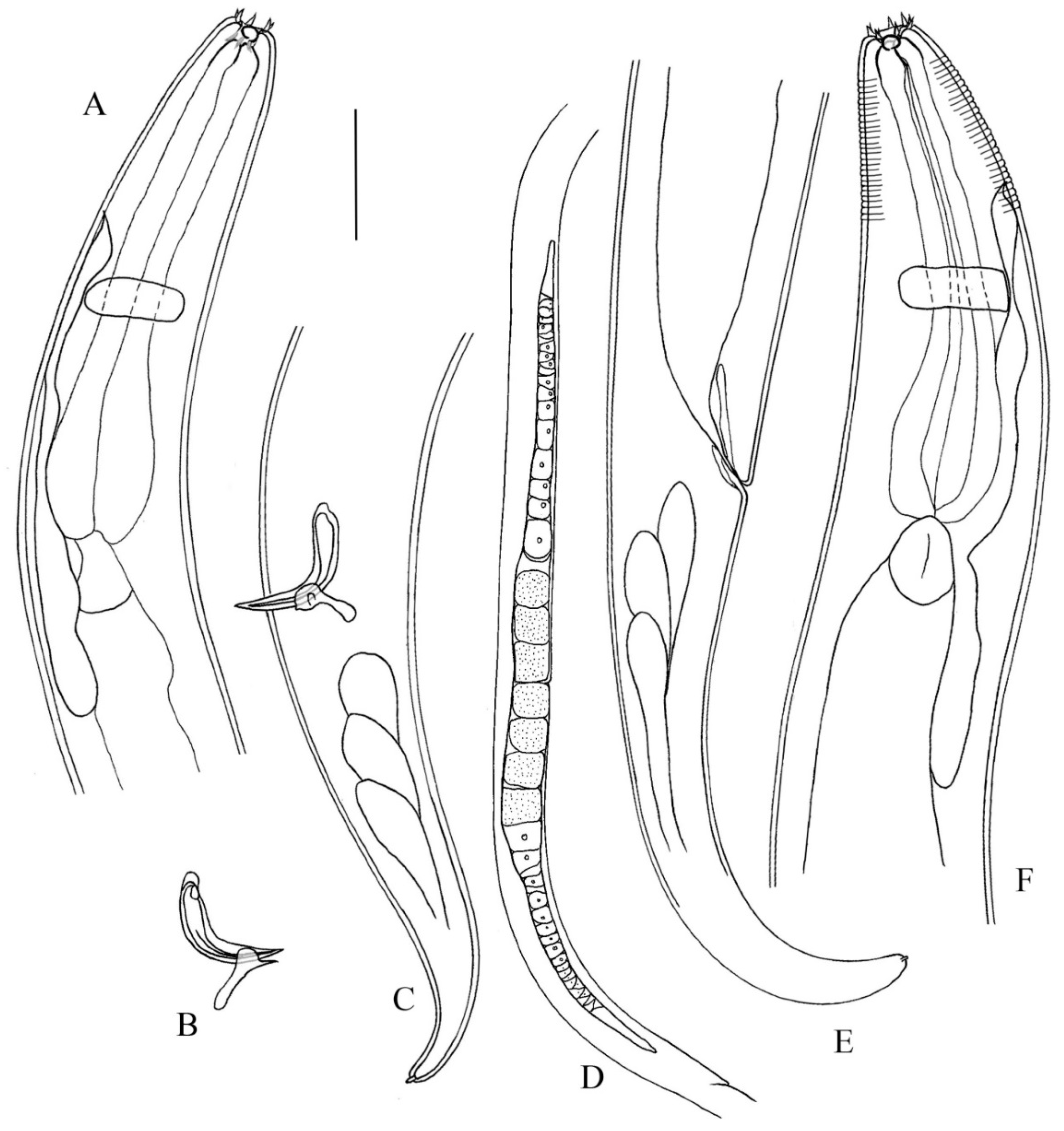
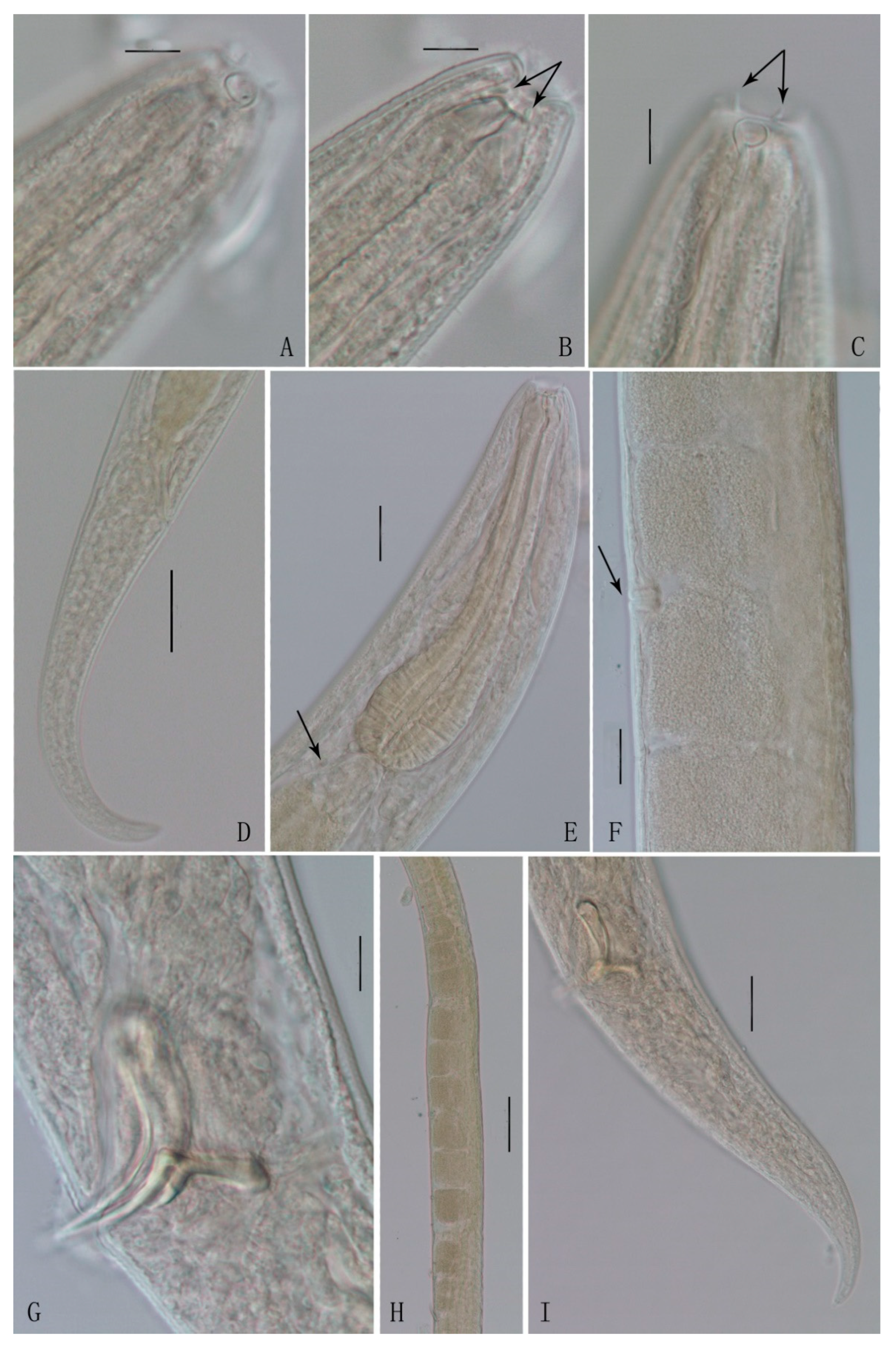
3.1. Description of Anticyathus Shenzhensis sp. nov.

3.1.1. Type Material

Four males and three females were collected from Station SZFT in July 2020. Holotype: ♂1 on slide number SZFT202007 2L104. Paratypes: ♂2 on slide number SZFT202007 2M115, ♂3 on slide number SZFT202007 2L104, ♂4 on slide number SZFT202007 3M116, ♀1 on slide number SZFT202007 2L108, ♀2 on slide number SZFT202007 3L115, ♀3 on slide number SZFT202007 2M106.

3.1.2. Etymology

The species is named after the seashore city, Shenzhen City, Guangdong Province, China, from where it was collected.

3.1.3. Type Locality and Habitat

All specimens were collected from a mangrove forest in the mangrove reservation of Shenzhen (22.52° N, 114.01° E), China. Characteristics of surface sediments of sampling stations are shown in Table 1.

3.1.4. Morphological Description

Body cylindrical, maximum body diameter 72–99 µm. Cuticle annulated, the annule begins a little posterior to the level of the base of the buccal cavity, about 13 µm from head end. Cuticle annule about 2.3 µm broad at anterior body part and 1.5 µm at midbody part. Ten cephalic setae in a circle, length about 2–4 µm. The setae appear to be at one level in submedian pairs. Head truncate, 17–18 µm in diameter. Buccal cavity length 5–10 µm and width 5–8 µm, lined with 6 thick, noncuticularized, longitudinal reinforcements. Amphids broken circles or cryptic spiral, length 6–7 µm, width 5–7 µm or 20–29% of corresponding body diameter. Amphids located at very anterior of head, anterior margin 2–3 µm from anterior end. Pharynx cylindrical, with anterior cuticularized, 0.07–0.10 times body length, gradually enlarging in posterior portion but without bulb. Nerve ring located at 51–59% of pharynx length. The excretory pore is anterior to the nerve ring, 69–98 µm from the anterior end. Renette cell extends posterior to the base of pharynx. Long clear cardia, about 25 µm long. Tail conical, tapering to bluntly rounded tip. Tail 2.5–3.4 anal body diameters long in male, 3.4–4.7 in female. The caudal glands are poorly visible, but they probably have an efferent duct-spinneret.
Male with two testes arranged in tandem, located at the ventral side of the body. Spicules small, arcuate, 41.0–48.5 µm long as chord and 51–59 µm long as arc. The distal ends pointed and gradually broaden to proximal, proximal slightly cepahlated. The gubernaculum has a dorsal–caudal apophysis, length 18–19 µm.
Female with two opposed, outstretched ovaries, the anterior is left of the intestine, the posterior shifted ventrally or towards the right. Vulva located at 52–56% of body length (Figure 1 and Figure 2, Table 2).

3.1.5. Differential Diagnosis

Anticyathus shenzhensis sp. nov. is characterized by ten cephalic setae length in a circle, about 2–4 µm; amphids located at very anterior of head, 2–3 μm from anterior end; tail conical, 2.5–3.4 abd in male and 3.4–4.7 abd in female; spicules arcuate, 0.71–1.04 abd, the distal ends pointed, gradually broaden towards proximal; gubernaculum has a dorsal–caudal apophysis, length 18–19 µm; female with two opposed, outstretched ovaries; vulva at 52–56% of body length.
From all valid species, this new species can be easily distinguished from A. tenuicaudatus Cobb, 1920 and A. septentrionalis (Cobb, 1914) Timm, 1978 by the tail (tail 8–11 abd, cylindroid in the posterior fourth in A. tenuicaudatus Cobb, 1920) [10] and the shape of spicule (description from Cobb [17]: spicule slender and uniform; description from Timm [18]: highly angular, distinctly cephalated, with head turned ventrally), respectively. It differs from A. boreicaspius, A. plicibucca, and A. trochus by male without pre-anal supplement (a pre-anal supplement present in A. boreicaspius Tchesunov 1978, A. plicibucca Tchesunov & Yushin, 1991 and A. trochus (Gerlach, 1957) Gerlach, 1963) [5,19,20]. Compared with A. primitivus, it can be distinguished by ten cephalic setae arranged in a circle (arranged in two circles in A. primitivus) and amphid and vulva located relative anterior of body (in A. primitivus: amphid at 6–7 μm from anterior end and vulva at 71% of body length) [9,21].

3.1.6. DNA Sequence Results

A total of 39 sequences of 18S rRNA gene were obtained from ten individuals of Anticyathus shenzhensis sp. nov. in this study. All sequences were deposited in GenBank. GenBank accession numbers are as follows: ON694124-ON694132, ON763151-ON763180.

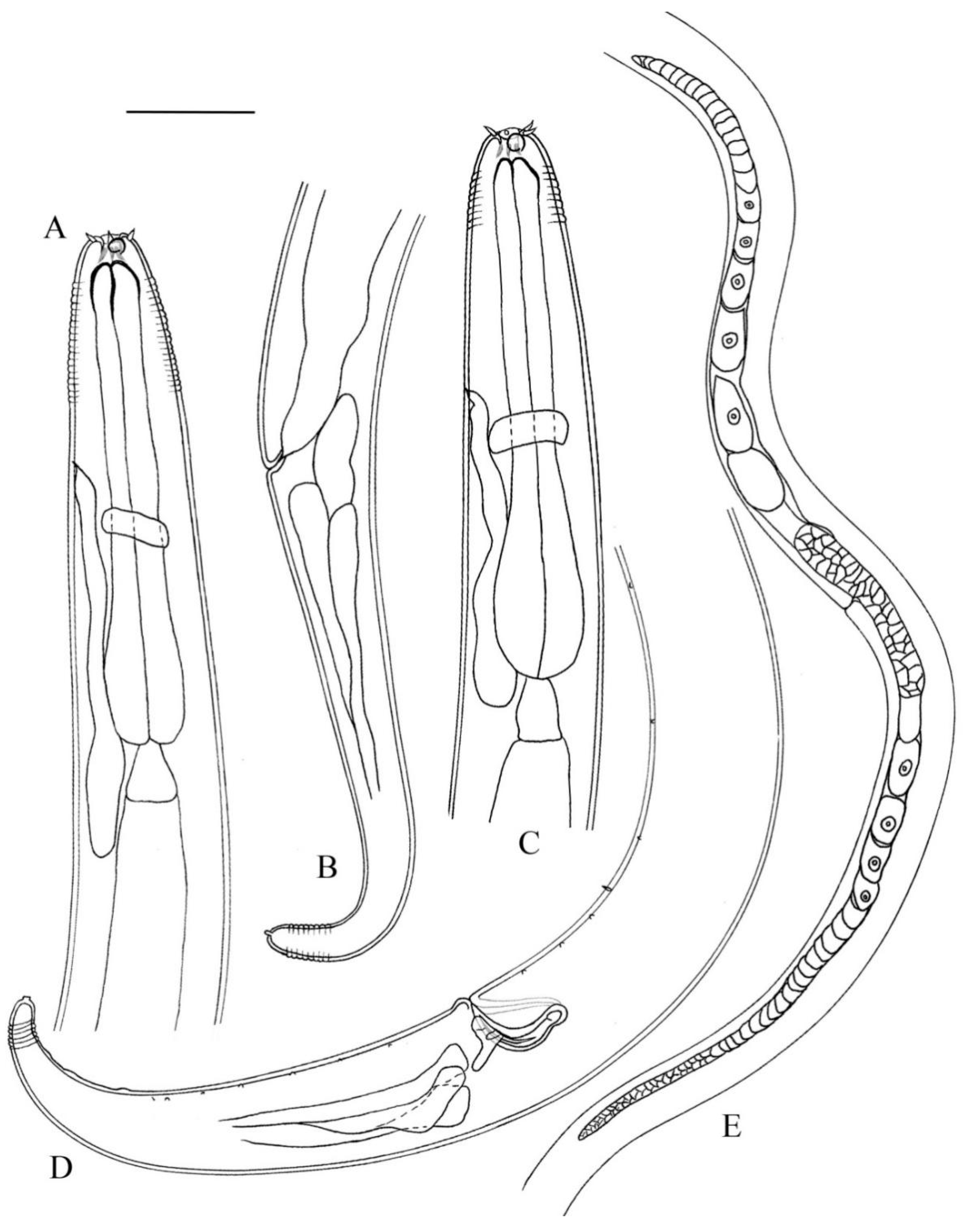
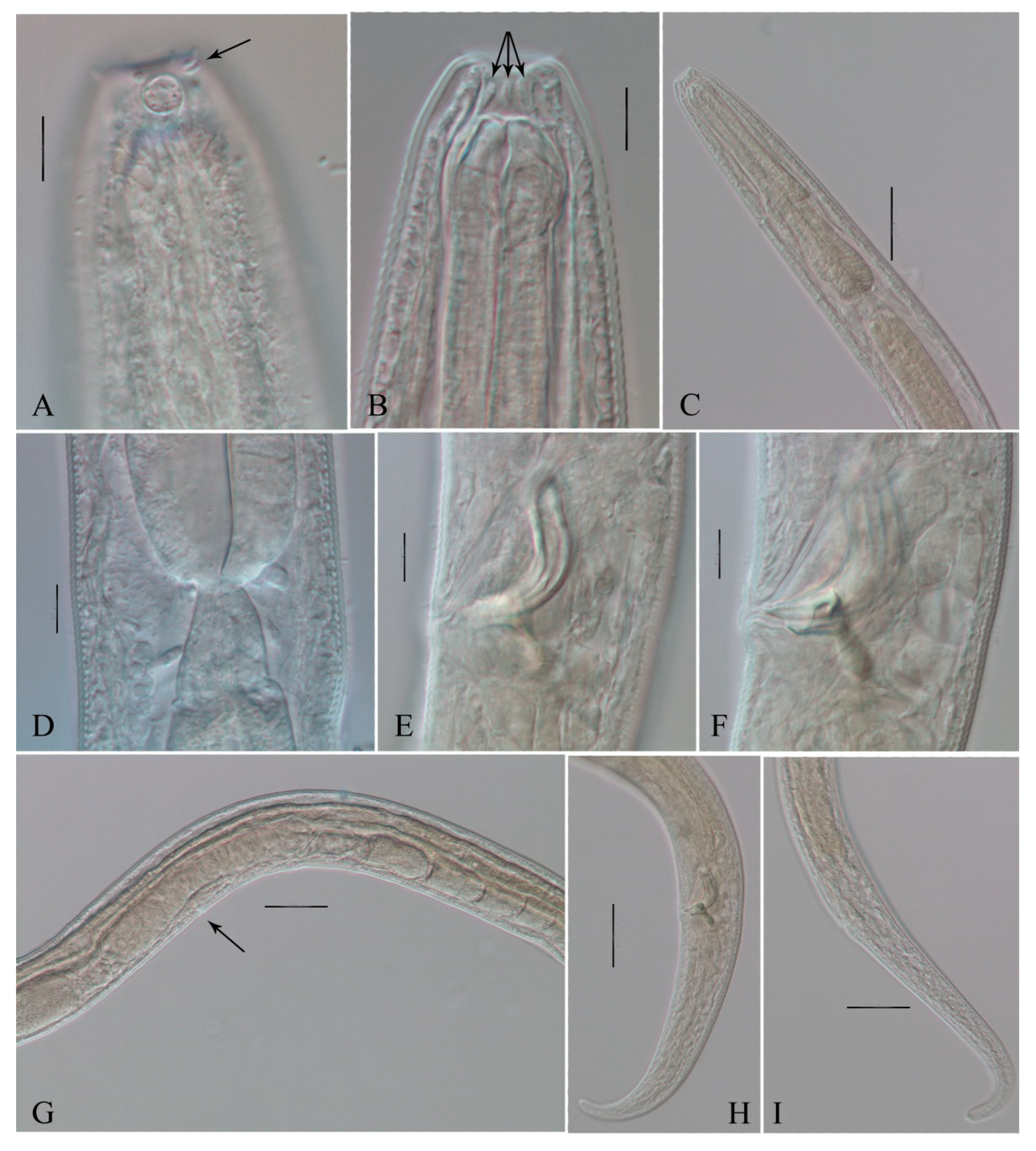
3.2. Description of Anticyathus communis sp. nov.

3.2.1. Type Material

Five males and two females were collected from Station ST 13L2, ST 13L3, ST 11H2, and ST 11H3 in September 2020. Holotype: ♂1 on slide number ST20200902 13L2 103. Paratypes: ♂2 on slide number ST20200902 13L2 103, ♂3 on slide number ST20200902 11H3 106, ♂4 and ♂5 on slide number ST20200902 13L2 108, ♀1 on slide number ST20200902 11H2 109, and ♀2 on slide number ST20200902 13L3 108.

3.2.2. Etymology

The specific name communis (“ordinary” in Latin) is derived from the usual body form of the new species.

3.2.3. Type Locality and Habitat

All specimens were collected from a mangrove forest in the mangrove reservation of Shantou (23.46° N, 116.87° E), China.

3.2.4. Morphological Description

Body cylindrical, maximum body diameter 53–66 µm. Cuticle annulated, the annule begins a little posterior to the level of the base of the buccal cavity, annule about 2.2 µm broad at anterior body part, and 1.3 µm at midbody part. Ten cephalic setae, length about 3–5 µm. The setae appear to be at one level in submedian pairs. Head truncate, 17–21 µm in diameter. Buccal cavity length 9–11 µm and width 7–9 µm, lined with 6 thick, non-cuticularized, longitudinal reinforcements. Amphids broken circles or cryptic spiral, width 5–6 µm or 20–32% of corresponding body diameter, length 6–7 µm. Amphids located at very anterior of head, anterior margin 1–2 µm from anterior end. Pharynx cylindrical, with anterior cuticularized, 0.09–0.11 times of body length, gradually enlarging in posterior portion but without bulb. Nerve ring located at 49–60% of pharynx length. The excretory pore is anterior to the nerve ring, 78–96 µm from the anterior end. Renette cell extends posterior to the base of pharynx. Long clear cardia, 17–27 µm long. Tail conical, tapering to bluntly rounded tip. Tail 3.6–4.5 anal body diameters long in male, 5.3–5.5 in female. The caudal glands are poorly visible, but they probably have an efferent duct-spinneret.
Male with two testes arranged in tandem, located at the ventral side of the body. Spicules arcuate, the distal ends pointed, gradually broaden to proximal, proximal distinctly cephalated, 31–37 µm long as chord and 43–49 µm long as arc. Gubernaculum “L”-shaped, consisting of sleeves around spicule and dorsal–caudal apophysis. Apophysis length 13–21 µm. Five to seven pre-anal and seven to ten post-anal almost invisible papilla or papillae-like setae, if the cuticle is thick, occur on each ventrally submedian line on the posterior body part of male. The tip of the tail with three slight elevations.
Female with two opposed, outstretched ovaries, the anterior is right to the intestine, and the posterior shifts to the left. Vulva at 52% of body length (Figure 3 and Figure 4, Table 3).

3.2.5. Differential and Diagnosis

Anticyathus communis sp. nov. most closely resembles A. shenzhensis sp. nov. by the similar body size, the location of amphid and vulva, the shape of the tail, the arrangement of the cephalic setae, and the absence of pre-anal supplement. However, it can be separated from A. shenzhensis sp. nov. by a more slender body (maximum body diameter 53–66, a = 33–45 vs. maximum body diameter 72–99, a = 24.5–30.6 in A. shenzhensis); a relatively shorter tail (3.6–4.5 abd in male and 5.3–5.5 abd in female vs. 2.5–3.4 in male and 3.4–4.7 in female in A. shenzhensis); a proximal spicule, significantly cephalated (proximal of spicule slightly cephalated in A. shenzhensis); posterior body part of male with five to seven pre-anal and seven to ten post-anal almost invisible papilla or papillae-like setae (A. shenzhensis sp. nov. without pre-anal or post-anal papilla).

4. Discussion

There are six species in this genus have been described up to now (Electronic database: http://nemys.ugent.be; http://www.marinespecies.org/index.php (accessed on 15 June 2022)). All valid species are listed below. A. primitivus was originally described as Linhomoeus (Eulinhomoeus) primitiva based on a single female [20]; Gerlach [19] added the description of male, and considered it to be synonymous Paralinhomoeus primitivus Allgén, 1934; A. trochus was first described as Prospharolaimus trochus by Gerlach in 1957. Gerlach [9] gave a dichotomous key to the genus Anticyathus, transferred Linhomoeus (Eulinhomoeus) primitiva and Prospharolaimus trochus to Anticyathus. A. septentrionalis was originally described as Laxus septentrionalis Cobb, 1914 [17], Timm (1978) [18] redescribed and illustrated it, and transferred it to Anticyathus.

4.1. List of Valid Anticyathus Species

Anticyathus boreicaspius Tchesunov 1978
Anticyathus plicibucca Tchesunov & Yushin, 1991
Anticyathus primitivus (Allgén, 1933) Gerlach, 1963 syn Linhomoeus primitiva Allgén, 1933; Paralinhomoeus primitivus Allgén, 1934
Anticyathus septentrionalis (Cobb, 1914) Timm, 1978
Anticyathus tenuicaudatus Cobb, 1920
Anticyathus trochus (Gerlach, 1957) Gerlach, 1963 syn Prospharolaimus trochus Gerlach, 1957
Figure 5 shows the pictorial key to 6 valid species as well as the two newly described species of the genus Anticyathus. Table 4 shows the differentiating characters of all known Anticyathus species.

4.2. Identification Key to Species of the Genus Anticyathus (emended after Gerlach, 1963) [9]

1. Tail 8–11 abd, cylindroid in the posterior fourth....................A. tenuicaudatus Cobb, 1920
- Tail shorter, conical......................................................................................................................2
2. Spicule relatively longer, 1.5 abd......................A. septentrionalis (Cobb, 1914) Timm, 1978
- Spicule relatively shorter, about 1 abd or less than 1 abd.....................................................3
3. Male with a pre-anal supplement............................................................................................4
- Male without pre-anal supplement...........................................................................................6
4. Cephalic sensilla setaceous-like, length 5.1 μm; amphid located posterior to the buccal cavity.............................................................................A. plicibucca Tchesunov & Yushin, 1991
- Cephalic sensilla reduced to blunt cone papillae or very short setae; amphid located at the base of buccal cavity................................................................................................................5
5. Body size smaller (body length 875–1044 μm, spicule length 23 μm), cuticle without annule………………………………………………………………....A. trochus (Gerlach, 1957)
Body size bigger (body length 2160–2480 μm, spicule length 43.7 and 47.5 μm), cuticle with annules……………………………………………………A. boreicaspius Tchesunov 1978
6. 10 cephalic sensilla arranged in one circle.............................................................................7
- 10 cephalic sensilla arranged in two circle................................A. prirnitivus (Allgén, 1933)
7. Tail relatively longer, 2.5–3.4 abd in male, 3.4–4.7 abd in female, without papilla or seatae............................................................................................................A. shenzhensis sp.nov.
- Tail relatively shorter, 3.6–4.5 abd in male, 5.3–5.5 abd in female, with papilla or seatae.............................................................................................................A. communis sp. nov.

4.3. Discussion and Notes on the Molecular Research of Anticyathus

The nucleotide divergence of genes could be used as a diagnostic tool for the discrimination of nematode species [13]. The combination of molecular with morphological approaches can help in the identification and resolution of evolutionary relationships within marine nematodes [22,23,24]. However, the number of marine nematode species with nucleotide sequences in databases is very low. By searching the Genbank database (Electronic database: https://www.ncbi.nlm.nih.gov/ [accessed on 15 June 2022]), a total of 207 nucleotide sequences belong of family Linhomoeidae have been deposited. No gene sequences of genus Anticyathus can be found in Genbank database to date. It seems that the molecular data of genus Anticyathus is still lacking, which limits the further resolution of evolutionary relationships between Anticyathus and other marine nematodes. A comprehensive comparative molecular database is needed. In the present work, 39 sequences of 18S rRNA gene were obtained from ten individuals of Anticyathus shenzhensis sp. nov. and provided to the Genbank database, serving as a foundation for further study.

Author Contributions

Conceptualization, data curation, writing—original draft preparation, and writing—review and editing, Y.C. and Y.G.; methodology, R.Z.; investigation, H.Z.; funding acquisition, Y.G. All authors have read and agreed to the published version of the manuscript.

Funding

This research was funded by the National Natural Science Foundation of China, grant number 31772416.

Institutional Review Board Statement

Not applicable.

Informed Consent Statement

Not applicable.

Data Availability Statement

Not applicable.

Acknowledgments

The authors thank Yi-Jia Shih for providing valuable comments on the manuscript and thank Pan Chao and Futian-CityU Mangrove Research & Development Centre for for great assistance in the sampling.

Conflicts of Interest

The authors declare no conflict of interest.

References

  1. Giere, O. Meiobenthology. In The Microscopic Motile Fauna of Aquatic Sediments, 2nd ed.; Springer: Berlin/Heidelberg, Germany, 2009. [Google Scholar]
  2. Soetaert, K.; Franco, M.; Lampadariou, N.; Muthumbi, A.; Steyaert, M.; Vandepitte, L.; Berghe, E.V.; Vanaverbeke, J.J.M.E.P. Factors affecting nematode biomass, length and width from the shelf to the deep sea. Mar. Ecol. Prog. Ser. 2009, 392, 123–132. [Google Scholar] [CrossRef]
  3. Ridall, A.; Ingels, J. Suitability of Free-Living Marine Nematodes as Bioindicators: Status and Future Considerations. Front. Ecol. Evol. 2021, 8, 685327. [Google Scholar] [CrossRef]
  4. Appeltans, W.; Ahyong, S.; Anderson, G.; Angel, M.; Artois, T.; Bailly, N.; Bamber, R.; Barber, A.; Bartsch, I.; Berta, A.; et al. The Magnitude of Global Marine Species Diversity. Curr. Biol. 2012, 22, 2189–2202. [Google Scholar] [CrossRef]
  5. Tchesunov, A.V.; Yushin, V.V. The Fine Structure of the Cephalic End of Two New Species of Nematodes of the Family Linhomoeidae from the Sea of Japan. Sov. J. Mar. Biol. 1991, 17, 88–94. [Google Scholar]
  6. Filipjev, I.N. The classification of the free-living nematodes and their relation to the parasitic nematodes. Smithson. Misc. Collect. 1934, 89, 1–63. [Google Scholar]
  7. Chitwood, B.G. North American marine nematodes. Tex. J. Sci. 1951, 4, 617–672. [Google Scholar]
  8. Wieser, W. Free-living marine nematodes III. Axonolaimoidea and Mohysteroidea. Acta Univ. Lund 1956, 52, 1–115. [Google Scholar]
  9. Gerlach, S.A. Über freilebende Meeresnematoden. Revision der Linhomoeidae. Zool. Jahrbücher Syst. Band 1963, 90, 599–658. [Google Scholar]
  10. Cobb, N.A. One hundred new nemas (Type species of 100 new genera). Contrib. Sci. Nematol. 1920, 9, 217–343. [Google Scholar]
  11. Fonseca, G.; Bezerra, T.N. Order Monhysterida Filipjev, 1929. In Handbook of Zoology: Gastrotricha, Cycloneuralia, Gnathifera, volume 2 Nematoda; Schmidt-Rhaesa, A., Ed.; De Gruyter: Berlin, Germany, 2014; Volume 2, pp. 435–465. [Google Scholar]
  12. Chen, Y.Z.; Guo, Y.Q. Three new and two known free-living marine nematode species of the family Ironidae from the East China Sea. Zootaxa 2015, 4018, 151. [Google Scholar] [CrossRef]
  13. Shih, Y.-J.; Chen, Y.-Z.; Guo, Y.-Q. A New Species of Free-living Marine Nematode (Ptycholaimellus: Chromadoridae: Chromadorida: Nematoda) from Mangrove Wetlands in China. Zool. Stud. 2022, 61, 20. [Google Scholar] [CrossRef]
  14. Bhadury, P.; Austen, M.C.; Bilton, D.T.; Lambshead, P.J.D.; Rogers, A.D.; Smerdon, G.R. Evaluation of combined morphological and molecular techniques for marine nematode (Terschellingia spp.) identification. Mar. Biol. 2008, 154, 509–518. [Google Scholar] [CrossRef]
  15. Bhadury, P.; Austen, M.C.; Bilton, D.T.; Lambshead, P.J.D.; Rogers, A.D.; Smerdon, G.R. Development and evaluation of a DNA-barcoding approach for the rapid identification of nematodes. Mar. Ecol. Prog. Ser. 2006, 320, 1–9. [Google Scholar] [CrossRef]
  16. Holterman, M.; van der Wurff, A.; van der Elsen, S.; van Megen, H.; Bongers, T.; Holovachov, O.; Bakker, J.; Helder, J. Phylum-wide analysis of SSU rDNA reveals deep phylogenetic relationships among nematodes and accelerated evolution toward crown clades. Mol. Biol. Evol. 2006, 23, 1792–1800. [Google Scholar] [CrossRef]
  17. Cobb, N.A. Antarctic marine free-living nematodes of the Shackleton expedition. Contrib. Sci. Nematol. 1914, 1, 1–33. [Google Scholar]
  18. Timm, R.W. Redescription of the marine nematodes of Shackleton’s British Antarctic Expedition of 1907–1909. Am. Geophys. Union Antarct. Res. Ser. 1978, 26, 237–255. [Google Scholar]
  19. Tchesunov, A.V. Free-living nematodes of the family Linhomoeidae from the Caspian Sea. Zool. Zhurnal 1978, 57, 1623–1631. [Google Scholar]
  20. Gerlach, S.A. Marine Nematoden aus dem Mangrove-Gebiet von Cananéia (Brasilianische Meeres-Nematoden III). Abh. Math. Nat. Kl. 1957, 5, 129–176. [Google Scholar]
  21. Allgén, C.A. Freilebende Nematoden aus dem Trondhjemsfjord. Capita Zool. 1933, 4, 1–162. [Google Scholar]
  22. Derycke, S.; Fonseca, G.; Vierstraete, A.; Vanfleteren, J.; Vincx, M.; Moens, T. Disentangling taxonomy within the Rhabditis (Pellioditis) marina (Nematoda, Rhabditidae) species complex using molecular and morhological tools. Zool. J. Linn. Soc. 2008, 152, 1–15. [Google Scholar] [CrossRef]
  23. Fonseca, G.; Derycke, S.; Moens, T. Integrative taxonomy in two free-living nematode species complexes. Biol. J. Linn. Soc. 2008, 94, 737–753. [Google Scholar] [CrossRef]
  24. Avó, A.P.; Daniell, T.J.; Roy, N.; Solange, O.; Jordana, B.; Helena, A. DNA barcoding and morphological identification of benthic nematodes assemblages of estuarine intertidal sediments: Advances in molecular tools for biodiversity assessment. Front. Mar. Sci. 2017, 4, 66. [Google Scholar] [CrossRef]
Figure 1. Anticyathus shenzhensis sp. nov.—holotype male and paratype female in lateral view. (A) lateral view of male head end; (B) lateral view of male copulatory apparatus; (C) lateral view of male tail region; (D) lateral view of female body part, showing ovaries; (E) lateral view of female tail region; (F) lateral view of female head end. Scale bar: (AC,E,F) = 50 μm; (D) = 250 μm.
Jmse 10 01107 g001
Figure 2. Anticyathus shenzhensis sp. nov.—DIC photomicrographs. (A) lateral view of male head end, showing amphid; (B) lateral view of female head end, showing buccal cavity and longitudinal reinforcements; (C) lateral view of female head end, showing amphid and cephalic setae; (D) lateral view of female tail region; (E) lateral view of female body part, showing pharynx and cardia; (F) lateral view of female body part, showing vulva; (G) lateral view of male body part, showing spicule, gubernaculum, and apophysis; (H) lateral view of female body part, showing ovary and eggs; (I) lateral view of male tail region. Scale bar: (AC,G) = 10 μm; (E,F,I) = 25 μm; (D) = 50 μm; (H) = 100 μm.
Jmse 10 01107 g002
Figure 3. Anticyathus communis sp. nov.—holotype male and paratype female in lateral view. (A) lateral view of female head end; (B) lateral view of female tail; (C) lateral view of male head end; (D) lateral view of male tail region; (E) lateral view of female body part, showing ovaries. Scale bar: (AD) = 50 μm; (E) = 125 μm.
Jmse 10 01107 g003
Figure 4. Anticyathus communis sp. nov.—DIC photomicrographs. (A) lateral view of male head end, showing amphid and cephalic setae; (B) lateral view of female head end, showing buccal cavity and longitudinal reinforcements; (C) lateral view of female anterior part, showing pharynx; (D) lateral view of male body part, showing cardia; (E) lateral view of male body part, showing spicule; (F) lateral view of male body part, showing gubernaculum and apophysis; (G) lateral view of female body part, showing ovary and vulva; (H): lateral view of male tail region; (I) lateral view of female tail region. Scale bar: (A,B,DF) = 10 μm; (C,GI) = 50 μm.
Jmse 10 01107 g004
Figure 5. Pictorial key for genus Anticyathus.
Jmse 10 01107 g005
Table 1. Characteristics of surface sediments of sampling stations.
StationTemperature
(°C)
pHSalinity
(‰)
Total Nitrogen
(mg·g−1)
Total Organic Carbon
(%)
SZFT 2L27.06.12.52.1413.93
SZFT 3L28.56.33.11.5014.90
SZFT 2M26.56.24.84.3411.69
SZFT 3M27.56.33.31.7516.66
Table 2. Morphometrics of Anticyathus shenzhensis sp.nov. (in µm).
CharactersHolotypeParatypes
♂1♂2♂3♂4♀1♀2♀3
Body length2232242221722399248620422255
Head diameter17181918181718
Length of cephalic setae2–32–32–342–32–32-3
Buccal cavity length81068675
Buccal cavity diameter5787967
Amphids from the anterior end2332232
Amphid length6567555
Amphid width5757566
Amphid cbd23232324252522
Amphid width/Amphid cbd0.200.290.220.280.220.220.27
Excretory pore from the anterior end84897498697984
Excretory pore cbd52635469585163
Nerve ring from the anterior end108117981209410699
Nerve ring cbd57695974675566
Pharynx length198211191203184205191
Pharynx cbd65777288786374
Maximum body diameter73847998997288
abd57656276545261
Tail length193189156196253209208
c′3.42.92.52.64.74.03.4
Spicule length as chord48414249---
Spicule length as arc59545154---
Spicule length as arc/abd1.040.830.820.71---
Gubernaculum14111013---
Length of apophysis19181919---
V′----129611181263
vbd----997288
V%----525556
a30.5828.8327.4924.4825.1128.3625.63
b11.2711.4811.3711.8313.519.9611.81
c11.5612.8113.9212.229.839.7710.84
Table 3. Individual measurements of Anticyathus communis sp. nov. (in µm).
CharactersHolotypeParatypes
♂1♂2♂3♂4♂5♀1♀2
Body length2462244723732266189221762242
Head diameter18192119171819
Length of cephalic setae4444354
Buccal cavity length1111111291011
Buccal cavity diameter7888897
Amphids from the anterior end1221222
Amphid length6766667
Amphid width6666656
Amphid cbd22202525222523
Amphidwidth/Amphid cbd0.260.320.250.230.270.200.25
Excretory pore from the anterior end96969378888986
Excretory pore cbd49515350434946
Nerve ring from the anterior end112111113101105117113
Nerve ring cbd50515453495349
Pharynx length221210217205201194209
Pharynx cbd53576060515756
Maximum body diameter55586565536659
abd49526054464240
Tail length220220219205176227215
c′4.54.33.63.83.95.55.3
Spicule length as chord3634343731--
Spicule length as arc4643444944--
Spicule length as arc/abd0.950.840.740.900.96--
Gubernaculum1212111212--
Length of apophysis2113131717--
V′-----11421177
vbd-----6557
V%-----5252
a44.9942.5436.4734.9535.6433.0438.30
b11.1611.6510.9311.039.4211.2110.73
c11.2211.1210.8611.0310.789.5710.43
Table 4. Differentiating characters of all known Anticyathus species (in µm).
SpeciesA. boreicaspiusA. plicibuccaA. primitivusA. septentrionalisA. tenuicaudatusA. trochusA. communis sp. nov.A. shenzhensis sp. nov.
References[19][5][9][21][17][18][10][20]This ArticleThis Article
Sex
Body length248021602140–3050255021721725200018002150–28702040–29905200650087510441892–24622176–22422172–24222042–2486
Amphids from the anterior end56.311.91576--------1–222–32–3
Amphid width6.9–7 8.56.896--------65–65–75–6
Maximum body diameter75506167.5554538 *37.8 *----30-53–6559–6673–9872–88
abd5043.751415230 *34 *23.4 *--88.4 *84.5 *262446–6040–4257–7652–61
Tail length150 *140 *--147130108 *84.6 *--728 *975 *--176–220215–227156–196208–253
c′33.23.8–4.143–44.3 *3.18 *3.62 *3.1–42.6–411.5 *8.24 *343.6–4.55.3–5.52.5–3.43.4–4.7
Spicule length as arc43.7/47.5-40-45-51 *-40-88 *-23-43–49-51–59-
Spicule length as arc/abd0.87/0.95 *-0.78-0.87-1.50- 1.00-0.88-0.74–0.96-0.71–1.04-
Length of apophysis--15---17 *-23-22 *---13–21-18–19-
V%---62.6%-71.01%-64.00%-51.4–58.6%-50% *-54%-52%-52–56%
a3343.25037.74038.353 *48 *50–6441–4947 *37 *292935.0–45.033.0–38.324.5–30.625.6–28.4
b6.55.915.4–18.215.68.57.3612.8 *13.2 *10.5–18.810.2–13.013.3 *20 *5.469.4–11.710.7–11.211.3–11.810.0–13.5
c15.515.918.2–23.322.61513.2718.5 *21.3 *13.7–19.415.7–26.87.14 *6.67 *11.19.210.8–11.29.6–10.411.6–13.99.8–10.8
Abbreviations: - data absent; * data calculated from descriptive formula.
Publisher’s Note: MDPI stays neutral with regard to jurisdictional claims in published maps and institutional affiliations.

