Article

Skin Culturable Microbiota in Farmed European Seabass (Dicentrarchuslabrax) in Two Aquacultures with and without Antibiotic Use

1
Ruđer Bošković Institute, 10000 Zagreb, Croatia
2
Orada Adriatic d.o.o., 51557 Cres, Croatia
3
Faculty of Agriculture, University of Zagreb, 10000 Zagreb, Croatia
*
Author to whom correspondence should be addressed.
J. Mar. Sci. Eng. 2022, 10(3), 303; https://doi.org/10.3390/jmse10030303
Submission received: 29 December 2021 / Revised: 31 January 2022 / Accepted: 11 February 2022 / Published: 22 February 2022
(This article belongs to the Special Issue Microbial Diseases of Marine Organisms)

Abstract

:
This study examined culturable skin microbiota that was associated with farmed European seabass (Dicentrarchus labrax). Healthy European seabass were sampled during summer commercial harvest from one conventional fish farm where antibiotics are used, and from another practicing a certified antibiotic-free fish aquaculture. Physicochemical and microbiological analysis of seawater and sediment were performed, as well as determination of culturable bacteria, including Vibrio, from skin swabs of European seabass and seawater and sediment at both farms. Samples were processed for isolation of bacteria and their characterization by molecular and antibiotic susceptibility tests. In both fish farms, most of the bacteria that were identified in the skin belonged to the genera Pseudomonas and Vibrio. Some of the microbiota that were identified are known to be pathogenic to fish: V. alginolyticus, V. anguillarum, and V. harveyi. Vibrio strains showed higher resistance to certain antibiotics compared to previous studies. This study provides, for the first time, information on the culturable skin bacteria that is associated with healthy European seabass under culture conditions with and without the use of antibiotics. This information will be useful in assessing how changes in culturable microbiota may affect the health of farmed European seabass, indicating a potential problem for fish health management during disease outbreaks.

1. Introduction

Common fish diseases and environmental pollution can lead to economic losses and poor and unprofitable production in aquaculture [1]. European seabass (Dicentrarchus labrax) is one of the most farmed fish species in Croatia and tops the list of most farmed fish species in the European Union [2]. Its cultivation is threatened by several common bacterial pathogens and the infections that are caused by them. For example, Photobacterium damselae causes photobacteriosis in European seabass with a mortality rate between 60% and 80% [1]. Vibrio species are ubiquitous in aquatic ecosystems, while many Vibrio species are serious opportunistic pathogens causing the most common bacterial diseases [3,4,5]. Control of bacterial pathogens in aquaculture production is routinely achieved by the administration of antibiotics. However, excessive antibiotic use has led to the emergence of antibiotic-resistant bacteria [1].
As the skin, along with the gills, is an organ that is involved in the primary defense of the organism from pathogens, it is important to investigate its microflora and how microflora disorders lead to disease development. Microflora refers to the microorganisms that are present on the mucous membranes and skin of fish [6]. Although microorganisms that are pathogenic to fish are part of the normal fish microflora, they do not cause disease unless the balance of the microflora is disturbed Disturbances in the balance (dysbiosis) are often caused by the specificities of aquaculture such as high stocking density and low oxygen concentration [7], but also by changes of physical and chemical conditions in water, temperature fluctuations, seasonality, climate variability, and the use of antibiotics [8]. The skin microbiota of the fish that are exposed to such stress is usually composed of microorganisms that are pathogenic to fish [7]. The viscoelasticity of the skin mucus blocks various bacteria, and upon contact with the bacterial pathogen, the fish excrete more mucus and alter the composition of the skin microflora [9].
Most of the research on European seabass microflora has focused on the gut microbiota that is exposed to conventional [10] and alternative diets [11], as well as on the effects of probiotics on the balance of the gut microbiota [12]. Several researchers focus on the seasonal survey of the marine aquaculture microbiome in a European seabass farm [13], the assessment of seawater microbial quality, and the health status of farmed European seabass [14], with more specific characterization of isolated Vibrio species [5]. The evaluation of the tissue-specific diversity of microbiomes within and between sea bass and sea bream [7], as well as the recent study of the effects of aging on the skin and gill microbiota of farmed European sea bass and sea bream, were done by Rosado et al. [7,15].
Although studies of the European seabass skin microbiota are not numerous [3,6,7], one study examined the basic diversity of skin microbiomes and gills of farmed seabass and gilthead sea bream [7], but the link to the history of antibiotic therapy remained unclear. Some recent reports have studied the health problems of European seabass in aquaculture [3,6], but only one correlates the abundance of bacteria with problems that are associated with subsequent antibiotic treatment with oxytetracycline [6]. The abundance of taxa belonging to the non-pathogenic marine group NS3a, and Polaribacter 4 decreased in the skin microbiome of diseased fish with oxytetracycline therapy [6], while the pathogens Pseudomonas and Stenotrophomonas increased significantly. However, there is a lack of available information on the microbiota in the skin of European seabass that are grown in farming practices without a history of antibiotic therapy. Due to the increasing importance of aquaculture, more research is needed in disease prevention and alternative treatments to prevent bacterial resistance to antibiotics [1].
In the present study, we characterized the culturable bacteria of the skin of healthy adult European seabass during summer commercial harvest in two cage fish farms. This included one conventional farm where antibiotics are used to treat disease, and the other practicing a certified antibiotic-free production. The main objectives of the research are: to compare the culturable bacteria of farmed European seabass between these two fish farms and to identify the potential opportunistic bacterial pathogens and determine their susceptibility to antibiotics.

2. Materials and Methods
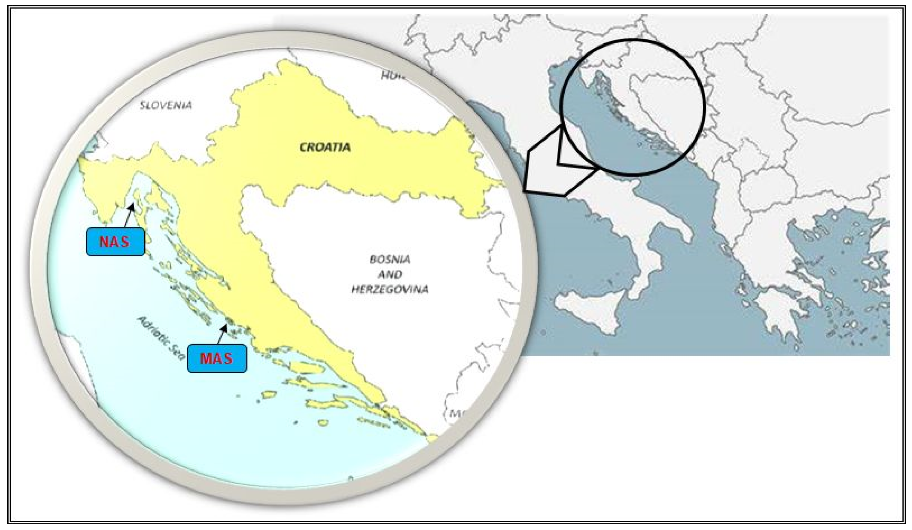
2.1. Sampling

This study was conducted on two floating cage fish farms, NAS farm in the northern, and MAS farm in the middle Adriatic (Figure 1). Both farms are in the semi open sea, with the NAS farm at depth of approximately 49 m, and MAS farm depth of approximately 22 m. The cages that are 10 m deep contain European seabass (Dicentrarchus labrax) and sea bream (Sparus aurata). Samples of fish, seawater, and sediments were collected during the summer at both farms. During the sampling year, the NAS farm was subjected to regular periodical control to determine antibiotic residues by an independent certification control service. Antibiotic-free production has been practiced since the beginning of aquaculture production on the NAS farm. The MAS farm differed from the NAS farm with respect to the usage of antibiotics. The antibiotics were administered at therapeutic dosage by a professional veterinarian at the farm based on a positive diagnosis. Usually, antibiotic treatment is conducted for the full period that is required for therapy and as little as necessary. The available information concerning antibiotic administration at the NAS farm have been undisclosed, however it is known that most used antibiotics in the Mediterranean countries are flumequine and oxytetracycline [16]. No clinical signs of fish disease were observed during sampling on both farms, and no antibiotic was used on the MAS farm at the time of sampling. The fish samples were obtained from cages of European seabass that were harvested for commercial purposes at the NAS and MAS farm. A total of 10 individual fish were obtained during commercial catch at each farm. One skin swab per fish was taken from 1 cm2 areas below the dorsal fin using sterile rods with a 1 cm cotton tip (Deltalab, Barcelona, Spain) for isolation and later bacterial identification. We specifically chose this area for swab collection because this area was not affected by fish handling during sampling which could have caused unwanted contamination. The samples were then serially diluted in 10 mL of sterile phosphate buffer saline (PBS, Sigma, St. Louis, MO, USA) [17]. During sampling, the health status of the fish was assessed and biometric data, namely, the total length and total weight, were recorded.
Sediment and seawater samples were collected from the NAS and MAS farm below the cages containing the European sea bass. Sediment samples (10 g of the top sediment layer) were collected from each farm using Ekman grab. Seawater samples were collected at four different depths (0.5 m below the surface, 6 m deep, 12 m deep, and 0.5 m above the bottom) using a Niskin water sampler and poured in sterile 0.5 L bottles. All the samples were serially diluted in 10 mL of sterile PBS solution (Sigma) and counts were determined by inoculating the undiluted and serially diluted samples on appropriate media for enumeration and isolation for microbial and molecular analysis (see below).

2.2. Physicochemical and Microbiological Analysis of Seawater and Sediment

The measurements of physicochemical parameters of seawater were carried out at both farms. The pH was measured electrometrically with portable digital SevenGo pro/Ion multiparameter probes (Mettler Toledo, Schwerzenbach, Switzerland) with an accuracy of 0.001 mg/L. The concentration of dissolved oxygen (mg/L), oxygen saturation of water (%), and the temperature (°C) were measured with a SevenGo pro/SG9 OptiOx probe (Mettler Toledo) with a precision of 0.01 mg/L. Conductivity and total dissolved solids (TDS) were measured with a SevenGo pro/conductivity probe (Mettler Toledo) with a precision of 0.1 mg/L.
The total coliforms and Escherichia coli were determined using Colilert-18™ and Quanti-Tray/2000 substrate technology (IDEXX, Westbrook, ME, USA). After incubation for 24 h at 35 °C, the appearance of a yellow color of the chamber allowed the evaluation of total coliforms, and fluorescence under UV light indicates the presence of E. coli. Enterococci were determined using Enterolert-E™ and Quanti-Tray/2000 (IDEXX). After incubation for 24 h at 41 °C, the appearance of fluorescence indicates the presence of enterococci [18]. Quanti-Tray/2000 indicates the most probable number of bacteria (MPN) in a 100 mL sample using the manufacturer’s reagents.

2.3. Number of Heterotrophic Bacteria and Vibrio Count

The number of heterotrophic bacteria (Heterotrophic Plate Count (HPC)) of the skin, seawater, and sediment, was determined by the spread plate method on Difco™ Marine Agar 2216 medium (BD, Sparks, MD, USA) that was incubated at 22 °C for 3–5 days. The spread plate method on a selective Thiosulfate-Citrate-Bile-Sucrose (TCBS) medium (BD), was used for the isolation of the bacteria of the genus Vibrio from fish skin swabs, sediment, and seawater samples. The plates were incubated at 22 °C for 24 h. The results are reported as the mean number of colony forming units (CFU) in 1 mL of sediment and seawater or per 1 cm2 of skin ± the standard deviation of two technical replicates by sample type [18]. Subsequently, two bacterial colonies representing different morphologies per plate on Marine Agar and TCBS agar were selected from each sample and transferred to Difco™ Tryptic Soy Agar (TSA) (BD) with the addition of 1% NaCl (Kemika, Zagreb, Croatia) (MTSA) plates to obtain a pure culture. After purification and plating on the MTSA plates, a total of 146 bacterial isolates were obtained.

2.4. DNA Isolation and PCR Amplification of Partial 16S rRNA Gene

A small amount of every purified bacterial colony was taken by a sterile loop and subjected to DNA isolation using GenEluteTM Mammalian Genomic DNA 132 Miniprep Kit (Sigma) according to the manufacturer’s instructions. Partial 16S rRNA gene sequence was amplified in PCR reaction mixtures containing 1× EmeraldAmp® GT PCR Master Mix (Takara, Shiga, Japan), 0.4 pmol/µL of primers 27F and 1492R (Wilson et al., 1990), 2 µL of DNA template, and nuclease-free water to the final volume of 50 µL. The reaction conditions were described previously [18]. Electrophoresis in 1.5% agarose gel was performed to check the presence of products of approximately 1450 bp length.

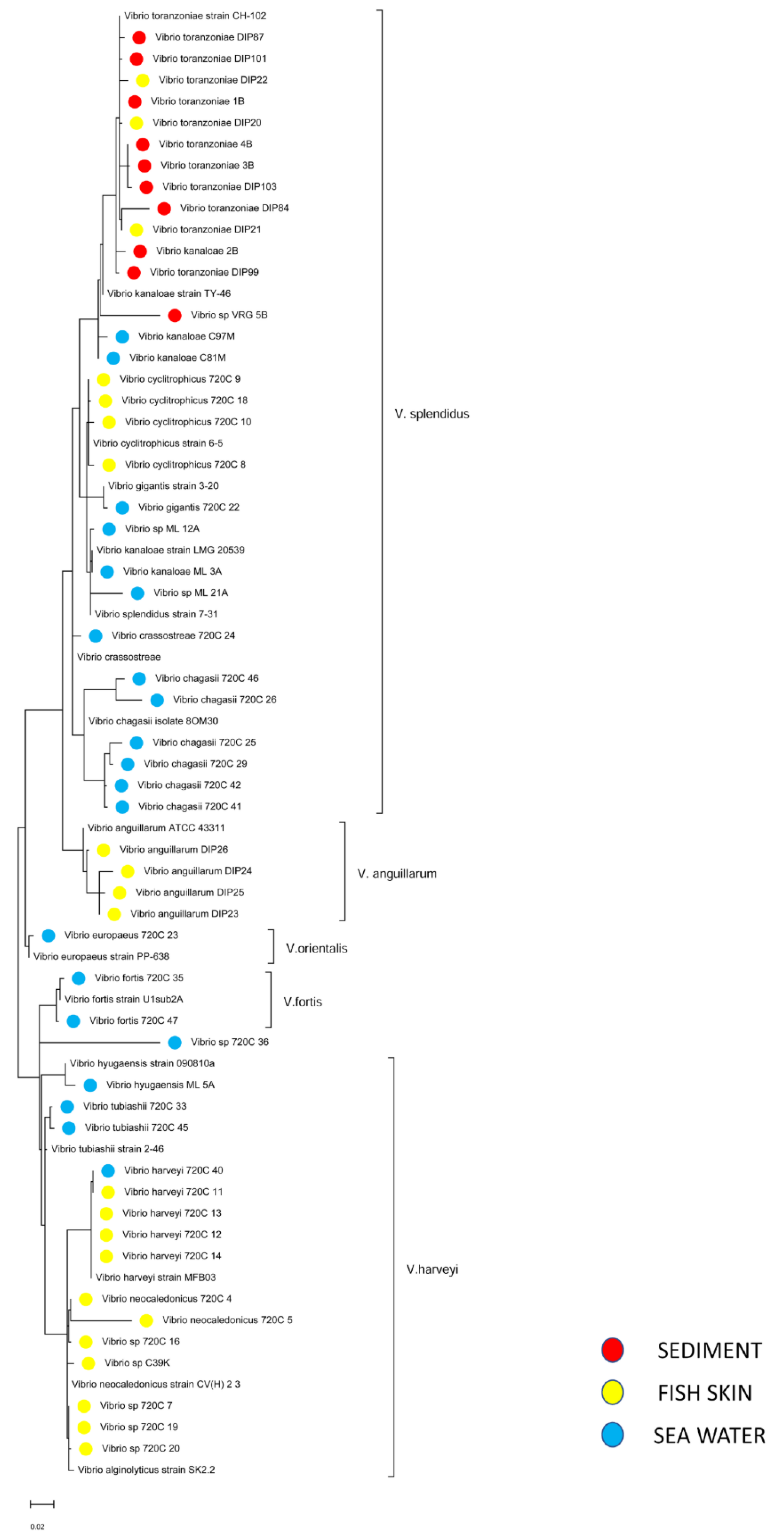
2.5. Sequencing and Phylogenetic Analysis of Vibrio sp.

The amplified PCR products were further sequenced commercially by Macrogen (Amsterdam, The Netherlands). The obtained sequences were edited manually and/or in BioEdit version 7.2.5. [19] and deposited in the GenBank under accession numbers: OL979296—OL979441. The sequences were analyzed by comparison with previously characterized 16S rRNA gene of the closest bacteria from the GenBank database using the NCBI BLAST program (Bethesda, MD, USA) and the percentage of similarity is highlighted.
To differentiate Vibrio species phylogenetic analysis that was based on the 16S rRNA gene sequence was conducted using MEGA11 [20]. The evolutionary history was inferred by using the maximum likelihood method and Kimura 2-parameter model. A discrete Gamma distribution was used to model the evolutionary rate differences among sites (5 categories (+G, parameter = 0.3935). The tree is drawn to scale, with branch lengths measured in the number of substitutions per site. This analysis involved 69 nucleotide sequences, 15 of which were references from GenBank (accession numbers shown in the Table S1 in Supplementary File). There was a total of 1724 positions per sequence in the final dataset.

2.6. Antimicrobial Susceptibility

The Kirby–Bauer disk diffusion method on BBL™ Mueller Hinton II agar (BD) was used to determine the antimicrobial susceptibility of the isolated bacterial strains. The most used antibiotics in aquaculture were selected. The following antimicrobial disks were used for the test (the amounts are given in micrograms in parentheses): ampicillin (10), streptomycin (10), gentamicin (10), chloramphenicol (30), ciprofloxacin (5), erythromycin (15), imipenem (10), oxytetracycline (30), sulfamethoxazole/trimethoprim (23.75/1.25), vancomycin (30) that is manufactured by BBL™ Sensi-Disk™, and enrofloxacin (5), florfenicol (30), and flumequine (30) that is manufactured by Thermo Scientific™ Oxoid (Hampshire, UK). The inoculum was prepared in 5 mL of sterile 0.85% suspension medium (BioMérieux). On the Vitek Systems ATB 1550 instrument (BioMérieux, Marcy l’Etoile, France), the turbidity for each inoculum was adjusted to 0.5 according to the McFarland value scale [14]. After 24 h incubation at 22 °C, the diameter of the zone of inhibition was measured with a ruler and the values were interpreted as sensitive, moderately sensitive, or resistant according to the manufacturer’s instructions.

2.7. Statistical Analysis

Statistical analysis was applied to detect the differences of heterotrophic bacteria and Vibrio content between the skin swabs, the seawater, and the sediment samples from both farms. Statistical significance of differences in the number of antibiotic-resistant bacterial isolates between the farms in each type of sample (skin, seawater, sediment) was also analyzed. The non-parametric Mann–Whitney U-test was applied for statistical analyses using SigmaPlot version 14.0 (Systat Software Inc., San Jose, CA, USA). The observed differences were statistically significant at p < 0.05. Venn diagrams were drawn to visualize the NAS and MAS culturable bacteria using a freely available web tool (http://bioinformatics.psb.ugent.be/webtools/Venn/, accessed on 15 December 2021).

3. Results

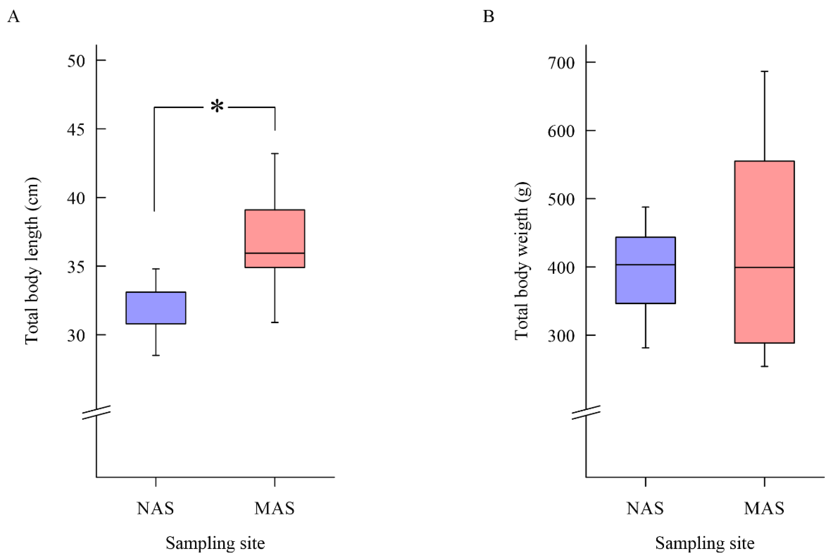
3.1. Results of Sea Bass Health Examination

An examination of the health status of the European seabass from both cage farms revealed no clinical signs of disease. In the NAS farm, the total length of the fish ranged from 28.5 cm to 34.8 cm and the total weight ranged from 281.5 g to 487.7 g. In the MAS farm, the total length of the sampled European seabass ranged from 30.9 cm to 43.2 cm, and the total weight ranged from 254.2 g to 686.5 g. The total body length was statistically significantly higher for the fish from farm MAS (p < 0.05) (Figure 2).

3.2. Physicochemical and Microbiological Analysis of Seawater

Table 1 shows the physicochemical parameters of the seawater from both fish farms.
The results of the microbiological analysis of the seawater and sediment from the two farms are presented in Table 2. Although the levels of total coliform bacteria in the seawater column were notably higher at the NAS farm, the difference between two farms was not statistically significant. The levels of E. coli were higher at the MAS farm, but also with no statistically significant difference (p > 0.05).
The studied parameters in the sediment samples had higher values in the samples from the MAS farm, except for the number of E. coli, which had the same value in the sediment samples from both farms.

3.3. Number of Heterotrophic Bacteria and Vibrio Count

The results of the microbiological analysis of skin swabs of European seabass from the NAS and the MAS farm are presented in Table 3. The total number of heterotrophic bacteria (HPC) from the skin swabs of European seabass between the farms was statistically significantly higher in the NAS antibiotics-free farm, as well as the number of bacteria of the genus Vibrio (p < 0.05).
The differences in the total number of heterotrophic bacteria in seawater between the farms were not statistically significant (p > 0.05). The number of bacteria of the genus Vibrio was higher in the seawater samples from the MAS farm, and the differences were statistically significantly higher than in the NAS farm (p < 0.05).

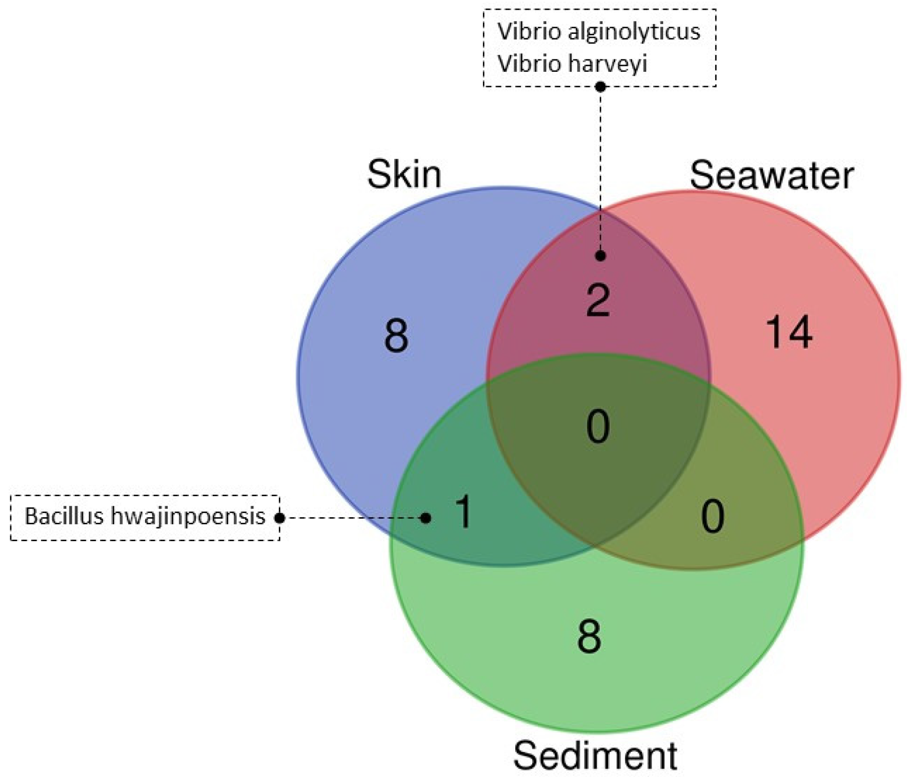
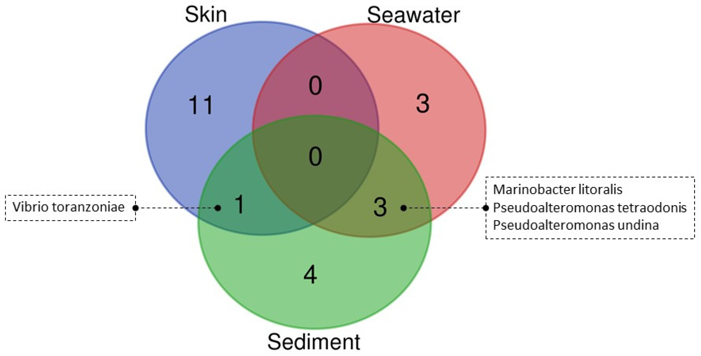
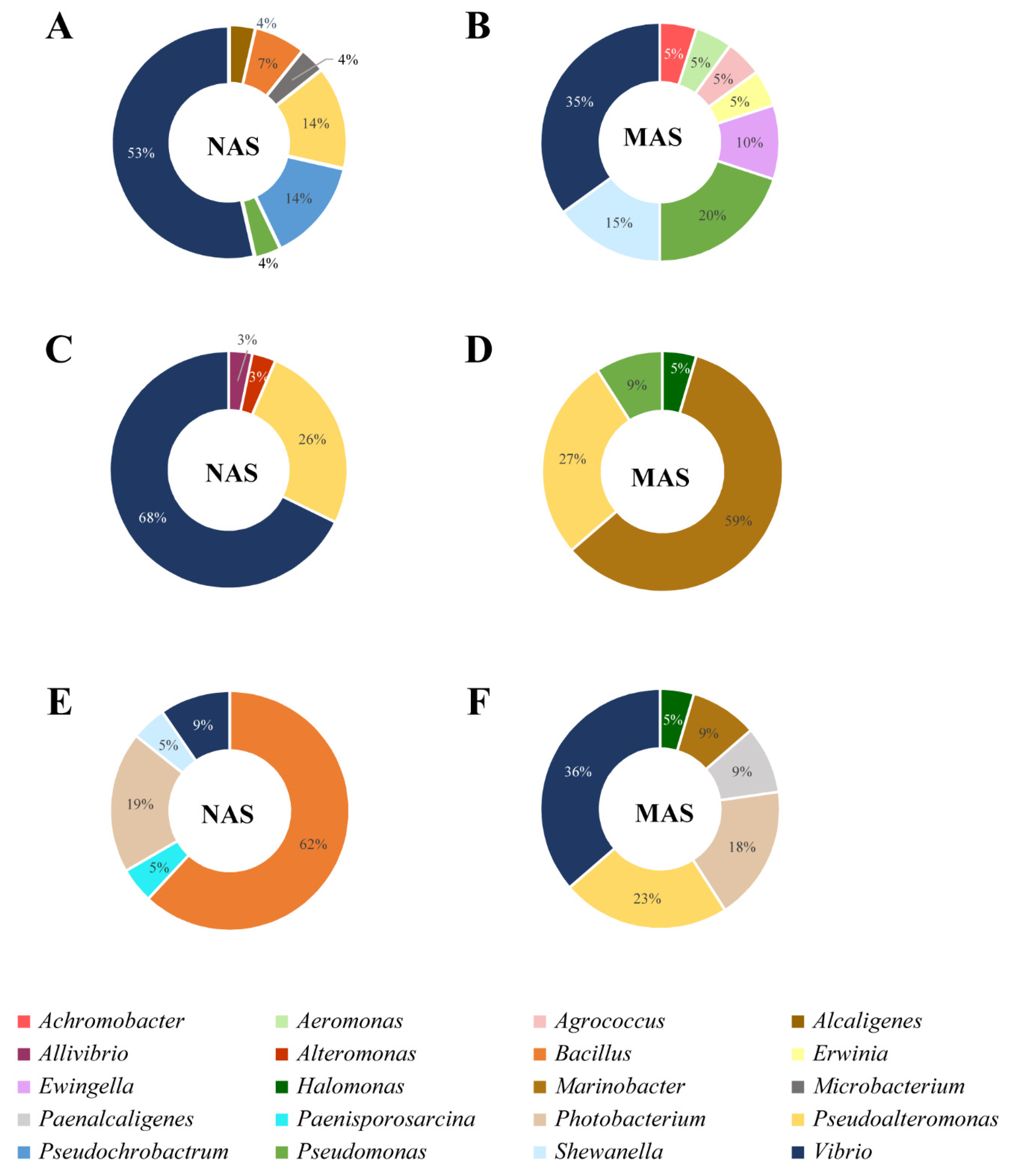
3.4. Culturable Microbiota

The 16S rRNA gene sequences were generated for 81 and 64 bacterial isolates from NAS and MAS farm, respectively. The results from the datasets of each fish farm were composed of three compartments: the skin swabs of European seabass, seawater, and sediments. Of the total number of bacterial isolates from the NAS farm, 28 were from skin swabs, 32 were from seawater, and 21 were from sediments. Based on the sequence comparison, 33 bacterial species were identified among the isolates from the NAS farm, which are listed in Table 4. Of them, two, namely V. alginolyticus and V. harveyi, were identified in both the skin and seawater. Only Bacillus hwajinpoensis was found in the skin and sediment (Figure 3).
At the MAS farm, 20 isolates out of 64 in total were isolated from the European seabass swabs, while 22 accounted for seawater and sediment isolates. A total of 22 bacterial species were identified among the isolates from all the sample types in the MAS farm (Table 5). Despite the difference of species number (Table 5) among the bacterial communities of the fish skin (11 species), seawater (3 species), and sediment (4 species), species Marinobacter litoralis, and two species from the genus Pseudoalteromonas (Pseudoalteromonas tetraodonis, Pseudoalteromonas undina) were detected in the seawater and sediment, whereas V. toranozoniae was identified in the skin and sediment (Figure 4).
A total of seven bacterial genera were identified by molecular analysis of bacterial isolates (N = 28) from skin swabs of European seabass from the NAS antibiotic-free farm. Figure 5A shows the occurrence of bacterial genera of skin swabs. The most common bacteria were bacteria of the genus Vibrio (53%), followed by the genera Pseudoalteromonas (14%) and Pseudochrobactrum (14%). V. alginolyticus (17.9%), and Pseudochrobactrum sacchaloryticum (14.3%), where V. cyclitrophicus (14.3%) and V. harveyi (14.3%) accounted for the largest number of the 11 bacterial species that were isolated (Table 4).
At the NAS antibiotics-free farm, the greatest diversity of bacterial genera (seven genera) was found in samples of skin swabs of European seabass. A total of five bacterial genera were detected in the sediment samples, while four bacterial genera were detected in the seawater samples. The most dominant bacterial genus that was identified in the seawater samples was Vibrio (65%), followed by Pseudoalteromonas (25%) (Figure 5C). Among the bacterial species, V. chagasii (18.8%), Pseudoalteromonas shioyasakiensis (9.4%), Pseudoalteromonas tetraodonis (9.4%), V. kanaloae (9.4%), and V. tubiashii (9.4%) were the most frequently identified.
In the sediment samples from the NAS farm, 21 bacterial isolates were obtained from five bacterial genera, among which the genus Bacillus (62%) dominated (Figure 5E). B. aquimaris (33.3%), Photobacterium aphoticum (19.0%), and B. hwajinpoensis (14.3%) corresponded to the most frequently identified bacterial species (Table 4).
V. alginolyticus and V. harveyi were isolated from European seabass skin swabs and seawater at the NAS antibiotic-free farm. B. hwajinpoensis was isolated from the skin swabs and sediment at the same farm. The seawater samples displayed the highest diversity of bacterial species (N = 16). A total of 11 bacterial species were identified from the skin swabs from the European seabass samples, while nine species were identified in the sediment samples (Table 4).
At the MAS conventional farm, among 20 bacterial isolates from skin swabs of European seabass, eight bacterial genera were identified, as shown in Figure 5B. The dominant bacterial genera were Vibrio (35%) and Pseudomonas (20%). Of the 12 bacterial species from the skin swabs of European seabass, V. anguillarum (20%), Shewanella arctica (15%), and V. toranzoniae (15%) were the most abundant (Table 5).
A total of four bacterial genera were identified in seawater samples from the MAS farm (22 bacterial isolates), of which the genus Marinobacter (59%) and the genus Pseudoalteromonas (27%) were the most abundant. The results of the presence of bacterial genera in the seawater samples are shown in Figure 5D. M. litoralis was most frequently identified bacterial species (59.1% of the total seawater isolates) (Table 5).
The identified bacterial genera from the sediment of the MAS farm and their representation are shown in Figure 5F. Among the six bacterial genera, Vibrio (36%), Pseudoalteromonas (23%), and Photobacterium (18%) were the most abundant. The most dominant bacterial species were V. toranzoniae (27.3%), Photobacterium lutimaris (18.2%), and Pseudoalteromonas tetraodonis (18.2%) (Table 5).
V. toranzoniae was identified in European seabass swab samples and in sediment samples from the MAS farm with antibiotic use. M. litoralis was identified in the seawater and in the sediment samples at this farm, as well as Pseudoalteromonas tetraodonis and Pseudoalteromonas undina. Other bacterial species were detected only in one sample type. Only the genus Pseudomonas and the genus Vibrio were identified from the swab samples at both farms.
To confirm the identity of Vibrio species and check their grouping within different clades, phylogenetic analysis based on 16S rRNA including reference sequences from GenBank was performed. As expected, phylogenetic analysis confirmed grouping into five clades: V. splendidus, V. anguillarum, V. orientalis, V. fortis, and V. harveyi as well as branching with the same species (Figure 6). In the Table 4 and Table 5 the percent of identity with the known Vibrio species from GenBank are shown.

3.5. Antimicrobial Resistance of Bacterial Isolates

The disc diffusion test for susceptibility to 13 antibiotics was performed using the total of 145 bacterial isolates from the two fish farms. The number of isolates that were resistant to individual antibiotics and the percentage of resistant isolates are shown in Table 6.
Most of the bacterial isolates from the skin swabs of European seabass from the NAS farm without antibiotic use showed resistance to vancomycin (85.7%), ampicillin (57.1%), enrofloxacin (39.3%), erythromycin (39.3%), and streptomycin (39.3%). All the isolates of V. alginolyticus were resistant to vancomycin and ampicillin (Table 7). The resistance of isolates to other antibiotics was inconsistent.
Most of the bacterial isolates from the skin swabs of European seabass from the MAS conventional farm with antibiotic use were resistant to ampicillin (100.0%), vancomycin (100.0%), erythromycin (75.0%), florfenicol (65.0%), and imipenem (65.0%). The predominant bacterial species in the samples was V. anguillarum and the isolates were resistant to enrofloxacin, ampicillin, erythromycin, vancomycin, and streptomycin (Table 7). No statistically significant differences in the number of antibiotic-resistant bacterial isolates were found between the two farms (p > 0.05).
Similar to the isolates from the skin swabs, most bacterial isolates from seawater from the NAS antibiotics-free farm showed resistance to vancomycin (84.4%), ampicillin (50.0%), enrofloxacin (31.3%), erythromycin (37.5%), and streptomycin (40.6%). The predominant bacterial species in the seawater samples was V. chagasii and all V. chagasii isolates showed resistance to vancomycin (Table 7).
Among the bacterial isolates from the farm seawater samples at the MAS farm, vancomycin resistance was the most common (90.9%). In seawater samples, the most common bacterial species was M. litoralis and all M. litoralis isolates were resistant to vancomycin, while resistance was not consistent among the other antibiotics. The differences in the number of resistant bacterial isolates between the two farms were not statistically significant (p > 0.05).
Most of the bacterial isolates from the sediment samples from the NAS antibiotics-free farm showed resistance to enrofloxacin (61.9%) and flumequine (52.9%). B. aquimaris was the most common isolate and most of the bacterial isolates (six out of seven) were resistant to enrofloxacin. For other antibiotics, the resistance of the bacterial isolates was not uniform.
As high as 86.4% of total bacterial isolates from the MAS farm sediment samples were resistant to vancomycin. This is also characteristic for the most numerous sediment isolates that were identified as V. toranzoniae (Table 7). In contrast to the results of resistance from the skin swabs and seawater isolates, the number of antibiotic-resistant bacterial isolates between the two farms were statistically significantly higher at the NAS antibiotic-free farm than in the MAS conventional farm (p < 0.05).

4. Discussion

The quality of seawater has a great influence on the success of fish farming. Stress that is caused by disturbances in seawater quality negatively affects fish growth and development [21]. The seawater physicochemical parameters that were measured at the NAS and MAS farms were consistent with previous studies on Adriatic Sea bass farms [17,22,23].
Microbiological analysis of the seawater samples and skin swabs of European seabass revealed a higher HPC and Vibrio count in the seawater samples from the MAS farm, but conversely higher HPC and Vibrio count from skin swabs from the NAS farm. This HPC count from the skin swabs of European seabass from the NAS farm (69.1 ± 21.7 CFU/cm2) is lower than a previously reported range between 102 and 104 CFU/cm2, at other geographical locations [8]. The higher Vibrio count in the seawater samples from the MAS farm can be explained by the influence of the higher seawater temperature which stimulates bacterial growth [22,24]. Interestingly, a higher Vibrio count was detected in samples from the fish skin than in the seawater of the NAS farm. This phenomenon was described by Vatsos [25] who documented the quantitative and qualitative differences between the culturable microbiota of the fish skin and that in the water from the host environment. Although the identification of Vibrio species is challenging due to similarities, both in phenotypic properties and 16S rRNA sequences, phylogenetic analyses can differentiate clades and subgroups affiliation [26]. That is why we used this analysis for the Vibrio species differentiation.
In this study, a similar number of bacterial genera was observed in each type of sample (skin swabs, seawater, sediment) after analyzing the composition of the culturable microbiota from the two investigated farms (under culture conditions with and without the use of antibiotics). This could be because each bacterial species grows under specific culture conditions. A total of four genera were found in the seawater samples from both farms, but with different prevalence. The number of bacterial species differed between the seawater samples from the NAS antibiotics-free farm and the MAS conventional farm, where 16 and 6 bacterial species, respectively, were identified. The diversity of bacterial species is strongly influenced by the quality of seawater in the farm [27]. Considering that the physicochemical and microbiological parameters in both farms showed seawater quality that is suitable for farming [17,23], the use of antibiotics at the MAS farm may have affected diversity of the bacterial species. Rosado et al. [6] stated that the use of antibiotics can affect the change in the composition of bacterial species in the skin microbiota, reducing the diversity of bacteria and the resistance of farmed fish to bacterial infections. Nevertheless, in this work, 12 bacterial species were identified from skin swabs in the MAS farm where antibiotics are regularly administered. At the NAS farm, where antibiotics are not used, 11 bacterial species were identified from the skin swabs. However, two common genera, Pseudomonas and Vibrio, were identified in the skin swab samples at both farms. Our observation agrees with those of Čož Rakovac et al. [28], who previously reported Pseudomonas isolates from wild and farmed sea bass in the Northern Adriatic Sea. These results are similar with those of recent study of skin microbiota of farmed European seabass in South Adriatic (Croatia and Montenegro), where most of the isolated bacteria comprised of Vibrio, Photobacterium, and Pseudomonas genera [16]. The skin is one of the main entry points for pathogens and infections, while the mucus of the fish skin has an important role in the immune system and has an antibacterial effect [1]. Therefore, one of the objectives of this study was to identify bacterial species from skin swabs of European seabass with particular emphasis on pathogenic bacteria to fish. An analysis of the identified bacterial species reveals some potentially pathogenic bacteria to fish [4]. According to the study of De Bruijn et al. [8], the microbiota of fish skin contains pathogenic bacteria to fish that do not cause disease unless the microbiota balance is disturbed. It is well known that antibiotic use can promote the proliferation of opportunistic pathogens [29], e.g., V. anguillarum in skin swabs from MAS conventional farm.
Pseudoalteromonas undina was isolated from skin swabs of European seabass from the NAS farm. P. undina is widely distributed in seawater, especially in farms that are rich in organic matter, but has not been reported as a pathogenic bacterium, except in a study by Pujalte et al. [30], in which it caused mortalities of sea bass. The opportunistic pathogens V. alginolyticus and V. harveyi have also been isolated from NAS farms. V. alginolyticus is known to cause vibriosis [17,31], while V. harveyi is also a potential threat for European seabass farming [5,32].
The samples of European seabass skin swabs from the MAS farm also contained some potentially pathogenic bacteria to fish. V. anguillarum is one of the causative agents of vibriosis in the Adriatic Sea [14], while V. toranzoniae has only been identified as a pathogen in diseased sea eels (Genypterus chilensis) in Chile [33]. In addition, Pseudomonas gessardii has been isolated and identified as a larval shrimp opportunistic pathogen in a recent study, while V. alginolyticus has been identified as the primary pathogen [31].
Considering the potential pathogenicity of V. alginolyticus, V. anguillarum, and V. harveyi, it is important to highlight their resistance to antibiotics, although this varied from isolate to isolate. V. alginolyticus from the skin swabs of European seabass from a NAS farm showed resistance to streptomycin and ampicillin. In previous studies, V. alginolyticus that was isolated from skin swabs of sea bass from three farms in the Adriatic Sea (Lim Bay, Lamjana and Mali Ston Bay) showed higher resistance to five (ampicillin, penicillin, piperacillin, sulfamethoxazole/trimethoprim, and trimethoprim) of the 13 antibiotics that were tested (ampicillin, streptomycin, gentamicin, imipenem, chloramphenicol, florfenicol, ciprofloxacin, enrofloxacin, erythromycin, oxytetracycline, sulfamethoxazole/trimethoprim, vancomycin, and flumequine) [17]. In addition, all the isolates of V. alginolyticus in this study were sensitive or moderately sensitive to sulfamethoxazole/trimethoprim, which is commonly used to treat vibriosis; this is consistent with the studies of Zorrilla et al. [34] who tested seven antimicrobial agents (ampicillin, amoxicillin, tetracycline, oxytetracycline, trimethoprim-sulphamethoxazole, oxolinic acid, and flumequine). Isolates of V. harveyi from the NAS farm were found to be resistant to ampicillin, erythromycin, and vancomycin. In a similar study, Veić [5] tested nine antibiotics (ampicilin, trimethoprim/sulfadiazine, chloramphenicol, oxytetracycline, enrofloxacin, flumequine, nalidixic acid, gentamicin, and neomycin) and described resistance to gentamicin and neomycin, as well as to ampicillin and erythromycin in all the strains, and intermediate sensitivity to nalidixic acid and oxytetracycline in the same V. harveyi strains that were isolated from diseased European seabass in the Adriatic Sea. Kang et al. [35] tested 16 antibiotics (ampicillin, cefotaxime, cefotetan, cephalothin, chloramphenicol, ciprofloxacin, cefepime, erythromycin, gentamicin, kanamycin, nalidixic acid, rifampicin, streptomycin, tetracycline, trimethoprim/sulfamethoxazole, and vancomycin) and reported that V. harveyi that was isolated from seawater in South Korea was resistant to ampicillin and vancomycin, and sensitive to erythromycin.
V. anguillarum that was isolated from the MAS conventional farm was resistant to ampicillin, enrofloxacin, erythromycin, streptomycin, and vancomycin. Resistance of V. anguillarum that was isolated from farmed rainbow trout (Oncorhynchus mykiss) was determined for cloxacillin, ampicillin, sulfamethoxazole/trimethoprim, and erythromycin [36]. Variations in results are commonly attributed to the difference between the tested organism as well as the sampling location. In a recent study that was carried out by Kapetanović et al. [14], V. anguillarum that was isolated from the skin and gills of European seabass (Mali Ston Bay) showed resistance to gentamicin, erythromycin, and streptomycin, which is not entirely consistent with the results of this study. Similarly, Veić [5] determined the resistance of isolated V. anguillarum strains from diseased European seabass in the Adriatic Sea to ampicillin and erythromycin. Thus, in our study, V. anguillarum was found to be more resistant, indicating the possible development of bacterial resistance to certain antibiotics to which resistance was not found in previous studies.
The results of antimicrobial resistance in sediment seem to be somewhat controversial, as resistance is higher in the sediment from the NAS farm where antibiotics are not used. Increased antibacterial resistance of bacteria in sediment is often the most sensitive environmental indicator of past antibiotic use [37]. Vibrionaceae were widely distributed in sediment samples on fish farms in the western Mediterranean and their resistance is known due to the antibiotics administered [38]. Interestingly, in our study, antibiotic resistance was detected in the microbiota of the NAS farm where antibiotics were not administered since the aquaculture production was established. Antibiotic resistance depends on the species and the location where the bacteria was isolated. It is well known that antibiotic resistance is higher along the coasts and in sheltered bays than in open waters but could also occur in more remote waters due to the influence of non-aquatic organisms or pollutants [39].
The resistant strains at the antibiotic-free farm could be of terrestrial/agricultural origin. In fact, the nearby island of Cres is known for its traditional and extensive farming of autochthonous sheep, which has previously been diagnosed clinical mastitis, enterotoxaemia, actinobacillosis, contagious ecthyma, foot rot, and Brucella ovis infection [40]. The use of various antibiotics for treatment of bacterial diseases in sheep, including those based on enrofloxacin and flumequine [40,41], might promote the development of antibiotic resistance that can potentially be carried towards surface waters and sediments by rainfall runoffs [42]. This should be considered as a potential risk for the release of bacterial pathogens into the water column and the spread of antibiotic resistance across different marine compartments and should be investigated in future studies. There is also the possibility that resistant bacteria were introduced on the NAS farm by fingerlings, which also should be investigated.
These results confirm the presence of some potentially pathogenic bacteria for fish. Bacterial fish diseases are usually treated with antibiotics. The most commonly used antibiotics include tetracyclines and fluoroquinolones. However, there is a risk of antibiotic resistance development, as multidrug-resistant bacteria can be easily spread in the marine environment. In addition, the inadequate treatment of fish diseases with antibiotics can lead to selection of bacteria that respond less to antibiotic treatments and increase the likelihood of disease outbreaks in fish farms. The need to reduce financial losses in European seabass aquaculture, overcome the risk of spreading antibiotic-resistant bacteria, and concerns about environmental impacts and consumer safety require alternative and sustainable control measures for frequent antibiotic therapy in aquaculture.
To our knowledge, this is the first study comparing the culturable microbiota at fish farms with and without the use of antibiotics. Our results should be extended in further studies with a larger number of samplings as well as antibiotic residues analyses in the water and sediment, all of which may affect the results. Before each stocking in the cages, the microbiota of the skin of fish fingerlings should be analyzed and included in further studies. Based on the preliminary results of the present study, further research is needed to confirm these findings and to investigate whether and how the microbiota differs seasonally in these farms with and without antibiotic use.

Supplementary Materials

The following supporting information can be downloaded at: https://www.mdpi.com/article/10.3390/jmse10030303/s1, Table S1: List of bacterial isolates/strains and their accession numbers from GenBank used for the phylogenetic tree construction (Figure 6, Results).

Author Contributions

Conceptualization, D.K., A.R. and A.G.; methodology, A.R., I.V.S., A.K., D.K. and A.G.; validation, I.B. and A.G.; field work, T.K., D.K., A.G. and I.B.; formal analysis, A.R., D.K., I.V.S. and F.B.; investigation, D.K., I.B., F.B. and A.G.; resources, D.K. and I.B.; writing—original draft preparation, A.R., D.K., I.V.S. and L.P.; writing—review and editing, A.K. and A.G.; visualization, A.R., D.K. and I.V.S.; supervision, I.V.S., L.P. and A.G.; project administration, T.K., D.K.; principal investigator, T.K.; funding acquisition, T.K. All authors have read and agreed to the published version of the manuscript.

Funding

This work has been fully supported by the Croatian Science Foundation’s funding of the under the project AqADAPT—Grant No IP-2018-01-3150, Croatian Academy of Sciences and Arts, and Croatian Ministry of Science and Education under the bilateral project with Germany (DAAD) EXAQUA.

Institutional Review Board Statement

Not applicable.

Informed Consent Statement

Not applicable.

Data Availability Statement

The data presented in this study are available on request from the corresponding author. The data are not publicly available because it’s an ongoing scientific project AqADAPT—Grant No IP-2018-01-31504.

Acknowledgments

This work has been fully supported by the Croatian Science Foundation’s funding of the under the project AqADAPT—Grant No IP-2018-01-3150. We also thank the Croatian Academy of Sciences and Arts for funding the project: The presence of resistant bacterial strains as part of the natural microflora of European seabass (Dicentrarchus labrax) cultured under conditions with and without the use of antibiotics: truths and prejudices. We thank our colleagues at the Helmholtz Centre for Environmental Research GmbH-UFZ, Leipzig, Germany, who collaborated on the Croatian-German bilateral project: Exploring the potential of phage and microbiomes to control pathogens in aquaculture (EXAQUA). The publication of this article was funded by the Croatian Ministry of Science and Education within the framework of the bilateral project with Germany (DAAD) EXAQUA. We thank the anonymous reviewers for their useful comments and suggestions for our article.

Conflicts of Interest

The authors declare no conflict of interest. The funders had no role in the design of the study; in the collection, analyses, or interpretation of data; in the writing of the manuscript, or in the decision to publish the results.

References

  1. Esteban, M.A. An Overview of the Immunological Defenses in Fish Skin. Int. Sch. Res. Netw. 2012, 2012, 853470. [Google Scholar] [CrossRef] [Green Version]
  2. FAO. 2021. Available online: https://www.fao.org/fishery/countrysector/naso_croatia/en (accessed on 8 November 2021).
  3. Camara-Ruiz, M.; Cerezo, I.M.; Guardiola, F.A.; Garcia-Beltran, J.M.; Balebona, M.C.; Morinigo, M.A.; Esteban, M.A. Alteration of the Immune Response and the Microbiota of the Skin during a Natural Infection by Vibrio harveyi in European Seabass (Dicentrarchus labrax). Microorganisms 2021, 9, 964. [Google Scholar] [CrossRef] [PubMed]
  4. Haenen, O.L.M.; Fouz, B.; Amaro, C.; Isern, M.M.; Mikkelsen, H.; Zrncic, S.; Travers, M.A.; Renault, T.; Wardle, R.; Hellström, A.; et al. Vibriosis in aquaculture. In Proceedings of the 16th EAFP Conference, Tampere, Finland, 4 September 2013; pp. 138–148. [Google Scholar]
  5. Veić, T. Characterization of Vibrio Species Isolated from European Sea Bass (Dicentrarhus Labrax, Linnaeus, 1758) Farmed on the Eastern Adriatic Sea. Master’s Thesis, University of Zagreb, Zagreb, Croatia, 2016. [Google Scholar]
  6. Rosado, D.; Xavier, R.; Severino, R.; Tavares, F.; Cable, J.; Perez-Losada, M. Effects of disease, antibiotic treatment and recovery trajectory on the microbiome of farmed seabass (Dicentrarchus labrax). Sci. Rep. 2019, 9, 18946. [Google Scholar] [CrossRef] [PubMed]
  7. Rosado, D.; Perez-Losada, M.; Severino, R.; Cable, J.; Xavier, R. Characterization of the skin and gill microbiomes of the farmed seabass (Dicentrarchus labrax) and seabream (Sparus aurata). Aquaculture 2018, 500, 57–64. [Google Scholar] [CrossRef]
  8. Bruijn, I.D.; Liu, Y.; Wiegertjes, G.F.; Raaijmakers, J.M. Exploring fish microbial communities to mitigate emerging diseases in aquaculture. FEMS Microbiol. Ecol. 2018, 94, 1–12. [Google Scholar] [CrossRef]
  9. Reverter, M.; Tapissier-Bontemps, N.; Lecchini, D.; Banaigs, B.; Sasal, P. Biological and Ecological Roles of External Fish Mucus: A Review. Fishes 2018, 3, 41. [Google Scholar] [CrossRef] [Green Version]
  10. Gatesoupe, F.-J.; Huelvan, C.; Le Bayon, N.; Le Delliou, H.; Madec, L.; Mouchel, O.; Quazuguel, P.; Mazurais, D.; Zambonino-Infante, J.-L. The highly variable microbiota associated to intestinal mucosa correlates with growth and hypoxia resistance of sea bass, Dicentrarchus labrax, submitted to different nutritional histories. BMC Microbiol. 2016, 16, 266. [Google Scholar] [CrossRef] [Green Version]
  11. Perez-Pascual, D.; Estelle, J.; Dutto, G.; Rodde, C.; Bernardet, J.-F.; Marchand, Y.; Duchaud, E.; Przybyla, C.; Ghigo, J.-M. Growth Performance and adaptability of European sea bass (Dicentrarchus labrax) gut microbiota to alternative diets free of fish products. Microorganisms 2020, 8, 1346. [Google Scholar] [CrossRef]
  12. Silvi, S.; Nardi, M.; Sulpizio, R.; Orpianesi, C.; Caggiano, M.; Carnevali, O.; Cresci, A. Effect of the addition of Lactobacillus delbrueckii subsp. delbrueckii on the gut microbiota composition and contribution to the well-being of European sea bass (Dicentrarchus labrax, L.). Microb. Ecol. Health Dis. 2008, 20, 53–59. [Google Scholar] [CrossRef] [Green Version]
  13. Roquigny, R.; Mougin, J.; Le Bris, C.; Bonnin-Jusserand, M.; Doyen, P.; Grard, T. Characterization of the marine aquaculture microbiome: A seasonal survey in a seabass farm. Aquaculture 2021, 531, 735987. [Google Scholar] [CrossRef]
  14. Kapetanovic, D.; Gavrilovic, A.; Jug-Dujakovic, J.; Smrzlic, I.V.; Kazazic, S.; Bojanic-Rasovic, M.; Kolda, A.; Pesic, A.; Peric, L.; Zunic, J.; et al. Assessment of microbial sea water quality and health status of farmed European seabass (Dicentrarchus labrax) in Eastern Adriatic sea (Montenegro and Croatia). Stud. Mar. 2019, 32, 52–64. [Google Scholar] [CrossRef]
  15. Rosado, D.; Pérez-Losada, M.; Pereira, A.; Severino, R.; Xavier, R. Effects of aging on the skin and gill microbiota of farmed seabass and seabream. Anim. Microbiome 2021, 3, 10. [Google Scholar] [CrossRef]
  16. Pepi, M.; Focardi, S. Antibiotic-Resistant Bacteria in Aquaculture and Climate Change: A Challenge for Health in the Mediterranean Area. Int. J. Environ. Res. Public Health 2021, 18, 5723. [Google Scholar] [CrossRef] [PubMed]
  17. Damir, K.; Irena, V.S.; Damir, V.; Emin, T. Occurrence, characterization and antimicrobial susceptibility of Vibrio alginolyticus in the Eastern Adriatic sea. Mar. Pollut. Bull. 2013, 75, 46–52. [Google Scholar] [CrossRef] [PubMed]
  18. Wilson, K.H.; Blitchington, R.B.; Greene, R.C. Amplification of bacterial 16S ribosomal DNA with polymerase chain reaction. J. Clin. Microbiol. 1990, 28, 1942–1946. [Google Scholar] [CrossRef] [PubMed] [Green Version]
  19. Hall, T.A. BioEdit: A user-friendly biological sequence alignment editor and analysis program for Windows 95/98/NT. Nucl. Acids. Symp. Ser. 1999, 41, 95–98. [Google Scholar]
  20. Tamura, K.; Stecher, G.; Kumar, S. MEGA11: Molecular Evolutionary Genetics Analysis Version 11. Mol. Biol. Evol. 2021, 38, 3022–3027. [Google Scholar] [CrossRef] [PubMed]
  21. Philipose, K.K.; Sharma, S.R.; Loka, J.; Damodaran, D.; Rao, G.S.; Vaidya, N.G.; Sonali, S.M.; Sadhu, N.; Dube, P. Observations on variations in physico-chemical water parameters of marine fish cage farm off Karwar. Indian J. Fish. 2012, 59, 83–88. [Google Scholar]
  22. Kapetanovic, D.; Smrzlic, I.V.; Valic, D.; Teskeredzic, Z.; Teskeredzic, E. Culturable microbiota associated with farmed Atlantic bluefin tuna (Thunnus thynnus). Aquat. Living Resour. 2017, 30, 30. [Google Scholar] [CrossRef] [Green Version]
  23. Matijevic, S.; Kuspilic, G.; Morovic, M.; Grbec, B.; Bogner, D.; Skejic, S.; Veza, J. Physical and chemical properties of the water column and sediments at sea bass/sea bream farm in the middle Adriatic (Maslinova bay). Acta Adriat. 2009, 50, 59–76. [Google Scholar]
  24. Roux, F.L.; Wegner, K.M.; Baker-Austin, C.; Vezzulli, L.; Osorio, C.R.; Amaro, C.; Ritchie, J.M.; Defoirdt, T.; Destoumieux-Garzón, D.; Blokesch, M.; et al. The emergence of Vibrio pathogens in Europe: Ecology, evolution, and pathogenesis (Paris, 11-12th March 2015). Front. Microbiol. 2015, 6, 830. [Google Scholar] [CrossRef] [PubMed] [Green Version]
  25. Vatsos, I.N. Standardizing the microbiota of fish used in research. Lab. Anim. 2017, 51, 353–364. [Google Scholar] [CrossRef] [PubMed] [Green Version]
  26. Franco, A.; Rückert, C.; Blom, J.; Busche, T.; Reichert, J.; Schubert, P.; Goesmann, A.; Kalinowski, J.; Wilke, T.; Kämpfer, P.; et al. High diversity of Vibrio spp. associated with different ecological niches in a marine aquaria system and description of Vibrio aquimaris sp. nov. Syst. Appl. Microbiol. 2020, 43, 126123. [Google Scholar] [CrossRef] [PubMed]
  27. Onianwah, I.F.; Stanley, H.O.; Oyakhire, M. Microorganisms in Aquaculture Development. Glob. Adv. Res. J. Microbiol. 2018, 7, 127–131. [Google Scholar]
  28. Čož-Rakovac, R.; Strunjak-Perović, I.; Topić Popović, N.; Hacmenjak, M.; Šimpraga, B.; Teskeredžić, E. Health status of wild and cultured sea bass in the northern Adriatic Sea. Vet. Med. 2002, 47, 222–226. [Google Scholar] [CrossRef] [Green Version]
  29. Romero, J.; Feijoo, C.G.; Navarrete, P. Antibiotics in Aquaculture—Use, Abuse and Alternatives. In Health and Environment in Aquaculture; Carvalho, E., Ed.; InTech: London, UK, 2012; ISBN 978-953-51-0497-1. Available online: http://www.intechopen.com/books/health-and-environment-in-aquaculture/antibioticsin-aquaculture-use-abuse-and-alternatives-23/12/2021 (accessed on 23 December 2021).
  30. Pujalte, M.J.; Sitjà-Bobadilla, A.; Macián, M.C.; Álvarez-Pellitero, P.; Garay, E. Occurrence and virulence of Pseudoalteromonas spp. in cultured gilthead sea bream (Sparus aurata L.) and European sea bass (Dicentrarchus labrax L.). Molecular and phenotypic characterisation of P. undina strain U58. Aquaculture 2007, 271, 47–53. [Google Scholar] [CrossRef]
  31. Hamza, F.; Kumar, A.R.; Zinjarde, S. Efficacy of cell free supernatant from Bacillus licheniformis in protecting Artemia salina against Vibrio alginolyticus and Pseudomonas gessardii. Microb. Pathog. 2018, 116, 335–344. [Google Scholar] [CrossRef]
  32. Vendramin, N.; Zrncic, S.; Padros, F.; Oraic, D.; Le Breton, A.; Zarza, C.; Olesen, N.J. Fish health in Mediterranean Aquaculture, past mistakes and future challenges. Bull. Eur. Assoc. Fish Pathol. 2016, 36, 38–45. [Google Scholar]
  33. Lasa, A.; Avendaño-Herrera, R.; Estrada, J.M.; Romalde, J.L. Isolation and identification of Vibrio toranzoniae associated with diseased red conger eel (Genypterus chilensis) farmed in Chile. Vet. Microbiol. 2015, 179, 327–331. [Google Scholar] [CrossRef]
  34. Zorrilla, I.; Chabrillón, M.; Arijo, S.; Dıaz-Rosales, P.; Martınez-Manzanares, E.; Balebona, M.C.; Morinigo, M.A. Bacteria recovered from diseased cultured gilthead sea bream (Sparus aurata L.) in southwestern Spain. Aquaculture 2003, 218, 11–20. [Google Scholar] [CrossRef]
  35. Kang, C.H.; Kim, Y.G.; Oh, S.J.; Mok, J.S.; Cho, M.H.; So, J.S. Antibiotic resistance of Vibrio harveyi isolated from seawater in Korea. Mar. Pollut. Bull. 2014, 86, 261–265. [Google Scholar] [CrossRef] [PubMed]
  36. Parin, U.; Erbaş, G.; Savasan, S.; Yuksel, H.T.; Gurpinar, S.; Kirkan, S. Antimicrobial resistance of Vibrio (Listonella) anguillarum isolated from rainbow trouts (Oncorhynchus mykiss). Indian J. Anim. Res. 2019, 53, 1522–1525. [Google Scholar] [CrossRef]
  37. Kummerer, K. Resistance in the environment. J. Antimicrob. Chemother. 2004, 54, 311–320. [Google Scholar] [CrossRef] [PubMed] [Green Version]
  38. Chelossi, E.; Vezzulli, L.; Milano, A.; Branzoni, M.; Fabiano, M.; Riccardi, G.; Banat, I.M. Antibiotic resistance of benthic bacteria in fish-farm and control sediments of the Western Mediterranean. Aquaculture 2003, 219, 83–97. [Google Scholar] [CrossRef]
  39. Baquero, F.; Martinez, J.-L.; Canton, R. Antibiotics and antibiotic resistance in water environments. Curr. Opin. Biotechnol. 2008, 19, 260–265. [Google Scholar] [CrossRef] [PubMed]
  40. Kostelic, A.; Artukovic, B.; Beck, R.; Benic, M.; Cergolj, M.; Stokovic, I.; Barac, Z. Diseases of sheep on Croatian islands. In Proceedings of the XVI. Congress of the Mediterranean Federation for Health and Production of Ruminants: FeMeSPRum, Zadar, Croatia, 26 April 2008; pp. 227–232. [Google Scholar]
  41. Official Gazette 21/2011 Ordinance on Pharmacologically Active Substances and Their Classification in Relation to the Maximum Permitted Levels of Residues in Food of Animal Origin. Available online: https://narodne-novine.nn.hr/clanci/sluzbeni/2005_03_29_510.html (accessed on 23 December 2021).
  42. Done, H.Y.; Venkatesan, A.K.; Halden, R.U. Does the Recent Growth of Aquaculture Create Antibiotic Resistance Threats Different from those Associated with Land Animal Production in Agriculture? AAPS J. 2015, 17, 513–524, Erratum in: AAPS J. 2016, 18, 1583. [Google Scholar] [CrossRef] [PubMed] [Green Version]
Figure 1. Sampling locations: NAS—marine fish farm in the north Adriatic and MAS—marine fish farm in the middle Adriatic.
Jmse 10 00303 g001
Figure 2. (A) The total body length (cm) and (B) the total body weight (g) of sea bass (N = 10) from sites NAS () and MAS (). Square boxes indicate the lower and upper quartiles and the whiskers represent the minimum and maximum data values (1.5 interquartile range). The medians are depicted by a solid line. * Significant difference (p < 0.05) between the sites.
Jmse 10 00303 g002
Figure 3. Venn diagram of the bacterial species that were identified in culturable microbiota of fish skin, seawater, and sediment associated with the NAS marine fish farm.
Jmse 10 00303 g003
Figure 4. Venn diagram of the bacterial species that were identified in culturable microbiota of fish skin, seawater, and sediment associated with marine fish farm MAS.
Jmse 10 00303 g004
Figure 5. Representation of the bacterial genera (percentage) in: (A)—European seabass skin swab samples from the NAS and (B)—the MAS farm; (C)—in seawater samples from the NAS farm and (D)—the MAS farm; (E)—in sediment samples from the NAS farm and (F)—the MAS farm.
Jmse 10 00303 g005
Figure 6. Phylogenetic analysis of studied Vibrio species that was inferred from partial 16S rRNA gene sequences using MEGA11 software—the clades are indicated by brackets.
Jmse 10 00303 g006
Table 1. Physicochemical analysis of seawater from the NAS and MAS fish farms.
Depth
(m)
Secchi (m)Sal. (ppt)Cond. (µS/cm)TDS (mg/L)Temp. (°C)pHDO (mg/L)DO (%)
NAS28 m
0.5 m 38.2551.537.322.78.357.0196.4
6 m 38.2651.537.321.78.366.9495.4
12 m 38.2751.537.319.78.376.9395.3
0.5 m above the bottom 38.4346.837.515.18.236.6283.7
MAS20 m
0.5 m 37.356.228.123.58.128.4899.2
6 m 37.156.028.023.08.138.0793.4
12 m 37.256.328.022.58.138.3595.5
0.5 m above the bottom 37.256.228.120.18.168.0287.6
NAS—North Adriatic Sea antibiotic-free farm; MAS—Middle Adriatic Sea conventional farm; Secchi—Secchi depth; Sal.—Salinity; Cond.—Conductivity; TDS—Total dissolved solids; Temp.—Temperature; DO (mg/L)—Dissolved oxygen values; DO%—Dissolved oxygen saturation.
Table 2. Microbiological analysis of seawater and sediment from the NAS and MAS farms.
NASMAS
Sample TypeTC
(MPN/100 mL)
EC
(MPN/100 mL)
EN
(MPN/100 mL)
TC
(MPN/100 mL)
EC
(MPN/100 mL)
EN
(MPN/100 mL)
Seawater
0.5 m487.0<10.0<10.088.025.510.0
6 m588.0<10.0<10.081.510.0< 10.0
12 m1034.0<10.0<10.020.010.0< 10.0
0.5 m above the bottom10.0<10.0<10.020.5< 10.0< 10.0
Sediment<10.0<10.0<10.015.0< 10.046.5
NAS—North Adriatic Sea antibiotics-free farm; MAS—Middle Adriatic Sea conventional farm; TC—Total coliforms; EC—E. coli; EN—Enterococci.
Table 3. Microbiological analysis of heterotrophic bacteria (HPC) and Vibrio count from skin swabs of European seabass, seawater, and sediment from NAS and MAS farm.
NASMAS
SkinWaterSedimentSkinWaterSediment
HPC (CFU/mL)69.1 ± 21.7278.75 ± 196.360 ± 14.136.2 ± 20.0480 ± 281.9120 ± 11.3
Vibrio (CFU/mL)38.1 ± 35.214 ± 3.63 ± 05 ± 7.155 ± 51.188 ± 4.2
Table 4. Results of molecular identification of bacterial isolates from the NAS antibiotic-free farm.
Sample Type Skin
(n = 28)
Seawater
(n = 32)
Sediment (n = 21)
SpeciesPercent Identity %No.%No.%No.%
Alcaligenes faecalis97.913.6
Aliivibrio finisterrensis 99.0 13.1
Alteromonas macleodii 99.6 26.3
Bacillus aquimaris 99.7–99.8 733.3
Bacillus horikoshii 99.6 14.8
Bacillus hwajinpoensis99.4–100.027.1 314.3
Bacillus idriensis 99.8 14.8
Bacillus tianshenii 99.7 14.8
Microbacterium oxydans99.613.6
Paenisporosarcina quisquiliarum99.5 14.8
Photobacterium aphoticum99.0–99.9 419.0
Pseudoalteromonas arabiensis 99.2–99.5310.7
Pseudoalteromonas hodoensis 99.5 13.1
Pseudoalteromonas phenolica 99.8 13.1
Pseudoalteromonas shioyasakiensis 99.4–99.7 39.4
Pseudoalteromonas tetraodonis 99.7–99.9 39.4
Pseudoalteromonas undina 100.013.6
Pseudochrobactrum saccharolyticum98.9–99.5414.3
Pseudomonas zhaodongensis99.413.6
Shewanella marinintestina99.9 14.8
Vibrio alginolyticus99.6–100.0517.926.3
Vibrio chagasii 98.2–98.8 618.8
Vibrio crassostreae 99.1 13.1
Vibrio cyclitrophicus 99.4–100.0414.3
Vibrio europaeus 99.7 13.1
Vibrio fortis 99.1–99.3 26.3
Vibrio gigantis 99.6 13.1
Vibrio harveyi 99.7–100.0414.313.1
Vibrio hyugaensis 99.7 13.1
Vibrio kanaloae 99.2–100.0 39.4
Vibrio neocaledonicus 99.827.1
Vibrio toranzoniae 98.5–99.9 29.5
Vibrio tubiashii 96.1–99.3 39.4
Table 5. Results of molecular identification of the bacterial isolates from the MAS conventional farm.
Sample Type Skin (n = 20)Seawater (n = 22)Sediment (n = 22)
SpeciesPercent
Identity %
No.%No.%No.%
Achromobacter spanius 98.715.0
Aeromonas molluscorum 99.715.0
Agrococcus sp. 98.915.0
Erwinia billingiae 99.615.0
Ewingella americana 99.3–99.4210.0
Halomonas aquamarina 99.5 14.5
Halomonas boliviensis 99.8 14.5
Marinobacter litoralis 99.4–100.0 1359.129.1
Paenalcaligenes suwonensis 99.6–99.7 29.1
Photobacterium lutimaris 98.4–98.8 418.2
Pseudoalteromonas tetraodonis99.9 29.1418.2
Pseudoalteromonas undina 99.5–99.8 418.214.5
Pseudomonas azotoformans99.815.0
Pseudomonas gessardii 99.815.0
Pseudomonas kribbensis 99.9 14.5
Pseudomonas poae 98.815.0
Pseudomonas sp. DSM 2814299.915.0
Pseudomonas zhaodongensis 99.7 14.5
Shewanella arctica 99.0–99.5315.0
Vibrio anguillarum 98.8–99.7420.0
Vibrio kanaloae96.3–99.5 29.1
Vibrio toranzoniae99.4–100.0315.0 627.3
Table 6. Differences in antibiotic resistance of the bacterial isolates from the NAS antibiotic-free European seabass farm versus farm MAS conventional farm.
NASMAS
AntibioticSkin Swabs (N = 28)Seawater (N = 32)Sediment (N = 21)Skin Swabs (N = 20)Seawater (N = 22)Sediment (N = 22)
No. of Isolates%No. of Isolates%No. of Isolates%No. of Isolates%No. of Isolates%No. of Isolates%
Enrofloxacin1139.31031.31361.91050.0313.629.1
Florfenicol828.639.4942.91365.0313.6313.6
Gentamicin414.3618.8419.0420.014.5--
Ampicillin1657.11650.0628.620100.0313.6731.8
Erythromycin1139.31237.5942.91575.0313.6627.3
Oxytetracycline932.113.1628.6735.0313.629.1
Sulfamethoxazole/Trimethoprim621.439.4314.31155.0627.329.1
Vancomycin2485.72784.4942.920100.02090.91986.4
Flumequine621.4412.51152.4840.0418.2418.2
Imipenem517.926.3628.61365.014.529.1
Ciprofloxacin932.1618.81047.6630.0418.2418.2
Streptomycin1139.31340.6628.61155.0313.629.1
Chloramphenicol414.313.1314.3840.029.114.5
N—number of analyzed bacterial isolates, No. of isolates—number of resistant bacterial isolates, %—percentage of resistant bacterial isolates.
Table 7. Antibiotic resistance of bacteria from the genus Vibrio.
V. alginolyticus (N = 7)V. toranzoniae (N = 11)V. anguillarum (N = 4)V. chagasii (N = 6)V. harveyi (N = 5)
AntibioticNo. of Isolates%No. of Isolates%No. of Isolates%No. of Isolates%No. of Isolates%
Enrofloxacin0 2184100240240
Florfenicol0 0 3750 240
Gentamicin0 0 2500 0
Ampicillin7100436410061005100
Erythromycin11421841004675100
Oxytetracycline1140 1250 0
Sulfamethoxazole/Trimethoprim0 191250 0
Vancomycin7100873410061005100
Flumequine0 327375117120
Imipenem1144363750 120
Ciprofloxacin114218250117360
Streptomycin1141941005832
Chloramphenicol0 2181250 0
N—number of analyzed bacterial isolates, No. of isolates—number of resistant bacte-rial isolates, %—percentage of resistant bacterial isolates.
Publisher’s Note: MDPI stays neutral with regard to jurisdictional claims in published maps and institutional affiliations.

