Article

Genotoxic Properties of Polystyrene (PS) Microspheres in the Filter-Feeder Mollusk Mytilus trossulus (Gould, 1850)

by
Victor Pavlovich Chelomin
,
Andrey Alexandrovich Mazur
,
Valentina Vladimirovna Slobodskova
,
Sergey Petrovich Kukla
* and
Nadezda Vladimirovna Dovzhenko
Il’ichev Pacific Oceanological Institute, Far Eastern Branch, Russian Academy of Sciences, 690041 Vladivostok, Russia
*
Author to whom correspondence should be addressed.
J. Mar. Sci. Eng. 2022, 10(2), 273; https://doi.org/10.3390/jmse10020273
Submission received: 31 December 2021 / Revised: 10 February 2022 / Accepted: 14 February 2022 / Published: 16 February 2022
(This article belongs to the Special Issue Ecotoxicology of Aquatic Plastic Debris)

Abstract

:
Microplastic pollution of the aquatic environment is one of the most serious environmental problems today. The potential environmental risks of such particles have become growing concerns in recent years, as direct or indirect exposure to these particles leads to adverse effects on marine organisms. In this study, we investigated the potential risk of polystyrene (PS) microspheres on the genome integrity of cells of different tissues (gills and digestive gland) of the filter-feeder mollusk Mytilus trossulus, using a comet assay. With the help of the comet assay, we estimated the level of genome destruction in the cells of two different mussel tissues after short-term exposure to polystyrene. It was discovered that, despite their chemical inertness, PS microspheres that are 0.9 µm in diameter, at a concentration of 106 particles/L, exhibit genotoxic properties, which are expressed as a two-fold increase in the level of cell DNA damage of the mussel’s digestive gland. It is noted that, after exposure to PS, about half of the mussel’s digestive gland cells experienced damage in 25–35% of their DNA. In addition, the proportion of cells with significant DNA damage (50%) was about 5%. Given the unique role of the genome, DNA damage in these cells may be the earliest stage in the development of biochemical events that lead to toxic effects. These findings provide a basis for studying specific biomarkers of microplastic contamination.

1. Introduction

Nowadays, the marine environment and the animals that inhabit it are greatly affected by human-generated waste, in addition to many other natural factors, among which plastic litter occupies a prominent place.
This problem has become particularly acute following the discovery of micro-sized plastic particles in the marine environment, as a result of the destruction of large plastic products. It is believed that, in the marine environment, large floating polymer fragments become rigid and brittle, as a result of photocatalytic autoxidation (involving UV, temperature, and oxygen), and break up into microscopic fragments of varying sizes under wave and wind activities. Such particles, ranging in size from a few millimeters to a few microns, have been defined in the literature as “microplastics”. With a size commensurate with the food particles of most marine hydrobionts, microplastic particles are actively or passively ingested by these marine hydrobionts, thus being incorporated into various food chains [1,2,3,4].
Recent studies have shown that synthetic polymers present in the aquatic environment are not inert, as previously thought, but pose a real ecotoxicological hazard to hydrobionts [1,5].
Fragments of plastic are likely to penetrate organisms, leading to extensive, serious toxicological damage, including the blockade of digestive enzyme secretion, a reduction in steroid hormone levels, disturbances in the immune system and reproductive function, stunted growth and development, and changes in eating behavior [6,7,8,9,10,11]. At the same time, the reasons for this diverse display of polymer toxicity are far from being understood, although the importance of research in this direction is undeniable, especially given that, nowadays, microplastic fragments are present in all ecosystems [1,12].
Bivalve filter-feeder mollusks, with the Pacific mussel Mytilus trossulus included among them, are of particular interest for studying the mechanisms underlying the toxic effects of plastic microparticles. During feeding, by filtering large volumes of water containing food organisms, mollusks extract, concentrate, and retain abiotic particles of anthropogenic origin for long periods of time, including plastic microparticles [13,14,15,16,17,18]. According to the literature on their effects on cellular energy metabolism, multiple toxic effects were observed when mollusks were exposed to plastic microparticles and fibers. In mussels, significant histological changes in gill and digestive tissues, inflammatory processes, disruption of lysosomal membrane integrity, and neurotoxic and genotoxic effects, all of which increase with exposure time, have been observed [19,20].
The present study aims to provide new insights into the potential role of microplastics as a source of genotoxic problems for marine organisms. A series of ecotoxicological studies on the relationship between microparticles of synthetic polymers and selected members of far eastern fauna have been carried out in this area.
The aim of this work was to investigate the potential risk of polystyrene microspheres on the genome integrity of gill and digestive gland cells, using the filter-feeder mollusk M. trossulus as an example.

2. Materials and Methods

2.1. Chemicals and Equipment

All reagents for the comet assays and general laboratory chemicals were provided by Sigma-Aldrich (Darmstadt, Germany). The horizontal electrophoresis chamber, slides, and coverslips were obtained from Thermo Fisher Scientific (Waltham, MA, USA). Electrophoresis was carried out using an ELF power supply from the Russian manufacturer DNA Technology (Moscow, Russia), designed for electrophoresis of nucleic acids and proteins in agarose and acrylamide gels. SYBR Green fluorescent dye was obtained from Lumiprobe Corporation (Cockeysville, MD, USA).

2.2. Description of the Experiment

Adult specimens of M. trossulus (4.5–5 cm in shell height) were selected from one drusen of one generation in Vostok Bay (Peter the Great Gulf, the Sea of Japan, Russia). Mollusks were allowed to acclimate (T 18–19 °C; pH 8.2 ± 0.1; salinity 32.52 ± 0.21 psu; O2 7.5 ± 0.3 mg/L; photoperiod 16 h light: 8 h dark) for 5 days before the experiment. After acclimation, all mollusks were divided into the following two groups: one group without treatment (control group) and the other with micro-PS (106 microspheres/L) for up to 3 days (experimental group). In the two groups, control and experimental, 3 parallel aquaria were used. Each had 6 mussels, with a stocking density of 1 mollusk per 2 L of sea water. During the experimental period, the seawater was changed every 12 h, and micropolystyrene (PS) dosing was repeated at each water change. Intensive air blowing was used to prevent microplastic particles from settling to the bottom and to maintain a stable oxygen concentration in the water. The mussels were not fed during either acclimatization (5 days) or the experiment (3 days). No M. trossulus mortality occurred during the experiment.

2.3. Preparation of the Micro-PS Solution

A working solution with a concentration of 106 microspheres/L (0.9 μm) was prepared using a commercial micro-PS solution with a concentration of 5% w/v (BaseLine ChromTech Research Centre, Tianjin, China; http://www.qiuhuan.com/product-unibead(E).html (accessed on 1 February 2022). To determine the working concentrations of microspheres in the solutions used, the microspheres were counted in a Goryaev chamber. The counting was carried out in triplicate for each aquarium. Every 3 h, water was taken from the aquaria to control the working concentrations of microplastic particles in the experimental aquaria (Supplementary File S1).

2.4. Comet Assay

To assess the degree of damage to the DNA of the cells of the gills and the digestive glands of M. trossulus, the comet assay adapted to marine organisms was used [21], which was described earlier, in detail, by the author of this study [22].
For the comet assay, the mollusks’ gills and digestive glands were removed and gently cut in a cold (4 °C) Ca2+- and Mg2+-free isotonic solution (500 mM NaCl, 12.5 mM KCl, 5 mM EDTA-Na2, and 20 mM Tris-HCl, pH 7.4). The cell suspension was filtered through a sieve with a mesh diameter of 40 mm from large tissue fragments. The cells in the filtrate were precipitated by centrifugation and resuspended in an isotonic solution to a concentration of 105 cells/mL.
Then, 50 μL of the cell suspension was added to 100 μL of 1% low-melting-point agarose (MP Biomedicals, Eschwege, Germany). After mixing, 50 μL of cell agarose suspension was added to a glass slide coated with 1% agarose and covered with a cover glass. After removing the cover glass, slides were placed in the lysis chamber for 1 h in the fridge (2.5 M NaCl; 0.1 M EDTA-Na2; 1% Triton X-100; 10% DMSO; 0.02 M Tris, pH 10). After washing, the slides were transferred to an electrophoretic buffer (pH 13) (300 mM NaOH and 1 mM EDTA-Na2) for 40 min. Then, electrophoresis was performed at 2 V/cm for 20 min.
Following neutralization, the slides were stained with SYBR Green I. The DNA comets were visualized and registered using an AxioImager A1 scanning fluorescence microscope (Carl Zeiss, Oberkochen, Germany) equipped with an AxioCam MRc digital camera (Carl Zeiss, Oberkochen, Germany). The resulting digital images were processed using CASP software v 1.2.2. (CASPLab, Wroclaw, Poland; https://casplab.com (accessed on 1 February 2022). The program allows for the calculation of various parameters of comets to determine the degree of DNA damage. The percentage of DNA in the comet’s tail (% DNA in tail) was determined for each comet. In the control and experimental groups, comets were counted for each mollusk in two parallels—1 mollusk = 2 glass slides (n = 12) containing at least 50 comets (n = 600)—and classified into one of the following five damage classes according to the migration distance and the fluorescence rate between the head and the tail of the nucleus: <5% DNA in the tail (class 0), 5–20% DNA in the tail (class 1), 20–40% DNA in the tail (class 2), 40–75% DNA in the tail (class 3), and >75% DNA in the tail (class 4). Based on the number of comets attributed to each class, the genetic damage index (GDI) was calculated for the experimental and control groups, as follows: (C1 + 2*C2 + 3*C3 + 4*C4)/(C0 + C1 + C2 + C3 + C4) [23].

2.5. Statistical Analysis
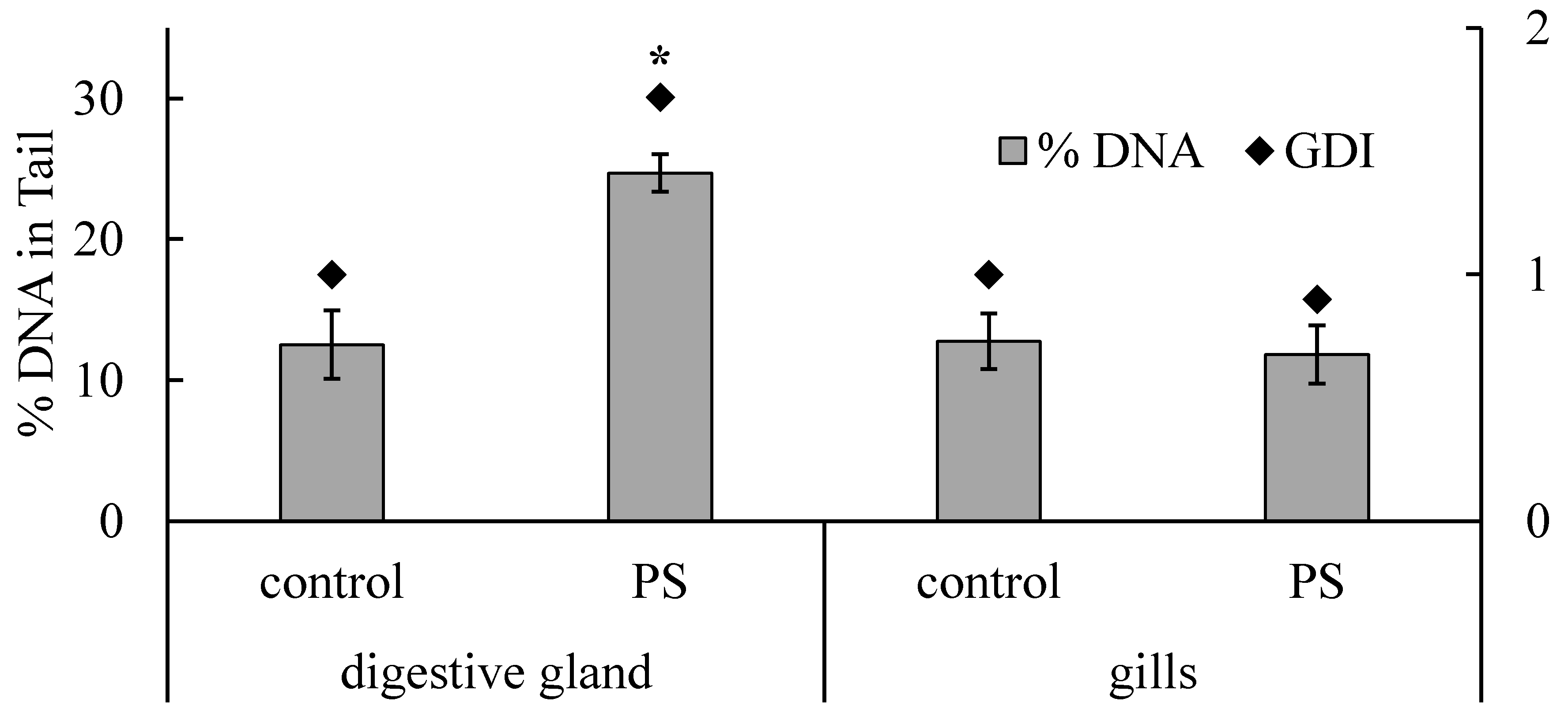
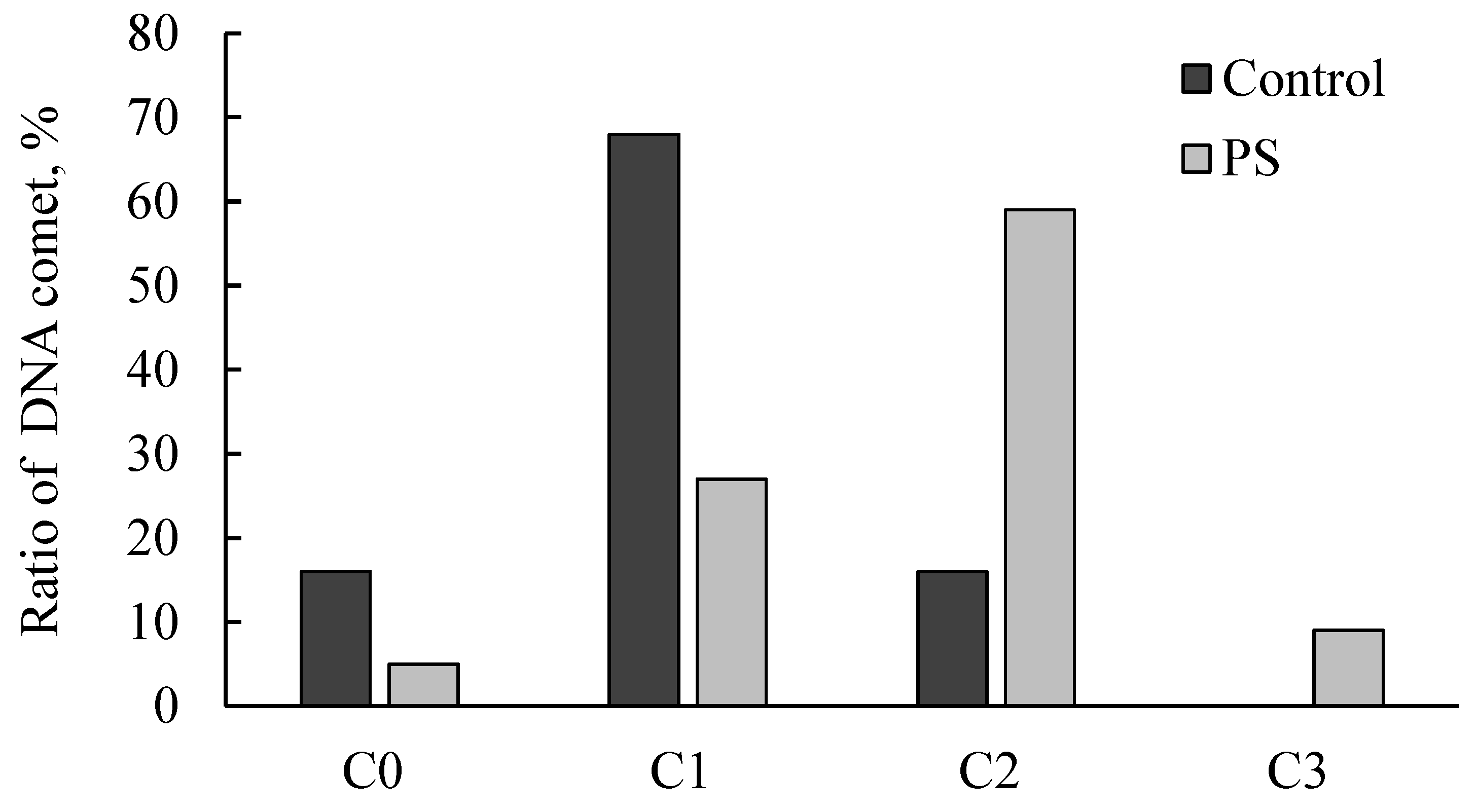
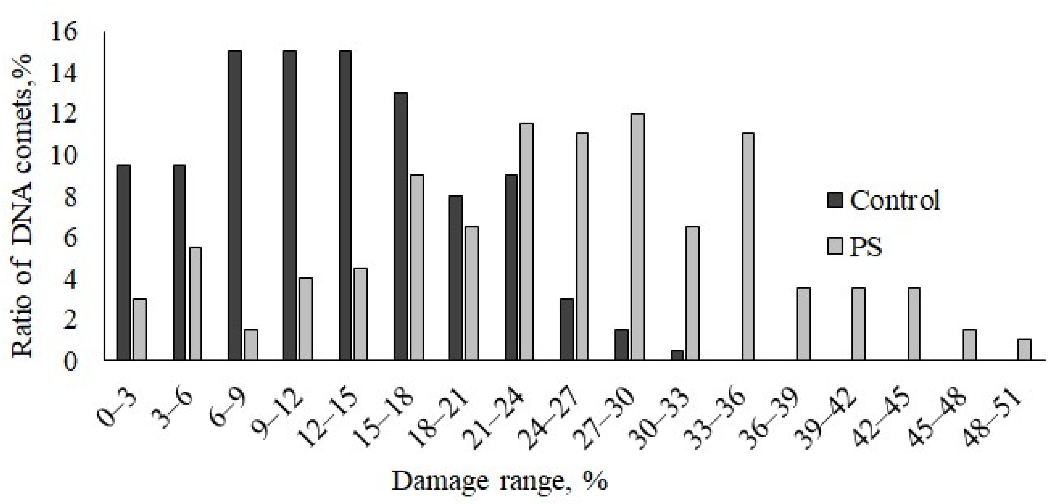
The experimental results were processed in MS Excel and STATISTICA 10 software packages (StatSoft, Tulsa, OK, USA). For the “% DNA in the tail” dates, nonparametric Kruskal–Wallis ANOVA followed by pair-wise Mann–Whitney tests were performed. A difference at p < 0.05 was considered statistically significant.

3. Results

As a result of the study, using cometary analysis, it became possible to assess the level of DNA damage in the cells of two tissues of the mussel M. trossulus, before and after short-term exposure to PS microparticles (Figure 1).
In the control mussels, a small level (~12%) of nuclear DNA fragmentation was observed in the gill and digestive gland cells, formed in normal cells during the functioning of life-supporting systems, due to the accumulation of alkali-labile sites and single- and double-stranded breaks. An analysis of the comet class distribution showed that, in the digestive gland cells of the control mollusks, the C0 (0–5% of DNA in the comet tail) and C1 (5–20%) classes prevailed, characteristic of intact and viable cells. The sum of comets in these classes (C0 + C1) was about 88%, whereas comets with DNA damage levels greater than 20% (C2 class) accounted for no more than 12% (Figure 2).
The exposure of mussels to a medium containing PS microparticles only resulted in a significant increase in DNA damage in the digestive gland cells (~25%), almost twice the control values, indicating damage to the genome integrity (Figure 1). In the experimental mussels, unlike the controls, the content of intact cells in the digestive gland cells was sharply reduced (less than 30%), and most of the comets were represented by the C2 class (~60%). In addition, comet cells with a high proportion of DNA damage appeared, belonging to the C3 class (40–75%), with a ratio of about 10% (Figure 2).

4. Discussion

Recent studies have demonstrated that microparticles of various polymers, although chemically inert, have negative toxic effects on a wide range of marine organisms, including polychaetes, mollusks, echinoderms, crustaceans, and fish [19,24,25,26]. By penetrating organisms, plastic fragments not only mechanically damage mucous tissues and block the digestive tract, but also cause severe disruption to fundamental physiological processes, including inhibiting the secretion of digestive enzymes, reducing filtering activity, and disturbing behavior, respiration and reproduction [27,28,29,30,31].
Under experimental conditions, the microparticles of polymers, particularly polystyrene, induced various sublethal effects in marine invertebrates at the molecular and biochemical levels. Even with short-term exposure, microplastic fragments have been shown to affect the activity of various enzymes and the antioxidant system, to induce the formation of ROS and stress proteins, and to stimulate the development of oxidative stress (lipid peroxidation and destabilization of lysosome membranes) and inflammatory processes [7,8,20,32,33].
At the same time, the reasons for this diverse expression of microplastic toxicity are far from being understood. Nevertheless, there is reason to believe that the molecular basis of all the identified physiological and biochemical changes is genome degradation, which may be enhanced in cells during the entry and concentration of microplastic particles. For DNA damage detection, we used the comet assay, which is widely used in toxicological studies, due to its high sensitivity [34,35,36].
Based on this experimental approach, we showed that, despite the chemical inertness, the microparticles of PS interacting with the digestive tissues of the bivalve M. trossulus exhibit biological activity, expressing genotoxic properties.
The effects of the genotoxic properties of plastic microparticles on marine invertebrates have been reported in a few articles. The first evidence of the genotoxicity of plastic microparticles, based on the DNA comet assay, was presented in experimental studies using marine bivalve mollusks. The number of DNA strand breaks increased in the hemocytes of the mussels Mytilus galloprovincialis and Scrobicularia plana after the absorption of plastic microparticles (PS and PE) [6,20]. Gonzalez-Soto and colleagues [37] observed the following biphasic DNA response to PS microparticles: after 7 days of exposure, a decrease in DNA damage was observed, compared with the control, but DNA damage increased dramatically after 26 days of exposure. This is supported by the work of Revel and colleagues [38], who showed that, in the presence of low concentrations of a mixture of polyethylene (PE) and polypropylene (PP) microparticles in Mytilus spp. mussel hemocytes, there was an increase in DNA molecule degradation. Furthermore, Berber and colleagues [39] showed that daily exposure of Neocaridina davidi shrimp to PS microspheres resulted in damage to their DNA, which was shown by the change in comet tail length relative to the control.
Sun and colleagues [40] summarized the available data in the literature and provided convincing evidence, based on a meta-analysis, that plastic microparticles at environmentally realistic concentrations (<1 mg/L, mean ~0.5 mg/L) exhibited marked genotoxic properties when interacting with aquatic organisms. This was particularly clear not only from the comet assay method, but also from the increase in the number of micronuclei.
At the same time, after bivalves, such as Chlamys farreri [41] and Crassostrea gigas [42], were exposed to PS microparticles, no significant changes in DNA integrity were detected. Furthermore, low-density polyethylene (LDPE) and polyvinyl chloride (PVC) microparticles were also found to show no genotoxicity when exposed to M. galloprovincialis and Perna perna mollusks [43,44].
Such contradictory data from the literature can be explained by the wide variation in the physicochemical characteristics of the microplastic particles used in the work presented; there was also wide variation in the conditions of the experiments and their duration. Furthermore, it should be noted that the authors of the above studies were limited to analyzing the effects of the genotoxic properties of microplastics on hemolymph cells and hemocytes, which are critical components of the immune and detoxification systems.
The importance of our findings consists of detecting DNA damage directly in the cells of the digestive gland, a critical element in the uptake of microplastics by bivalves after exposure to PS microparticles. For clarity, the experimental data obtained are presented in a diagram that characterizes the distribution of cells according to the degree of nuclear DNA damage at intervals of 3% (Figure 3).
From the analysis of these diagrams, it appears that, in the control mollusks, the highest number of cells had a damage range of 6 to 18%, with the maximum DNA content (about 30%) in the comet tail being observed in only 5% of the cells. However, after exposure to polystyrene microparticles, about half of the mussels’ digestive gland cells had DNA damage in the range of 25 to 35%. In addition, DNA damage >50% was observed in about 5% of the studied comets, which indicates a significant genotoxic effect.
It is also important that, in our experiment, after exposing the mussels to PS microparticles, the digestive gland cells were more sensitive to the effects than the gill cells. By the final stage, the level of DNA damage in the digestive gland cells of the experimental mussels significantly increased, almost twice as much, while no significant changes were observed in the gill cells (Figure 1). In all likelihood, such significant differences in the response of the cells of these two tissue types to PS microparticle exposure are due to the features of the physiological and biochemical systems of feeding, and due to the assimilation of various particles, characteristic of filter mollusks and mussels, in particular.
In our experiments, we found a decrease in the working concentration of PS over a 12 h period (Supplementary File S1). This change is probably due to partial sedimentation, as well as uptake and sorption, of the PS microspheres by mussels. Although we did not study the localization of the PC microparticles in our experiments, we relied on extensive data from the literature on this issue. Generalized data from the literature on the mussels Mytilus edulis and M. galloprovincialis suggest that the gills and digestive systems play major roles in the uptake of MF microparticles. It has been found that, when in contact with the gill surfaces of mussels, microplastics are adsorbed by mucus and then transported by cilia movement to the mouth opening, and penetrate the digestive system and hemolymph, where the translocation of these particles into cells occurs [20,27,32,45]. In the digestive gland, PS microspheres have been shown to enter cells via endocytosis, and to be taken up by lysosomes [27].
This is supported by experimental data on the bioaccumulation of plastic microparticles in mussels, mainly in the digestive gland and, to a much lesser extent, in the gills and hemolymph of mussels [43,46,47]. Similar studies on another representative of the bivalve filter-feeder mollusk, the oyster Crassostrea gigas, also confirm this trend; fluorescent PS microparticles were only found in the digestive glands of experimental mollusks [48].
This short list of examples, together with our experimental results, suggests that digestive gland cells become the primary target for exposure to PS microparticles. Given the unique role of the genome, DNA damage to these cells may be the earliest step in the development of biochemical events that lead to toxic effects.

5. Conclusions

The genotoxic effect of PS microparticles on the digestive system cells of M. trossulus was shown for the first time. This study provided evidence that polystyrene microspheres exhibit genotoxic properties. The exposure of mussels to water containing polystyrene microparticles resulted in a significant increase in DNA damage in digestive gland cells, exceeding control values by almost two times. Our studies were performed under controlled laboratory conditions, using concentrations of microparticles of PS that were many times higher than the actual concentrations. However, a reasonable point of view is that dangerous consequences of toxicological character not only depend on the pollution level, lifetime, and processes of particle dispersion, but are also conditioned by the possibility of microplastic accumulation in sea organisms. Therefore, the results obtained suggest that the digestive gland cells of filter-feeder mollusks, which can concentrate microplastics at relatively low concentrations of these particles in the external environment, may be at risk from the steadily increasing amount of pollution in marine environments by plastic wastes.

Supplementary Materials

The following supporting information can be downloaded at https://www.mdpi.com/article/10.3390/jmse10020273/s1: Supplementary File S1: concentration of PS in the exposure medium.

Author Contributions

Conceptualization, V.P.C., A.A.M. and N.V.D.; methodology, A.A.M., V.V.S. and S.P.K.; soft-ware, S.P.K.; validation, A.A.M. and V.V.S.; formal analysis, S.P.K.; investigation, A.A.M., V.V.S. and S.P.K.; resources, V.P.C.; data curation, V.P.C. and N.V.D.; writing—original draft preparation, V.P.C.; writing—review and editing, V.P.C., A.A.M. and V.V.S.; visualization, A.A.M.; supervision, V.P.C. and N.V.D.; project administration, V.P.C.; funding acquisition, V.P.C. All authors have read and agreed to the published version of the manuscript.

Funding

This research was supported by the state assignment for research work of V.I. Il’ichev Pacific Oceanological Institute, FEB RAS (No. 121-21500052-9).

Institutional Review Board Statement

Not applicable.

Informed Consent Statement

Not applicable.

Data Availability Statement

Not applicable.

Conflicts of Interest

The authors declare no conflict of interest.

References

  1. Lusher, A. Microplastics in the marine environment: Distribution, interactions and effects. In Marine Anthropogenic Litter; Springer: Cham, Switzerland, 2015; pp. 245–308. [Google Scholar] [CrossRef] [Green Version]
  2. Rochman, C.M.; Tahir, A.; Williams, S.L.; Baxa, D.V.; Lam, R.; Miller, J.T.; Teh, F.-C.; Werorilangi, S.; Teh, S.J. Anthropogenic debris in seafood: Plastic debris and fibers from textiles in fish and bivalves sold for human consumption. Sci. Rep. 2015, 5, 14340. [Google Scholar] [CrossRef]
  3. Galloway, T.S.; Cole, M.; Lewis, C. Interactions of microplastic debris throughout the marine ecosystem. Nat. Ecol. Evol. 2017, 1, 116. [Google Scholar] [CrossRef] [PubMed]
  4. Santana, M.F.M.; Moreira, F.T.; Turra, A. Trophic transference of microplastics under a low exposure scenario: Insights on the likelihood of particle cascading along marine food-webs. Mar. Pollut. Bull. 2017, 121, 154–159. [Google Scholar] [CrossRef]
  5. Prokic, M.; Radovanović, T.B.; Gavrić, J.P.; Faggio, C. Ecotoxicological effects of microplastics: Examination of biomarkers, current state and future perspectives. TrAC Trends Anal. Chem. 2019, 111, 37–46. [Google Scholar] [CrossRef]
  6. Ribeiro, F.; Garcia, A.R.; Pereira, B.P.; Fonseca, M.; Mestre, N.C.; Fonseca, T.G.; Ilharco, L.M.; Bebianno, M.J. Microplastics effects in Scrobicularia plana. Mar. Pollut. Bull. 2017, 122, 379–391. [Google Scholar] [CrossRef] [PubMed]
  7. Détrée, C.; Gallardo-Escárate, C. Single and repetitive microplastics exposures induce immune system modulation and homeostasis alteration in the edible mussel Mytilus galloprovincialis. Fish Shellfish Immunol. 2018, 83, 52–60. [Google Scholar] [CrossRef]
  8. Varó, I.; Perini, D.A.; Torreblanca, A.; Garcia, Y.; Bergami, E.; Vannuccini, M.L.; Corsi, I. Time-dependent effects of polystyrene nanoparticles in brine shrimp Artemia franciscana at physiological, biochemical and molecular levels. Sci. Total Environ. 2019, 675, 570–580. [Google Scholar] [CrossRef] [PubMed]
  9. Pannetier, P.; Morin, B.; Le Bihanic, F.; Dubreil, L.; Clérandeau, C.; Chouvellon, F.; Van Arkel, K.; Danion, M.; Cachot, J. Environmental samples of microplastics induce significant toxic effects in fish larvae. Environ. Int. 2020, 134, 105047. [Google Scholar] [CrossRef]
  10. Roda, J.F.B.; Lauer, M.M.; Risso, W.E.; Martinez, C.B.R. Microplastics and copper effects on the neotropical teleost Prochilodus lineatus: Is there any interaction? Comp. Biochem. Physiol. Part A Mol. Integr. Physiol. 2020, 242, 110659. [Google Scholar] [CrossRef]
  11. Thomas, M.; Jon, B.; Craig, S.; Edward, R.; Ruth, H.; John, B.; Dick, V.A.; Heather, L.A.; Matthew, S. The world is your oyster: Low-dose, long-term microplastic exposure of juvenile oysters. Heliyon 2020, 6, e03103. [Google Scholar] [CrossRef] [Green Version]
  12. Avio, C.G.; Gorbi, S.; Regoli, F. Plastics and microplastics in the oceans: From emerging pollutants to emerged threat. Mar. Environ. Res. 2017, 128, 2–11. [Google Scholar] [CrossRef] [PubMed]
  13. Li, J.; Qu, X.; Su, L.; Zhang, W.; Yang, D.; Kolandhasamy, P.; Li, D.; Shi, H. Microplastics in mussels along the coastal waters of China. Environ. Pollut. 2016, 214, 177–184. [Google Scholar] [CrossRef]
  14. Faggio, C.; Tsarpali, V.; Dailianis, S. Mussel digestive gland as a model tissue for assessing xenobiotics: An overview. Sci. Total Environ. 2018, 636, 220–229. [Google Scholar] [CrossRef] [PubMed]
  15. Kolandhasamy, P.; Su, L.; Li, J.; Qu, X.; Jabeen, K.; Shi, H. Adherence of microplastics to soft tissue of mussels: A novel way to uptake microplastics beyond ingestion. Sci. Total Environ. 2018, 610–611, 635–640. [Google Scholar] [CrossRef] [PubMed]
  16. Renzi, M.; Guerranti, C.; Blašković, A. Microplastic contents from maricultured and natural mussels. Mar. Pollut. Bull. 2018, 131, 248–251. [Google Scholar] [CrossRef]
  17. Qu, X.; Su, L.; Li, H.; Liang, M.; Shi, H. Assessing the relationship between the abundance and properties of microplastics in water and in mussels. Sci. Total Environ. 2018, 621, 679–686. [Google Scholar] [CrossRef]
  18. Ward, J.E.; Zhao, S.Y.; Holohan, B.A.; Mladinich, K.M.; Griffin, T.W.; Wozniak, J.; Shumway, S.E. Selective ingestion and egestion of plastic particles by the blue mussel (Mytilus edulis) and Eastern Oyster (Crassostrea virginica): Implications for using bivalves as bioindicators of microplastic pollution. Environ. Sci. Technol. 2019, 53, 8776–8784. [Google Scholar] [CrossRef]
  19. Van Cauwenberghe, L.; Claessens, M.; Vandegehuchte, M.B.; Janssen, C.R. Microplastics are taken up by mussels (Mytilus edulis) and lugworms (Arenicola marina) living in natural habitats. Environ. Pollut. 2015, 199, 10–17. [Google Scholar] [CrossRef]
  20. Avio, C.G.; Gorbi, S.; Milan, M.; Benedetti, M.; Fattorini, D.; D’Errico, G.; Pauletto, M.; Bargelloni, L.; Regoli, F. Pollutants bioavailability and toxicological risk from microplastics to marine mussels. Environ. Pollut. 2015, 198, 211–222. [Google Scholar] [CrossRef]
  21. Mitchelmore, C.L.; Birmelin, C.; Livingstone, D.R.; Chipman, J.K. Detection of DNA strand breaks in isolated mussel (Mytilus edulis L.) digestive gland cells using the “Comet” assay. Ecotoxicol. Environ. Saf. 1998, 41, 51–58. [Google Scholar] [CrossRef]
  22. Slobodskova, V.V.; Solodova, E.E.; Slinko, E.N.; Chelomin, V.P. Evaluation of the genotoxicity of cadmium in gill cells of the clam Corbicula japonica using the comet assay. Russ. J. Mar. Biol. 2010, 36, 311–315. [Google Scholar] [CrossRef]
  23. Cavaş, T.; Könen, S. In vivo genotoxicity testing of the amnesic shellfish poison (domoic acid) in piscine eryth-rocytes using the micronucleus test and the comet assay. Aquat. Toxicol. 2008, 90, 154–159. [Google Scholar] [CrossRef] [PubMed]
  24. Gunaalan, K.; Fabbri, E.; Capolupo, M. The hidden threat of plastic leachates: A critical review on their impacts on aquatic organisms. Water Res. 2020, 184, 116170. [Google Scholar] [CrossRef] [PubMed]
  25. Berlino, M.; Mangano, M.; De Vittor, C.; Sarà, G. Effects of microplastics on the functional traits of aquatic benthic organisms: A global-scale meta-analysis. Environ. Pollut. 2021, 285, 117174. [Google Scholar] [CrossRef] [PubMed]
  26. Steer, M.; Cole, M.; Thompson, R.C.; Lindeque, P.K. Microplastic ingestion in fish larvae in the western English Channel. Environ. Pollut. 2017, 226, 250–259. [Google Scholar] [CrossRef] [PubMed]
  27. Browne, M.A.; Dissanayake, A.; Galloway, T.S.; Lowe, D.M.; Thompson, R.C. Ingested microscopic plastic translocates to the circulatory system of the mussel, Mytilus edulis (L.). Environ. Sci. Technol. 2008, 42, 5026–5031. [Google Scholar] [CrossRef] [PubMed]
  28. Jeong, C.B.; Kang, H.M.; Lee, M.C.; Kim, D.H.; Han, J.; Hwang, D.S.; Souissi, S.; Lee, S.-J.; Shin, K.-H.; Park, H.G.; et al. Adverse effects of microplastics and oxidative stress-induced MAPK/Nrf2 pathway-mediated defense mechanisms in the marine copepod Paracyclopina nana. Sci. Rep. 2017, 7, 41323. [Google Scholar] [CrossRef] [Green Version]
  29. Pinsino, A.; Bergami, E.; Della Torre, C.; Vannuccini, M.L.; Addis, P.; Secci, M.; Dawson, K.A.; Matranga, V.; Corsi, I. Amino-modified polystyrene nanoparticles affect signaling pathways of the sea urchin (Paracentrotus lividus) embryos. Nanotoxicology 2017, 11, 201–209. [Google Scholar] [CrossRef]
  30. Anbumani, S.; Kakkar, P. Ecotoxicological effects of microplastics on biota: A review. Environ. Sci. Pollut. Res. 2018, 25, 14373–14396. [Google Scholar] [CrossRef]
  31. Revel, M.; Yakovenko, N.; Caley, T.; Guillet, C.; Châtel, A.; Mouneyrac, C. Accumulation and immunotoxicity of microplastics in the estuarine worm Hediste diversicolor in environmentally relevant conditions of exposure. Environ. Sci. Pollut. Res. 2018, 27, 3574–3583. [Google Scholar] [CrossRef]
  32. Paul-Pont, I.; Lacroix, C.; Fernández, C.G.; Hégaret, H.; Lambert, C.; Le Goïc, N.; Frère, L.; Cassone, A.-L.; Sussarellu, R.; Fabioux, C.; et al. Exposure of marine mussels Mytilus spp. to polystyrene microplastics: Toxicity and influence on fluoranthene bioaccumulation. Environ. Pollut. 2016, 216, 724–737. [Google Scholar] [CrossRef] [PubMed] [Green Version]
  33. Gambardella, C.; Morgana, S.; Ferrando, S.; Bramini, M.; Piazza, V.; Costa, E.; Garaventa, F.; Faimali, M. Effects of polystyrene microbeads in marine planktonic crustaceans. Ecotoxicol. Environ. Saf. 2017, 145, 250–257. [Google Scholar] [CrossRef] [PubMed]
  34. Frenzilli, G.; Nigro, M.; Lyons, B.P. The Comet assay for the evaluation of genotoxic impact in aquatic environments. Mutat. Res./Rev. Mutat. Res. 2009, 681, 80–92. [Google Scholar] [CrossRef] [PubMed]
  35. Smith, M.A.; Fernandez-Triana, J.; Roughley, R.; Hebert, D.N. DNA barcode accumulation curves for understudied taxa and areas. Mol. Ecol. Resour. 2009, 9, 208–216. [Google Scholar] [CrossRef] [PubMed]
  36. Martins, M.; Costa, P.M. The comet assay in Environmental Risk Assessment of marine pollutants: Applications, assets and handicaps of surveying genotoxicity in non-model organisms. Mutagenesis 2015, 30, 89–106. [Google Scholar] [CrossRef] [Green Version]
  37. Gonzalez-Soto, N.; Hatfield, J.; Katsumiti, A.; Duroudier, N.; Lacave, J.M.; Bilbao, E.; Orbea, A.; Navarro, E.; Cajaraville, M.P. Impacts of dietary exposure to different sized polystyrene microplastics alone and with sorbed benzo [a] pyrene on biomarkers and whole organism responses in mussels Mytilus galloprovincialis. Sci. Total Environ. 2019, 684, 548–566. [Google Scholar] [CrossRef]
  38. Revel, M.; Lagarde, F.; Perrein-Ettajani, H.; Bruneau, M.; Akcha, F.; Sussarellu, R.; Rouxel, J.; Costil, K.; Decottignies, P.; Cognie, B.; et al. Tissue-specific biomarker responses in the blue mussel Mytilus spp. exposed to a mixture of microplastics at environmentally relevant concentrations. Front. Environ. Sci. 2019, 7, 33. [Google Scholar] [CrossRef]
  39. Berber, A.A. Genotoxic evaluation of polystyrene microplastic. Sak. Univ. J. Sci. 2019, 23, 358–367. [Google Scholar] [CrossRef] [Green Version]
  40. Sun, T.; Zhan, J.; Li, F.; Ji, C.; Wu, H. Evidence-based meta-analysis of the genotoxicity induced by microplastics in aquatic organisms at environmentally relevant concentrations. Sci. Total Environ. 2021, 783, 147076. [Google Scholar] [CrossRef]
  41. Xia, B.; Zhang, J.; Zhao, X.; Feng, J.; Teng, Y.; Chen, B.; Sun, X.; Zhu, L.; Sun, X.; Qu, K. Polystyrene microplastics increase uptake, elimination and cytotoxicity of decabromodiphenyl ether (BDE-209) in the marine scallop Chlamys farreri. Environ. Pollut. 2020, 258, 113657. [Google Scholar] [CrossRef]
  42. Revel, M.; Châtela, A.; Perrein-Ettajani, H.; Bruneau, M.; Akcha, F.; Sussarellu, R.; Rouxel, J.; Costil, K.; Decottignies, P.; Cognie, B.; et al. Realistic environmental exposure to microplastics does not induce biological effects in the Pacific oyster Crassostrea gigas. Mar. Pollut. Bull. 2020, 150, 110627. [Google Scholar] [CrossRef] [PubMed]
  43. Pittura, L.; Avio, C.G.; Giuliani, M.E.; d’Errico, G.; Keiter, S.H.; Cormier, B.; Gorbi, S.; Regoli, F. Microplastics as vehicles of environmental PAHs to marine organisms: Combined chemical and physical hazards to the Mediterranean mussels, Mytilus galloprovincialis. Front. Mar. Sci. 2018, 5, 103. [Google Scholar] [CrossRef] [Green Version]
  44. Santana, M.F.; Moreira, F.T.; Pereira, C.D.; Abessa, D.M.; Turra, A. Continuous exposure to microplastics does not cause physiological effects in the cultivated mussel Perna perna. Arch. Environ. Contam. Toxicol. 2018, 74, 594–604. [Google Scholar] [CrossRef] [PubMed] [Green Version]
  45. Von Moos, N.; Burkhardt-Holm, P.; Köhler, A. Uptake and effects of microplastics on cells and tissue of the blue mussel Mytilus edulis L. after an experimental exposure. Environ. Sci. Technol. 2012, 46, 11327–11335. [Google Scholar] [CrossRef]
  46. Sıkdokur, E.; Belivermiş, M.; Sezer, N.; Pekmez, M.; Bulan, Ö.K.; Kılıç, Ö. Effects of microplastics and mercury on manila clam Ruditapes philippinarum: Feeding rate, immunomodulation, histopathology and oxidative stress. Environ. Pollut. 2020, 262, 114247. [Google Scholar] [CrossRef]
  47. Wang, S.; Hu, M.; Zheng, J.; Huang, W.; Shang, Y.; Fang, J.K.-H.; Shi, H.; Wang, Y. Ingestion of nano/micro plastic particles by the mussel Mytilus coruscus is size dependent. Chemosphere 2021, 263, 127957. [Google Scholar] [CrossRef]
  48. Sussarellu, R.; Suquet, M.; Thomas, Y.; Lambert, C.; Fabioux, C.; Arsenault-Pernet, E.-J.; Le Goïc, N.; Quillien, V.; Mingant, C.; Epelboin, Y.; et al. Oyster reproduction is affected by exposure to polystyrene microplastics. Proc. Natl. Acad. Sci. USA 2016, 113, 2430–2435. [Google Scholar] [CrossRef] [Green Version]
Figure 1. Assessment of DNA damage in gill and digestive gland cells from control and experimental groups (n = 12; n = 600). * Difference from the control is significant (p < 0.05).
Jmse 10 00273 g001
Figure 2. Induction of DNA strand breaks, represented as a percentage of comet damage classes in digestive gland cells from control and experimental groups (n = 12; n = 600).
Jmse 10 00273 g002
Figure 3. Distribution of DNA comets at 3% damage intervals in digestive gland cells (n = 12; n = 600).
Jmse 10 00273 g003
Publisher’s Note: MDPI stays neutral with regard to jurisdictional claims in published maps and institutional affiliations.

